Article

Simple Fabrication and Biological Evaluation of Ulva australis-Derived Marine Carbon Dots with Anti-Inflammation, Anti-Oxidation, and Anti-Adipogenesis Features

1
Department of Biomaterial Research, National Marine Biodiversity Institute of Korea, 75 Jangsan-ro, 101 Beon-gil, Janghang-eup, Seocheon-gun 33662, Chungcheongnam-do, Republic of Korea
2
Department of Bioindustrial Strategy, National Marine Biodiversity Institute of Korea, 75 Jangsan-ro, 101 Beon-gil, Janghang-eup, Seocheon-gun 33662, Chungcheongnam-do, Republic of Korea
3
Department of Biological Application & Technology, National Marine Biodiversity Institute of Korea, 75 Jangsan-ro, 101 Beon-gil, Janghang-eup, Seocheon-gun 33662, Chungcheongnam-do, Republic of Korea
*
Authors to whom correspondence should be addressed.
Current address: Department of Marine Ecological Biotechnology, Kunsan National University, 558 Daehak-ro, Gunsan-si 54150, Jeonbuk-do, Republic of Korea.
J. Mar. Sci. Eng. 2025, 13(10), 1878; https://doi.org/10.3390/jmse13101878
Submission received: 4 August 2025 / Revised: 18 September 2025 / Accepted: 27 September 2025 / Published: 1 October 2025
(This article belongs to the Special Issue Advances in Marine Natural Products)

Abstract

The overabundance of Ulva australis (U. australis), a green macroalga widespread along the coastline of Jeju Island, Republic of Korea, presents a growing ecological challenge, as it can cause unpleasant odors and disturb the ecological balance. Hence, we report a sustainable valorization strategy for converting U. australis biomass into marine carbon dots (MCDs) via a facile hydrothermal carbonization process. The synthesis requires no hazardous reagents or complex instrumentation and yields highly water-dispersible MCDs with excitation-dependent fluorescence properties. Comprehensive in vitro and in vivo assessments revealed the multifunctional bioactivity of the synthesized MCDs. Moreover, in vivo fluorescence imaging at seven days post-fertilization revealed the preferential accumulation of MCDs along the vertebral column, implying a possible affinity for mineralized tissues and suggesting their utility in skeletal imaging applications. Collectively, these findings underscore the potential of U. australis-derived MCDs as biocompatible and multifunctional nanomaterials with broad biomedical applications.

1. Introduction

Oxidative stress, inflammation, and obesity are interconnected pathological processes contributing to the onset and progression of chronic diseases. Oxidative stress results from an imbalance between the production of reactive oxygen species (ROS) and the antioxidant defense system of the body, leading to cellular damage and metabolic dysfunction [1,2]. Mounting evidence suggests that oxidative stress not only plays a pivotal role in the pathogenesis of obesity but also exacerbates its complications, including insulin resistance, dyslipidemia, and endothelial dysfunction [3]. Inflammation is a fundamental physiological response essential for host defense and tissue repair. In its acute form, inflammation is self-limiting and resolves within days as tissue homeostasis is restored. However, when inflammation becomes chronic, it shifts from protective to pathogenic. Chronic inflammation is characterized by the persistent activation of immune cells, such as macrophages, neutrophils, and lymphocytes, which secrete an array of cytokines, chemokines, and lipid mediators. These signaling molecules propagate a continuous inflammatory response, disrupting the balance between proinflammatory and anti-inflammatory mechanisms and ultimately leading to tissue damage, fibrosis, and even carcinogenesis [4,5,6]. Understanding the complex interplay between acute and chronic inflammation is critical for developing targeted therapies to treat chronic diseases [7,8,9]. Obesity, defined as excessive accumulation of adipose tissue, is now recognized as a state of chronic low-grade inflammation and oxidative stress. It significantly increases the risk of developing metabolic disorders, such as type 2 diabetes, cardiovascular disease, and certain types of cancer [10,11]. The simultaneous presence of oxidative stress and inflammation in obese individuals suggests a synergistic relationship that accelerates the disease progression. Therefore, novel strategies that modulate the oxidative and inflammatory responses are urgently required to combat obesity and its associated conditions. Nanotechnology has emerged as a promising avenue to address these challenges. In particular, nano-antioxidants, which are nanoscale materials with intrinsic radical-scavenging properties, have demonstrated potential for mitigating oxidative stress. These nanomaterials offer several advantages, including improved stability, bioavailability, and targeted delivery [12,13,14,15]. Carbon dots (CDs), also known as C-dots, have garnered significant attention because of their unique physicochemical and biological properties. CDs are zero-dimensional carbon-based nanomaterials exhibiting excitation-dependent fluorescence, low toxicity, high water dispersibility, and robust photostability. They possess inherent antioxidant capabilities, effectively scavenging both reactive oxygen and nitrogen species such as nitric oxide (NO) and nitrogen dioxide (NO2), which are known to disrupt cellular homeostasis [16,17,18]. Owing to their versatile functionality and biocompatibility, CDs have been explored in diverse biomedical applications, including biosensing, drug delivery, photodynamic therapy, and photocatalysis [19,20,21,22]. Notably, their green synthesis from natural resources has enhanced their environmental and economic appeal. Marine organisms, including algae, offer a sustainable and underutilized resource for the synthesis of CDs via environmentally friendly hydrothermal methods [22,23,24,25,26]. In this context, we report the synthesis and bioactivity of marine carbon dots (MCDs) derived from Ulva australis Areschoug 1854, a green macroalga that proliferates along the eastern coastline of Jeju Island, Republic of Korea. U. australis has been identified as a marine waste species, with more than 22,000 tons collected since 2020. Owing to their high salt content, conventional disposal strategies such as incineration or landfilling are environmentally and economically impractical. Most existing studies have focused on the inherent properties and biochemical composition of U. australis, whereas its potential for development into high-value nanomaterials remains largely unexplored [27,28,29,30]. In this proof-of-concept study, we synthesized MCDs from U. australis using a simple hydrothermal carbonization method and evaluated their biological activities. Specifically, we investigated its (i) anti-inflammatory activity attributed to the inhibition of nitrite production in lipopolysaccharide (LPS)-stimulated RAW264.7 macrophages, (ii) antioxidant capacity via intracellular ROS reduction in macrophages, and (iii) anti-adipogenic effect on 3T3-L1 preadipocyte differentiation. This study not only introduces a novel approach for marine biomass utilization but also highlights the therapeutic potential of marine-derived nanomaterials as multifunctional agents to combat inflammation, oxidative stress, and obesity-related disorders.

2. Materials and Methods

2.1. Materials

The U. australis used in this study was collected from near the Ojori coastal area in Seongsan-eup, Seogwipo-si, Jeju Island, Republic of Korea (33°28′21.5″ N 126°54′49.2″ E). Minisart® NML syringe filters with 0.22 μm pores were purchased from Sartorius AG (Göttingen, Germany). UltraPureTM DNase/RNase-free distilled water was obtained from Thermo Fisher Scientific (Waltham, MA, USA). A transmission electron microscopy (TEM) grid coated with a formvar film stabilized by an evaporated carbon layer (TED PELLA Inc., Redding, CA, USA) was used. All chemicals including dimethyl sulfoxide (DMSO), 2′,7′-dichlorofluorescin diacetate (DCFDA), LPS, dexamethasone, 3-Isobutyl-1-methylxanthine (IBMX), and insulin were purchased from Sigma-Aldrich (St. Louis, MO, USA). The model cell lines, including RAW264.7 and 3T3-L1, were obtained from the American Type Culture Collection (ATCC, Manassas, VA, USA). Their cryopreserved vials were either stored immediately at −80 °C or thawed according to the protocol specified by the supplier and cultured in the appropriate growth medium prior to experimental use.

2.2. Synthesis of CDs Using U. australis and Other Marine Algae

MCDs can be easily fabricated using U. Australis and other seaweeds via a single-pot hydrothermal reaction. U. australis (0.5 g) and two other algae—green and red algae—were thoroughly cleaned using running tap water to remove any remaining salts and sand. Subsequently, the seaweeds were dried at 50 °C. The cleaned and dried U. australis and other algae were processed in a mini-pulverizer to obtain fine powders. The fine powders were mixed with 20 mL of UltraPureTM DNase/RNase-free distilled water, and the solution mixtures were introduced into hydrothermal autoclaving reactors manufactured by TEFIC Biotech Co. (Xi’an, China). Subsequently, the reactors were maintained at 180 °C for 6 h and then cooled to 25 °C. The resulting MCD solutions were centrifuged using an Ohaus FrontierTM 5718R centrifuge (Parsippany, NJ, USA) at 5500 rpm for 30 min. Their supernatants were then filtered through a syringe filter with a 0.22 μm pore size. Finally, the pH-adjusted MCD solutions (with a pH range of 6–6.5) were stored at room temperature (approximately 25 °C) until use. In the functional analysis tests, including anti-inflammation and adipogenesis of carbon dots derived from marine organisms in this study, the MCDs synthesized using U. australis were used as the target group whereas those derived from the two other seaweeds served as the control groups (R-MCDs and G-MCDs).

2.3. Morphological Properties of MCDs from U. australis

TEM images of MCDs derived from U. australis were acquired using a JEM-ARM200F microscope operated at 80 kV (JEOL, Ltd., Tokyo, Japan). A suitably diluted MCD solution was deposited onto a TEM grid coated with formvar film and carbon. The particle sizes were quantified using ImageJ software v.1.54n and the particle size distribution (PSD) was determined using OriginPro 2024 software (version 10.1; OriginLab Corp., Northampton, MA, USA). Fourier-transform infrared (FT-IR) spectroscopy, X-ray photoelectron spectroscopy (XPS), and X-ray diffraction (XRD) analyses were performed on the MCDs. FT-IR was conducted using an attenuated total reflection method with an FT/IR 4600 spectrometer (JASCO Corp., Tokyo, Japan) and covered the spectral range from 700 cm−1 to 4000 cm−1. XPS measurements were conducted using a PHI5000 VersaProbe III photoelectron spectrometer (Ulvac-Phi, Inc., Kanagawa, Japan) equipped with an Al Kα radiation source (hv = 1486.6 eV). The XPS spectra were calibrated using the Au 4f7/2 peak at 84.0 eV and the Cu 2p3/2 peak at 932.7 eV as internal standards. In addition, the background of the XPS spectra was treated using the Shirley method. XRD analysis was performed using a MiniFlex 600 desktop diffractometer (Rigaku Corp., Tokyo, Japan) with a Cu Kα radiation source (λ = 1.5406 Å). Prior to the analysis, the MCD solution was freeze-dried using a FreeZone drying machine (Labconco, Kansas City, MO, USA) at −80 °C for one week. Subsequently, FT-IR, XPS, and XRD analyses were performed on the lyophilized MCD powders.

2.4. Investigation of Optical Characteristics of MCDs from U. australis

Photoluminescence (PL) spectra were measured using a FluoroMax-4 spectrometer (HORIBA, Ltd., Kyoto, Japan) within the range of 270–700 nm by varying the excitation range between 250–550 nm in increments of 10 nm. The UV–vis absorption spectra were obtained for 220–800 nm using BioSpectrometer® Basic (Eppendorf, Hamburg, Germany). Also, the optical bandgap (Eg) of the MCDs was determined from UV-vis absorption spectrum using Tauc analysis. Fluorescent images of the MCDs were captured using a commercial digital camera (EOS M6, Canon, Tokyo, Japan) under a commercial LED light of 430–440 nm (purchased online in Korea) or UV light of 365 nm (VL-6. LC; Vilber, Collégien, France). The photoluminescence quantum yield (PLQY, Φ) of the synthesized MCDs was determined using quinine sulfate (literature Φ = 0.54 in 0.1 M H2SO4, refractive index η = 1.33) as a reference. Five different concentrations of both quinine sulfate and the MCD suspended in UltraPureTM DNase/RNase-free distilled water (η = 1.33) were prepared to achieve varying absorbance values at 340 nm, and the fluorescence spectra were measured under 340 nm of excitation. The integrated emission intensities were plotted against the corresponding absorbance. The slope (gradient m) of each linear fit was obtained, and the PLQY of the MCDs was calculated according to the following relation:
Φ MCDs = Φ QS ( m MCDs / m QS ) ( η MCDs 2 / η QS 2 )

2.5. Functional Bioactivity Assays of MCDs

2.5.1. Anti-Inflammatory Activity

Cell viability was assessed using an MTT assay. RAW264.7 macrophages were exposed to varying concentrations (10–500 µg/mL) of MCDs for 24 h. After removing the cell media, thiazolyl blue tetrazolium bromide (0.5 mg/mL in 200 µL) was added. Following incubation for 4 h, 100 µL of DMSO was added to each well and incubated for 30 min. The absorbance was measured at 570 nm using a microplate reader. For the Griess assay to measure nitric oxide (NO) production, RAW264.7 macrophages were seeded into a 96-well plate for 24 h, followed by LPS treatment for 1 h with varying sample concentrations. A mixture of 1% sulfanilamide in 5% phosphoric acid and 0.1% N-1-naphthylethylene-diamine dihydrochloride was added to the medium of each sample. After 20 min of incubation, the absorbance was measured at 540 nm [31].

2.5.2. Anti-Adipogenic Activity

An MTT assay was conducted to determine the cytotoxicity of the MCDs in 3T3-L1 preadipocytes. Cells were treated for 24 h with MCDs at concentrations ranging from 10 to 500 µg/mL, after which the medium was discarded and replaced with 0.5 mg/mL of MTT solution. Following an incubation for 4 h, DMSO (100 µL) was added to each well to solubilize the resulting formazan crystals. After an additional incubation for 30 min, absorbance was read at 570 nm using a microplate spectrophotometer (SpectraMax i3X System, Molecular Devices, San Jose, CA, USA) [32]. To evaluate adipogenic differentiation and lipid accumulation, post-confluent 3T3-L1 preadipocytes were induced to differentiate adipocytes using a differentiation cocktail containing dexamethasone, IBMX (3-isobutyl-1-methylxanthine), and insulin. After six days of differentiation, the adipocytes were fixed and stained with 0.5% Oil Red O solution. Microscopic images were captured at 40× magnification. To quantitatively assess the lipid accumulation, the absorbance was measured at 510 nm after incubation with 100% isopropanol.

2.5.3. Antioxidant Activity

The ability of U. australis-based carbon dots to reduce intracellular ROS generation was investigated using RAW264.7 macrophages. The cells were seeded at a density of 3 × 10⁵ cells/mL in a 6-well plate and cultured for 24 h. Following treatment with varying concentrations of MCDs, the cells were exposed to LPS for 1 h. After 24 h of culture, the cells were washed, centrifuged, and then incubated with 10 μM DCFDA (2′,7′-dichlorofluorescein diacetate) diluted in a medium at 37 °C for 30 min. Subsequently, we evaluated the ROS inhibition using the BD Accuri™ C6 Plus Flow Cytometer system (BD Biosciences, Franklin Lakes, NJ, USA). Remarkably, LPS-treated RAW264.7 macrophages exhibited an elevated fluorescence intensity owing to ROS formation. However, treatment with U. australis-based carbon dots resulted in a concentration-dependent decrease in the fluorescence intensity, approaching the levels observed in the LPS-untreated group. These findings highlight the effective ROS-suppressive properties of perforated algae-based carbon dots in cellular environments.

2.6. Toxicity and Activity Test in Zebrafish

Adult zebrafish were maintained at 28.5 °C under a 14/10 h light/dark cycle. Wild-type embryos were obtained through natural mating of adult fish and raised in egg water, which was prepared using 60 µg/mL sea salt (Sigma-Aldrich, St. Louis, MO, USA) dissolved in distilled water. The MCDs were also dissolved in the distilled water. For the cytotoxicity test, embryos at the blastula stage (5 h post-fertilization (hpf)) were placed in a 24-well plate (five embryos per well) containing 2 mL egg water. Normal (DW) and MCD solution at 10 µg/mL (MCDs-10), 100 µg/mL (MCDs-100), and 1000 µg/mL (MCDs-1000) were added to the wells and the embryos were incubated at 28.5 °C. Morphological phenotypes were observed under a stereomicroscope (Leica S6D, Leica Microsystems, Wetzlar, Germany). For fluorescence imaging, the larvae were anesthetized with tricaine (MS-222, Sigma-Aldrich, St. Louis, MO, USA) and mounted in 3% methyl cellulose (Sigma-Aldrich, St. Louis, MO, USA). The mounted larvae were imaged using a fluorescence microscope (Leica DM6 B, Leica Microsystems, Wetzlar, Germany) and digital camera system (Leica sCMOS, Leica Microsystems, Wetzlar, Germany). All zebrafish experiments were performed in compliance with institutional ethical guidelines (MABIK-IACUC Approval No. MAB-23-03).

2.7. Statistical Analysis

Statistical analyses were performed using one-way analysis of variance (ANOVA), followed by Duncan’s multiple range test in SigmaPlot software v.12.0 (Systat Software Inc., San Jose, CA, USA). Normality of the data was examined with the Shapiro–Wilk test, and homogeneity of variance was assessed using the Levene median test. All experiments were carried out in triplicate with three technical replicates. Results are presented as mean ± SD (n = 3), and statistical significance was set at p < 0.05. These analyses correspond to the experiments described in Section 2.5.1 and Section 2.5.2.

3. Results and Discussion

3.1. Morphological Properties of MCDs

The morphology of the MCDs was investigated using TEM imaging (Figure 1a). The MCDs exhibited quasi-spherical shapes with sizes of approximately 50–80 nm and a peak at approximately 65 nm (Figure 1b). We propose that the larger size and wider size distribution observed in this study are influenced by various factors, including the organic constituents of U. australis (used as the raw starting material), operating temperature, and reaction time. They are much larger than typical CDs, which are known to be 10 nm or less in size; however, CDs with such a large size have often been reported. Although the literature reports particle sizes of up to 60 nm, these particles typically have nanocrystallites or amorphous nanostructures interlinked with sp2 carbon and oxygen/nitrogen-based functional groups [33,34,35,36,37].
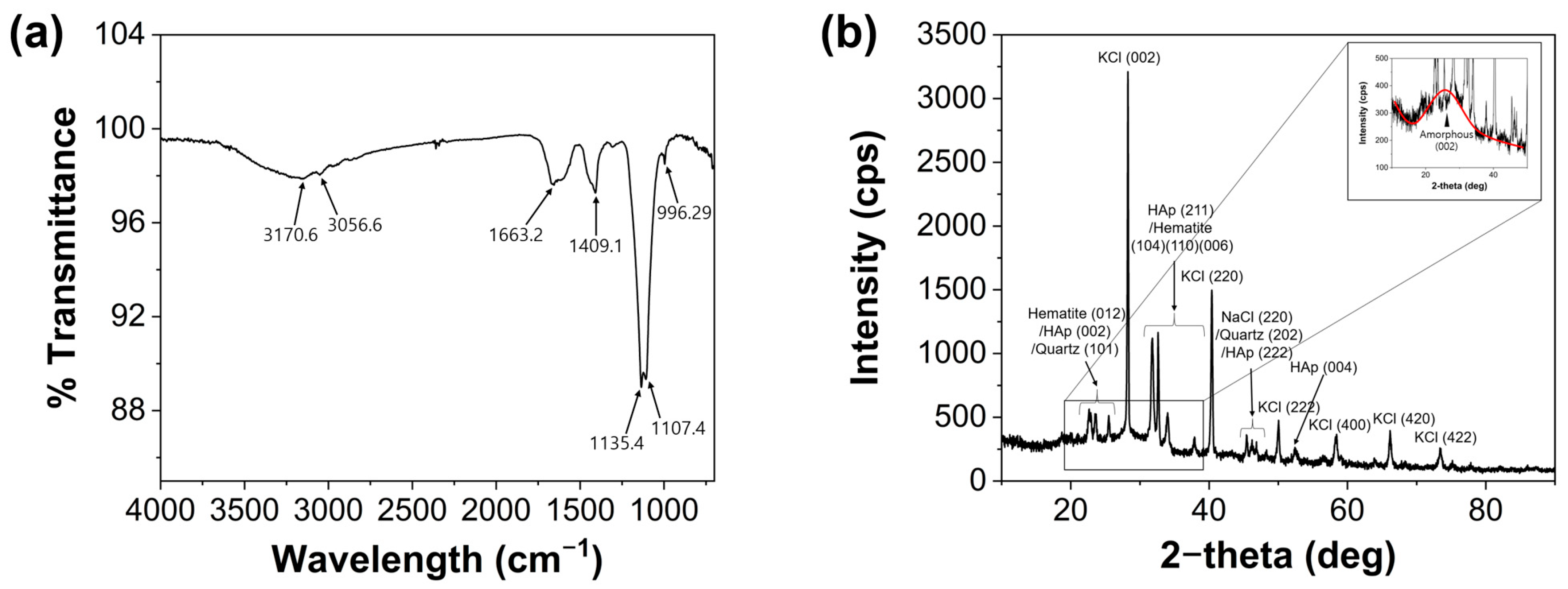
FT-IR spectra (Figure 2a) were obtained to determine the chemical composition of the MCDs. The spectrum of the MCDs displayed an O-H/N-H stretching vibration peak at 3170.6 cm−1, with a minor C-H stretching vibration at 3056.6 cm−1. The absorption band at 1663.2 cm−1 is mainly associated with C=O stretching. However, its irregular, double-shouldered profile may reflect a faint overlap with C=C vibrations, but the effect appears negligible compared to the dominant C=O stretching, which is consistent with the absence of clear C=C-related features in the XRD and XPS analyses. Similarly, the bands observed around 1135.4 and 1107.4 cm−1 exhibit a broad and irregular profile that is characteristic of complex carbonaceous matrices, where multiple vibrational modes overlap. These signals are therefore more appropriately regarded as composite bands rather than distinct assignments to individual functional groups [38,39]. The peaks at 1409.1 and 996.29 cm−1 were attributed to C-C bending and C-H bending, respectively. XRD was performed to further validate the FT-IR spectra. The XRD pattern of the as-synthesized MCDs is shown in Figure 2b. A broad hump centered at 2θ ≈ 26.0° corresponds to the (002) reflection of amorphous carbon. Based on Bragg’s law using Cu Kα radiation (λ = 1.5406 Å), this feature yields a d-spacing of approximately 0.342 nm. This value is consistent with those commonly reported for biomass-derived carbon dots and suggests the presence of disordered short-range graphitic stacking embedded within amorphous domains [40]. In addition to this broad feature, several sharp diffraction peaks are observed, indicating the presence of residual inorganic components. Among these, KCl is the most prominent, with characteristic reflections at 28.26°, 40.38°, 50.16°, 58.41°, 66.18°, and 73.36°, which correspond to the (200), (220), (222), (400), (420), and (422) planes, respectively. A distinct peak at 45.46° is also present and can be attributed to the (220) plane of NaCl, indicating the presence of sodium chloride as an additional residual salt. Minor impurity-related peaks are observed as well and can be assigned to α-Fe2O3 (hematite), which shows reflections at 23.66° for the (012) plane, 32.62° for the (104) plane, 34.81° for the (110) plane, and 37.94° for the (006) plane. Additional peaks are associated with hydroxyapatite, appearing at 24.52° for the (002) plane, 31.82° for the (211) plane, 46.84° for the (222) plane, and 52.42° for the (004) plane. Quartz is also detected, with reflections at 25.51° for the (101) plane and 46.21° for the (202) plane. These findings collectively indicate that the crystalline features in the XRD pattern are primarily due to residual inorganic salts and minerals such as KCl, NaCl, iron oxides, and silica. This suggests that the washing or dialysis procedures applied during synthesis were not sufficiently rigorous, allowing these inorganic impurities to remain and significantly influence the overall diffraction profile [41,42,43,44,45].
XPS was conducted to interpret the morphological properties of the MCDs and to compare the results with those of the FT-IR assignments (Figure 3). XPS analysis revealed the presence of carbon (C 1s; 285.02 eV), nitrogen (N 1s; 400.27 eV), and oxygen (O 1s; 532.24 eV) in the MCDs. The atomic composition was C = 63.88%, N = 7.26%, and O = 26.90%. As shown in Figure 3a, the structure of the MCDs from U. australis consisted mainly of carbon and oxygen. The percentages of other elements such as Na, Mg, S, and Ca were insignificant and were not considered. High-resolution narrow XPS spectra were plotted with the prediction data from Gaussian deconvolution using the OriginPro 2024 program (Figure 3b–d). Notably, the C1s high-resolution spectrum exhibited peaks corresponding to sp2 C–C (284.3 eV; 47.94 at.%), C-O/C-N (285.77 eV; 36.18 at.%), and carbonyl C=O (287.68 eV; 15.88 at.%). The O 1s spectrum displayed peaks attributed to C=O (531.23 eV; 62.36 at.%) and C-O (532.49 eV; 37.64 at.%). Additionally, the deconvoluted N1s spectrum revealed peaks associated with pyridinic N (399.33 eV; 91.72 at.%), and pyrrolic N (401.23 eV; 8.28 at.%). The XPS results mainly measured the compositional bonds related to carbon and oxygen, which could be the main factor in the long-term colloidal stability of the as-fabricated MCDs from U. australis.

3.2. Optical Properties of MCDs

A solution of MCDs was prepared by hydrothermally reacting fine U. australis powder with ultrapure water. The resulting solution appeared clear and yellowish-brown to the naked eye in daylight. Upon exposure to UV light (365 nm) and commercial LED light (430–440 nm), the MCD solution exhibited coral blue and yellow-green fluorescence, respectively (Figure 4a). The optical properties of the MCDs were characterized using UV–vis absorption and excitation-dependent fluorescence spectra, as shown in Figure 4b,c. The UV–vis absorption spectrum displayed two distinct peaks at 273 nm and 321 nm, corresponding to π-π* transitions of the sp2 carbon bond, along with a shoulder peak between 300 to 330 nm attributed to n-π* transitions of the C=O bond [46]. In addition, the MCDs exhibited excitation-dependent PL, with the emission maximum shifting from blue to red with increase in the excitation wavelength. The excitation wavelength-dependent PL behavior of the MCDs is similar to that of typical CDs. This behavior indicates that the PL of the MCDs is influenced by their surface state [23,24]. The unique excitation-dependent PL mechanism, involving band-to-band transitions of electrons from the conduction to valence band, is induced by defects resulting from the surface passivation of the carbonized MCDs. Specifically, the emission maximum of the MCDs occurred at approximately 475 nm at an excitation wavelength of 400 nm. The PLQY is a critical parameter for assessing the emission behavior of luminescent nanomaterials. It was determined using the relative method, in which the fluorescence intensities and absorbance values of the synthesized MCDs were compared with those of quinine sulfate in 0.1 M H2SO4 as a reference standard [47]. For both the quinine sulfate and the MCD suspensions at five different concentrations, absorbance at 340 nm and fluorescence emission spectra under 340nm excitation were measured. The resulting data were plotted and fitted by linear regression (Figure S1), from which the PLQY of the MCDs was calculated to be 8.31%.

3.3. Anti-Inflammatory Activity of MCDs in RAW264.7 Macrophages

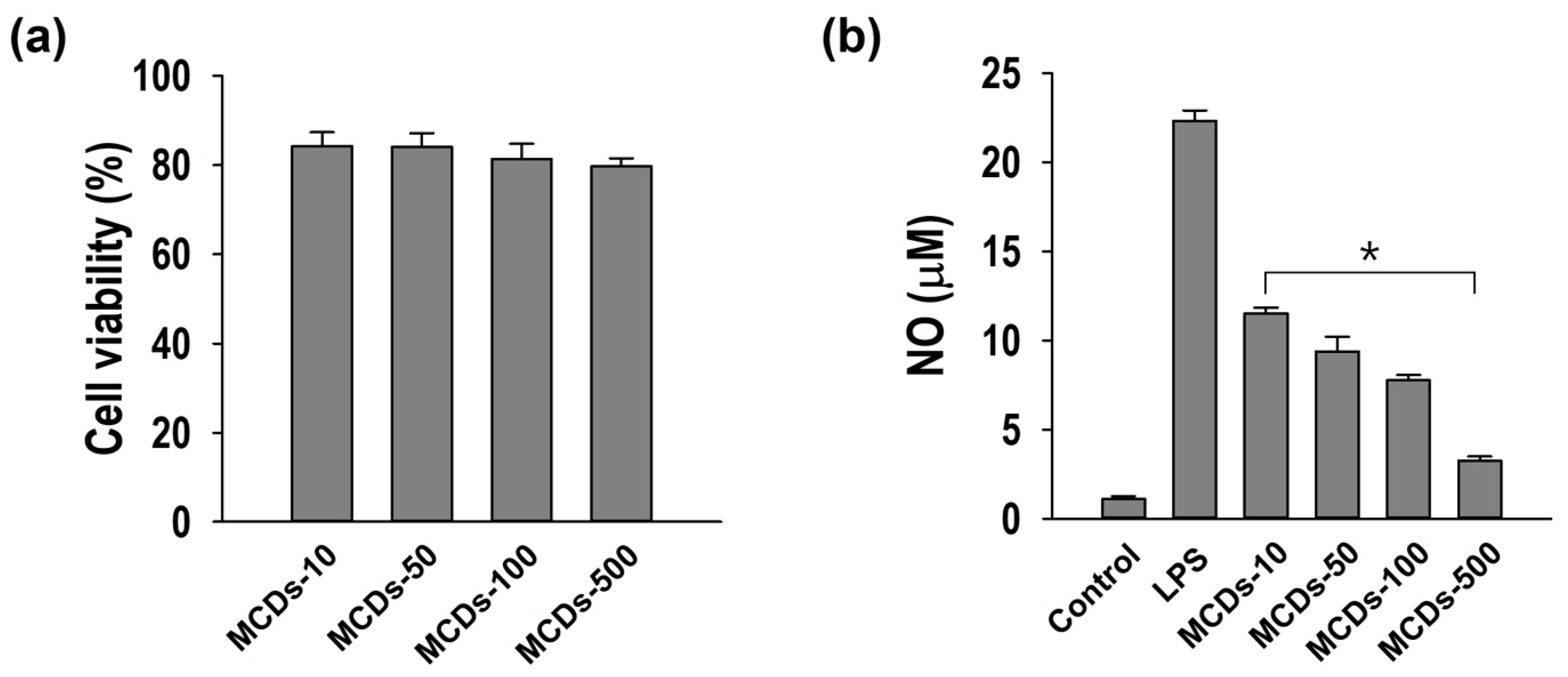
To evaluate the cytocompatibility of the MCDs, RAW264.7 macrophages were treated with varying concentrations (10, 50, 100, and 500 µg/mL) of the MCDs. Cell viability remained consistently above 80% across all tested concentrations, indicating the low cytotoxicity and high biocompatibility of the MCDs, even at higher doses (Figure 5a,b). No dose-dependent decrease in cell viability was observed. To assess the anti-inflammatory potential of the MCDs, LPS-stimulated RAW264.7 macrophages were treated with the same concentrations of the MCDs, and nitrite accumulation was measured as an indicator of NO production. The results demonstrated a clear dose-dependent suppression of the nitrite levels upon MCD treatment, suggesting effective inhibition of proinflammatory NO production. The cytotoxicity assay revealed that the MCDs were well-tolerated by the RAW264.7 macrophages at concentrations of up to 500 µg/mL, maintaining over 80% cell viability. This indicates a favorable safety profile suitable for further biomedical exploration. Importantly, the absence of a dose-dependent cytotoxic response implies a wide therapeutic window for these nanomaterials. In the context of inflammation, excessive nitric oxide production by activated macrophages is a hallmark of inflammatory response. Our findings show that the MCDs significantly attenuate nitrite production in a concentration-dependent manner, reflecting their anti-inflammatory activity. Lipopolysaccharide (LPS)-mediated stimulation of macrophages initiates key inflammatory signaling cascades, notably the NF-κB and MAPK pathways, which in turn drive the transcriptional activation of inducible nitric oxide synthase (iNOS). This enzyme facilitates the conversion of L-arginine into nitric oxide (NO), a reactive molecule that is swiftly oxidized to nitrite and nitrate in aqueous environments. As such, elevated nitrite concentrations in the culture medium are widely recognized as a surrogate marker for inflammatory NO output. In our investigation, we observed that macrophage carbon dots (MCDs) attenuated nitrite accumulation in a dose-dependent manner, implying that these nanomaterials may disrupt either the expression or enzymatic function of iNOS, thereby curbing excessive NO production during inflammatory activation [48]. These results are in agreement with prior reports of CDs modulating immune responses, likely via interaction with inflammatory signaling pathways such as NF-κB or MAPK [49]. Overall, these findings suggest that the MCDs not only possess low cytotoxicity but also exert measurable anti-inflammatory effects, making them strong candidates for development as nanotherapeutics for inflammation-related disorders. Their dual characteristics of safety and functional efficacy support their potential applications in anti-inflammatory skin care, wound healing, and immunomodulatory formulations. The results for the two control CDs derived from red and green algae (referred to as R-MCDs and G-MCDs) are presented in Figure S2 of the Supplementary Information. Although both carbon dots exhibited anti-inflammatory activity, their effects were weaker compared to those of the MCDs. Comparative evaluations revealed that MCDs consistently outperformed R-MCDs and G-MCDs in terms of anti-inflammatory efficacy across all tested concentrations. At the highest concentration of 500 µg/mL, MCDs inhibited nitric oxide (NO) production by approximately 84%, whereas R-MCDs and G-MCDs showed only 57–59% inhibition—a notable disparity of around 25%. Even at the intermediate dose of 100 µg/mL, MCDs achieved roughly 66% suppression of NO, compared to about 45% by the other carbon dot variants, underscoring a difference of nearly 20%. These findings suggest that MCDs possess enhanced anti-inflammatory properties, likely attributable to the unique physicochemical characteristics imparted by U. australis-based CDs, distinguishing them from other marine algae-derived CDs.

3.4. Anti-Adipogenic Effects of MCDs in 3T3-L1 Adipocytes

The anti-obesity potential of MCDs was explored by exposing 3T3-L1 preadipocytes to concentrations of 10, 50, 100, and 500 µg/mL throughout the adipogenic induction phase. Cell viability remained consistently above 80% across all treatment groups, indicating the absence of any significant cytotoxic effect induced by the MCDs at the tested concentrations (Figure 6a). Oil Red O staining combined with phase-contrast microscopy revealed dose-dependent inhibition of intracellular lipid accumulation in the MCD-treated cells, which was not observed in the untreated controls (Figure 6b). This reduction in adipogenic activity was particularly evident at 100 and 500 µg/mL, where the cells displayed markedly fewer lipid droplets. Quantitative image analysis further confirmed this trend, demonstrating a significant decrease in lipid content per cell in proportion to the MCD concentration (Figure 6c). These outcomes indicate a clear anti-adipogenic effect of the MCDs, which may be attributed to interference with the regulatory pathways involved in adipocyte maturation. It is plausible that the MCDs affect the expression or activity of key transcription factors such as peroxisome proliferator-activated receptor gamma (PPARγ) and CCAAT/enhancer-binding protein alpha (C/EBPα), which are essential for adipogenesis [50]. Although further mechanistic validation is required, the observed suppression of lipid accumulation indicates the potential regulatory role of the MCDs in adipocyte differentiation. In conjunction with their antioxidant and anti-inflammatory properties, the ability of the MCDs to inhibit fat-cell formation underscores their multifunctionality. Synthesized from U. australis, a marine macroalga considered an environmental nuisance, these carbon dots present a sustainable and cost-effective platform for the development of anti-obesity nanotherapeutics with low toxicity and high biological efficacy. In addition, the adipogenic activities of R-MCDs and G-MCDs were examined under identical experimental conditions. Detectable activity was observed only at the higher concentrations of 100 and 500 µg/mL. Quantitative comparison revealed that MCDs exhibited markedly stronger anti-adipogenic effects across all concentrations. At 500 µg/mL, MCDs inhibited lipid accumulation by ~60%, whereas R-MCDs and G-MCDs achieved only 40–45% inhibition, a difference of about 15–20%. Even at 100 µg/mL, MCDs suppressed lipid accumulation by ~50%, while R-MCDs and G-MCDs showed only 25–30% inhibition. These results highlight the superior bioactivity of U. australis-derived MCDs compared to carbon dots from other algal sources.

3.5. Inhibition of LPS-Induced ROS by MCDs

The antioxidant potential of MCDs synthesized from U. australis was evaluated by monitoring the intracellular ROS levels in RAW264.7 macrophage cells (Figure 7a). Following lipopolysaccharide (LPS) stimulation, a pronounced increase in the DCFDA fluorescence intensity was observed, signifying elevated ROS generation and confirming successful induction of oxidative stress in the macrophages. However, when the cells were pretreated with varying concentrations of MCDs (125, 250, and 500 μg/mL), prior to LPS exposure, a noticeable and progressive reduction in fluorescence intensity was recorded (Figure 7b). This attenuation of fluorescence clearly indicated that MCDs play a role in reducing the intracellular ROS levels. The fluorescence intensity decreased in a concentration-dependent manner, with higher doses of MCDs resulting in greater ROS suppression. Notably, at 500 μg/mL, the fluorescence intensity approached the levels observed in the untreated cells, suggesting a robust antioxidant effect at this dose. Quantitative analysis of the mean fluorescence intensity further supported these findings. The LPS-only group exhibited the highest ROS-related fluorescence, whereas the MCD-treated group showed a marked reduction. Although the suppression was not uniform across the concentrations, a consistent downward trend in the ROS levels was evident, reflecting a dose-dependent protective effect. The fact that even the lowest concentration (125 μg/mL) resulted in a measurable reduction suggests that the MCDs possess significant antioxidant activity, even at relatively low doses. These results reinforce the notion that CNDs derived from marine algae can effectively mitigate oxidative stress in immune cells. The antioxidant properties observed in the MCDs may be attributed to a combination of molecular and structural features inherent to carbon dots. Specifically, their radical-scavenging ability is considered to originate from mechanisms involving hydrogen atoms or electron transfer, the formation of radical adducts on conjugated sp2-carbon domains, and presence of oxygen-rich or nitrogen-doped surface sites. These features allow the carbon dots to effectively neutralize various ROS through resonance stabilization and surface-mediated interactions. Overall, the findings highlight the clear dose-dependent ROS-scavenging effect of U. australis-based MCDs and their potential as natural biocompatible antioxidant agents [17]. These nanomaterials hold promise for further development in biomedical applications targeting oxidative stress-related diseases. Future studies investigating their interactions with cellular redox pathways and long-term biological effects could further validate their therapeutic potential.

3.6. Toxicity and Activity of MCDs in Zebrafish

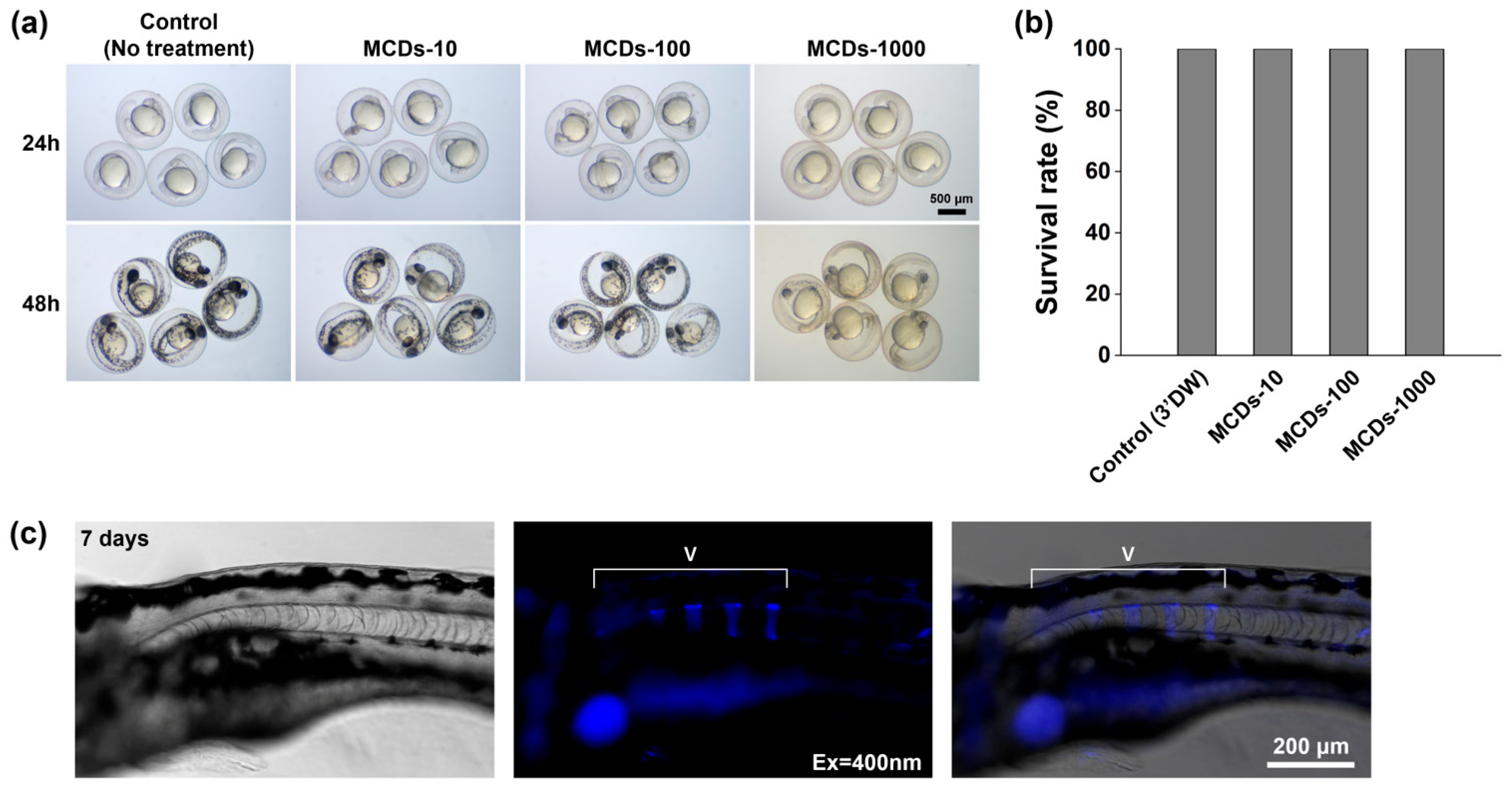
To evaluate the biocompatibility of MCDs, zebrafish embryos were exposed to three different concentrations of MCDs (10 µg/mL, 100 µg/mL, and 1000 µg/mL) at 5 hpf. Throughout the embryonic developmental period, all treatment groups showed 100% survival rates, indicating no apparent acute toxicity across all the tested concentrations (Figure 8a). An assessment of the developmental effects following exposure to the MCDs revealed no significant morphological abnormalities at 24 or 48 hpf or up to 7 days post-fertilization (dpf), indicating that the synthesized MCD exhibited high biocompatibility in vivo (Figure 8b). Interestingly, although the overall embryonic morphology remained unaffected, a distinct reduction in melanin pigmentation was clearly observed in the MCDs-1000 group at 48 hpf. Embryos exposed to this relatively higher concentration exhibited visibly reduced pigmentation in the dorsal and ocular regions when compared with that of the controls and other dilution groups. This suggests the potential inhibitory effect of MCDs on melanin synthesis and melanosome development during embryogenesis in zebrafish. At 7 dpf, in vivo fluorescence imaging revealed distinct MCD-derived signals localized along the vertebral column in the zebrafish larvae (Figure 8c). The strongest emission was observed in the axial skeletal region, suggesting a possible association between the MCDs and developing bone tissue. No off-target accumulation or abnormal distribution of fluorescence was detected, further supporting the biocompatibility of the MCDs. These findings demonstrate the high in vivo biocompatibility and promising imaging potential of the MCDs, as evaluated using zebrafish embryos and larvae as in vivo models. The absence of lethality or teratogenicity across all tested concentrations highlights the nontoxic nature of the MCDs during the early developmental stages. Notably, the suppression of melanin pigmentation by MCDs-1000 (1000 µg/mL) raises the possibility that the MCDs may interfere with melanogenesis-related pathways, potentially because of their interaction with tyrosinase activity or melanosome transport proteins. Although the exact mechanism remains to be elucidated, this observation is consistent with previous reports indicating that nanomaterials affect the development and migration of pigment cells [51]. Future studies should examine the expression profiles of pigmentation-related genes (e.g., mitfa, tyr, dct) following exposure to MCDs to identify the underlying molecular mechanisms. The in vivo fluorescence signals observed along the vertebrae at 7 dpf suggest that the MCDs preferentially accumulate or bind to bone-forming tissues. Given that vertebral development involves active calcium deposition and mineralization, the observed fluorescence may indicate the affinity of the MCDs for calcium-rich matrices. This raises the possibility that MCDs can be engineered as fluorescent probes for skeletal imaging or as calcium ion indicators in live vertebrates. Further studies using calcium-binding assays, mineral staining (e.g., alizarin red and calcein), and colocalization with osteogenic-specific markers are essential to validate this hypothesis. Overall, our findings demonstrate the low toxicity and high potential of MCDs as bioimaging agents in vertebrate models. Moreover, the unexpected suppression of pigmentation and skeletal localization of MCDs open new avenues for their application in developmental biology, dermatology, and bone imaging diagnostics.

4. Conclusions

In this study, we successfully synthesized multifunctional MCDs from U. australis, a marine algal species considered an environmental nuisance on Jeju Island. The synthesis process is simple and sustainable, requiring only washing, drying, and powdering of the biomass without specialized conditions or equipment. The resulting MCDs, produced in an aqueous solution, exhibited excitation-dependent fluorescence and demonstrated anti-inflammatory, antioxidant, and anti-adipogenic properties in multiple in vitro assays. The biocompatibility and in vivo tracking of the MCDs were evaluated in zebrafish embryos. No developmental toxicity or mortality was observed at any of the tested concentrations, confirming their safety during early embryogenesis. Notably, exposure to a higher concentration (1000 µg/mL) resulted in suppressed melanin pigmentation, indicating that the MCDs may influence melanogenesis. In addition, strong fluorescence signals were observed along the vertebral column at 7 dpf, suggesting the potential of the MCDs to bind to calcium-rich skeletal tissues, thus enabling their application to bone-targeted bioimaging. These results highlight the dual role of the MCDs as functional nanomaterials with biomedical relevance and as a representative example of value-added utilization of problematic marine biomass. This study contributes to the growing interest in carbon dots derived from underutilized natural resources, thereby promoting a circular bioeconomic approach. Specifically, U. australis, which is regarded as waste in Jeju Island, Republic of Korea, demonstrates considerable promise as a low-cost, ecofriendly precursor for nanomaterial development. Given the demonstrated biological activities and favorable safety profile, the MCDs synthesized in this study hold promise for application to functional cosmetics, nutraceuticals, and biomedical imaging. This research establishes the foundation for further exploration of marine-biomass-based nanomaterials, emphasizing sustainability and environmental responsibility in the development of functional materials. Nonetheless, the present outcome has certain limitations. The biological performance of the MCDs was evaluated primarily through in vitro and early-stage in vivo models. Therefore, further studies are necessary to validate their long-term safety, efficacy, and functional stability in preclinical and clinical settings. In addition, comprehensive assessments related to formulation, delivery, and regulatory compliance should be undertaken to support their potential industrial applications.

5. Patents

A Korean domestic application has been filed for the content described in this paper (patent number: 10-2024-0070109).

Supplementary Materials

The following supporting information can be downloaded at: https://www.mdpi.com/article/10.3390/jmse13101878/s1, Figure S1: Linear regression plot used for PLQY calculation of the MCDs.; Figure S2: Anti-inflammatory effects of R-MCDs and G-MCDs.; Figure S3: Anti-adipogenic effects of R-MCDs and G-MCDs in 3T3-L1 preadipocytes.

Author Contributions

Conceptualization, K.W.K.; methodology, K.W.K.; software, K.W.K.; validation, K.W.K., G.-W.O. and S.-H.J.; formal analysis, K.W.K., G.-W.O. and S.-H.J.; investigation, K.W.K., G.-W.O. and S.-H.J.; resources, K.W.K., G.-W.O., S.-H.J., S.-C.K., J.-Y.K., D.Y., D.-M.J. and G.C.; data curation, K.W.K. and G.C.; writing—original draft preparation, K.W.K.; writing—review and editing, K.W.K., G.-W.O., S.-H.J., S.-C.K., J.-Y.K., D.Y., D.-M.J., D.-S.L. and G.C.; visualization, K.W.K.; supervision, K.W.K. and G.C.; project administration, K.W.K., D.-S.L. and G.C.; funding acquisition, D.-S.L. and G.C. All authors have read and agreed to the published version of the manuscript.

Funding

This work was funded by an in-house project of the National Marine Biodiversity Institute of Korea (MABIK), grant number 2025M00500.

Institutional Review Board Statement

All zebrafish experiments were conducted in accordance with the institutional guidelines and approved by the Institutional Animal Care and Use Committee (IACUC) of the National Marine Biodiversity Institute of Korea (MABIK) (Approval No. MAB-23-03).

Data Availability Statement

The datasets presented in this article are not readily available because they are part of an ongoing domestic patent application process.

Acknowledgments

We would like to thank J.H. Won for assisting in the collection of U. australis from Jeju Island, and J.Y. Je for helping with the acquisition of statistical analysis data.

Conflicts of Interest

The authors declare no conflicts of interest. The funders had no role in the design of the study; in the collection, analyses, or interpretation of data; in the writing of the manuscript; or in the decision to publish the results.

Abbreviations

The following abbreviations are used in this manuscript:
CDsCarbon dots
DCFDA2′,7′-dichlorofluorescin diacetate
DMSODimethyl sulfoxide
FT-IRFourier-transform infrared
XPSX-ray photoelectron spectroscopy
XRDX-ray diffraction
HApHydroxyapatite
IBMXDexamethasone, 3-Isobutyl-1-methylxanthine
MCDsMarine carbon dots
LPSLipopolysaccharide
iNOSInducible nitric oxide synthase
PLPhotoluminescence
TEMTransmission electron microscopy
ROSReactive oxygen species

References

  1. Naomi, R.; Teoh, S.H.; Embong, H.; Balan, S.S.; Othman, F.; Bahari, H.; Yazid, M.D. The role of oxidative stress and inflammation in obesity and its impact on cognitive impairments—A narrative review. Antioxidants 2023, 12, 1071. [Google Scholar] [CrossRef] [PubMed]
  2. Fernández-Sánchez, A.; Madrigal-Santillán, E.; Bautista, M.; Esquivel-Soto, J.; Morales-González, A.; Esquivel-Chirino, C.; Durante-Montiel, I.; Sánchez-Rivera, G.; Valadez-Vega, C.; Morales-González, J.A. Inflammation, oxidative stress, and obesity. Int. J. Mol. Sci. 2011, 12, 3117–3132. [Google Scholar] [CrossRef]
  3. Furukawa, S.; Fujita, T.; Shimabukuro, M.; Iwaki, M.; Yamada, Y.; Nakajima, Y.; Nakayama, O.; Makishima, M.; Matsuda, M.; Shimomura, I. Increased oxidative stress in obesity and its impact on metabolic syndrome. J. Clin. Investig. 2004, 114, 1752–1761. [Google Scholar] [CrossRef]
  4. Chen, L.; Deng, H.; Cui, H.; Fang, J.; Zuo, Z.; Deng, J.; Li, Y.; Wang, X.; Zhao, L. Inflammatory responses and inflammation-associated diseases in organs. Oncotarget 2018, 9, 7204–7218. [Google Scholar] [CrossRef]
  5. Meizlish, M.L.; Franklin, R.A.; Zhou, X.; Medzhitov, R. Tissue homeostasis and inflammation. Annu. Rev. Immunol. 2021, 39, 557–581. [Google Scholar] [CrossRef] [PubMed]
  6. Bender, E.C.; Tareq, H.S.; Suggs, L.J. Inflammation: A matter of immune cell life and death. npj Biomed. Innov. 2025, 2, 7. [Google Scholar] [CrossRef]
  7. Furman, D.; Campisi, J.; Verdin, E.; Carrera-Bastos, P.; Targ, S.; Franceschi, C.; Ferrucci, L.; Gilroy, D.W.; Fasano, A.; Miller, G.W.; et al. Chronic inflammation in the etiology of disease across the life span. Nat. Med. 2019, 25, 1822–1832. [Google Scholar] [CrossRef]
  8. Ahmed, A.U. An overview of inflammation: Mechanism and consequences. Front. Biol. 2011, 6, 274–281. [Google Scholar] [CrossRef]
  9. Varela, M.L.; Mogildea, M.; Moreno, I.; Lopes, A. Acute inflammation and metabolism. Inflammation 2018, 41, 1115–1127. [Google Scholar] [CrossRef]
  10. Müller, T.D.; Blüher, M.; Tschöp, M.H.; DiMarchi, R.D. Anti-obesity drug discovery: Advances and challenges. Nat. Rev. Drug Discov. 2022, 21, 201–223. [Google Scholar] [CrossRef] [PubMed]
  11. Seong, J.; Kang, J.Y.; Sun, J.S.; Kim, K.W. Hypothalamic Inflammation and Obesity: A Mechanistic Review. Arch. Pharm. Res. 2019, 42, 379–392. [Google Scholar] [CrossRef]
  12. Khalil, I.; Yehye, W.A.; Etxabide Etxeberria, A.; Alhadi, A.A.; Masoomi Dezfooli, S.; Muhd Julkapli, N.B.; Basirun, W.J.; Seyfoddin, A. Nanoantioxidants: Recent Trends in Antioxidant Delivery Applications. Antioxidants 2020, 9, 24. [Google Scholar] [CrossRef]
  13. Samrot, A.V.; Singh, S.P.R.; Deenadhayalan, R.; Rajesh, V.V.; Padmanaban, S.; Radhakrishnan, K. Nanoparticles, a Double-Edged Sword with Oxidant as Well as Antioxidant Properties—A Review. Oxygen 2022, 2, 591–604. [Google Scholar] [CrossRef]
  14. Kumar, H.; Bhardwaj, K.; Nepovimova, E.; Kuča, K.; Dhanjal, D.S.; Bhardwaj, S.; Bhatia, S.K.; Verma, R.; Kumar, D. Antioxidant Functionalized Nanoparticles: A Combat against Oxidative Stress. Nanomaterials 2020, 10, 1334. [Google Scholar] [CrossRef]
  15. Shah, S.T.; Chowdhury, Z.Z.; Simarani, K.; Basirun, W.J.; Badruddin, I.A.; Hussien, M.; Alrobei, H.; Kamangar, S. Nanoantioxidants: The Fourth Generation of Antioxidants—Recent Research Roadmap and Future Perspectives. Coatings 2022, 12, 1568. [Google Scholar] [CrossRef]
  16. Gedda, G.; Sankaranarayanan, S.A.; Putta, C.L.; Gudimella, K.K.; Rengan, A.K.; Girma, W.M. Green Synthesis of Multi-Functional Carbon Dots from Medicinal Plant Leaves for Antimicrobial, Antioxidant, and Bioimaging Applications. Sci. Rep. 2023, 13, 6371. [Google Scholar] [CrossRef] [PubMed]
  17. Innocenzi, P.; Stagi, L. Carbon dots as oxidant–antioxidant nanomaterials, understanding the structure–properties relationship. A critical review. Nano Today 2023, 50, 101837. [Google Scholar] [CrossRef]
  18. Ji, Z.; Sheardy, A.; Zeng, Z.; Zhang, W.; Chevva, H.; Allado, K.; Yin, Z.; Wei, J. Tuning the functional groups on carbon nanodots and antioxidant studies. Molecules 2019, 24, 152. [Google Scholar] [CrossRef] [PubMed]
  19. Behi, M.; Gholami, L.; Naficy, S.; Palomba, S.; Dehghani, F. Carbon dots: A novel platform for biomedical applications. Nanoscale Adv. 2022, 4, 353–376. [Google Scholar] [CrossRef] [PubMed]
  20. Kang, Z.; Lee, S.-T. Carbon dots: Advances in nanocarbons applications. Nanoscale 2019, 11, 19214–19224. [Google Scholar] [CrossRef]
  21. Cui, L.; Ren, X.; Sun, M.; Liu, H.; Xia, L. Carbon dots: Synthesis, properties and applications. Nanomaterials 2021, 11, 3419. [Google Scholar] [CrossRef]
  22. Rajesh, G.A.; John, V.L.; Santhosh, A.P.; Ambika, A.K.N.; Puthiyedath, V.T. Carbon Dots from Natural Sources for Biomedical Applications. Part. Part. Syst. Charact. 2022, 39, 2200017. [Google Scholar] [CrossRef]
  23. Kim, K.W.; Choi, T.-Y.; Kwon, Y.M.; Kim, J.Y.H. Simple Synthesis of Photoluminescent Carbon Dots from a Marine Polysaccharide Found in Shark Cartilage. Electron. J. Biotechnol. 2020, 47, 36–42. [Google Scholar] [CrossRef]
  24. Kim, K.W.; Kwon, Y.M.; Kim, S.Y.; Kim, J.Y.H. One-Pot Synthesis of UV-Protective Carbon Nanodots from Sea Cauliflower (Leathesia difformis). Electron. J. Biotechnol. 2022, 56, 22–30. [Google Scholar] [CrossRef]
  25. Lin, X.; Xiong, M.; Zhang, J.; He, C.; Ma, X.; Zhang, H.; Kuang, Y.; Yang, M.; Huang, Q. Carbon Dots Based on Natural Resources: Synthesis and Applications in Sensors. Microchem. J. 2020, 159, 105604. [Google Scholar] [CrossRef]
  26. Xiang, S.; Tan, M. Carbon Dots Derived from Natural Sources and Their Biological and Environmental Impacts. Environ. Sci. Nano 2022, 9, 3206–3225. [Google Scholar] [CrossRef]
  27. Lee, Y.-H.; Kim, H.-R.; Yeo, M.-H.; Kim, S.C.; Hyun, H.-B.; Ham, Y.-M.; Jung, Y.-H.; Kim, H.-S.; Chang, K.-S. Anti-obesity potential of Sargassum horneri and Ulva australis extracts: Study in vitro and in vivo. Appl. Sci. 2023, 13, 8951. [Google Scholar] [CrossRef]
  28. Li, G.-L.; Wang, R.-R.; Hou, Y.-X.; Guo, W.-J.; Wang, G.-B.; Liu, K.; Liu, Y.; Wu, W. Sterols from the green alga Ulva australis and their human recombinant aldose reductase iinhibitory activities. Mar. Drugs 2017, 15, 299. [Google Scholar] [CrossRef] [PubMed]
  29. Trentin, R.; Custódio, L.; Rodrigues, M.J.; Moschin, E.; Sciuto, K.; da Silva, J.P.; Moro, I. Biotechnological potential of the invasive alga Ulva australis collected in the Venice Lagoon (Italy). Algal Res. 2023, 69, 102969. [Google Scholar] [CrossRef]
  30. Lee, H.; Kim, G.; Depuydt, S.; Shin, K.; Han, T.; Park, J. Metal toxicity across different thallus sections of the green macroalga, Ulva australis. Toxics 2023, 11, 548. [Google Scholar] [CrossRef]
  31. Wu, X. Guidelines for anti-inflammatory assays in RAW264.7 cells. Food Saf. Health 2025, 3, 128–136. [Google Scholar] [CrossRef]
  32. Kraus, N.A.; Ehebauer, F.; Zapp, B.; Rudolphi, B.; Kraus, B.J.; Kraus, D. Quantitative assessment of adipocyte differentiation in cell culture. Adipocyte 2016, 5, 351–358. [Google Scholar] [CrossRef]
  33. Truskewycz, A.; Yin, H.; Halberg, N.; Lai, D.T.H.; Ball, A.S.; Truong, V.K.; Rybicka, A.M.; Cole, I. Carbon dot therapeutic platforms: Administration, distribution, metabolism, excretion, toxicity, and therapeutic potential. Small 2022, 18, e2106342. [Google Scholar] [CrossRef]
  34. Mansuriya, B.D.; Altintas, Z. Carbon dots: Classification, properties, synthesis, characterization, and applications in health care—An updated review (2018–2021). Nanomaterials 2021, 11, 2525. [Google Scholar] [CrossRef]
  35. Manzoor, S.; Dar, A.H.; Dash, K.K.; Singh, S.; Choudhury, M.D.; Nayak, B. Carbon dots: A novel photoluminescent nanomaterial for multifaceted applications. Appl. Food Res. 2023, 3, 100263. [Google Scholar] [CrossRef]
  36. Yuan, F.; Li, X.; Li, Y.; Fan, L.; Yang, S. Engineering Carbon Dots for Photonics and Optoelectronics. Nano Today 2016, 11, 565–586. [Google Scholar] [CrossRef]
  37. Sun, X.; Lei, Y. Fluorescent Carbon Dots and Their Sensing Applications. TrAC Trends Anal. Chem. 2017, 89, 163–180. [Google Scholar] [CrossRef]
  38. Tucureanu, V.; Matei, A.; Avram, A.M. FTIR Spectroscopy for Carbon Family Study. Crit. Rev. Anal. Chem. 2016, 46, 502–520. [Google Scholar] [CrossRef] [PubMed]
  39. Zavidovskiy, I.A.; Streletskiy, O.A.; Nuriahmetov, I.F.; Nishchak, O.Y.; Savchenko, N.F.; Tatarintsev, A.A.; Pavlikov, A.V. Highly Selective Polyene-Polyyne Resistive Gas Sensors: Response Tuning by Low-Energy Ion Irradiation. J. Compos. Sci. 2023, 7, 156. [Google Scholar] [CrossRef]
  40. Ozyurt, D.; Al Kobaisi, M.; Hocking, R.K.; Fox, B. Properties, synthesis, and applications of carbon dots: A review. Carbon Trends 2023, 12, 100276. [Google Scholar] [CrossRef]
  41. Chen, J.; Liu, J.; Deng, H.; Yao, S.; Wang, Y. Regulatory synthesis and characterization of hydroxyapatite nanocrystals by a microwave-assisted hydrothermal method. Ceram. Int. 2020, 46, 2185–2193. [Google Scholar] [CrossRef]
  42. Ismail, S.N.; Ali, E.; Alwan, B.J.; Abd, A.N. Potassium Chloride Nanoparticles: Synthesis, Characterization, and Study the Antimicrobial Applications. Macromol. Symp. 2022, 401, 2100312. [Google Scholar] [CrossRef]
  43. Damodar, D.; Kumar, S.K.; Martha, S.K.; Deshpande, A.S. Nitrogen-doped graphene-like carbon nanosheets from commercial glue: Morphology, phase evolution and Li-ion battery performance. Dalton Trans. 2018, 47, 12218–12227. [Google Scholar] [CrossRef]
  44. Qureshi, A.A.; Javed, S.; Javed, H.M.A.; Jamshaid, M.; Ali, U.; Akram, M.A. Systematic Investigation of Structural, Morphological, Thermal, Optoelectronic, and Magnetic Properties of High-Purity Hematite/Magnetite Nanoparticles for Optoelectronics. Nanomaterials 2022, 12, 1635. [Google Scholar] [CrossRef]
  45. Zhang, C.; Xu, Z.; Hu, Y.; He, J.; Tian, M.; Zhou, J.; Zhou, Q.; Chen, S.; Chen, D.; Chen, P.; et al. Novel insights into the hydroxylation behaviors of α-quartz (101) surface and its effects on the adsorption of sodium oleate. Minerals 2019, 9, 450. [Google Scholar] [CrossRef]
  46. Meng, W.; Bai, X.; Wang, B.; Liu, Z.; Lu, S.; Yang, B. Biomass-derived carbon dots and their applications. Energy Environ. Mater. 2019, 2, 172–192. [Google Scholar] [CrossRef]
  47. Quraishi, S.; Plappert, S.; Ungerer, B.; Taupe, P.; Gindl-Altmutter, W.; Liebner, F. Preparation and Characterization of Bacterial Cellulose-Carbon Dot Hybrid Nanopaper for Potential Sensing Applications. Appl. Sci. 2019, 9, 107. [Google Scholar] [CrossRef]
  48. Farahani, A.; Farahani, A.; Kashfi, K.; Ghasemi, A. Inducible nitric oxide synthase (iNOS): More than an inducible enzyme? Rethinking the classification of NOS isoforms. Pharmacol. Res. 2025, 216, 107781. [Google Scholar] [CrossRef]
  49. Kumar, M.; Chinnathambi, S.; Bakhori, N.; Abu, N.; Etezadi, F.; Thangavel, V.; Packwood, D.; Sivaniah, E.; Pandian, G.N. Biomass-derived carbon dots as fluorescent quantum probes to visualize and modulate inflammation. Sci. Rep. 2024, 14, 12665. [Google Scholar] [CrossRef]
  50. Habelreeh, H.H.; Athinarayanan, J.; Periasamy, V.S.; Alshatwi, A.A. Maillard reaction-derived S-doped carbon dots promotes downregulation of PPARγ, C/EBPα, and SREBP-1 genes in-vitro. Molecules 2024, 29, 2008. [Google Scholar] [CrossRef]
  51. Choi, T.Y.; Kim, J.H.; Ko, D.H.; Kim, C.H.; Hwang, J.S.; Ahn, S.; Kim, S.Y.; Kim, C.D.; Lee, J.H.; Yoon, T.J. Zebrafish as a new model for phenotype-based screening of melanogenic regulatory compounds. Pigment Cell Res. 2007, 20, 120–127. [Google Scholar] [CrossRef] [PubMed]
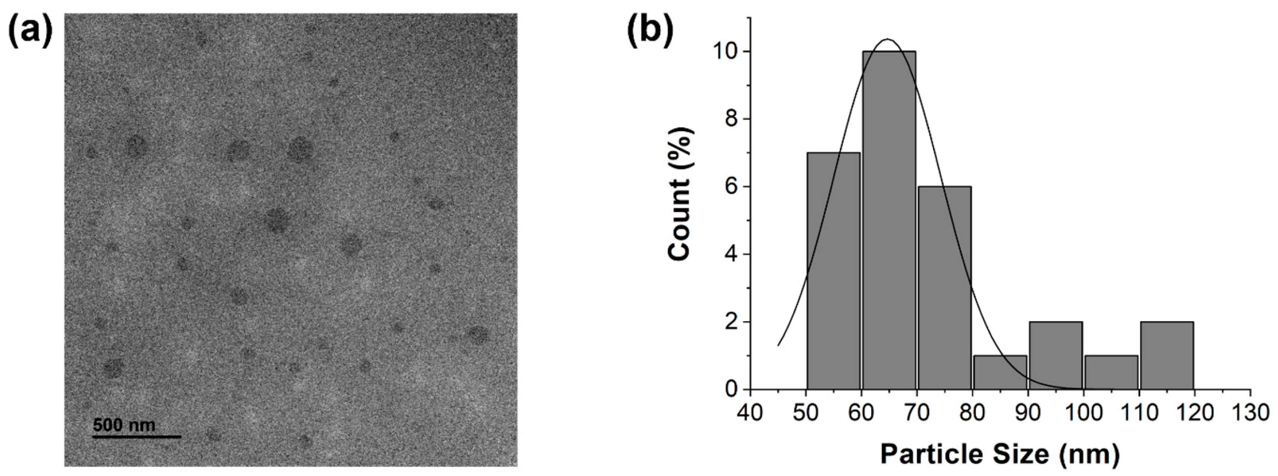
Figure 1. (a) TEM images of MCDs, revealing quasi-spherical morphology; (b) PSD histogram of MCDs derived from TEM image analysis, showing a size range of 50–80 nm.
Jmse 13 01878 g001
Figure 2. (a) FT-IR spectrum of MCDs showing characteristic peaks for O–H/N–H and C–H stretching, a dominant C=O band with minor possible C=C overlap, and composite bands characteristic of carbonaceous structures, as well as C-C and C-H bending; (b) XRD pattern of the as-synthesized carbon dots, showing a broad peak at 26.0°, indicative of amorphous carbon, along with sharp diffraction peaks of residual inorganic salts and minerals. The inset shows the amorphous hump, where the red line is included only as a visual guide to highlight the overall peak trend.
Jmse 13 01878 g002
Figure 3. XPS analysis of MCDs derived from U. australis. (a) Survey spectrum confirming the presence of C, N, and O as major elements; (bd) High-resolution spectra of O 1s, N 1s, and C 1s showing dominant surface functional groups such as C=O, C–O, pyridinic N, and sp2 carbon species. These results align with the FT-IR data, indicating oxygenated and nitrogenous surface chemistry important for MCD stability.
Jmse 13 01878 g003
Figure 4. Optical properties of MCDs synthesized from U. australis. (a) MCD solutions exhibit coral blue and yellowish-green fluorescence under UV (365 nm) and LED (430–440 nm) light, respectively; (b) UV–vis absorption shows peaks at 273 and 321 nm, attributed to π–π* and n–π* transitions; (c) Excitation-dependent PL reveals a redshift in emission, with a peak near 475 nm at 400 nm excitation.
Jmse 13 01878 g004
Figure 5. Anti-inflammatory effects of MCDs. (a) Cell viability of RAW264.7 macrophages treated with MCDs at various concentrations (10, 50, 100, and 500 μg/mL) for 24 h, assessed via MTT assay. Cell viability remained above 80% across all concentrations, indicating low cytotoxicity and high biocompatibility; (b) NO production in LPS-stimulated RAW264.7 macrophages treated with the same concentrations of MCDs. Nitrite levels, measured using the Griess assay, exhibited a concentration-dependent decrease, demonstrating the anti-inflammatory potential of the MCDs. All data are shown as mean ± SD (n = 3). Groups labeled with distinct letters differ significantly at p < 0.05. An asterisk (*) indicates statistically significant differences relative to the corresponding control group.
Jmse 13 01878 g005
Figure 6. Anti-adipogenic effects of MCDs in 3T3-L1 preadipocytes. (a) Cell viability following treatment with MCDs at various concentrations (10, 50, 100, and 500 μg/mL) throughout the adipogenic induction period. Viability remained above 80% in all groups, indicating minimal cytotoxicity; (b) Quantification of intracellular lipid accumulation using Oil Red O staining. A dose-dependent reduction in lipid content was observed, with statistically significant suppression at higher concentrations; (c) Representative phase-contrast microscopy images of Oil Red O-stained 3T3-L1 cells showing reduced lipid droplet formation in MCD-treated cells when compared with that in the untreated control group. The decrease in red-stained lipid droplets correlates with increasing MCD concentration. All data are shown as mean ± SD (n = 3). Groups labeled with distinct letters differ significantly at p < 0.05. An asterisk (*) indicates statistically significant differences relative to the corresponding control group.
Jmse 13 01878 g006
Figure 7. Antioxidant effects of U. australis-derived MCDs in LPS-stimulated RAW264.7 macrophages. (a) Flow cytometry analysis of intracellular ROS levels measured by DCFDA fluorescence. LPS treatment markedly increased the ROS production when compared with that in the untreated group, whereas pretreatment with MCDs (125, 250, and 500 μg/mL) led to a progressive reduction in the fluorescence intensity, indicating decreased ROS accumulation; (b) Quantitative analysis of mean fluorescence intensity. LPS-induced ROS generation was significantly attenuated by MCD treatment in a concentration-dependent manner, with the highest dose (500 μg/mL) approaching baseline levels.
Jmse 13 01878 g007
Figure 8. Developmental toxicity and bioimaging assessment of MCDs in zebrafish embryos. (a) Light microscopic images of zebrafish embryos treated with MCDs from 5 hpf to 7 dpf. No significant morphological changes are observed at 24 hpf, whereas melanocyte formation is suppressed in a dose-dependent manner at 48 hpf; (b) Survival rate of zebrafish embryos following exposure to MCDs for 7 days; (c) At 7 dpf, MCD-treated larvae exhibit fluorescence, with visible accumulation along the spinal vertebral column (V).
Jmse 13 01878 g008
Disclaimer/Publisher’s Note: The statements, opinions and data contained in all publications are solely those of the individual author(s) and contributor(s) and not of MDPI and/or the editor(s). MDPI and/or the editor(s) disclaim responsibility for any injury to people or property resulting from any ideas, methods, instructions or products referred to in the content.