Article

FOXO-like Gene Is Involved in the Regulation of 20E Pathway through mTOR in Eriocheir sinensis

1
College of Aquaculture and Life Science, Dalian Ocean University, Dalian 116026, China
2
Key Laboratory of Marine Bio-Resources Restoration and Habitat Reparation in Liaoning Province, Dalian Ocean University, Dalian 116023, China
*
Authors to whom correspondence should be addressed.
These authors contributed equally to this work.
J. Mar. Sci. Eng. 2023, 11(6), 1225; https://doi.org/10.3390/jmse11061225
Submission received: 26 April 2023 / Revised: 9 June 2023 / Accepted: 12 June 2023 / Published: 14 June 2023
(This article belongs to the Special Issue New Techniques in Marine Aquaculture)

Abstract

:
The Forkhead Box O (FOXO) gene plays a key role in various biological processes, such as growth, metabolism, development, immunity and longevity. Molting is an essential process for crustacean growth, which is mainly regulated by 20-hydroxyecdysone (20E) and molt-inhibiting hormone (MIH). Although the role of FOXO in regulating the immune response of crustaceans is well documented, its involvement in controlling crustacean molting remains unclear. In this study, a FOXO-like gene (designed as EsFOXO-like) was identified in Eriocheir sinensis, and the regulation of the 20E pathway by EsFOXO-like was also investigated. The coding sequence of EsFOXO-like was 852 bp, which consisted of 283 amino acids including a conserved Forkhead (FH) domain. EsFOXO-like shared high similarity with FOXO genes from other crustaceans, and the mRNA expression levels of the EsFOXO-like gene were highest in the hepatopancreas and lowest in the hemocytes. However, transcription and protein expression of the EsFOXO-like gene were found to be up-regulated only during the pre-molt stage in the hepatopancreas, with lower expression levels observed at the post-molt stage. To explore the role of EsFOXO-like in the 20E pathway, EsFOXO-like was firstly inhibited by a specific FOXO inhibitor (AS1842856) and then through an EsFOXO-like dsRNA injection, respectively, and the results showed that the relative expression levels of EsFOXO-like were notably decreased in the hepatopancreas after both the inhibitor and dsRNA treatments. The 20E concentration, the mRNA expression levels of the 20E receptors including the ecdysone receptor (EcR) and the retinoid-X receptor (RXR) and EsmTOR transcription in the AS1842856 group or the EsFOXO-RNAi group were all significantly higher than that in the control group, while the mRNA expression level of EsMIH was significantly decreased after EsFOXO-like inhibition. To further investigate whether the EsFOXO-like acts through mTOR or not, Rapamycin was administered to inhibit mTOR activity in EsFOXO-like inhibited crabs. The results revealed a significant reduction in the concentration of 20E and the expression level of EsMIH in the AS1842856 + Rapamycin group compared to the AS1842856 + DMSO group, accompanied by an increase in EsEcR and EsRXR expression. These findings collectively suggest that EsFOXO-like regulates the 20E pathway through mTOR, which offered valuable insights into the understanding of the molting process in crustaceans.

1. Introduction

The Forkhead Box O (FOXO) protein, which belongs to the Forkhead transcription factor family, was initially identified as a significant downstream molecular target of the insulin pathway [1,2]. FOXO is known to participate in numerous physiological responses that determine development, metabolism, cell cycle, apoptosis and longevity [3,4]. The FOXO gene is evolutionarily conserved with a typical Forkhead (FH) DNA domain, which consists of three α-vehicles, three β-sands and two wing-like loops [5,6]. Until now, four FOXO homologous genes, including FOXO1, FOXO3α, FOXO4 and FOXO6, were identified in higher animals [7,8]. The distribution characteristics of the four FOXO genes are different as they are involved in the regulation of various biological processes [9]. However, only one FOXO gene was identified in invertebrates [10].
In recent studies of insects, the FOXO gene has been demonstrated to be involved in the regulation of development and growth [11]. For instance, FOXO overexpression in the adipose body could prolong the lifespan of Drosophila melanogaster [12]. Additionally, in Bactrocera doralis, the weight of the larvae body was significantly increased after FOXO inhibition [13]. Moreover, in Blattella germanica, BgFOXO had an inhibitory effect on juvenile hormone (JH) biosynthesis in the case of nutrient deficiency [14]. The activation of FOXO through inhibition of insulin signaling led to reproductive diapause and, ultimately, resulted in the cessation of JH production in Culex pipiens [15]. In addition, it has been demonstrated that FOXO is involved in the metamorphosis of insects. Knocking down the expression of FOXO in Helicoverpa armigera larvae led to a molting failure [16]. Similarly, Tribolium castaneum larvae with silenced FOXO genes exhibited a significant delay in pupation [17]. Ecdysone, also known as 20-hydroxyecdysone (20E), has been found to stimulate FOXO transcription factor activity, resulting in increased expression of acid lipase-1 and subsequent promotion of fat decomposition in Bombyx mori [18]. In H. armigera, it has been observed that 20E can activate FOXO to facilitate protein hydrolysis during the molting cycle [16]. In larvae of Tribolium castaneum, RNA interference of the FOXO gene also led to delayed pupation, reduced levels of 20E and decreased expression of both the prothymotropic hormone (PTTH) and the spook (spo) gene, which are crucial for ecdysone biosynthesis [17]. Studies on the physiological role of FOXO in crustaceans are mainly focused on the immune response. It has been reported that the expression of FOXO was significantly decreased in the intestine of crabs after hepatopancreatic necrosis disease (HPND) stimulation [19]. In the Chinese mitten crab, FOXO has also been found to have a positive impact on the expression levels of genes coding antimicrobial peptides (AMPs) [19]. In Marsupenaeus japonicas, FOXO was discovered to upregulate the expression of AMPs via the IMD pathway as well as enhance the phagocytosis of hemocytes against pathogenic bacteria [1,3]. Despite the known involvement of FOXO in various physiological processes in crustaceans, it remained unclear whether the gene plays a role in the regulation of molting.
Molting is an important biological process closely related to the growth and development of crustaceans [20]. The molting cycle could be divided into three vital stages including pre-molt, post-molt and inter-molt [21,22]. As the primary component of ecdysteroids, 20-hydroxyecdysone (20E) plays a crucial role in mediating the changes that occur during the molting process [23]. The signals mediated by 20E are transduced via the binding of an isomeric dimer complex consisting of an Ecdysone receptor (EcR) and a Retinoid-X receptor (RXR) [24,25,26]. In addition, 20E and molt-inhibiting hormone (MIH) are mutually antagonistic and jointly regulate the molting process [27,28]. Recent studies demonstrated that the mammalian target of rapamycin (mTOR) is essential for the production of ecdysteroids in arthropods [29]. The mTOR gene has been shown to stimulate the synthesis of 20E and simultaneously downregulate the expression of molting inhibiting hormone (MIH) signaling genes [30]. Inhibition of mTOR expression by Rapamycin has been shown to impair ecdysteroid secretion in the prothoracic gland of insects [31,32]. Moreover, it is worth noting that FOXO could block rapamycin complex 1 (mTORC1) signal transduction in mammals, and the inactivation of FOXO alleviated mTORC1 inhibition [33]. However, whether FOXO regulates 20E synthesis and expression of molting-related genes through mTOR still remains unknown. In insects, it has been discovered that mTOR can enhance 20E production by regulating the size of the prothoracic and the molting glands [34,35,36]. Here, we speculated that FOXO might activate the ecdysone signaling pathway through mTOR activation, thereby regulating the occurrence of molting.
The Chinese mitten crab, Eriocheir sinensis, is one of the most important aquaculture crustaceans in China [37]. So far, the roles of 20E and its receptors, i.e., EsEcR, EsRXR as well as EsMIH, in the regulation of molting have been studied in this species [22,38,39,40]. It was found that EsEcR and EsRXR were highly expressed in the hepatopancreas at pre-molt and lowly expressed at post-molt, while EsMIH was highly expressed in eyestalk and showed the opposite expression pattern to that of EsEcR and EsRXR [40]. In this study, a FOXO-like molecule containing an FH domain was identified and characterized in E. sinensis (designated as EsFOXO-like), with the objectives to (1) examine its expression pattern at three molting stages, (2) investigate the impact of EsFOXO-like on the concentration of 20E and the expression levels of genes related to molting (EsEcR, EsRXR and EsMIH) and (3) explore the involvement of mTOR in the regulation of EsFOXO-like and its impact on the 20E pathway. Overall, these findings would be helpful for understanding the role of FOXO in the molting of crustaceans.

2. Materials and Methods

2.1. Crabs and Sample Preparation

Healthy Chinese mitten crabs (E. sinensis) weighing about 10 g were provided by a crab farm in Lianyungang, Jiangsu province. The crabs were cultivated in aerated water at 25–26 °C for at least one week to acclimate to the test conditions.
Nine crabs were selected for measuring the tissue distribution of EsFOXO mRNA expression levels. Hemocytes, heart, hepatopancreas, stomach, muscles, gills, and eyestalks were collected. Meanwhile, the hepatopancreas was collected from three crabs at each molting stage, including the post-molt stage, the inter-molt stage and the pre-molt stage [21,22]. Additionally, hemolymph (about 750 μL) was collected from the walking legs of the Chinese mitten crabs with a pre-cooling anticoagulant solution [41]. The hemolymph from three crabs was pooled into one sample and then centrifuged at 800× g, 4 °C for 10 min to collect the hemocytes and serum. In total, three replicate samples were processed for the analyses of tissue distribution of the EsFOXO-like mRNA, and three replicate samples were processed for each molting stage (made from 9 individual crab samples for each molting stage). The tissues were stored at −80 °C in TRIzol reagent (Invitrogen, Carlsbad, CA, USA). Three crabs were pooled together as one parallel, and there were three parallels (including 9 individuals) for sampling at each molting stage and in various tissues [42,43].

2.2. Identification and Sequence Analysis of EsFOXO-like

A gene encoding FOXO (designed as EsFOXO-like) was identified by screening and downloading the genome of E. sinensis from NCBI (NCBI accession No. CL100111224_L02). Briefly, all the gene sequences annotated as FOXO were searched and obtained based on the E. sinensis genome annotation results. Then, the sequence alignment and domain analysis was performed, and, finally, the EsFOXO-like gene was screened. The primers EsFOXO-F and EsFOXO-R were designed to clone the ORF of EsFOXO-like according to the sequence of EsFOXO-like (Table 1). The Takara Ex Taq® DNA Polymerase (RR001Q, Takara, Otsu, Shiga, Japan) was used as the polymerase in the PCR reaction system. PCR amplification of hepatopancreas cDNA was performed as follows: one cycle at 95 °C for 5 min and 35 cycles at 95 °C for 30 s, 57 °C for 30 s, 72 °C for 1 min and 72 °C for 10 min. The PCR product was gel-purified and inserted into a pMD19-T simple vector (Takara) and verified by DNA sequencing. The recombinant plasmid (pMD19-T-EsFOXO-like) was transformed into competent cells of Escherichia coli Trans5α (TransGen Biotech, Beijing, China), as follows: The recombinant plasmid (5 μL) and Escherichia coli Trans5α (100 μL) were mixed and then placed on ice for 45 min, at 42 °C in a water bath for 90 s and on ice again for 2 min immediately. The transformants were incubated in Luria–Bertani (LB) medium, and three positive clones were selected and sequenced. The validated sequence of EsFOXO-like has been submitted with the GenBank accession number OR115551.
The cDNA sequence of EsFOXO-like was blasted against the GenBank database (www.ncbi.nlm.nih.gov/blast) (accessed on 8 November 2022). The amino acid sequence and protein domain of EsFOXO-like were analyzed by the Expert Protein Analysis System (https://web.expasy.org/translate/) (accessed on 15 November 2022) and the online SMART tool (http://smart.embl-heidelberg.de/) (accessed on 20 November 2022), respectively. Multiple sequence alignment was performed with the Clustal X multiple alignment program. The phosphorylation sites were predicted by NetPhos-3.1 tool (https://services.healthtech.dtu.dk/services/NetPhos-3.1/) (accessed on 1 March 2023). The phylogenetic tree of the FOXO gene was constructed by using the maximum likelihood method and the MEGA 11.0 software. The reliability of the branching was tested using 1000 bootstrap samples.

2.3. FOXO Inhibitor and Rapamycin Treatment

In order to investigate the influence of EsFOXO-like on both the concentration of 20E and the expression levels of genes associated with molting, the expression level of EsFOXO-like was experimentally suppressed. For this purpose, the FOXO inhibitor AS1842856 (S8222, Selleck, UT, USA) was used, for which the inhibitory effect time was set to 24 h [44,45]. AS1842856 was dissolved in dimethyl sulfoxide (DMSO) (1% diluted in PBS) (Beyotime). Eighteen crabs at the inter-molt stage were divided into AS1842856 and DMSO groups. The crabs from each group were injected with 100 μL AS1842856 (1 μg/μL) and DMSO, respectively. The inhibitor and DMSO were injected into the hemolymph from the membrane of the third posterior walking leg on the right with slight modifications to the previously described methods [46]. Then, at 24 h post-injection of AS1842856 and DMSO, the hepatopancreas was collected and used for the detection of the mRNA expression of EsFOXO, EsEcR, EsRXR and EsmTOR. The eyestalks were also collected for detecting the mRNA expression of EsMIH. Finally, the upper liquid of hemolymph after centrifugation was obtained as serum, and the serum was isolated for the detection of ecdysone concentration.
Rapamycin (HY-10219, MCE, Dallas, TX, USA) was used to inhibit mTOR activity [47]. According to a previous study, it has been confirmed that mTOR could stimulate 20E synthesis [30]. Moreover, the mTOR inhibitor Rapamycin can cause impaired secretion of 20E [29]. Therefore, to explore whether EsFOXO-like regulates the 20E pathway through EsmTOR or not, rapamycin was administrated to EsFOXO-like inhibited crabs. Rapamycin was also dissolved in DMSO [48], and the effect time for Rapamycin was set to 12 h [49]. Eighteen inter-molt crabs were divided into the AS1842856 + Rapamycin group and the AS1842856 + DMSO group. Overall, 100 μL AS1842856 (1 μg/μL) was administered to EsFOXO-like inhibited crabs 24 h after the AS1842856 injection. Then, the crabs in the AS1842856 + Rapamycin group and the AS1842856 + DMSO group received 100 μL Rapamycin (2 μg/μL) and DMSO injection, respectively. At 12 h after injection, the mRNA expression levels of EsEcR and EsRXR in the hepatopancreas, EsMIH expression levels in the eyestalk and the 20E concentration in serum were collectively detected.

2.4. RNA Isolation, cDNA Synthesis and Quantitative Real-Time PCR Analysis

Total RNA was extracted from different tissues using the TRIzol reagent (Invitrogen, Carlsbad, CA, USA). The cDNA was synthesized with 1 μg of total RNA using the Prime Script™ RT reagent Kit with gDNA Eraser (Takara, Otsu, Shiga, Japan) according to the protocol of the manufacturer. The cDNA was synthesized by a primer mix containing Oligo dT and random hexamers, and the synthesis was conducted at 37 °C for 15 min, 85 °C for 5 s. The cDNA was stored at −80 °C for quantitative real-time PCR (qRT-PCR) analysis.
The qRT-PCR reactions were performed using the SYBR® Premix Ex Tap™ (Takara, Otsu, Shiga, Japan) with a PCR amplification procedure of 95 °C for 30 s, 40 cycles at 95 °C for 5 s and 60 °C for 30 s on the ABI PRISM 7500 Sequence Detection System. An incremental increase of 0.5 °C/5 s was conducted for melting curve analyses. The standard curve was performed using ten-fold dilutions of the cDNA templates from the hepatopancreas for each primer pair to determine the efficiency of each primer. In addition, non-template controls were tested to check for primer-dimers and contaminated samples. The gene-specific primers of EsEcR (GenBank accession No. KF732874.1) and Esβ-actin (No. HM053699) were designed according to our previous studies [42,50] (Table 1). The gene-specific primers of EsFOXO-like, EsmTOR (No. MT920347.1), EsMIH (No. DQ341280.1) and EsRXR (No. MK604180.1) were designed using the qRT-PCR primer design website (https://www.genscript.com/tools/real-time-pcr-taqman-primer-design-tool) (accessed on 13 December 2022). Additionally, in the study, Esβ-actin acted as an internal control [46,51,52,53]. The specificity of primers was evaluated by 1% agarose gel electrophoresis and melting curve analysis. The relative expression levels were normalized to the control samples using the comparative threshold cycle (2−△△Ct) method [54].

2.5. Detection of 20-Hydroxyecdysone (20E) Concentration

The separated serum was used to detect ecdysone concentration, and it was measured using the crab ecdysone ELISA Kit according to the manufacturer’s protocol [42]. The plates were first precoated with purified ecdysone antibodies. After that, 10 μL of serum (diluted 1:5) and standard samples were incubated in the thermostat bath at 37 °C for 30 min, the plate was washed five times and then the chromogenic agent and stop buffer were added. The absorbance (450 nm) was measured using a microtiter plate reader (BioTek, Winooski, VT, USA). Lastly, the 20E concentration of the samples was calculated by the standard curve that was constructed based on absorbance and standard concentration.

2.6. RNA Interference Assay

The double-stranded RNA (dsRNA) of EsFOXO-like and EGFP were synthesized according to the method described in a previous report [42]. T7 promoter-linked primers, including EsFOXO-RNAi-F and EsFOXO-RNAi-R and EGFP-RNAi-F and EGFP-RNAi-R (Table 1), were used to amplify the DNA fragment of EsFOXO-like and EGFP, respectively. The fragments were used as templates to synthesize dsRNA. The dsRNA was synthesized by using the in vitro Transcription T7 Kit (for siRNA synthesis) (6140, Takara) according to the instructions. Additionally, the RNA integrity was examined by electrophoresis, and the concentration was quantified using the NanoDrop 2000 spectrophotometer (Thermo Fisher Scientific, Wilmington, DE, USA). The dsRNAs of EsFOXO-like and EGFP were dissolved in PBS at a final concentration of 1 μg μL−1.
Twenty-seven crabs were randomly divided into three groups (PBS group, EGFP-RNAi group and EsFOXO-like-RNAi group) with nine individuals in each group to investigate the RNAi efficacy. The EGFP-RNAi group was employed as the control group according to previous studies [42]. The crabs in the PBS group, the EGFP-RNAi group and the EsFOXO-like-RNAi group received an injection of 100 μL PBS, EGFP dsRNA and EsFOXO-like dsRNA, respectively. The hepatopancreas, eyestalk and serum were collected from three crabs in each group at 24 h after the dsRNA or PBS injection. There were three replicates for each group, and the tissues from three crabs were taken as one replicate. To evaluate the RNAi efficacy, the mRNA and protein expression levels the in hepatopancreas were detected by qRT-PCR and western blotting, respectively. In addition, the mRNA expression levels of EsEcR, EsRXR and EsmTOR in the hepatopancreas and the EsMIH expression level in the eyestalk together with 20E concentration in serum were also analyzed after knocking down EsFOXO-like mRNA expression.

2.7. Western Blotting Analysis

Western blotting was used to detect the protein expression levels of EsFOXO-like with the commercial FOXO1 antibody (ab52857, Abcam, Cambridge, MA, USA). The proteins were extracted from the hepatopancreas using the Protein Extraction Kit (Biyotime, Beijing, China) according to the protocol. Samples were separated by 12% sodium dodecyl sulphate-polyacrylamide gel electrophoresis (SDS-PAGE) and transferred onto nitrocellulose membranes. The membranes were blocked with 3% Bovine Serum Albumin (BSA) (Sangon Biotech, Shanghai, China) in TBST (10 mM Tris-HCl, pH 7.5, 150 mM NaCl, 0.2% Tween-20) at 4 °C overnight. Then, the membranes were incubated with 1/3000 diluted FOXO1A antibody or beta-tubulin rabbit antibody (Beyotime Biotechnology, Beijing, China) in TBST with 3% BSA for 2 h at room temperature, and this was followed with washing in TBST three times. Next, the membranes were incubated with HRP-labeled goat anti-rabbit IgG (Beyotime Biotechnology, Beijing, China) at a ratio of 1:1000 (diluted in 3% BSA) for 1 h at room temperature. After three washes, the membranes were incubated in the western blot chemiluminescence HRP substrate (Beyotime Biotechnology, Beijing, China) in the dark for 2 min, and the protein bands were visualized using the chemiluminescent imaging system (Amersham Imager 600, USA). The relative protein expression levels of EsFOXO-like were analyzed by ImageJ software (http://rsb.info.nih.gov/ij/) (accessed on 17 March 2023). All bands were digitalized by using the ImageJ software.

2.8. Statistical Analysis

Data were firstly checked for normality of distribution and homogeneity of variances using the Shapiro–Wilk and Levene’s tests, respectively. The Mann–Whitney U test was also used for the non-normally distributed data. The Student’s t test and one-way ANOVA analysis were used for the normally distributed data. A Turkey multiple comparison test was also used after the one-way ANOVA analysis. Finally, the data were graphed by Origin 8.1. Significant differences were accepted at p < 0.05, and the data were shown as mean ± SD (N = 3) (* p < 0.05 and ** p < 0.01).

3. Results

3.1. Sequence and Phylogenetic Analysis of EsFOXO-like Gene

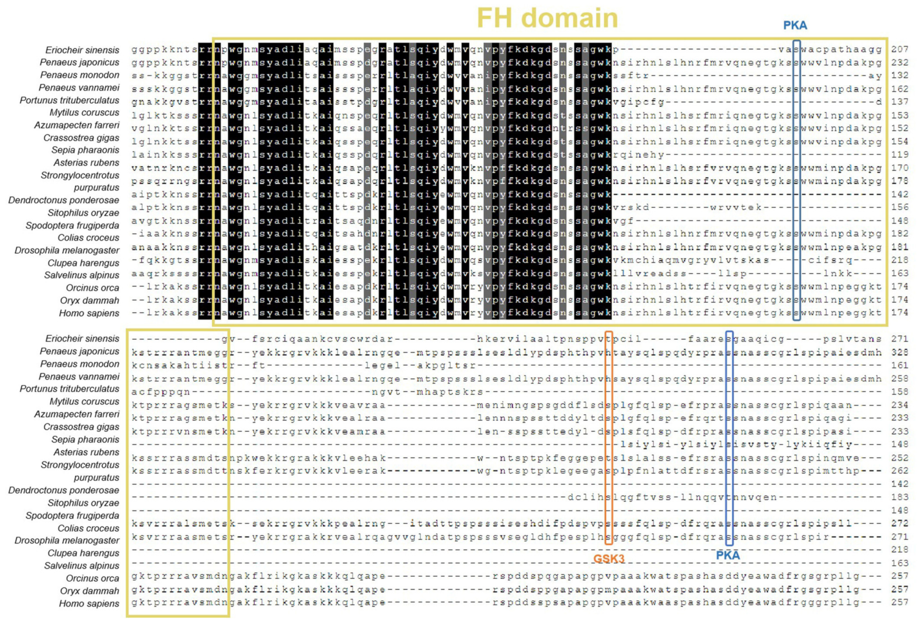
The ORF sequence of EsFOXO-like was found to be 852 bp long and encoded a polypeptide consisting of 283 amino acids (Figure 1a). Notably, the EsFOXO-like protein was found to contain a Forkhead (FH) domain between amino acids 140 and 208 (Figure 1b), which bears similar protein kinase A (PKA) phosphorylation modification sites. The EsFOXO-like protein has also been found to have a glycogen synthase kinase 3 (GSK3) phosphorylation modification site and a PKA phosphorylation modification site outside the FH domain (Figure 2). The deduced amino acid sequence of EsFOXO-like shared high similarity with those FOXOs from other invertebrates species (Figure 2), such as Penaeus japonicus (MW080526.1, 83.51%), Penaeus monodon (XP_037792098.1, 88.37%), Portunus trituberculatus (MPC 39137.1, 88.55%), Penaeus vannamei (XP_027228067.1, 75.00%), Clupea harengus (XP_031428678.1, 83.33%) and Salvelinus alpinus (XP_023824765.1, 85.45%) (Table 2).
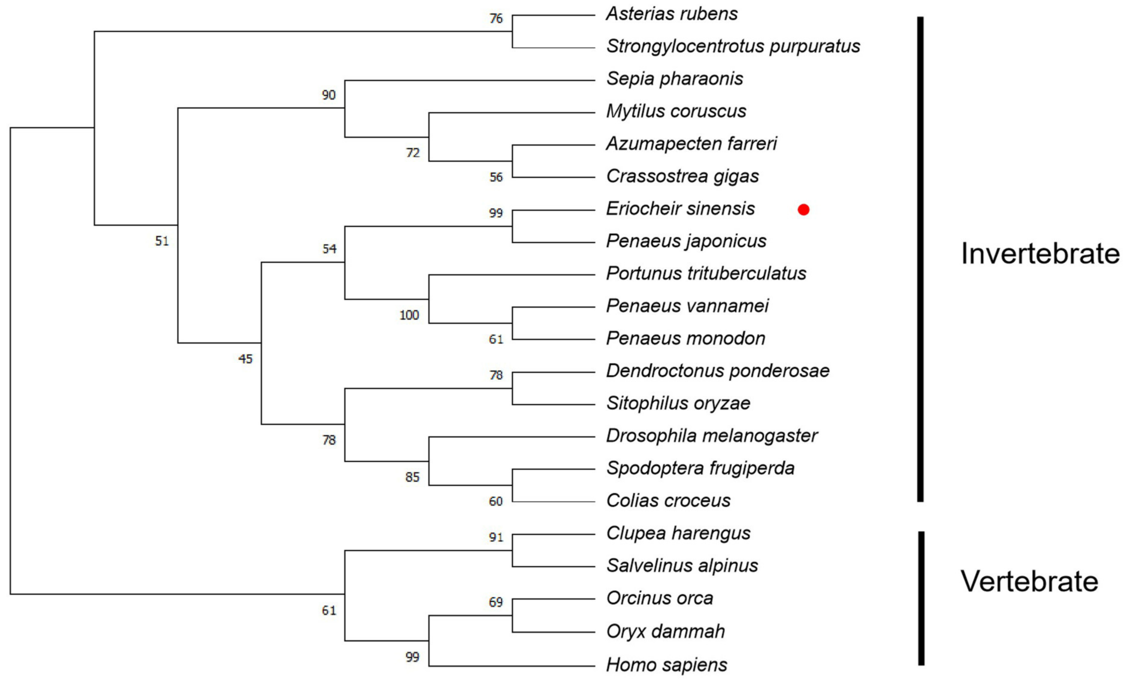
In the phylogenetic tree of FOXOs, all the selected FOXO members were divided into invertebrate and vertebrate branches. EsFOXO-like was firstly clustered with FOXO from Penaeus japonicus, and shared a closer evolutionary relationship with FOXO from Portunus trituberculatus, Penaeus vannamei and Penaeus monodon, enabling categorization into the invertebrate FOXO branch. The other FOXOs from Clupea harengus, Salvelinus alpinus, Orcinus orca, Orys dammah and Homo sapiens were clustered into the vertebrate clade (Figure 3).

3.2. The Distribution of EsFOXO-like Gene in Different Tissues

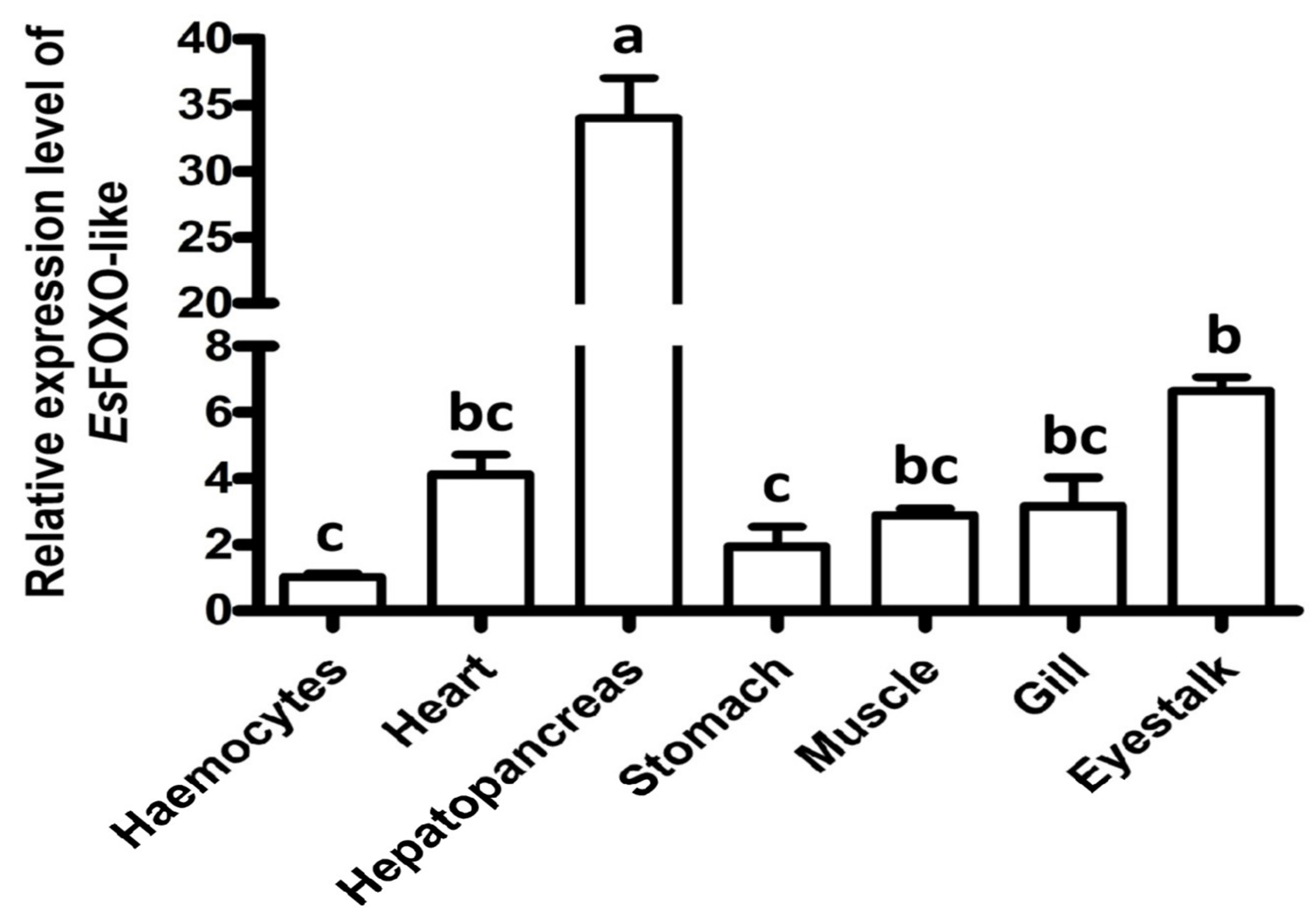
The EsFOXO-like gene mRNA transcripts were detected in different tissues, including the hepatopancreas, hemocytes, stomach, heart, gill, muscle and eyestalk. The highest levels of EsFOXO-like mRNA transcript were detected in the hepatopancreas (33.65-fold more than that in hemocytes, p < 0.05). The recorded mRNA relative expression levels of EsFOXO-like in the heart, stomach, muscles, gills, and eyestalks were 4.06-fold, 1.92-fold, 2.86-fold, 3.12-fold and 6.58-fold of that in hemocytes (p < 0.05), respectively (Figure 4).

3.3. The EsFOXO-like mRNA and Protein Expression Characteristic in Hepatopancreas at Three Molting Stages

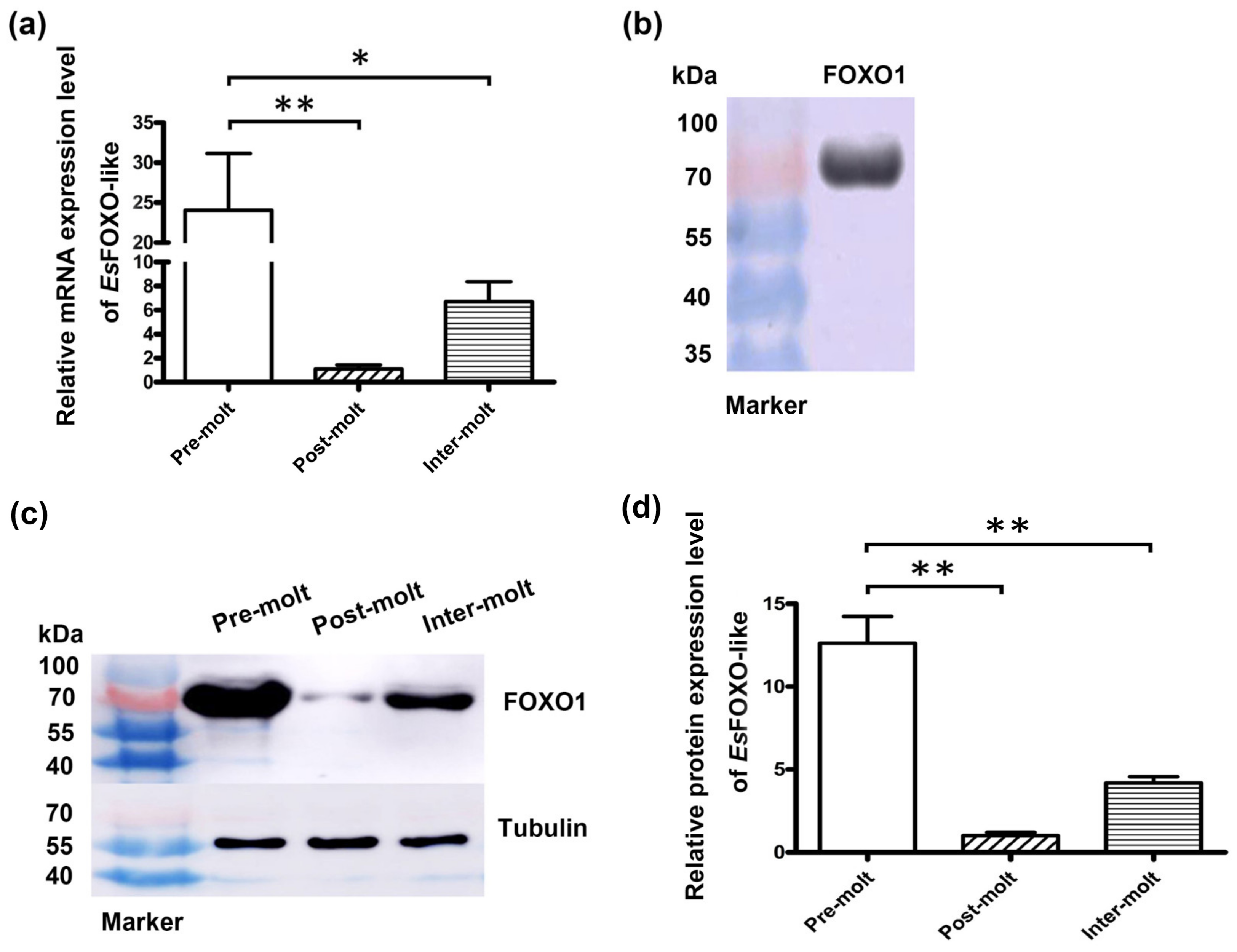
The mRNA and protein expression of EsFOXO-like in hepatopancreas at pre-molt, post-molt and inter-molt were measured. It was shown that EsFOXO-like was highly expressed at the pre-molt stage and lowly expressed at the post-molt stage (Figure 5a). The expression level at pre-molt was 22.01-fold (p < 0.01) and 3.58-fold (p < 0.05) of that at post-molt stage and inter-molt stage, respectively.
The results showed that there was a specific band of about 70 kDa (Figure 5b) detected with FOXO1 antibody in the hepatopancreas by western blotting (Figure 5b), indicating that the FOXO1 antibody was relatively specific. Additionally, the EsFOXO-like protein in the hepatopancreas was increased at the pre-molt stage and decreased at the post-molt stage. Specifically, the protein expression levels of EsFOXO-like at the pre-molt stage were 12.62-fold (p < 0.01) and 3.03-fold (p < 0.01) of that at the post-molt stage and the inter-molt stage, respectively (Figure 5c,d).

3.4. The EsFOXO-like mRNA and Protein Expression Levels after Inhibition of EsFOXO-like

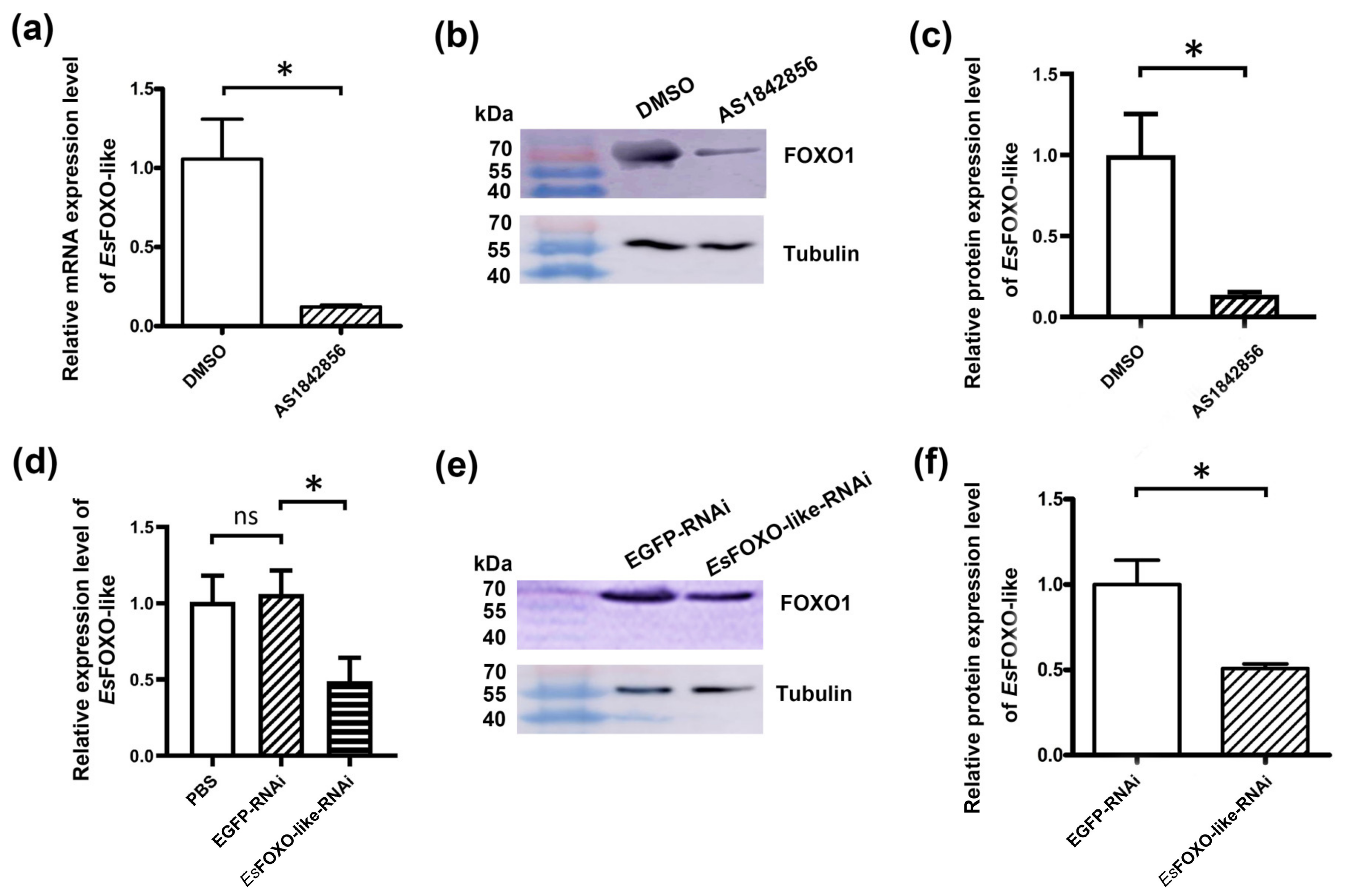
The inhibitory effect of the FOXO inhibitor (AS1842856) on EsFOXO-like expression in hepatopancreas was evaluated by qRT-PCR and western blotting, respectively. After AS1842856 injection, the EsFOXO-like mRNA transcripts in the AS1842856 group were significantly decreased, representing 0.11-fold (p < 0.05) of that in the DMSO group (Figure 6a). The western blotting assay showed that the band against the EsFOXO-like antibody in AS1842856 injected crabs was thinner compared to that in the PBS group (Figure 6b). The protein expression level of EsFOXO-like in the hepatopancreas was also decreased (Figure 6c) in the AS1842856 group (0.14-fold of that in the PBS group, p < 0.05).
Furthermore, the RNAi assay was employed to reduce the expression of EsFOXO-like mRNA, and the silencing efficiency of EsFOXO-like was detected by qRT-PCR and western blotting, respectively. The mRNA expression of EsFOXO-like in the hepatopancreas was significantly decreased 24 h after the injection of EsFOXO-like dsRNA, which was 0.46-fold compared with that of the EGFP-RNAi group (p < 0.05) (Figure 6d). No significant difference in the EsFOXO-like expression level was observed between the EGPF-RNAi group and the PBS group (Figure 6d). Similarly, the western blotting assay demonstrated that the intensity values of the EsFOXO-like band in EsFOXO-like-RNAi crabs decreased significantly (0.51-fold of that in the EGFP-RNAi group; p < 0.05) (Figure 6e,f).

3.5. The 20E Concentration and Molting-Related Genes mRNA Expression Levels after EsFOXO-like Inhibition

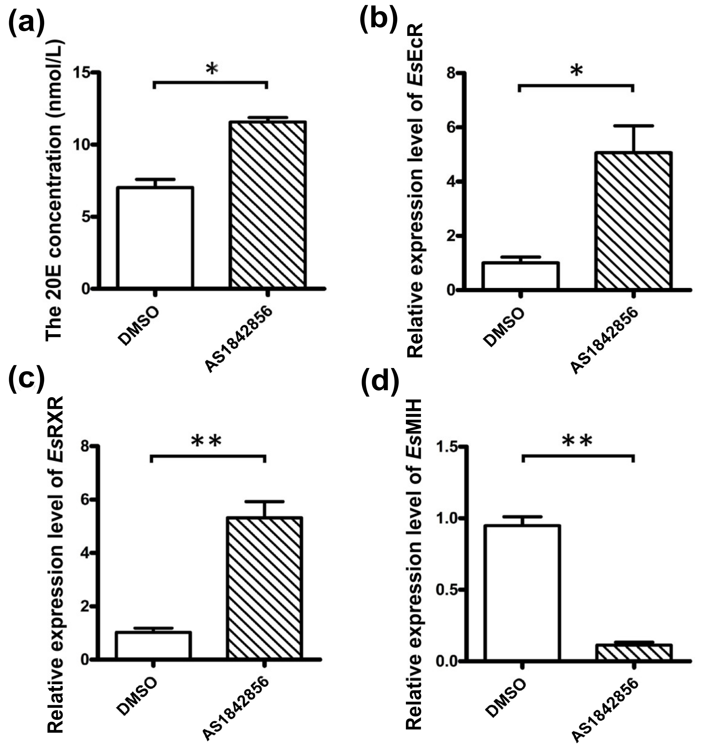
To investigate the regulation of EsFOXO-like gene in the 20E pathway, the 20E concentration and mRNA transcripts of EsEcR, EsRXR and EsMIH were detected after AS1842856 and EsFOXO-like dsRNA injection, respectively. The 20E concentration was significantly increased in the AS1842856 group, which was 1.65-fold (p < 0.05) of that in the DMSO group (Figure 7a). Similarly, the EsEcR and EsRXR mRNA expression levels in the hepatopancreas were both significantly increased in the AS1842856 group (5.03- and 5.18-fold compared to that in the DMSO group; p < 0.05) (Figure 7b,c). Conversely, the EsMIH mRNA expression level in eyestalk decreased significantly, which was 0.12-fold compared to that in the DMSO group (p < 0.01) (Figure 7d).
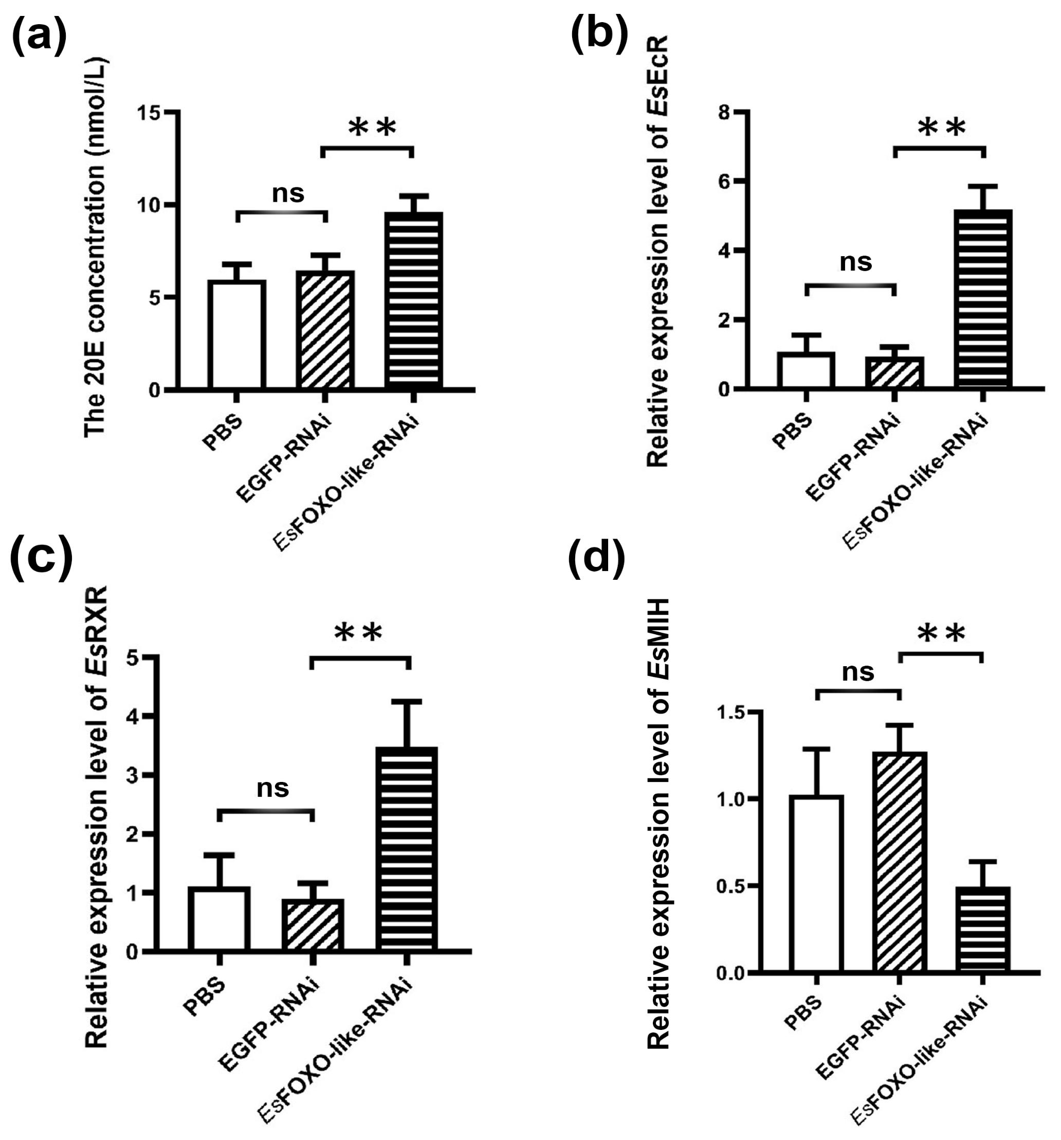
After EsFOXO-like was knocked down by RNAi, the 20E concentration and the mRNA expression levels of EsEcR and EsRXR in the hepatopancreas were significantly up-regulated, which were 1.49-fold (p < 0.01), 5.59-fold (p < 0.01) and 3.88-fold (p < 0.01) of that in the EGFP-RNAi group, respectively (Figure 8a–c). On the contrary, the EsMIH mRNA expression in eyestalk was significantly lower than that (0.39-fold, p < 0.01) in the EGFP-RNAi group (Figure 8d).

3.6. The mRNA Expression Level of EsmTOR after EsFOXO-like Inhibition

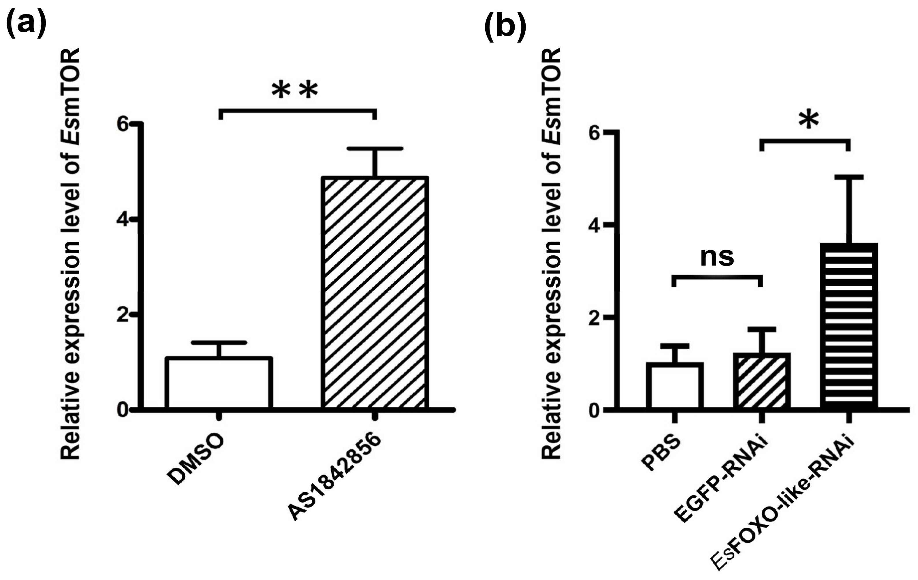
In order to study whether or whether not EsFOXO-like regulates the 20E pathway through EsmTOR, the expression level of EsmTOR was first detected after EsFOXO-like inhibition by AS1842856 treatment or dsRNA-EsFOXO-like injection. The EsmTOR mRNA expression levels in the hepatopancreas were up-regulated after injection of both AS1842856 and dsRNA-EsFOXO-like. The mRNA transcript of EsmTOR in the AS1842856 group was 4.49-fold (p < 0.01) that of the DMSO group (Figure 9a). Similarly, the relative expression level of EsmTOR in the hepatopancreas was significantly (p < 0.05) increased in the EsFOXO-RNAi group, which was 2.91-fold compared to that in the EGFP-RNAi group (Figure 9b).

3.7. The EsmTOR mRNA Transcripts after AS1842856 and Rapamycin Injection

To further investigate whether EsFOXO-like regulates the 20E pathway through EsmTOR or not, we evaluated the expression level of EsmTOR in the hepatopancreas after inhibiting EsFOXO-like by AS1842856 injection, followed by administering Rapamycin. The mRNA expression level of EsmTOR in the hepatopancreas was decreased after injection of AS1842856 + Rapamycin, and the EsmTOR transcript in the AS1842856 + Rapamycin group was 0.21-fold of that in the AS1842856 + DMSO group (p < 0.05) (Figure 10).

3.8. The 20E Concentration and EsEcR, EsRXR and EsMIH mRNA Expression Levels in AS1842856 + Rapamycin Group after Inhibiting mTOR

To investigate the role of EsmTOR in the regulation of the 20E pathway through EsFOXO-like, the concentration of 20E and the mRNA expression levels of EsEcR, EsRXR, and EsMIH were measured in the crabs following AS1842856 and Rapamycin injections. The results showed that the 20E concentration in serum, as well as the EsMIH mRNA expression level in eyestalk, were significantly decreased in the AS1842856 + Rapamycin group, which was 0.81-fold (p < 0.05) and 0.23-fold (p < 0.05) of that in AS1842856 + DMSO group for the respective components analyzed (Figure 11a,d). On the contrary, the expression levels of EsEcR (5.78-fold, p < 0.05) and EsRXR (3.69-fold, p < 0.05) in the hepatopancreas in the AS1842856 + Rapamycin group were significantly higher than that in the AS1842856 + DMSO group (Figure 11b,c).

4. Discussion

FOXO plays a key regulatory role in many physiological, metabolic and immunoregulatory responses [55,56,57]. The FOXO gene is conserved from lower yeast to higher humans [58]. So far, there are four FOXO members found in mammals [2], and only one FOXO member has been identified in invertebrates [59]. In this study, a FOXO-like transcription factor (EsFOXO-like) was identified and characterized in E. sinensis. The ORF of EsFOXO-like was 852 bp and encoded a 283 amino acid polypeptide. The EsFOXO-like sequence perfectly contains a typical Forkhead (FH) domain [19]. The DBD domain, also named the FH domain, was found to be highly conserved in different species [60,61]. The FH domain has been confirmed to interact with p53 to stabilize it from degradation, which is required to induce apoptosis [62]. Phylogenetic analysis showed that EsFOXO-like exhibits high similarity to crustacean FOXO genes and clusters together with them, suggesting that EsFOXO-like is a member of the FOXO family in crustaceans.
In mammals, different FOXO genes had various tissue expression patterns. For instance, FOXO1 and FOXO3α are expressed in multiple tissues, while FOXO4 is predominantly expressed in the kidney and muscle, and FOXO6 exhibits high expression in the liver [9]. Our results showed that the EsFOXO-like transcripts were expressed in various tissues, such as hepatopancreas, hemocytes, heart, stomach, gills, muscles, and eyestalks, with the highest level of expression in the hepatopancreas, which is similar to the distribution of FOXO genes in crustaceans [3,19]. The hepatopancreas of crustaceans has been identified to serve as a vital immunologic and metabolic organ [63]. Furthermore, it is considered to be one of the crucial organs involved in the molting process of crustaceans [22,64,65]. Moreover, the function of four different FOXO genes is also varied in vertebrates. For instance, the global loss of FOXO1 could cause the death of embryonic cells [66]. Furthermore, the overall deletion of FOXO3 can affect lymphatic proliferation and extensive organ inflammation [67]. Additionally, the loss of FOXO4 can exacerbate colitis induced by inflammatory stimuli [68]. Lastly, FoxO6 is preferentially enriched in the hippocampus, and the deletion of the FOXO6 gene can lead to impaired memory consolidation that showed a reduced ability to form long-term contextual and object recognition memories in mice [69]. Recently, many studies have demonstrated that FOXO regulates molting and metamorphosis in insects [16]. Although FOXO has been identified in several crustacean species, most studies only focus on its regulatory role in the immune response against bacterial invasions [3,19]. Thus, the potential involvement of FOXO in growth still needs to be further investigated.
Molting is a typical biological trait that directly determines the behavior and physiological processes in arthropods [70,71,72,73]. Generally, the molting cycle of crustaceans has been divided into three main stages: inter-molt, pre-molt and ecdysis [21,22]. We found that EsFOXO-like mRNA expression was different at each molting stage, with the highest level at pre-molt and the lowest level at post-molt. In H.armigera, the FOXO protein in the fat body was up-regulated during the fifth-larvae molting stage and metamorphosis when compared to the feeding stage. This difference indicated that the expression of FOXO also increased during molting and metamorphosis [16]. In insects, 20E and juvenile hormone are mutually antagonistic and jointly regulate molting processes. Usually, the juvenile hormone (JH) concentration decreases sharply in the juvenile developmental stage, and the increase in 20E concentration causes pupation or adult morphogenesis [74]. It has been discovered that FOXO plays a role in regulating the degradation of juvenile hormone (JH) to control the growth of B. mori [11]. Additionally, in Tribolium castaneum, it has been observed that 20E up-regulates FOXO expression, promoting its nuclear migration, where FOXO subsequently regulates protease factors [75]. In H. armigera, 20E also up-regulates FOXO expression which, in turn, induces the expression of Carboxypeptidase A to regulate the final proteolysis step during the molting process [16]. In our study, it was observed that the mRNA transcripts and protein expression of EsFOXO-like reached their highest levels during the pre-molt stage, suggesting its potential involvement in the molting process.
Although FOXO has been demonstrated to play a key role in the regulation of metamorphosis and growth of insects, the involvement of FOXO in the regulation of molting in crustaceans is unclear. The role of EsFOXO-like in regulating crab molting was explored in our research by evaluating the 20E concentration and expression levels of molting-related genes after AS1842856 [44] and EsFOXO-like dsRNA injections. In this study, the EsFOXO-like mRNA expression was significantly decreased after injections of AS1842856 or EsFOXO-like dsRNA. In mammals, AS1842856, which served as a FOXO1 inhibitor, could inhibit the protein activity of FOXO1 but had no effect on the mRNA transcription of FOXO1 [44,45,76]. It has also been demonstrated that treatment with AS1842856 displayed similar effects to FOXO1 knockdown in pancreatic progenitors [76]. Until now, the potential role of AS1842856 on FOXO genes has not been reported in crustaceans. In this study, our results showed that the mRNA and protein expression levels of EsFOXO-like were both significantly decreased after AS1842856 injection. This observation might be related to the differences in metabolism between crustaceans and mammals. Similarly, in the study of 3T3-L1 preadipocytes cells, it has also been found that the level of total FOXO1 protein was reduced after AS1842856 treatment [77]. Additional investigation revealed a significant increase in 20E concentration after the inhibition of EsFOXO-like. In addition, our study showed that when EsFOXO-like was inhibited, the EsEcR and EsRXR expressions were significantly up-regulated, while the EsMIH expression was significantly down-regulated. In crustaceans, molting is promoted by 20E through its dependence on EcR and RXR, which leads to the activation of molting-related genes [78]. EcR and RXR proteins bind together to form a complex, which then interacts with the 20E hormone to create an active trimer. This trimer is responsible for regulating the growth and molting process in crustaceans [79]. On the other hand, the MIH hormone has been shown to have a negative effect on the synthesis of 20E [23]. Although the studies on the regulation of FOXO on EcR, RXR and MIH expressions have not been reported in crustaceans, it has been studied in insects. For example, it has been found that FOXO could directly interact with Ultraspiracle (Usp), the ecdysone receptor, and FoxO/Usp complexes suppress 20E biosynthesis in Drosophila [80]. Additionally, JH regulated the growth rate via inhibition of the 20E pathway, and it is also dependent on FOXO [81]. While it has been reported that FOXO inhibition does not affect the expression levels of EcRB1 and USP1, it could significantly block the molting of larvae in H. armigera [16]. These results collectively suggested that FOXO was closely related to the physiological process of molting in E. sinensis by inhibiting the 20E signal. However, whether FOXO affects the 20E signal through related genes or signaling pathways remains to be further explored.
Multiple studies have shown that mTOR, a key regulator of cellular metabolism, plays a role in controlling the synthesis of 20E [30,31,32]. Research has shown that the activation of Y-organ ecdysteroidogenesis is dependent on the activity of mTOR and that mTORC1 is primarily responsible for driving the increase in 20E concentration [48,82]. Therefore, in order to explore the mechanism of FOXO regulation in the 20E pathway in crabs, we first measured the EsmTOR mRNA expression level after FOXO inhibition. The results showed that EsmTOR transcription was significantly increased in the AS1842856-injected group and the EsFOXO-like-RNAi group. In mammals, the PI3K–AKT–FOXO–mTOR pathway has been verified to be a regulator of metabolism and somatic growth [83]. It has also been found that loss of FOXO function resulted in precocious differentiation in tissues with high mTOR activity on nutrient restriction in Drosophila [84]. Moreover, the cells that lost the function of FOXO have higher mTOR activity and are more sensitive to 20E response, possibly due to a similar inhibitory action of FOXO on the 20E receptor complex [84]. Moreover, Rapamycin, an effective and specific mTOR activity inhibitor, was injected into crabs to explore the function of mTOR in the FOXO regulation of the 20E pathway in this study [85]. Injections of AS1842856 and Rapamycin resulted in reduced expression of the EsmTOR transcript, and the AS1842856 + Rapamycin group exhibited a significantly lower concentration of 20E compared to the AS1842856 + DMSO group. Similarly, in Manduca sexta, Rapamycin resulted in delayed larval molting and reduced 20E production [86]. In B. moriit, it has been found that rapamycin could also inhibit 20E synthesis and secretion [32]. Although accumulating evidence has suggested that mTOR could stimulate 20E synthesis and inhibit the expression levels of MIH signaling-related genes [29,30]. 20E is transduced via binding of an isomeric dimer complex consisting of EcR and RXR [25], but the impact of mTOR on EcR and RXR is unknown. Notably, mTOR signaling plays an essential role in the regulation of growth, body size and aging of arthropods. It has also been confirmed that the insulin receptor (InR) and phosphoinositide 3-kinase (PI3K) genes, involved in growth and development progress, showed progressively increasing mRNA levels in rapamycin-treated crabs [29]. Similarly, the InR and PI3K could promote EcR and USP (RXR in crustaceans) expression in insects [87]. Therefore, we speculate that the increase in EsEcR and EsRXR expression observed in the AS1842856- + Rapamycin-inhibited group in this study might be due to up-regulation of the insulin and PI3K pathway. Furthermore, 20E and its receptors have been confirmed to negatively affect MIH expression [27,28]. Thus, the decrease of EsMIH expression also observed in the AS1842856 + Rapamycin inhibited group might be due to the increased expression of EsEcR and EsRXR. It might be also associated with the sampling time after rapamycin injection. For instance, in Gecarcinus lateralis, rapamycin did not block or reverse the decrease in MIH signaling genes until the seventh day after rapamycin injection [29]. Overall, these results suggest that FOXO could regulate the 20E pathway through mTOR in mitten crabs.

5. Conclusions

In summary, a Forkhead Box O-like (named EsFOXO-like) was identified in E. sinensis, with the highest expression in the hepatopancreas at pre-molt stage and lowest expression at post-molt stage. It was also found that the 20E and EsMIH mRNA expressions were both significantly down-regulated after EsFOXO-like inhibition, while the expression of EsEcR and EsRXR showed the opposite expression pattern. Taken together, these results suggest that EsFOXO-like has a negative effect on 20E signaling by inhibiting mTOR. Molting is an essential process for the growth of crustaceans, and FOXO served as a key transcription factor. Its role in regulating molting had not yet been studied in crustaceans. Our current results reveal the inhibitory function of FOXO in the molting of crustaceans. Future research should be performed on the regulatory mechanisms involved in the promotion of molting and growth by inhibiting FOXO.

Supplementary Materials

The following supporting information can be downloaded at: https://www.mdpi.com/article/10.3390/jmse11061225/s1, Table S1: The original data for analysis in this manuscript.

Author Contributions

Conceptualization, J.L. (Jiaming Li) and S.H.; methodology, J.L. (Jiaming Li) and Y.M.; software, Z.Y. and F.W.; validation, J.L. (Jialin Li) and Y.M.; formal analysis, J.L. and F.W.; investigation, J.L. (Jialin Li) and Z.Y.; writing—original draft preparation, J.L. (Jiaming Li) and Y.M.; writing—review and editing, Y.J., D.Y., Q.Y. and S.H.; supervision, Q.Y. and S.H.; project administration, S.H.; funding acquisition, Q.Y. and S.H. All authors have read and agreed to the published version of the manuscript.

Funding

This work was supported by research grants from Educational Department of Liaoning Province (20220078), Natural Resources Department of Liaoning Province (20220001-18) and Educational Department of Liaoning Province (S202210158002X).

Institutional Review Board Statement

The animal study protocol conformed with the Animal Care Committee of Dalian Ocean University.

Informed Consent Statement

Not applicable.

Data Availability Statement

Data is contained within the article. All the data are available from the corresponding author upon request.

Acknowledgments

We are grateful to all the members for their help and support in this experiment.

Conflicts of Interest

The authors declare no conflict of interests.

References

  1. Calissi, G.; Lam, E.W.; Link, W. Therapeutic strategies targeting FOXO transcription factors. Nat. Rev. Drug Discov. 2021, 20, 21–38. [Google Scholar] [CrossRef] [PubMed]
  2. Jiramongkol, Y.; Lam, E.W. FOXO transcription factor family in cancer and metastasis. Cancer Metastasis Rev. 2020, 39, 681–709. [Google Scholar] [CrossRef]
  3. Li, C.; Hong, P.P.; Yang, M.C.; Zhao, X.F.; Wang, J.X. FOXO regulates the expression of antimicrobial peptides and promotes phagocytosis of hemocytes in shrimp antibacterial immunity. PLoS Pathog. 2021, 17, e1009479. [Google Scholar] [CrossRef] [PubMed]
  4. Arden, K.C. FOXO animal models reveal a variety of diverse roles for FOXO transcription factors. Oncogene 2008, 27, 2345–2350. [Google Scholar] [CrossRef] [PubMed] [Green Version]
  5. Boura, E.; Silhan, J.; Herman, P.; Vecer, J.; Sulc, M.; Teisinger, J.; Obsilova, V.; Obsil, T. Both the N-terminal loop and wing W2 of the forkhead domain of transcription factor Foxo4 are important for DNA binding. J. Biol. Chem. 2007, 282, 8265–8275. [Google Scholar] [CrossRef] [Green Version]
  6. Clark, K.L.; Halay, E.D.; Lai, E.; Burley, S.K. Co-crystal structure of the HNF-3/fork head DNA-recognition motif resembles histone H5. Nature 1993, 364, 412–420. [Google Scholar] [CrossRef]
  7. Jacobs, F.M.; van der Heide, L.P.; Wijchers, P.J.; Burbach, J.P.; Hoekman, M.F.; Smidt, M.P. FoxO6, a novel member of the FoxO class of transcription factors with distinct shuttling dynamics. J. Biol. Chem. 2003, 278, 35959–35967. [Google Scholar] [CrossRef] [Green Version]
  8. Arden, K.C.; Biggs, W.H. Regulation of the FoxO family of transcription factors by phosphatidylinositol-3 kinase-activated signaling. Arch. Biochem. Biophys. 2002, 403, 292–298. [Google Scholar] [CrossRef]
  9. van der Vos, K.E.; Coffer, P.J. The extending network of FOXO transcriptional target genes. Antioxid. Redox Signal. 2011, 14, 579–592. [Google Scholar] [CrossRef]
  10. Dong, X.; Zhai, Y.; Zhang, J.; Sun, Z.; Chen, J.; Chen, J.; Zhang, W. Fork head transcription factor is required for ovarian mature in the brown planthopper, Nilaparvata lugens (Stål). BMC Mol. Biol. 2011, 12, 53. [Google Scholar] [CrossRef] [Green Version]
  11. Zeng, B.; Huang, Y.; Xu, J.; Shiotsuki, T.; Bai, H.; Palli, S.R.; Huang, Y.; Tan, A. The FOXO transcription factor controls insect growth and development by regulating juvenile hormone degradation in the silkworm, Bombyx mori. J. Biol. Chem. 2017, 292, 11659–11669. [Google Scholar] [CrossRef] [Green Version]
  12. Giannakou, M.E.; Goss, M.; Jünger, M.A.; Hafen, E.; Leevers, S.J.; Partridge, L. Long-lived Drosophila with overexpressed dFOXO in adult fat body. Science 2004, 305, 361. [Google Scholar] [CrossRef]
  13. Wu, Y.B.; Yang, W.J.; Xie, Y.F.; Xu, K.K.; Tian, Y.; Yuan, G.R.; Wang, J.J. Molecular characterization and functional analysis of BdFoxO gene in the oriental fruit fly, Bactrocera dorsalis (Diptera: Tephritidae). Gene 2016, 578, 219–224. [Google Scholar] [CrossRef]
  14. Süren-Castillo, S.; Abrisqueta, M.; Maestro, J.L. FoxO inhibits juvenile hormone biosynthesis and vitellogenin production in the German cockroach. Insect Biochem. Mol. Biol. 2012, 42, 491–498. [Google Scholar] [CrossRef] [Green Version]
  15. Sim, C.; Denlinger, D.L. Insulin signaling and FOXO regulate the overwintering diapause of the mosquito Culex pipiens. Proc. Natl. Acad. Sci. USA 2008, 105, 6777–6781. [Google Scholar] [CrossRef] [Green Version]
  16. Cai, M.J.; Zhao, W.L.; Jing, Y.P.; Song, Q.; Zhang, X.Q.; Wang, J.X.; Zhao, X.F. 20-Hydroxyecdysone activates Forkhead box O to promote proteolysis during Helicoverpa armigera molting. Development 2016, 143, 1005–1015. [Google Scholar] [CrossRef] [Green Version]
  17. Lin, X.; Yu, N.; Smagghe, G. FoxO mediates the timing of pupation through regulating ecdysteroid biosynthesis in the red flour beetle, Tribolium castaneum. Gen. Comp. Endocrinol. 2018, 258, 149–156. [Google Scholar] [CrossRef]
  18. Hossain, M.S.; Liu, Y.; Zhou, S.; Li, K.; Tian, L.; Li, S. 20-Hydroxyecdysone-induced transcriptional activity of FoxO upregulates brummer and acid lipase-1 and promotes lipolysis in Bombyx fat body. Insect Biochem. Mol. Biol. 2013, 43, 829–838. [Google Scholar] [CrossRef]
  19. Zhao, Y.; Nie, X.; Han, Z.; Liu, P.; Xu, H.; Huang, X.; Ren, Q. The forkhead box O transcription factor regulates lipase and anti-microbial peptide expressions to promote lipid catabolism and improve innate immunity in the Eriocheir sinensis with hepatopancreatic necrosis disease. Fish Shellfish Immunol. 2022, 124, 107–117. [Google Scholar] [CrossRef]
  20. Yu, Y.Q.; Ma, W.M.; Yang, W.J.; Yang, J.S. The complete mitogenome of the lined shore crab Pachygrapsus crassipes Randall 1840 (Crustacea: Decapoda: Grapsidae). Mitochondrial DNA 2014, 25, 263–264. [Google Scholar] [CrossRef]
  21. Gao, Y.; Zhang, X.; Wei, J.; Sun, X.; Yuan, J.; Li, F.; Xiang, J. Whole Transcriptome Analysis Provides Insights into Molecular Mechanisms for Molting in Litopenaeus vannamei. PLoS ONE 2015, 10, e0144350. [Google Scholar] [CrossRef] [PubMed] [Green Version]
  22. Huang, S.; Wang, J.; Yue, W.; Chen, J.; Gaughan, S.; Lu, W.; Lu, G.; Wang, C. Transcriptomic variation of hepatopancreas reveals the energy metabolism and biological processes associated with molting in Chinese mitten crab, Eriocheir sinensis. Sci. Rep. 2015, 5, 14015. [Google Scholar] [CrossRef] [PubMed] [Green Version]
  23. Chang, E.S.; Mykles, D.L. Regulation of crustacean molting: A review and our perspectives. Gen. Comp. Endocrinol. 2011, 172, 323–330. [Google Scholar] [CrossRef] [PubMed]
  24. Aranda, A.; Pascual, A. Nuclear hormone receptors and gene expression. Physiol. Rev. 2001, 81, 1269–1304. [Google Scholar] [CrossRef] [Green Version]
  25. Nakagawa, Y.; Henrich, V.C. Arthropod nuclear receptors and their role in molting. FEBS J. 2009, 276, 6128–6157. [Google Scholar] [CrossRef] [Green Version]
  26. Techa, S.; Chung, J.S. Ecdysone and retinoid-X receptors of the blue crab, Callinectes sapidus: Cloning and their expression patterns in eyestalks and Y-organs during the molt cycle. Gene 2013, 527, 139–153. [Google Scholar] [CrossRef]
  27. Techa, S.; Chung, J.S. Ecdysteroids regulate the levels of Molt-Inhibiting Hormone (MIH) expression in the blue crab, Callinectes sapidus. PLoS ONE 2015, 10, e0117278. [Google Scholar] [CrossRef]
  28. Mykles, D.L.; Chang, E.S. Hormonal control of the crustacean molting gland: Insights from transcriptomics and proteomics. Gen. Comp. Endocrinol. 2020, 294, 113493. [Google Scholar] [CrossRef]
  29. Shyamal, S.; Das, S.; Guruacharya, A.; Mykles, D.L.; Durica, D.S. Transcriptomic analysis of crustacean molting gland (Y-organ) regulation via the mTOR signaling pathway. Sci. Rep. 2018, 8, 7307. [Google Scholar] [CrossRef] [Green Version]
  30. Mykles, D.L. Signaling Pathways That Regulate the Crustacean Molting Gland. Front. Endocrinol. 2021, 12, 674711. [Google Scholar] [CrossRef]
  31. Gu, S.H.; Young, S.C.; Lin, J.L.; Lin, P.L. Involvement of PI3K/Akt signaling in PTTH-stimulated ecdysteroidogenesis by prothoracic glands of the silkworm, Bombyx mori. Insect Biochem. Mol. Biol. 2011, 41, 197–202. [Google Scholar] [CrossRef]
  32. Gu, S.H.; Yeh, W.L.; Young, S.C.; Lin, P.L.; Li, S. TOR signaling is involved in PTTH-stimulated ecdysteroidogenesis by prothoracic glands in the silkworm, Bombyx mori. Insect Biochem. Mol. Biol. 2012, 42, 296–303. [Google Scholar] [CrossRef]
  33. Lin, A.; Yao, J.; Zhuang, L.; Wang, D.; Han, J.; Lam, E.W.; Gan, B. The FoxO-BNIP3 axis exerts a unique regulation of mTORC1 and cell survival under energy stress. Oncogene 2014, 33, 3183–3194. [Google Scholar] [CrossRef] [Green Version]
  34. Teleman, A.A. Molecular mechanisms of metabolic regulation by insulin in Drosophila. Biochem. J. 2009, 425, 13–26. [Google Scholar] [CrossRef] [Green Version]
  35. Mirth, C.K.; Shingleton, A.W. Integrating body and organ size in Drosophila: Recent advances and outstanding problems. Front. Endocrinol. 2012, 3, 49. [Google Scholar] [CrossRef] [Green Version]
  36. Rewitz, K.F.; Yamanaka, N.; O’Connor, M.B. Developmental checkpoints and feedback circuits time insect maturation. Curr. Top. Dev. Biol. 2013, 103, 1–33. [Google Scholar]
  37. He, J.; Wu, X.; Li, J.Y.; Huang, Q.; Huang, Z.H.; Cheng, Y.X. Comparison of the culture performance and profitability of wild-caught and captive pond-reared Chinese mitten crab (Eriocheir sinensis) juveniles reared in grow-out ponds: Implications for seed selection and genetic selection programs. Aquaculture 2014, 434, 48–56. [Google Scholar] [CrossRef]
  38. Zhang, Y.; Sun, Y.; Liu, Y.; Geng, X.; Wang, X.; Wang, Y.; Sun, J.; Yang, W. Molt-inhibiting hormone from Chinese mitten crab (Eriocheir sinensis): Cloning, tissue expression and effects of recombinant peptide on ecdysteroid secretion of YOs. Gen. Comp. Endocrinol. 2011, 173, 467–474. [Google Scholar] [CrossRef]
  39. Chen, H.; Gu, X.; Zeng, Q.; Mao, Z.; Liang, X.; Martyniuk, C.J. Carbamazepine disrupts molting hormone signaling and inhibits molting and growth of Eriocheir sinensis at environmentally relevant concentrations. Aquat. Toxicol. 2019, 208, 138–145. [Google Scholar] [CrossRef]
  40. Li, C.; Huang, L.; Zhang, Y.; Guo, X.; Cao, N.; Yao, C.; Duan, L.; Li, X.; Pang, S. Effects of triazole plant growth regulators on molting mechanism in Chinese mitten crab (Eriocheir sinensis). Fish Shellfish Immunol. 2022, 131, 646–653. [Google Scholar] [CrossRef]
  41. Söderhäll, K.; Smith, V.J. Separation of the haemocyte populations of Carcinus maenas and other marine decapods, and prophenoloxidase distribution. Dev. Comp. Immunol. 1983, 7, 229–239. [Google Scholar] [CrossRef] [PubMed]
  42. Huang, S.; Yi, Q.; Lian, X.; Xu, S.; Yang, C.; Sun, J.; Wang, L.; Song, L. The involvement of ecdysone and ecdysone receptor in regulating the expression of antimicrobial peptides in Chinese mitten crab, Eriocheir sinensis. Dev. Comp. Immunol. 2020, 111, 103757. [Google Scholar] [CrossRef] [PubMed]
  43. Yang, W.; Liu, C.; Xu, Q.; Qu, C.; Sun, J.; Huang, S.; Kong, N.; Lv, X.; Liu, Z.; Wang, L.; et al. Beclin-1 is involved in the regulation of antimicrobial peptides expression in Chinese mitten crab Eriocheir sinensis. Fish Shellfish Immunol. 2019, 89, 207–216. [Google Scholar] [CrossRef] [PubMed]
  44. Xiao, Q.; Liu, H.; Wang, H.S.; Cao, M.T.; Meng, X.J.; Xiang, Y.L.; Zhang, Y.Q.; Shu, F.; Zhang, Q.G.; Shan, H.; et al. Histone deacetylase inhibitors promote epithelial-mesenchymal transition in Hepatocellular Carcinoma via AMPK-FOXO1-ULK1 signaling axis-mediated autophagy. Theranostics 2020, 10, 10245–10261. [Google Scholar] [CrossRef] [PubMed]
  45. Shi, Y.; Fan, S.; Wang, D.; Huyan, T.; Chen, J.; Chen, J.; Su, J.; Li, X.; Wang, Z.; Xie, S.; et al. FOXO1 inhibition potentiates endothelial angiogenic functions in diabetes via suppression of ROCK1/Drp1-mediated mitochondrial fission. Biochim. Biophys. Acta Mol. Basis Dis. 2018, 1864, 2481–2494. [Google Scholar] [CrossRef] [PubMed]
  46. Zhou, K.; Qin, Y.; Song, Y.; Zhao, K.; Pan, W.; Nan, X.; Wang, Y.; Wang, Q.; Li, W. A novel Ig domain-containing C-type lectin triggers the intestine-hemocyte axis to regulate antibacterial immunity in crab. J. Immunol. 2022, 208, 2343–2362. [Google Scholar] [CrossRef]
  47. Bulut-Karslioglu, A.; Biechele, S.; Jin, H.; Macrae, T.A.; Hejna, M.; Gertsenstein, M.; Song, J.S.; Ramalho-Santos, M. Inhibition of mTOR induces a paused pluripotent state. Nature 2016, 540, 119–123. [Google Scholar] [CrossRef] [Green Version]
  48. Abuhagr, A.M.; MacLea, K.S.; Mudron, M.R.; Chang, S.A.; Chang, E.S.; Mykles, D.L. Roles of mechanistic target of rapamycin and transforming growth factor-β signaling in the molting gland (Y-organ) of the blackback land crab, Gecarcinus lateralis. Comp. Biochem. Physiol. A Mol. Integr. Physiol. 2016, 198, 15–21. [Google Scholar] [CrossRef] [Green Version]
  49. Zhao, C.; Peng, C.; Wang, P.; Yan, L.; Fan, S.; Qiu, L. Identification of a Shrimp E3 Ubiquitin Ligase TRIM50-like involved in restricting White Spot Syndrome virus proliferation by its mediated autophagy and ubiquitination. Front. Immunol. 2021, 12, 682562. [Google Scholar] [CrossRef]
  50. Chen, X.; Wang, J.; Yue, W.; Huang, S.; Chen, J.; Chen, Y.; Wang, C. Structure and function of the alternatively spliced isoforms of the ecdysone receptor gene in the Chinese mitten crab, Eriocheir sinensis. Sci. Rep. 2017, 7, 12993. [Google Scholar] [CrossRef] [Green Version]
  51. Sun, Q.; Lin, S.; Zhang, M.; Gong, Y.; Ma, H.; Tran, N.T.; Zhang, Y.; Li, S. SpRab11a-regulated exosomes inhibit bacterial infection through the activation of antilipopolysaccharide factors in crustaceans. J. Immunol. 2022, 209, 710–722. [Google Scholar] [CrossRef]
  52. Wang, F.; Yang, Z.; Li, J.; Ma, Y.; Tu, Y.; Zeng, X.; Wang, Q.; Jiang, Y.; Huang, S.; Yi, Q. The involvement of hypoxia inducible factor-1α on the proportion of three types of haemocytes in Chinese mitten crab under hypoxia stress. Dev. Comp. Immunol. 2023, 140, 104598. [Google Scholar] [CrossRef]
  53. Wang, M.; Zhou, J.; Su, S.; Tang, Y.; Xu, G.; Li, J.; Yu, F.; Li, H.; Song, C.; Liang, M.; et al. Comparative transcriptome analysis on the regulatory mechanism of thoracic ganglia in Eriocheir sinensis at post-molt and inter-molt Stages. Life 2022, 12, 1181. [Google Scholar] [CrossRef]
  54. Schmittgen, T.D.; Livak, K.J. Analyzing real-time PCR data by the comparative C(T) method. Nat. Protoc. 2008, 3, 1101–1108. [Google Scholar] [CrossRef]
  55. van der Vos, K.E.; Coffer, P.J. FOXO-binding partners: It takes two to tango. Oncogene 2008, 27, 2289–2299. [Google Scholar] [CrossRef] [Green Version]
  56. Wang, S.; Xia, P.; Huang, G.; Zhu, P.; Liu, J.; Ye, B.; Du, Y.; Fan, Z. FoxO1-mediated autophagy is required for NK cell development and innate immunity. Nat. Commun. 2016, 7, 11023. [Google Scholar] [CrossRef]
  57. Iyer, S.; Ambrogini, E.; Bartell, S.M.; Han, L.; Roberson, P.K.; de Cabo, R.; Jilka, R.L.; Weinstein, R.S.; O’Brien, C.A.; Manolagas, S.C.; et al. FOXOs attenuate bone formation by suppressing Wnt signaling. J. Clin. Investig. 2013, 123, 3409–3419. [Google Scholar] [CrossRef]
  58. Barbieri, M.; Bonafè, M.; Franceschi, C.; Paolisso, G. Insulin/IGF-I-signaling pathway: An evolutionarily conserved mechanism of longevity from yeast to humans. Am. J. Physiol. Endocrinol. Metab. 2003, 285, E1064–E1071. [Google Scholar] [CrossRef] [Green Version]
  59. Park, D.; Hahm, J.H.; Park, S.; Ha, G.; Chang, G.E.; Jeong, H.; Kim, H.; Kim, S.; Cheong, E.; Paik, Y.K. A conserved neuronal DAF-16/FoxO plays an important role in conveying pheromone signals to elicit repulsion behavior in Caenorhabditis elegans. Sci. Rep. 2017, 7, 7260. [Google Scholar] [CrossRef] [Green Version]
  60. Obsil, T.; Obsilova, V. Structure/function relationships underlying regulation of FOXO transcription factors. Oncogene 2008, 27, 2263–2275. [Google Scholar] [CrossRef] [Green Version]
  61. Mazet, F.; Yu, J.K.; Liberles, D.A.; Holland, L.Z.; Shimeld, S.M. Phylogenetic relationships of the Fox (Forkhead) gene family in the Bilateria. Gene 2003, 316, 79–89. [Google Scholar] [CrossRef] [PubMed]
  62. Wang, F.; Marshall, C.B.; Yamamoto, K.; Li, G.Y.; Plevin, M.J.; You, H.; Mak, T.W.; Ikura, M. Biochemical and structural characterization of an intramolecular interaction in FOXO3a and its binding with p53. J. Mol. Biol. 2008, 384, 590–603. [Google Scholar] [CrossRef] [PubMed]
  63. Rőszer, T. The invertebrate midintestinal gland (“hepatopancreas”) is an evolutionary forerunner in the integration of immunity and metabolism. Cell Tissue Res. 2014, 358, 685–695. [Google Scholar] [CrossRef] [PubMed]
  64. Xu, Z.; Liu, A.; Li, S.; Wang, G.; Ye, H. Hepatopancreas immune response during molt cycle in the mud crab, Scylla paramamosain. Sci. Rep. 2020, 10, 13102. [Google Scholar] [CrossRef] [PubMed]
  65. Liu, M.; Ni, H.; Zhang, X.; Sun, Q.; Wu, X.; He, J. Comparative transcriptomics reveals the immune dynamics during the molting cycle of swimming crab Portunus trituberculatus. Front. Immunol. 2022, 13, 1037739. [Google Scholar] [CrossRef]
  66. Hosaka, T.; Biggs, W.H.; Tieu, D.; Boyer, A.D.; Varki, N.M.; Cavenee, W.K.; Arden, K.C. Disruption of forkhead transcription factor (FOXO) family members in mice reveals their functional diversification. Proc. Natl. Acad. Sci. USA 2004, 101, 2975–2980. [Google Scholar] [CrossRef] [Green Version]
  67. Lin, L.; Hron, J.D.; Peng, S.L. Regulation of NF-kappaB, Th activation, and autoinflammation by the forkhead transcription factor Foxo3a. Immunity 2004, 21, 203–213. [Google Scholar] [CrossRef] [Green Version]
  68. Zhou, W.; Cao, Q.; Peng, Y.; Zhang, Q.J.; Castrillon, D.H.; DePinho, R.A.; Liu, Z.P. FoxO4 inhibits NF-kappaB and protects mice against colonic injury and inflammation. Gastroenterology 2009, 137, 1403–1414. [Google Scholar] [CrossRef] [Green Version]
  69. Salih, D.A.M.; Rashid, A.J.; Colas, D.; de la Torre-Ubieta, L.; Zhu, R.P.; Morgan, A.A.; Santo, E.E.; Ucar, D.; Devarajan, K.; Cole, C.J.; et al. FoxO6 regulates memory consolidation and synaptic function. Genes Dev. 2012, 26, 2780–2801. [Google Scholar] [CrossRef] [Green Version]
  70. Panganiban, G.; Sebring, A.; Nagy, L.; Carroll, S. The development of crustacean limbs and the evolution of arthropods. Science 1995, 270, 1363–1366. [Google Scholar] [CrossRef]
  71. Hopkins, P.M.; Chung, A.C.-K.; Durica, D.S. Limb Regeneration in the Fiddler Crab, Uca pugilator: Hormonal and Growth Factor Control. Am. Zool. 2001, 41, 389–398. [Google Scholar] [CrossRef] [Green Version]
  72. Morris, S.; Postel, U.; Mrinalini; Turner, L.M.; Palmer, J.; Webster, S.G. The adaptive significance of crustacean hyperglycaemic hormone (CHH) in daily and seasonal migratory activities of the Christmas Island red crab Gecarcoidea natalis. J. Exp. Biol. 2010, 213 Pt 17, 3062–3073. [Google Scholar] [CrossRef] [Green Version]
  73. Jung, H.; Lyons, R.E.; Hurwood, D.A.; Mather, P.B. Genes and growth performance in crustacean species: A review of relevant genomic studies in crustaceans and other taxa. Rev. Aquacult. 2013, 5, 77–110. [Google Scholar] [CrossRef]
  74. Li, K.; Jia, Q.Q.; Li, S. Juvenile hormone signaling—A mini review. Insect. Sci. 2019, 26, 600–606. [Google Scholar] [CrossRef]
  75. Ji, C.; Zhang, N.; Jiang, H.; Meng, X.; Ge, H.; Yang, X.; Xu, X.; Qian, K.; Park, Y.; Zheng, Y.; et al. 20-hydroxyecdysone regulates expression of methioninesulfoxide reductases through transcription factor FOXO in the red flour beetle, Tribolium castaneum. Insect Biochem. Mol. Biol. 2021, 131, 103546. [Google Scholar] [CrossRef]
  76. Yu, F.; Wei, R.; Yang, J.; Liu, J.; Yang, K.; Wang, H.; Mu, Y.; Hong, T. FoxO1 inhibition promotes differentiation of human embryonic stem cells into insulin producing cells. Exp. Cell Res. 2018, 362, 227–234. [Google Scholar] [CrossRef]
  77. Zou, P.; Liu, L.; Zheng, L.; Liu, L.; Stoneman, R.E.; Cho, A.; Emery, A.; Gilbert, E.R.; Cheng, Z. Targeting FoxO1 with AS1842856 suppresses adipogenesis. Cell Cycle 2014, 13, 3759–3767. [Google Scholar] [CrossRef] [Green Version]
  78. Durica, D.S.; Arthur, C.-K.C.; Hopkins, P.M. Characterization of EcR and RXR gene homologs and receptor expression during the molt cycle in the crab, Uca pugilator. Am. Zool. 1999, 39, 758–773. [Google Scholar] [CrossRef] [Green Version]
  79. Riddiford, L.M.; Hiruma, K.; Zhou, X.; Nelson, C.A. Insights into the molecular basis of the hormonal control of molting and metamorphosis from Manduca sexta and Drosophila melanogaster. Insect Biochem. Mol. Biol. 2003, 33, 1327–1338. [Google Scholar] [CrossRef]
  80. Koyama, T.; Rodrigues, M.A.; Athanasiadis, A.; Shingleton, A.W.; Mirth, C.K. Nutritional control of body size through FoxO-Ultraspiracle mediated ecdysone biosynthesis. eLife 2014, 3, e03091. [Google Scholar] [CrossRef] [Green Version]
  81. Mirth, C.K.; Tang, H.Y.; Makohon-Moore, S.C.; Salhadar, S.; Gokhale, R.H.; Warner, R.D.; Koyama, T.; Riddiford, L.M.; Shingleton, A.W. Juvenile hormone regulates body size and perturbs insulin signaling in Drosophila. Proc. Natl. Acad. Sci. USA 2014, 111, 7018–7023. [Google Scholar] [CrossRef] [PubMed] [Green Version]
  82. Abuhagr, A.M.; Maclea, K.S.; Chang, E.S.; Mykles, D.L. Mechanistic target of rapamycin (mTOR) signaling genes in decapod crustaceans: Cloning and tissue expression of mTOR, Akt, Rheb, and p70 S6 kinase in the green crab, Carcinus maenas, and blackback land crab, Gecarcinus lateralis. Comp. Biochem. Physiol. A Mol. Integr. Physiol. 2014, 168, 25–39. [Google Scholar] [CrossRef] [PubMed]
  83. Goldbraikh, D.; Neufeld, D.; Eid-Mutlak, Y.; Lasry, I.; Gilda, J.E.; Parnis, A.; Cohen, S. USP1 deubiquitinates Akt to inhibit PI3K-Akt-FoxO signaling in muscle during prolonged starvation. EMBO Rep. 2020, 21, e48791. [Google Scholar] [CrossRef] [PubMed]
  84. Nowak, K.; Gupta, A.; Stocker, H. FoxO restricts growth and differentiation of cells with elevated TORC1 activity under nutrient restriction. PLoS Genet. 2018, 14, e1007347. [Google Scholar] [CrossRef] [Green Version]
  85. Benjamin, D.; Colombi, M.; Moroni, C.; Hall, M.N. Rapamycin passes the torch: A new generation of mTOR inhibitors. Nat. Rev. Drug Discov. 2011, 10, 868–880. [Google Scholar] [CrossRef]
  86. Kemirembe, K.; Liebmann, K.; Bootes, A.; Smith, W.A.; Suzuki, Y. Amino acids and TOR signaling promote prothoracic gland growth and the initiation of larval molts in the tobacco hornworm Manduca sexta. PLoS ONE 2012, 7, e44429. [Google Scholar] [CrossRef] [Green Version]
  87. Deng, P.; Xu, Q.Y.; Fu, K.Y.; Guo, W.C.; Li, G.Q. RNA interference against the putative insulin receptor substrate gene chico affects metamorphosis in Leptinotarsa decemlineata. Insect. Biochem. Mol. Biol. 2018, 103, 1–11. [Google Scholar] [CrossRef]
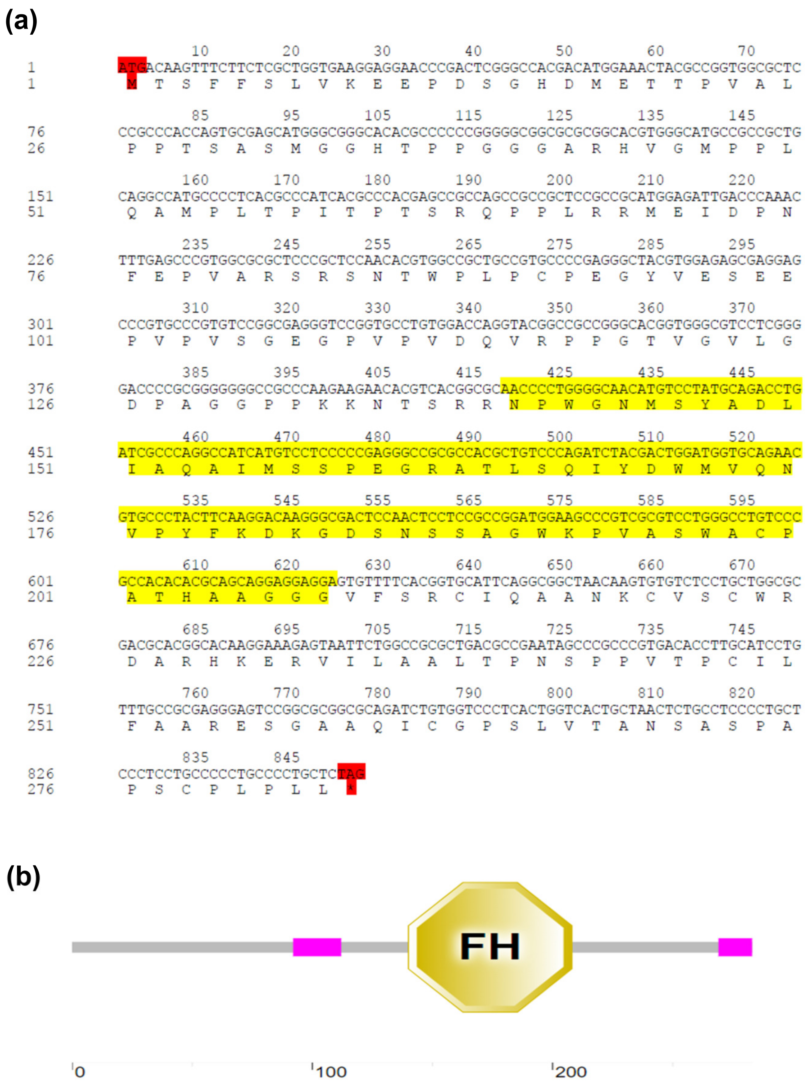
Figure 1. Sequence features and domain architecture of EsFOXO-like. (a) Nucleotide sequence and deduced amino acid sequence of EsFOXO-like. The number of amino acids and nucleotides are listed on the left of the sequence. The start codon (ATG) and stop codon (TAG) are marked in red. The FH domain is marked in yellow. (b) SMART analysis of EsFOXO-like. The EsFOXO-like protein domain contained two low complexity regions (pink boxes) and a conserved FH domain (yellow polygon).
Jmse 11 01225 g001
Figure 2. Multiple sequence alignment of the FOXOs from both vertebrates and invertebrates. The amino acids shaded in dark are identical residues between all analyzed sequences, and relatively conserved amino acids are shown in gray. The FH domain is marked in the yellow box. Sequence information of the FOXO proteins is listed in Table 2. PKA: protein kinase A; GSK3: glycogen synthase kinase 3.
Jmse 11 01225 g002
Figure 3. Phylogenetic relationship of FOXO sequences. The phylogenetic tree of FOXO gene was constructed based on the amino acid sequences of EsFOXO-like and another twenty FOXOs (Table 2) using maximum likelihood method implemented in the Mega 11.0 software. The bootstrap values of the branches were obtained by testing the tree 1000 times and are shown as percentage numbers.
Jmse 11 01225 g003
Figure 4. The mRNA expression level of EsFOXO-like in different tissues. The data were analyzed statistically using one-way ANOVA analysis. Each bar represents the mean ± standard deviation of three independent biological replicates (Supplementary Table S1). Different lowercase letters indicate significant differences (p < 0.05).
Jmse 11 01225 g004
Figure 5. The mRNA and protein expression patterns of EsFOXO-like in hepatopancreas at different molt stages. (a) The relative mRNA expression level of EsFOXO-like in hepatopancreas at different molt stages by qRT-PCR. (b) The specific antibody detection of FOXO1 antibody in hepatopancreas. (c) The protein expression levels of EsFOXO-like in hepatopancreas at different molt stages using western blotting. (d) Statistical analysis of western blot performed in (c). Each bar represents the mean ± standard deviation of three independent biological replicates (Supplementary Table S1). The results were analyzed using one-way ANOVA. Asterisks indicate significant differences (* p < 0.05 and ** p < 0.01).
Jmse 11 01225 g005
Figure 6. The mRNA and protein expression levels of EsFOXO-like gene in hepatopancreas after being injected with AS1842856 and EsFOXO-like dsRNA. (a) The EsFOXO-like mRNA expression level in hepatopancreas in AS1842856 injected crabs. (b,c) The protein expression levels of EsFOXO-like in hepatopancreas in AS1842856 and DMSO group. (c) Statistical analysis of blot performed in (b). (d) The mRNA expression level of EsFOXO-like in hepatopancreas after knockdown of EsFOXO-like by RNAi. (e,f) The protein expression levels of EsFOXO-like in hepatopancreas in EsFOXO-like-RNAi group and EFPG-RNAi group. (f) Statistical analysis of blot performed in (e). Each bar represents the mean ± standard deviation of three independent biological replicates (Supplementary Table S1). Asterisks indicate significant differences (* p < 0.05), “ns” = not significant. The results were analyzed using Student’s t test.
Jmse 11 01225 g006
Figure 7. The 20E concentration and mRNA expression levels of EsEcR, EsRXR and EsMIH in the AS1842856 group. (a) The 20E concentration in serum in AS1842856 injected crabs. The data were analyzed using Mann–Whitney U test. (bd) Relative expression levels of EsEcR and EsRXR in the hepatopancreas and EsMIH expression level in the eyestalk of crabs injected with AS1842856. The DMSO group was the control group. The data were analyzed by Student’s t test. Each bar represents the mean ± standard deviation of three independent biological replicates (Supplementary Table S1). Asterisks indicate significant differences (* p < 0.05, ** p < 0.01).
Jmse 11 01225 g007
Figure 8. The 20E concentration and mRNA expression levels of EsEcR, EsRXR and EsMIH after EsFOXO-like was knocked down by RNAi. (a) The 20E concentration in serum in EsFOXO-like-RNAi group. (bd) Relative expression levels of EsEcR and EsRXR in the hepatopancreas and EsMIH expression level in the eyestalk of EsFOXO-like-RNAi crabs group. The EFPG-RNAi group was the control group. Each bar represents the mean ± standard deviation of three independent biological replicates. Asterisks indicate significant differences (** p < 0.01), “ns” = not significant. The data were analyzed by one-way ANOVA.
Jmse 11 01225 g008
Figure 9. The expression level of EsmTOR gene in hepatopancreas after EsFOXO-like inhibition. (a) The EsmTOR expression level in AS1842856-injected crabs. The data were analyzed by Student’s t test. (b) The EsmTOR expression level in EsFOXO-like-RNAi group. The data were analyzed by one-way ANOVA. Each bar represents the mean ± standard deviation of three independent biological replicates (Supplementary Table S1). Asterisks indicate significant differences (* p < 0.05 and ** p < 0.01), “ns” = not significant.
Jmse 11 01225 g009
Figure 10. The expression level of EsmTOR gene in hepatopancreas after injection with AS1842856 and Rapamycin. AS1842856 + DMSO were injected and treated as the control group. Each bar represents the mean ± standard deviation of three independent biological replicates (Supplementary Table S1). Asterisks indicate significant differences (* p < 0.05). The data were analyzed by Student’s t test.
Jmse 11 01225 g010
Figure 11. The 20E concentration and mRNA expression levels of EsEcR, EsRXR and EsMIH in AS1842856 + Rapamycin group. (a) The 20E concentration in serum in AS1842856- and Rapamycin-injected crabs. (bd) The relative expression levels of EsEcR and EsRXR in hepatopancreas and EsMIH expression level in eyestalk in AS1842856 + Rapamycin group. The AS1842856 + DMSO group was treated as the control group. Each bar represents the mean ± standard deviation of three independent biological replicates (Supplementary Table S1). Asterisks indicate significant differences (* p < 0.05). The data were analyzed by Student’s t test.
Jmse 11 01225 g011
Table 1. Sequences of the primers used in the study.
Primer NameSequences (5′–3′)Tm (°C)Size (bp) Efficiency (%)
Cloning primers
EsFOXO-FATGACAAGTTTCTTCTCGCT52.5852
EsFOXO-RCTAGAGCAGGGGCAGGGG62.3
qRT-PCR primers
EsFOXO-like-FGGCTACGTGGAGAGCGAGGA60.6142 99%
EsFOXO-like-RCCTGGGCGATCAGGTCTGC60.0
EsEcR-FGAGAGAACAGAAAAAGGCACGA57.3105 102%
EsEcR-RATGGCTGACATTGGACTAATGG58.8
EsMIH-FTGAAGACTGCGCCAACATCT56.290 103%
EsMIH-RCGTGAGGTCGTCCTTCTGTG60.4
EsRXR-FAGGCTTCAGGTTCCACTCGC58.3131 98%
EsRXR-RGTGTACGCTGCCCTGGAGGA60.1
EsmTOR-FCTTGAGGAGTTCTACCCTGCGT60.5115 101%
EsmTOR-RGGACCACCTGGGCAAGGTAT56.9
Esβ-actin-FGCATCCACGAGACCACTTACA56.4223 102%
Esβ-actin-RCTCCTGCTTGCTGATCCACATC60.1
RNAi primers
EGFP-RNAi-FTAATACGACTCACTATAGGGCGACGTAAACGGCCACAAGT
EGFP-RNAi-RTAATACGACTCACTATAGGGCTTGTACAGCTCGTCCATGC
EsFOXO-RNAi-FTAATACGACTCACTATAGGGATGACAAGTTTCTTCTCGCTGGTGA
EsFOXO-RNAi-RTAATACGACTCACTATAGGGATCAGGTCTGCATAGGACATGTTGC
Tm: Annealing temperature; Size: Amplicon size.
Table 2. The FOXO genes used in the alignment of EsFOXO-like.
SpeciesGene NameAccession NumberQuery Cover %Identity %
Azumapecten farreriFOXO-like proteinQFR 39803.14353.66
Crassostrea gigasforkhead box protein OXM 011416057.34360.16
Mytilus coruscusFOXO3CAC 5392548.14357.03
Sepia pharaonisFOXO3CAE 1327826.13960.53
Asterias rubensforkhead box protein O3-likeXP 033635399.14354.48
Strongylocentrotus purpuratusforkhead transcription factor ODQ 286746.24349.30
Dendroctonus ponderosaeforkhead box protein O isoform X3XP 048524044.15251.23
Sitophilus oryzaeforkhead box protein O isoform X2XP 030755757.15059.17
Drosophila melanogasterforkhead box, sub-group O
isoform C
NP 996204.14255.37
Spodoptera frugiperdaforkhead box protein O-like
isoform X2
XP 035439085.14454.33
Colias croceusforkhead box protein O isoform X1 XP 045494104.15447.67
Penaeus vannameiforkhead box protein O-like
isoform X2
XP 027228067.14075.00
Penaeus monodonforkhead box protein O4-likeXP 037792098.14088.37
Portunus trituberculatusForkhead box protein O MPC 39137.14288.55
Penaeus japonicusFork box protein O MW080526.16783.51
Clupea harengusforkhead box protein O1-B-like XP 031428678.13083.33
Salvelinus alpinusforkhead box protein O1 XP 023824765.14585.45
Orcinus orcaforkhead box protein O6XP 033277871.14352.67
Oryx dammahforkhead box protein O6XP 040087454.14352.67
Homo sapiensforkhead box protein O6NP 001278210.24349.63
Disclaimer/Publisher’s Note: The statements, opinions and data contained in all publications are solely those of the individual author(s) and contributor(s) and not of MDPI and/or the editor(s). MDPI and/or the editor(s) disclaim responsibility for any injury to people or property resulting from any ideas, methods, instructions or products referred to in the content.