Article

Effects of Iron Valence on the Growth, Photosynthesis, and Fatty Acid Composition of Phaeodactylum tricornutum

1
School of Life Sciences, Ludong University, Yantai 264025, China
2
Yantai Engineering & Technology College, Yantai 264025, China
3
School of Resources and Environmental Engineering, Ludong University, Yantai 264025, China
*
Author to whom correspondence should be addressed.
J. Mar. Sci. Eng. 2023, 11(2), 316; https://doi.org/10.3390/jmse11020316
Submission received: 9 December 2022 / Revised: 21 January 2023 / Accepted: 29 January 2023 / Published: 2 February 2023
(This article belongs to the Special Issue Algal Cultivation and Breeding)

Abstract

:
Iron is a limiting factor that controls the phytoplankton biomass of the ocean and plays an important role in the lipid production of microalgae. Elucidating the effects of different iron valences on microalgae is helpful for their commercial production. We investigated the growth, photosynthesis, and fatty acid profile of the model diatom Phaeodactylum tricornutum cultured with depleted Fe, Fe2+, Fe2+/Fe3+, and Fe3+. Samples were taken every 24 h for 8 days, and their cell density, photosynthetic pigment content, chlorophyll fluorescence, total fatty acid content, and fatty acid composition were analyzed. The cell densities of the Fe2+ and Fe2+/Fe3+ groups were significantly higher than those of the control and Fe3+ groups (p < 0.05). They were 1.26 times and 1.23 times higher than those in the Fe-depleted group. The contents of chlorophyll a and c in the Fe2+ group were significantly higher than those in the Fe-depleted group (p < 0.05). The chlorophyll fluorescence results show that Fe2+ enhanced the photosynthesis of P. tricornutum to a greater extent than Fe3+. On the eighth day of harvest, Fv/Fm and Y(II) in the Fe2+ group were 0.672 and 0.476, respectively, being 1.10 and 1.19 times greater than those in the Fe3+ group and 1.15 and 1.33 times greater than those in the Fe-depleted group, respectively. Compared with the control group, the levels of saturated fatty acids of the Fe2+/Fe3+ and Fe3+ groups were significantly higher (p < 0.05) at 21.36 ± 1.24% and 21.20 ± 0.13%, respectively. The levels of polyunsaturated fatty acids of the Fe2+/Fe3+ group were significantly lower (p < 0.05) at 29.82 ± 2.75%. Our results show that P. tricornutum exhibited physiological plasticity, including changes in photosynthetic activities and shifts in fatty acid composition, in response to different iron valences and that Fe2+ was more beneficial to the biomass production of this species than Fe3+. These findings are applicable to the production of biomass and polyunsaturated fatty acids.

1. Introduction

Iron is one of the most abundant elements on Earth, and it is present in multiple forms in aquatic environments, including in solutions and colloids [1]. Like the three biogenic elements of nitrogen, phosphorus, and carbon, iron plays an important role in phytoplankton physiology [2]. Iron is both an essential element for phytoplankton growth and a major factor controlling marine phytoplankton biomass. It is essential for multiple metabolic pathways, including electron transport, oxygen metabolism, nitrate utilization, and respiration. It is especially important in several photosynthetic activities [3], as the photosynthetic electron transfer of phytoplankton requires the participation of iron [4]. Iron is a key component of the cytochrome b6-f complex, which is an enzyme present in the thylakoid membrane of chloroplasts that plays an important role in photosynthesis through electron transfer between photosystems I and II [5]. Therefore, iron deficiency adversely affects the growth, photosynthesis, and lipid accumulation of phytoplankton [4,6].
Different iron sources have different effects on phytoplankton growth [7]. Polat et al. showed that FeCl3 inhibited the growth of Auxenochlorella protothecoides [8], whereas FeSO4 helped it obtain the highest biomass, and cells cultured with Fe-EDTA had the highest specific growth rate. They also reported that the fatty acid yield of A. protothecoides cultured with FeCl3 was higher than that of cells cultured with FeSO4 and Fe-EDTA. In another study, Rizwan et al. showed that Dunaliella tertiolecta cultured with FeH8N2O8S2 exhibited good growth but a lower lipid content, whereas cells cultured with Fe-EDTA had a higher lipid content than cells cultured with other iron sources [9]. Iron sources also affected the photosynthesis of microalgae. Ferric EDTA was reported to enhance the mean intensity of chlorophyll fluorescence, compared with ferric citrate in Phaeodactylum tricornutum [10]. The results were different for Anabaena flosaquae. When the concentration of iron is in the range of 0.1–0.8 mg L−1, iron species have more significant influences on Anabaena bloom compared with iron concentration. The promoting effect was in the order of ferric ammonium citrate >EDTA-Fe> iron ions > iron oxalate, and among the four iron sources, A. flosaquae had the strongest adsorption capacity for iron ions [11].
The diatom P. tricornutum has become a model species in the biotechnology field because it is a biofuel precursor and a recombinant protein expression host due to its biosynthetic capacity and high growth rates [12]. This species can grow photoautotrophically by using CO2 and light as carbon and energy sources, respectively. It can store carbon and energy in the form of neutral lipids, especially triacylglycerols. This diatom has potential as an energy source for biodiesel production. Moreover, it may also be a source of omega-3, long-chain polyunsaturated fatty acids. In addition, as much as 35–40% of the total fatty acid content (TFAC) of P. tricornutum comes from natural accumulation of eicosapentaenoic acid (EPA) [13,14], which makes it a potential candidate for EPA production. Like other phytoplankton species, the growth performance and lipid accumulation of P. tricornutum are usually affected by iron concentrations [15]. Hayward reported that iron deficiency led to growth restriction and reduced pigmentation and photochemical efficiency in this species [16]. More recently, iron limitation (0.04 µM iron) has been shown to decrease photosystem II content in P. tricornutum, and the decline in the content of cytochrome c550 under iron-limiting conditions is accompanied by a decrease in the binding of this protein to photosystem II, and also the extrinsic PsbO subunit [17]. Other experiments have shown that adding an appropriate amount of iron (FeCl3·6H2O, 12 and 60 mg L−1) can promote the growth of Chlorella sorokiniana [18]. Chen et al. found that both iron deficiency and iron excess enhanced lipid accumulation in P. tricornutum [19]. Zhao et al. reported that the addition of iron at concentrations in the range of 1.2 × 10⁻⁸ to 1.2 × 10⁻⁵ mol L−1 resulted in growth rates of P. tricornutum that were similar between adjacent iron concentration gradients and that the trend between FeCl3 and FeSO4 was similar [20]. Additionally, Kosakowska et al. found that Fe3+ affected the qualitative and quantitative composition of P. tricornutum pigments [21]. Liang et al. used different iron sources (FeCl3, C6H5FeO7, FeSO4) to culture P. tricornutum mutant strains and found that FeSO4 at 0.5 mg L−1 was optimal for growth. Higher FeSO4 concentrations (1 mg L−1) promoted EPA accumulation [22].
To date, most studies on the effect of iron on P. tricornutum have focused on the iron source type (chelated or unchelated, FeCl3 or FeSO4) and concentration, especially under iron depletion. The effects of different iron valences on the growth and physiology are rarely reported. P. tricornutum is a promising algae species for biofuel production and usually cultivated with enriched iron. It is important to know which valence form of iron is the best source for the bulk culture of this species. We hypothesize that Fe2+ is more effective in promoting the growth, photosynthetic yield, and fatty acid accumulation of P. tricornutum. Therefore, we tested the effects of different iron valences (Fe-depleted, Fe2+, Fe2+/Fe3+, and Fe3+) under replete concentrations on the growth, photosynthesis, and fatty acid composition of P. tricornutum. The purpose of this study was to identify the valence of iron that would result in the highest fatty acid accumulation and photosynthetic yield. The results are applicable to the production of biomass and PUFAs.

2. Materials and Methods

2.1. Algal Strain and Medium Composition

P. tricornutum was obtained from the Microalgae Culture Center, Ocean University of China (Qingdao, China). The alga was maintained in f/2 medium [23] with laboratory-prepared artificial sea water to avoid the potential influence of iron in natural seawater. The artificial seawater [24] was filtered and sterilized to prepare f/2 culture medium without the addition of silicon. Iron was added to the medium in the form of FeSO4·7H2O or FeCl3·6H2O at a final concentration of 10−6 mol L−1. The experiments were divided into four groups: control (Fe-depleted, Ctrl), ferrous (Fe2+), ferrous/ferric (each half, Fe2+/Fe3+), and ferric (Fe3+). Each treatment was replicated four times.

2.2. Culture and Growth

Algal cultures in the log phase were harvested via centrifugation at 4000× g for 10 min and washed twice with artificial sea water, and the pellets were then resuspended in 500 mL of medium to generate the inoculum. Cells were grown at 20 °C under cool white fluorescent tubes (100 μmol m–2 s−1) with a photoperiod of 12 h light:12 h dark for 8 days, and they were manually shaken periodically. The initial inoculation density of all experimental groups was 106 cells mL−1. Daily sampling for analysis was conducted 1 h after the end of the light cycle. Cell numbers were counted under a microscope using the Neubauer-improved chamber (Marienfeld, Lauda-Konigshofen, Germany), and cell growth was recorded. At the end of the growth experiment, cells were harvested via centrifugation at 4000× g for 10 min and washed twice with artificial seawater. Cell pellets were then prepared for the analysis of photosynthetic pigments and fatty acid composition.

2.3. Photosynthesis

Each day, the photosynthesis of 2 mL cultures from each group was measured using a pulse-amplitude-modulated fluorometer (Water-PAM; Walz, Effeltrich, Germany). First, each sample was dark-adapted for 15 min, and then the minimum (Fo) and maximum (Fm) fluorescence were immediately determined as the maximal photochemical efficiency of photosystem II (PSII) (Fv/Fm). The actinic light was then turned on to promote photosynthesis, and the following parameters were determined: the effective photochemical efficiency of PSII in the light [Y(II) = yield = Fv/Fm = (Fm’ − Fo)/Fm], the electron transport rate (ETR = Y(II)·PAR·0.84·0.5), and nonphotochemical quenching (NPQ). All the determinations were carried out in darkness. Based on the results of Ralph [25], we derived cardinal points of a rapid light curve (α, Ek and ETRmax) to describe the photosynthetic capacity of P. tricornutum, its light adaptation state, and its capacity to tolerate short-term changes in light.

2.4. Photosynthetic Pigments

Chlorophyll (Chl-a, c) and total carotenoid were extracted according to Tucker [26]. The pigment concentrations were determined spectrophotometrically with a TU-1800 spectrophotometer (Pgeneral Ltd., Beijing, China) using coefficients reported in Jeffrey and Humphrey [27] and Strickland and Parsons [28]:
Chl-a (mg mL−1) = 11.47 × (A664 − A750) − 0.40 × (A630 − A759),
Chl-c (mg mL−1) = 24.34 × (A630 − A750) − 0.40 × (A664 − A759),
Carotenoid (mg mL−1) = 7.6 × ((A480 − A750) − 1.49 × (A510 − A750))

2.5. Fatty Acid Analysis

The fatty acid composition of each sample was determined according to our previous report [29]. A 0.1 g wet pellet with 0.1 mg glyceryl triheptadecanoate as the internal standard was transesterified using a sequential combination of base and acid catalysts. Fatty acid methyl esters were then analyzed using a gas chromatograph (GC-2010; Shimadzu, Tokyo, Japan) equipped with a fused silica capillary column (Supelco SP-2560: 100 m × 0.25 mm, film thickness 0.20 μm; Bellefonte, PA, USA). Injector and flame ionization detector temperatures were controlled at 260 °C with nitrogen as the carrier gas. The column temperature was programmed from 140 to 240 °C at a rate of 4 °C min−1. Fatty acids were identified by comparing the relative retention time with the reference standards (Merck, Rahway, NJ, USA).

2.6. Statistical Analysis

Experimental results are expressed as the mean ± standard deviation (SD) of the four replicates. The Kolmogorov–Smirnov test and Levene’s F-test were used to check normality and heterogeneity of variances. Data with parametric distribution were subjected to one-way analysis of variance. The Duncan multiple-range test [30] or least-significant difference test [31] was performed to identify significant differences between any two means that differed at p < 0.05. All statistical analyses were performed using IBM SPSS statistics version 20.0 (IBM, Armonk, NY, USA).

3. Results

3.1. Cell Density

The cell density clearly increased from 24 to 48 h, during which the specific growth rate of the Fe2+ group changed the most, reaching 0.278 d−1. The cell density of the Fe3+ group was lower than that of the control group until day 5, but it showed a slight increase compared with the control group from day 6 to day 8. On the eight day, the cell densities among the treatments were significantly different. As shown in Figure 1b, the Fe2+ group had the highest density, followed by the Fe2+/Fe3+ group. Both groups had significantly higher values than the control and Fe3+ groups (p < 0.05). However, the cell density under the Fe3+ treatment was not significantly different from that of the control group (p > 0.05).

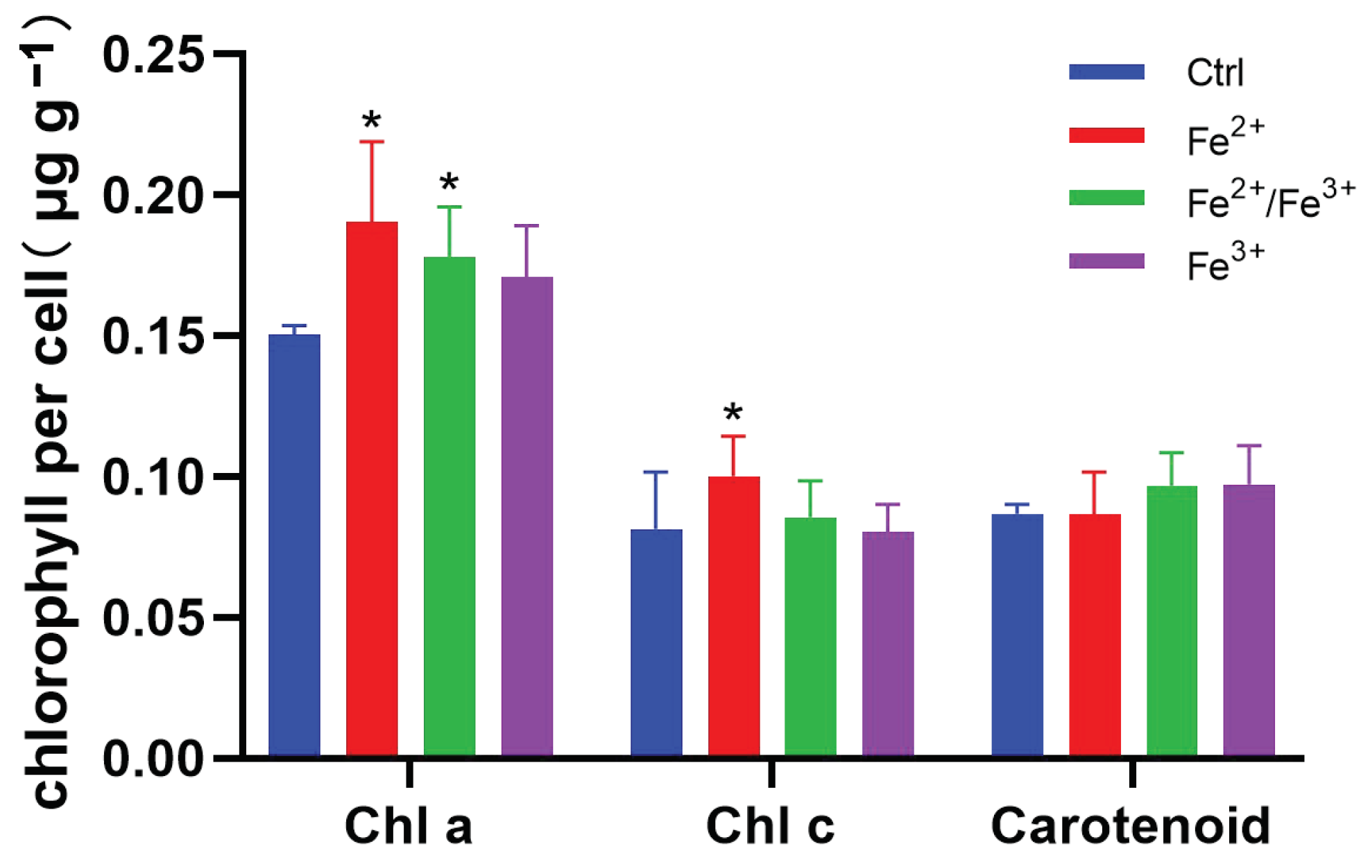
3.2. Photosynthetic Pigments

Chl-a content was significantly higher in the Fe2+ and Fe2+/Fe3+ groups than in the control group (p < 0.05), with increases of 26.4% and 19.8% by the end of day 8, respectively (Figure 2). Chl-c content was significantly higher in Fe2+-treated cells than in the control group (p < 0.05), but there was no significant difference in Chl-c concentration between the control and Fe2+/Fe3+ or Fe3+ groups (p > 0.05). The highest carotenoid concentrations were found in the Fe3+ group and then the Fe2+/Fe3+ group; however, the differences among all groups were not statistically significant (p > 0.05).

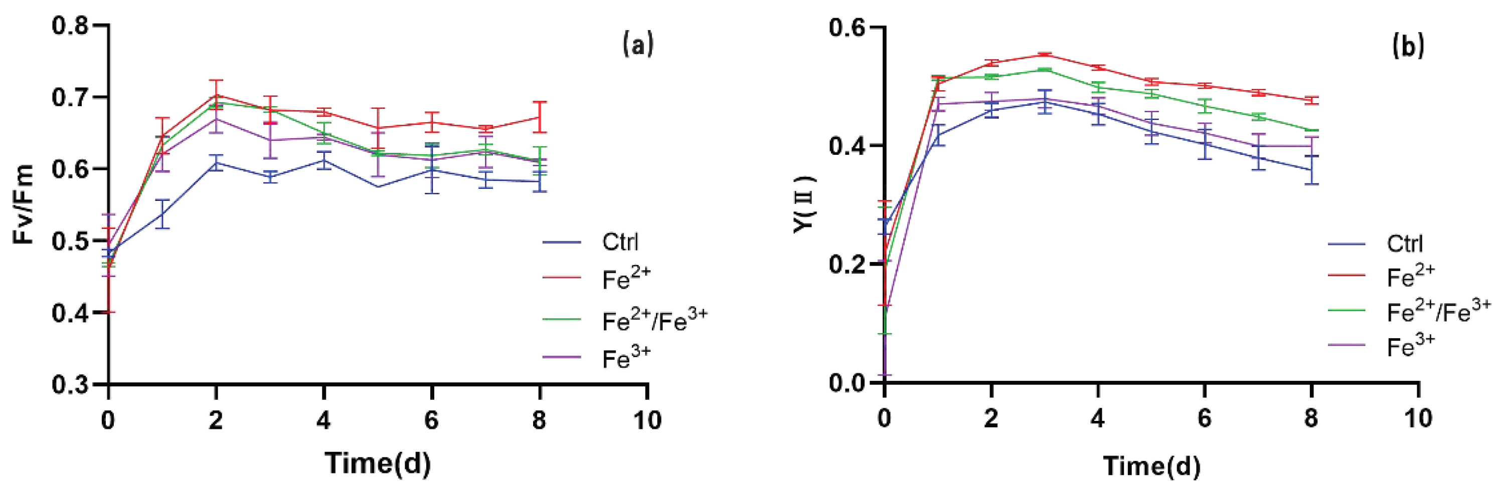
3.3. Chl Fluorescence

Both Fv/Fm and Y(II) increased immediately after iron treatment, and the rate of increase was highest in the first 24 h (Figure 3). Fv/Fm peaked at 48 h in all iron treatment groups (Figure 3a), followed by a downward trend until it leveled off. The Fv/Fm increase rate of the Fe2+ group was always higher than that of the other groups during the experimental period, and this difference was statistically significant from day 4 until the end of the experiment (p < 0.05). The difference between the Fe2+/Fe3+ and Fe3+ groups began to appear on day 1, peaked on day 3, and then gradually decreased until the end of the experiment. Y(II) showed a similar trend of increasing first and then decreasing, with the maximum value appearing on day 3 (Figure 3b). The results clearly show that Fe2+ and Fe2+/Fe3+ had a significant promoting effect on Fv/Fm and Y(II), and the effect of Fe2+ was better than that of Fe2+/Fe3+. Additionally, the NPQ values of the Fe2+ and Fe2+/Fe3+ groups were significantly lower than that of the control group on day 4 (p < 0.05), and the values had decreased by 60.4% and 64.1%, respectively, by the end of the experiment (Figure 4).
The light–response curves of all treatments showed a trend of rising first and then gradually becoming stable (Figure 5). During this process, the advantage of the Fe2+ group gradually became significant. The saturation irradiance (Ek) of the Fe2+/Fe3+ and Fe3+ groups was lower than that of the control group (Table 1). The smallest Ek occurred in the Fe3+ group (144.38 ± 11.68 μmol photons m–2 s−1), and the largest Ek was about 1.3 times higher in the Fe2+ group. In addition, the α values of all Fe-treated groups were significantly higher than those of the control group (p < 0.05), and the value of the Fe2+ group (α = 0.23) was significantly higher than those of the other two iron groups (α = 0.21, p < 0.05). The trend for ETRmax was similar to that for the α value (Table 1).

3.4. Total Fatty Acid Content (TFAC) and Fatty Acid Composition

No significant differences in the TFAC were found among the experimental groups (p < 0.05, Table 2). The fatty acid compositions of all treatments on day 8 were analyzed, and the main constituents were C14:0 (6.03–6.24%), C16:0 (13.03–14.42%), C16:1n-7 (19.80–21.78%), C18:1n-9 (7.34–7.81%), and EPA (23.86–25.84%) in each treatment (Table 2). There were no significant differences in the C14:0, C16:1n-7, or C18:1n-9 percentage contents among the four groups. The C16:0 percentage content of Fe2+/Fe3+ was significantly higher than that of the control (p < 0.05). The EPA percentage contents of Fe2+/Fe3+ and Fe3+ groups were found to be significantly lower than those of the control (p < 0.05), whereas there were no significant differences between the Fe2+ group and the control (p > 0.05).
A gradual upward trend was found in the levels of saturated fatty acids (SFAs) with increasing iron valences, and those of the Fe2+/Fe3+ and Fe2+ groups were significantly higher than those of the control (p < 0.05). The levels of monounsaturated fatty acids (MUFAs) showed a similar trend, with a significantly higher value in the Fe2+/Fe3+ group than in the Fe2+ group (p < 0.05). However, the levels of polyunsaturated fatty acids (PUFAs) showed the opposite trend, and those of the Fe2+/Fe3+ groups were significantly lower than those of the control (p < 0.05). The ratio of docosahexaenoic acid (DHA) to EPA remained steady and did not differ significantly between the control and experimental groups (Table 2).

4. Discussion

4.1. Effects of Different Iron Valences on the Growth of P. tricornutum

The growth of P. tricornutum cells treated with Fe2+ was better than that of cells treated with Fe3+, Fe2+/Fe3+, or no iron. Zhou et al. compared the effects of Fe2+ and Fe3+ on the growth of natural mixed algal colonies and single-species colonies of Scenedesmus quadricauda and reported that Fe2+ exhibited higher biological time-effectiveness than Fe3+ for algal reproduction [32], which is consistent with our finding. However, our growth results are inconsistent with those of Zhao et al., who reported a similar growth rate between FeCl3 and FeSO4 at the same iron concentration [20], most likely because we used artificial seawater instead of natural seawater throughout the experimental period. We also found that, compared to the P. tricornutum cells under the Fe3+ conditions, the growth of the control group did not show signs of significant iron limitation. A similar result was reported by Liang et al. [22].
On the one hand, P. tricornutum could minimize the need for iron by replacing iron-rich proteins with iron-free proteins to withstand the pressure of iron limitation [20]. At the same time, we speculate that certain metal ions in the experiment replaced the function of iron. According to Kothamasi and Kothamasi, cobalt is a possible competitive inhibitor of iron uptake because of its similar size [33]. The presence of cobalt in the f/2 medium used in our experiment may have narrowed the advantage of the iron-treated groups over the control group. In addition, Rico et al. studied iron-limited diatom cells and found that copper can replace iron in certain metabolic functions [34], such as some high-affinity iron transport systems. Another response of phytoplankton to iron deprivation is the substitution of Cyt c553 (one Fe atom) with copper-dependent plastocyanin (no Fe atoms) and the partial replacement of ferredoxin (two Fe atoms) with flavodoxin (no Fe atoms) [35]. In another study, Peers and Price suggested that selection pressure imposed by Fe limitation resulted in the use of a copper protein for photosynthesis in an oceanic diatom, and this biochemical switch reduced the need for Fe and increased the requirement for copper [36].
The P. tricornutum genome contains at least three clusters of Fe-responsive genes. One of the Fe-regulated clusters contains the gene encoding a copper tyrosinase (TYR1) and a protein of unknown function (ISIP1) [37]. Because the role of copper nutrition in iron-limited diatoms has become increasingly apparent [38], we infer that the addition of Cu2+ in f/2 medium may play a role in replacing iron.

4.2. Effects of Different Iron Valences on Photosynthesis of P. tricornutum

Chlorophyll is an important pigment involved in photosynthesis, and its content indicates the stress resistance of plants to a certain extent [39,40]. Although iron is not directly involved in the composition of chlorophyll, it is the precursor of chlorophyll synthesis and plays a key role in the activation of various enzymes in the process of chlorophyll synthesis [41,42,43]. Therefore, iron deficiency may be sufficient to retard chlorophyll synthesis [44]. Carotenoids are important pigments that play a major role in the protection of photosynthetic eukaryotes against photooxidative processes because of their antioxidant activity [45,46]. Although the synthesis pathway of carotenoids does not depend on iron, iron reduction may allow their formation to actively compete with chlorophyll synthesis for glycine, thereby reducing the amount of glycine incorporated into carotenes and xanthophylls [47]. In the present study, iron significantly increased the production of Chl- a in P. tricornutum cells, and it also affected the content of Chl-c and carotenoids, which is consistent with the above conclusions. At the same time, statistical analysis showed that the enhancing effect of Fe2+ treatment on Chl-a content was better than that of Fe3+. This may be because the pyrrole ring is a chlorophyll precursor [48], and in the dark, ferrous stimulate the formation of porphyrin rings [49,50,51].
Photosynthetic parameters are commonly used to evaluate the effects of environmental stress on microalgae, and they can reflect algal growth status [52]. In our study, the values of Fv/Fm, ETR, and Y(II) of P. tricornutum cells treated with iron were all higher than those of the control group (Figure 3 and Figure 5), indicating that iron has important effects on the photosynthesis of P. tricornutum cells. Similar results were reported for Anacystis nidulans [53], D. tertiolecta [54], and Beta vulgaris leaves [55]. NPQ is fluorescence quenching that is caused by heat dissipation, and it reflects the ability of plants to dissipate excess light energy into heat; therefore, it is a photoprotective ability [56]. Decreased NPQ was detected in iron-treated P. tricornutum (Figure 4), suggesting an increased ability of cells to dissipate excess energy in the form of heat and fluorescence. This would have a positive protective effect on plant photosynthetic machinery. We also found that the ETRmax was higher in the presence of Fe2+ than in the no-iron or Fe3+ groups (Figure 5). These results show that Fe2+ had a certain positive effect on improving the potential maximum relative electron transfer rate of P. tricornutum. Fe2+ treatment also resulted in greater α and Ek values of P. tricornutum cells (Figure 5), which suggests a higher light intensity tolerance.
Virtually all light reactions take place in the thylakoid membranes in oxygenic organisms. These membranes contain two photochemical systems (PSII and PSI), their associated membrane-intrinsic or membrane-bound light-harvesting protein complexes, the cytochrome b6-f complex, and ATP synthase [57]. The cytochrome b6-f complex is the photosynthetic electron transport mediator between PSII and PSI, transferring electrons from water to NADP+ [58]. Iron supply plays an important role in the structure and function of thylakoid membranes and the photochemical energy conversion process [59,60]. Zhao et al. experimentally demonstrated that iron affects the synthesis of the cytochrome b6-f complex in P. tricornutum [20]. This complex contains heme iron and ironululin protein [61], and iron accomplishes the transfer of electrons in the process of photosynthesis via the mutual conversion of Fe2+ and Fe3+ [62]. The electron transfer process in photosynthesis can thus be affected by many iron-containing compounds [63,64]. In our study, the electron transfer rate of P. tricornutum treated with iron was higher than that not treated with iron, and the enhancing effect of Fe2+ treatment was better than that seen for the Fe2+/Fe3+- and Fe3+-treated groups. Therefore, we concluded that Fe2+ has a better promoting effect on electron transport between the PS II and PS I of P. tricornutum than Fe3+, most likely because Fe3+ interrupted the electron transport between PS II and PS I [65].

4.3. Effects of Different Iron Valences on Lipid Accumulation and Fatty Acid Composition of P. tricornutum

The rate of lipid production is an essential feature that must be considered in the large-scale microalgal lipid production process because it takes into account both biomass production and lipid content [5]. For sustainable biodiesel production, the process should be able to produce lipids at the highest productivity level with the highest cellular lipid content [66]. Liu et al. found that adding Fe3+ directly to Chlorella vulgaris in the late growth stage did not induce lipid accumulation in cells, but increased cell density; however, re-inoculation into Fe3+-containing medium after centrifugation induced lipid accumulation in the cells [67]. In our study, the method of reseeding after centrifugation was used, and no obvious increase in TFAC content was found (Table 2). This may be because the cultivation conditions necessary for maximizing biomass productivity are not always consistent with those leading to the highest lipid production [5]. Savvidou et al. previously showed that the TFAC of Nannochloropsis oceanica increased under iron deficiency compared with the control group containing Fe3+ [68]. In other studies, severe iron stress caused Chlamydomonas reinhardtii cells to accumulate a significant amount of lipids, specifically triacylglycerols, by compromising the growth [69,70]. However, in the present study, we clearly showed that iron deficiency did not enhance the TFAC content and even obtained the same growth rate as Fe3+, and the Fe2+ and Fe2+/Fe3+-containing medium promote growth but not lipid accumulation. We inferred that the response of P. tricornutum to iron deficiency is different from that of the green algae. P. tricornutum has been reported to be highly tolerant to Fe limitation and to be able to maintain steady-state growth at an Fe ion concentration in the range of 10–30 pmol L−1 [30], which suggest P. tricornutum should have the ability to utilize and recycle endocellular ferrous iron. This possibility of ferrous recycling via maintaining a steady lipid content in P. tricornutum should be further elucidated.
Studies have shown that the production of PUFAs in certain microalgae could be qualitatively and quantitatively improved under iron-deficient or low-iron conditions [71]. For example, N. oceanica cells cultured under iron-limiting conditions had increased PUFA content [68]. However, iron deficiency reduced the production of PUFAs in Chlamydomonas but increased the production of SFAs [69]. The results of our study are consistent with the report of [68] (Table 2), and further studies are needed to elucidate the various responses of different species. We also found that, compared with the control group, the protection of EPA and PUFAs by Fe2+ was better than that provided by Fe2+/Fe3+ and Fe3+ (Table 2). This was most likely because Fe2+ can combine with a conserved histidine cluster in fatty acid desaturases to form an enzyme active center, thereby catalyzing the formation of fatty acid double bonds [72,73].

5. Conclusions

In this study, we investigated the effects of different iron valences on various physiological indices of P. tricornutum at the iron concentration of 10−6 mol L−1. The results have shown that the valences of iron have a statistically significant effect on biomass accumulation and fatty acid quality. We found that Fe2+ was better than Fe3+ at improving the growth and photosynthesis of P. tricornutum at the enriched iron concentration. During this period, compared with the other groups, Fe2+ showed a higher specific growth rate of 0.278 d−1, while the other groups showed no significant change from the control group (Fe-depleted group). On the eighth day of harvest, Fv/Fm and Y(II) in the Fe2+ group were 1.10 and 1.19 times those in the Fe3+ group, respectively. Fe3+ improved the production of SFAs in P. tricornutum, which increased by 0.51–1.33%, compared to the iron-deficient and Fe2+ group, whereas it seemed to have a certain tendency to decrease PUFA accumulation when added to the medium. Based on the results, Fe2+ should be the first choice for biomass and EPA production by P. tricornutum, and Fe3+ blending should be avoided. We hope that our experiment can provide some reference for future research on how to improve microalgae yield and PUFA production.

Author Contributions

Conceptualization, H.W., Q.S. and H.Q.; methodology, H.W. and H.Q.; software, Q.S.; validation, H.W., B.G. and H.Q.; formal analysis, H.W., Y.Z. (Yanmin Zhuang) and C.W.; investigation, H.W. and H.Q.; resources, S.T. and H.Q.; data curation, Q.S.; writing—original draft preparation, H.W. and H.Q.; writing—review and editing, H.W., Y.Z. (Yancui Zhao) and H.Q.; visualization, Q.S.; supervision, H.Q.; project administration, S.T. and H.Q.; funding acquisition, H.Q. All authors have read and agreed to the published version of the manuscript.

Funding

This research was funded by the National Natural Science Foundation of China (grant no. 32072997), (grant no. 42006117), Talent Induction Program for Youth Innovation Teams in Colleges and Universities of Shandong Province (2022-2024), the open project of Rongcheng Marine Industrial Technology Research Institute, Ludong University (KF20180003), Major Agricultural Application Technology Innovation Project of Shandong Province (SD2019YY010), and Natural Science Foundation of Shandong Province (ZR2020QC042). The funders had no role in study design, data collection and analysis, decision to publish, or preparation of the manuscript.

Institutional Review Board Statement

Not applicable.

Informed Consent Statement

Not applicable.

Data Availability Statement

Not applicable.

Acknowledgments

The authors would like to thank Qingrong Huang and Yanhua Wang for their help on the experiment preparation.

Conflicts of Interest

The authors declare no conflict of interest.

References

  1. Duverger, A.; Berg, J.S.; Busigny, V.; Guyot, F.; Bernard, S.; Miot, J. Mechanisms of Pyrite Formation Promoted by Sulfate-Reducing Bacteria in Pure Culture. Front. Earth Sci. 2020, 8, 588310. [Google Scholar] [CrossRef]
  2. Hudson, R.J.M.; Morel, F.M.M. lron transport in marine phytoplankton: Kinetics of cellular and medium coordination reactions. Limnol. Oceanogr. 1990, 35, 1002–1020. [Google Scholar] [CrossRef]
  3. Cook, G.; Teufel, A.; Kalra, I.; Li, W.; Wang, X.; Priscu, J.; Morgan-Kiss, R. The Antarctic psychrophiles Chlamydomonas spp. UWO241 and ICE-MDV exhibit differential restructuring of photosystem I in response to iron. Photosynth. Res. 2019, 141, 209–228. [Google Scholar] [CrossRef]
  4. Terauchi, A.M.; Peers, G.; Kobayashi, M.C.; Niyogi, K.K.; Merchant, S.S. Trophic status of Chlamydomonas reinhardtii influences the impact of iron deficiency on photosynthesis. Photosynth. Res. 2010, 105, 39–49. [Google Scholar] [CrossRef] [PubMed]
  5. Rajabi Islami, H.; Assareh, R. Enhancement effects of ferric ion concentrations on growth and lipid characteristics of freshwater microalga Chlorococcum oleofaciens KF584224.1 for biodiesel production. Renew. Energy 2020, 149, 264–272. [Google Scholar] [CrossRef]
  6. de Baar, H.; Gieskes, W.; van Leeuwe, M.; van Oijen, T. Effects of iron limitation on photosynthesis and carbohydrate metabolism in the Antarctic diatom Chaetoceros brevis (Bacillariophyceae). Eur. J. Phycol. 2004, 39, 161–171. [Google Scholar] [CrossRef]
  7. Das, P.; Thaher, M.; Abdulquadir, M.; Al-jabri, H.; Alghasal, G. Optimization of iron dosage for microalgal biomass production as a feedstock for biofuel. Biofuels 2019, 12, 569–577. [Google Scholar] [CrossRef]
  8. Polat, E.; Yüksel, E.; Altınbaş, M. Effect of different iron sources on sustainable microalgae-based biodiesel production using Auxenochlorella protothecoides. Renew. Energy 2020, 162, 1970–1978. [Google Scholar] [CrossRef]
  9. Rizwan, M.; Mujtaba, G.; Lee, K. Effects of iron sources on the growth and lipid/carbohydrate production of marine microalga Dunaliella tertiolecta. Biotechnol. Bioprocess Eng. 2017, 22, 68–75. [Google Scholar] [CrossRef]
  10. Botebol, H.; Sutak, R.; Scheiber, I.F.; Blaiseau, P.L.; Bouget, F.Y.; Camadro, J.M.; Lesuisse, E. Different iron sources to study the physiology and biochemistry of iron metabolism in marine micro-algae. Biometals 2014, 27, 75–88. [Google Scholar] [CrossRef]
  11. Qiu, Y.; Wang, Z.; Liu, F.; Liu, J.; Zhou, T. Effect of different kinds of complex iron on the growth of Anabaena flos-aquae. Environ. Technol. 2019, 40, 2889–2896. [Google Scholar] [CrossRef] [PubMed]
  12. Bañuelos-Hernandez, B.; Beltrán-López, J.; Rosales-Mendoza, S. Production of biopharmaceuticals in microalgae. In Handbook of Marine Microalgae; Kim, S.-K., Ed.; Academic Press: Boston, MA, USA, 2015; pp. 281–298. [Google Scholar]
  13. Cui, Y.; Thomas-Hall, S.R.; Chua, E.T.; Schenk, P.M. Development of high-level omega-3 eicosapentaenoic acid (EPA) production from Phaeodactylum tricornutum. J. Phycol. 2021, 57, 258–268. [Google Scholar] [CrossRef] [PubMed]
  14. Kadalag, N.L.; Pawar, P.R.; Prakash, G. Co-cultivation of Phaeodactylum tricornutum and Aurantiochytrium limacinum for polyunsaturated omega-3 fatty acids production. Bioresour. Technol. 2022, 346, 126544. [Google Scholar] [CrossRef]
  15. Wang, Z.; Mou, J.; Qin, Z.; He, Y.; Sun, Z.; Wang, X.; Lin, C.S.K. An auxin-like supermolecule to simultaneously enhance growth and cumulative eicosapentaenoic acid production in Phaeodactylum tricornutum. Bioresour. Technol. 2022, 345, 126564. [Google Scholar] [CrossRef] [PubMed]
  16. Hayward, J. Studies on the growth of Phaeodactylum tricornutum III. The Effect of iron on growth. J. Mar. Biol. Assoc. UK 1968, 48, 295–302. [Google Scholar] [CrossRef]
  17. Castell, C.; Díaz-Santos, E.; Heredia-Martínez, L.G.; López-Maury, L.; Ortega, J.M.; Navarro, J.A.; Roncel, M.; Hervás, M. Iron deficiency promotes the lack of photosynthetic cytochrome c(550) and affects the binding of the luminal extrinsic subunits to photosystem II in the diatom Phaeodactylum tricornutum. Int. J. Mol. Sci. 2022, 23, 2138. [Google Scholar] [CrossRef]
  18. Charria-Girón, E.; Amazo, V.; De Angulo, D.; Hidalgo, E.; Villegas-Torres, M.F.; Baganz, F.; Caicedo Ortega, N.H. Strategy for managing industrial anaerobic sludge through the heterotrophic cultivation of Chlorella sorokiniana: Effect of iron addition on biomass and lipid production. Bioengineering 2021, 8, 82. [Google Scholar] [CrossRef]
  19. Chen, Y.; Qiu, Y.; Zhang, W.; Lu, S.; Liu, T. Effect of nutrient elements on growth and lipid accumulation of Phaeodactylum tricornutum. Biomass Chem. Eng. 2011, 45, 1–6. [Google Scholar]
  20. Zhao, P.; Gu, W.; Huang, A.; Wu, S.; Liu, C.; Huan, L.; Gao, S.; Xie, X.; Wang, G. Effect of iron on the growth of Phaeodactylum tricornutum via photosynthesis. J. Phycol. 2018, 54, 34–43. [Google Scholar] [CrossRef]
  21. Kosakowska, A.; Lewandowska, J.; Stoń, J.; Burkiewicz, K. Qualitative and quantitative composition of pigments in Phaeodactylum tricornutum (Bacillariophyceae) stressed by iron. Biometals 2004, 17, 45–52. [Google Scholar] [CrossRef]
  22. Liang, J.; Jiang, X.; Ye, L.; Han, Q. Effects of nitrogen, phosphorus and iron on the growth, total lipid content and fatty acid composition of Phaeodactylum tricornutum mutant strain. Chin. J. Ecol. 2016, 35, 189–198. (In Chinese) [Google Scholar] [CrossRef]
  23. Guillard, R.R.; Ryther, J.H. Studies of marine planktonic diatoms. I. Cyclotella nana Hustedt, and Detonula confervacea (cleve) Gran. Can. J. Microbiol. 1962, 8, 229–239. [Google Scholar] [CrossRef] [PubMed]
  24. Bidwell, J.; Spotte, S.H. Artificial Seawaters: Formulas and Methods; Jones & Bartlett Learning: Burlington, MA, USA, 1985. [Google Scholar]
  25. Ralph, P.J.; Gademann, R. Rapid light curves: A powerful tool to assess photosynthetic activity. Aquat. Bot. 2005, 82, 222–237. [Google Scholar] [CrossRef]
  26. Tucker, A. Pigment extraction as a method of quantitative analysis of phytoplankton. Trans. Am. Microsc. Soc. 1949, 68, 21. [Google Scholar] [CrossRef]
  27. Jeffrey, S.W.; Humphrey, G.F. New spectrophotometric equations for determining chlorophylls a, b, c1, and c2 in higher plants, algae and natural phytoplankton. Biochem. Physiol. Pflanz. 1975, 8, 53–59. [Google Scholar] [CrossRef]
  28. Strickland, J.D.H.; Parsons, T.R. A Practical Handbook of Seawater Analysis; Fisheries Research Board of Canada: Ottawa, ON, Canada, 1968. [Google Scholar]
  29. Qiao, H.; Cong, C.; Sun, C.; Li, B.; Wang, J.; Zhang, L. Effect of culture conditions on growth, fatty acid composition and DHA/EPA ratio of Phaeodactylum tricornutum. Aquaculture 2016, 452, 311–317. [Google Scholar] [CrossRef]
  30. Duncan, D.B.J.B. Multiple range and multiple F tests. Biometrics 1955, 11, 1–42. [Google Scholar] [CrossRef]
  31. Williams, L.J.; Abdi, H.H. Fisher’s Least Significant Difference (LSD) Test. 2010. Available online: https://www.graphpad.com/guides/prism/latest/statistics/stat_fishers_lsd.htm (accessed on 16 April 2022).
  32. Zhou, T.; Wang, Z.; Hu, Q.; Liu, L.; Luo, K. Effects of Fe2+ and Fe3+ on algal proliferation in a natural mixed algal colony in algae-rich raw water in southern China. J. Residuals Sci. Technol. 2016, 13, 15–22. [Google Scholar] [CrossRef]
  33. Kothamasi, D.; Kothamasi, S. Cobalt interference in iron-uptake could inhibit growth in Pseudomonas aeruginosa. World J. Microbiol. Biotechnol. 2004, 20, 755–758. [Google Scholar] [CrossRef]
  34. Rico, M.; López, A.; Santana-Casiano, J.M.; Gonzàlez, A.G.; Gonzàlez-Dàvila, M. Variability of the phenolic profile in the diatom Phaeodactylum tricornutum growing under copper and iron stress. Limnol. Oceanogr. 2013, 58, 144–152. [Google Scholar] [CrossRef]
  35. Behrenfeld, M.J.; Milligan, A.J. Photophysiological expressions of iron stress in phytoplankton. Annu. Rev. Mar. Sci. 2013, 5, 217–246. [Google Scholar] [CrossRef]
  36. Peers, G.; Price, N.M. Copper-containing plastocyanin used for electron transport by an oceanic diatom. Nature 2006, 441, 341–344. [Google Scholar] [CrossRef]
  37. Allen, A.E.; Laroche, J.; Maheswari, U.; Lommer, M.; Schauer, N.; Lopez, P.J.; Finazzi, G.; Fernie, A.R.; Bowler, C. Whole-cell response of the pennate diatom Phaeodactylum tricornutum to iron starvation. Proc. Natl. Acad. Sci. USA 2008, 105, 10438–10443. [Google Scholar] [CrossRef] [PubMed]
  38. Maldonado, M.T.; Allen, A.E.; Chong, J.S.; Lin, K.; Leus, D.; Karpenko, N.; Harris, S.L. Copper-dependent iron transport in coastal and oceanic diatoms. Limnol. Oceanogr. 2006, 51, 1729–1743. [Google Scholar] [CrossRef]
  39. Jung, H.-S.; Niyogi, K. Molecular analysis of photoprotection of photosynthesis. In Advances in Photosynthesis and Respiration; Springer: Berlin/Heidelberg, Germany, 2006; pp. 127–143. [Google Scholar]
  40. Yoshioka, N.; Imanishi, Y.; Yasuda, K.; Sakai, A. Effects of chloroplast dysfunction in a subpopulation of leaf mesophyll cells on photosynthetic and respiratory activities of a whole leaf: A study using variegated leaves of Hedera helix L. Plant Morphol. 2009, 21, 87–91. [Google Scholar] [CrossRef]
  41. Spiller, S.C.; Castelfranco, A.M.; Castelfranco, P.A. Effects of iron and oxygen on chlorophyll biosynthesis: I. In vivo observations on iron and oxygen-deficient plants. Plant Physiol. 1982, 69, 107–111. [Google Scholar] [CrossRef]
  42. Woolhouse, H.W. Toxicity and tolerance in the responses of plants to metals. In Physiological Plant Ecology III: Responses to the Chemical and Biological Environment; Lange, O.L., Nobel, P.S., Osmond, C.B., Ziegler, H., Eds.; Springer: Berlin/Heidelberg, Germany, 1983; pp. 245–300. [Google Scholar]
  43. Franck, V.; Bruland, K.; Hutchins, D.; Brzezinski, M.A. Iron and zinc effects on silicic acid and nitrate uptake kinetics in three high-nutrient, low-chlorophyll (HNLC) regions. Mar. Ecol. -Prog. Ser. 2003, 252, 15–33. [Google Scholar] [CrossRef]
  44. Marsh, H.V., Jr.; Evans, H.J.; Matrone, G. Investigations of the role of iron in chlorophyll metabolism. II. Effect of iron deficiency on chlorophyll synthesis. Plant Physiol. 1963, 38, 638–642. [Google Scholar] [CrossRef] [PubMed]
  45. Thomas, P.; Mythili, J.B.; Shivashankara, K.S. Effects of photo-oxidative loss of FeNa2EDTA and of higher iron supply on chlorophyll content, growth and propagation rate in triploid watermelon cultures. Vitr. Cell. Dev. Biol. Plant 2000, 36, 537–542. [Google Scholar] [CrossRef]
  46. Stahl, W.; Sies, H. Antioxidant activity of carotenoids. Mol. Asp. Med. 2004, 24, 345–351. [Google Scholar] [CrossRef]
  47. Ortega Cabello, L.; Pérez-Méndez, H.I.; Manjarrez-Alvarez, N.; Solís-Oba, A.; López-Luna, A. Efect of iron salts on Rhodococcus sp. and Grodonia sp. on carotenoid production. Rev. Mex. Ing. Quim. 2017, 16, 1–10. [Google Scholar] [CrossRef]
  48. Jahn, D.; Verkamp, E.; Söll, D. Glutamyl-transfer RNA: A precursor of heme and chlorophyll biosynthesis. Trends Biochem. Sci. 1992, 17, 215–218. [Google Scholar] [CrossRef]
  49. Duggan, J.; Gassman, M. Induction of porphyrin synthesis in etiolated bean leaves by chelators of iron. Plant Physiol. 1974, 53, 206–215. [Google Scholar] [CrossRef] [PubMed]
  50. Miller, G.W.; Denney, A.; Pushnik, J.; Yu, M.H. The formation of delta-Aminolevulinate a precursor of chlorophyll, in barley and the role of iron. J. Plant Nutr. 1982, 5, 289–300. [Google Scholar] [CrossRef]
  51. Pushnik, J.C.; Miller, G.W.; Manwaring, J.H. The role of iron in higher plant chlorophyll biosynthesis, maintenance and chloroplast biogenesis. Plant Nutr. 1984, 7, 733–758. [Google Scholar] [CrossRef]
  52. Guo, R.; Lu, D.; Liu, C.; Hu, J.; Wang, P.; Dai, X. Toxic effect of nickel on microalgae Phaeodactylum tricornutum (Bacillariophyceae). Ecotoxicology 2022, 31, 746–760. [Google Scholar] [CrossRef]
  53. Guikema, J.A. Fluorescence induction characteristics of Anacystis nidulans during recovery from iron deficiency. J. Plant Nutr. 1985, 8, 891–908. [Google Scholar] [CrossRef]
  54. Vassiliev, I.R.; Kolber, Z.; Wyman, K.D.; Mauzerall, D.; Shukla, V.K.; Falkowski, P.G. Effects of iron limitation on photosystem II composition and light utilization in Dunaliella tertiolecta. Plant Physiol. 1995, 109, 963–972. [Google Scholar] [CrossRef]
  55. Morales, F.; Abadía, A.; Abadía, J. Chlorophyll fluorescence and photon yield of oxygen evolution in iron-deficient sugar beet (Beta vulgaris L.) leaves. Plant Physiol. 1991, 97, 886–893. [Google Scholar] [CrossRef]
  56. Ruban, A.V. Quantifying the efficiency of photoprotection. Philos. Trans. R. Soc. Lond. Ser. B Biol. Sci. 2017, 372, 20160393. [Google Scholar] [CrossRef]
  57. Garab, G. Structural changes and non-photochemical quenching of chlorophyll a fluorescence in oxygenic photosynthetic organisms. In Non-Photochemical Quenching and Energy Dissipation in Plants, Algae and Cyanobacteria; Demmig-Adams, B., Garab, G., Adams Iii, W., Govindjee, Eds.; Springer: Dordrecht, The Netherlands, 2014; pp. 343–371. [Google Scholar]
  58. Dubey, A.; Kumar, N.; Sanyal, I. Targets of NO in plastids. In Nitric Oxide in Plant Biology; Pratap Singh, V., Singh, S., Tripathi, D.K., Romero-Puertas, M.C., Sandalio, L.M., Eds.; Academic Press: Cambridge, MA, USA, 2021; pp. 331–344. [Google Scholar]
  59. Zhu, M.; Mu, X.; Li, R. The role of iron in primary productivity in the sea. J. Oceanograpgy Huanghai Bohai Seas 1997, 15, 50–56. (In Chinese) [Google Scholar]
  60. Varsano, T.; Kaftan, D.; Pick, U. Effects of iron deficiency on thylakoid membrane structure and composition in the alga Dunaliella salina. Plant Nutr. 2003, 26, 2197–2210. [Google Scholar] [CrossRef]
  61. Malone, L.A.; Proctor, M.S.; Hitchcock, A.; Hunter, C.N.; Johnson, M.P. Cytochrome b6f—Orchestrator of photosynthetic electron transfer. Biochim. Biophys. Acta Bioenerg. 2021, 1862, 148380. [Google Scholar] [CrossRef] [PubMed]
  62. Roach, T.; Krieger-Liszkay, A. Regulation of photosynthetic electron transport and photoinhibition. Curr. Protein Pept. Sci. 2014, 15, 351–362. [Google Scholar] [CrossRef] [PubMed]
  63. Est, A. Photosynthesis: Electron transfer chemistry. In Wiley Encyclopedia of Chemical Biology; Begley, T.P., Ed.; Wiley: Hoboken, NJ, USA, 2008. [Google Scholar] [CrossRef]
  64. Kroh, G.E.; Pilon, M. Regulation of iron homeostasis and use in chloroplasts. Int. J. Mol. Sci. 2020, 21, 3395. [Google Scholar] [CrossRef]
  65. Kráľová, K.; Masarovičová, E.; Šeršeň, F.; Ondrejkovičová, I. Effect of different Fe(III) compounds on photosynthetic electron transport in spinach chloroplasts and on iron accumulation in maize plants. Chem. Pap. 2008, 62, 358. [Google Scholar] [CrossRef]
  66. Singh, B.; Guldhe, A.; Rawat, I.; Bux, F. Towards a sustainable approach for development of biodiesel from plant and microalgae. Renew. Sustain. Energy Rev. 2014, 29, 216–245. [Google Scholar] [CrossRef]
  67. Liu, Z.-Y.; Wang, G.-C.; Zhou, B.-C. Effect of iron on growth and lipid accumulation in Chlorella vulgaris. Bioresour. Technol. 2008, 99, 4717–4722. [Google Scholar] [CrossRef]
  68. Savvidou, M.G.; Boli, E.; Logothetis, D.; Lymperopoulou, T.; Ferraro, A.; Louli, V.; Mamma, D.; Kekos, D.; Magoulas, K.; Kolisis, F.N. A Study on the effect of macro- and micro-nutrients on Nannochloropsis oceanica growth, fatty acid composition and magnetic harvesting efficiency. Plants 2020, 9, 660. [Google Scholar] [CrossRef]
  69. Urzica, E.I.; Vieler, A.; Hong-Hermesdorf, A.; Page, M.D.; Casero, D.; Gallaher, S.D.; Kropat, J.; Pellegrini, M.; Benning, C.; Merchant, S.S. Remodeling of membrane lipids in iron-starved Chlamydomonas. J. Biol. Chem. 2013, 288, 30246–30258. [Google Scholar] [CrossRef]
  70. Devadasu, E.; Subramanyam, R. Enhanced lipid production in Chlamydomonas reinhardtii caused by severe iron deficiency. Front. Plant Sci. 2021, 12, 615577. [Google Scholar] [CrossRef] [PubMed]
  71. Carpio, R.; de Leon, R.; Martinez-Goss, M. Growth, lipid content, and lipid profile of the green alga, Chlorella vulgaris Beij., under different concentrations of Fe and CO2. Eng. Sci. Technol. 2015, 10, 19–30. [Google Scholar]
  72. Aitzetmüller, K.; Tsevegsüren, N. Seed fatty acids, «front-end»-desaturases and chemotaxonomy—A case study in the Ranunculaceae. Plant Physiol. 1994, 143, 538–543. [Google Scholar] [CrossRef]
  73. Kachroo, P.; Shanklin, J.; Shah, J.; Whittle, E.J.; Klessig, D.F. A fatty acid desaturase modulates the activation of defense signaling pathways in plants. Proc. Natl. Acad. Sci. USA 2001, 98, 9448–9453. [Google Scholar] [CrossRef] [Green Version]
Figure 1. (a) Growth curve of Phaeodactylum tricornutum cultured under different iron valence treatments. (b) Growth of P. tricornutum cells under different iron valence treatments on day 8. Column values (mean ± SD of four replicates) with different lowercase letters are significantly different (p < 0.05).
Jmse 11 00316 g001
Figure 2. Chlorophyll content per cell of Phaeodactylum tricornutum under different iron valence treatments on day 8. Data are means (±SD), n = 4. Significant differences (labeled with asterisk) compared with the Ctrl were determined using the least-significant difference test, where p < 0.05 is considered statistically significant.
Jmse 11 00316 g002
Figure 3. (a) Day-to-day variation in maximum photochemical efficiency of photosystem II in dark-adapted Phaeodactylum tricornutum. (b) Day-to-day variation in PSII photochemical efficiency (YII) of P. tricornutum under different iron valence treatments. Error bars represent ±SD of four replicates.
Jmse 11 00316 g003
Figure 4. Day-to-day variation in nonphotochemical quenching (NPQ) of Phaeodactylum tricornutum under different iron valence treatments. Error bars represent ±SD of four replicates.
Jmse 11 00316 g004
Figure 5. Light–response curve of Phaeodactylum tricornutum under different iron valence treatments on day 8. Data are means (±SD), n = 4.
Jmse 11 00316 g005
Table 1. The parameters of light–response curve of Phaeodactylum tricornutum under different iron valence treatments on day 8. Values (mean ± SD of four replicate) within the same row with different lowercase letters are significantly different (p < 0.05).
ParameterCtrlFe2+Fe2+/Fe3+Fe3+
α0.18 ± 0.01 a0.23 ± 0.00 b0.21 ± 0.01 c0.20 ± 0.01 c
Ek152.72 ± 1.67 a188.95 ± 2.12 b148.31 ± 18.40 a144.38 ± 11.68 a
ETRmax27.53 ± 1.31 a42.80 ± 1.08 b31.48 ± 3.15 c29.03 ± 2.14 a c
Table 2. Fatty acid composition (% of total fatty acid) and total fatty acid content (TFAC, mg g−1) of Phaeodactylum tricornutum under different iron valence treatments on day 8. Values (mean ± SD of four replicate) within the same row with different lowercase letters are significantly different (p < 0.05).
Fatty AcidCtrlFe2+Fe2+/Fe3+Fe3+
C14:06.03 ± 0.026.24 ± 0.026.10 ± 0.006.14 ± 0.01
C16:013.03 ± 0.04 a13.72 ± 0.08 ab14.42 ± 1.09 b14.21 ± 0.14 ab
C16:1n-719.97 ± 0.0319.80 ± 0.8021.78 ± 3.6220.76 ± 0.09
C16:21.19 ± 0.00 ab1.26 ± 0.00 b1.12 ± 0.01 a1.19 ± 0.01 ab
C16:30.67 ± 0.00 a0.59 ± 0.00 b0.67 ± 0.00 a0.60 ± 0.00 ab
C16:42.37 ± 0.002.34 ± 0.012.18 ± 0.072.16 ± 0.01
C18:00.81 ± 0.030.74 ± 0.000.84 ± 0.010.86 ± 0.01
C18:1n-97.66 ± 0.007.34 ± 0.017.61 ± 0.097.81 ± 0.27
C18:1n-71.61 ± 0.011.59 ± 0.001.65 ± 0.001.69 ± 0.00
C18:2n-61.80 ± 0.001.96 ± 0.011.74 ± 0.112.26 ± 0.16
C18:3n-60.74 ± 0.00 a0.77 ± 0.00 a0.63 ± 0.00 b0.64 ± 0.00 b
C18:3n-30.22 ± 0.000.21 ± 0.000.21 ± 0.000.23 ± 0.00
C18:4n-30.59 ± 0.00 a0.52 ± 0.00 b0.42 ± 0.00 c0.38 ± 0.00 c
C20:3n-3--0.04 ± 0.000.02 ± 0.00
ARA1.02 ± 0.00 a1.21 ± 0.00 b1.15 ± 0.00 b1.15 ± 0.00 b
C20:4n-30.51 ± 0.00 ab0.57 ± 0.00 a0.50 ± 0.00 bc0.43 ± 0.00 c
C24:02.02 ± 0.002.07 ± 0.001.94 ± 0.001.97 ± 0.01
EPA25.84 ± 0.74 a25.13 ± 0.12 ab23.86 ± 1.53 b23.96 ± 0.24 b
C22:5n-30.49 ± 0.00 a0.49 ± 0.00 a0.39 ± 0.00 b0.42 ± 0.00 b
DHA0.94 ± 0.000.91 ± 0.000.89 ± 0.010.91 ± 0.00
others12.48 ± 0.3312.54 ± 0.2711.88 ± 0.8112.21 ± 0.49
SFAs19.87 ± 0.07 a20.69 ± 0.15 ab21.36 ± 1.24 b21.20 ± 0.13 b
MUFAs29.25 ± 0.03 ab28.73 ± 0.64 b31.04 ± 3.09 a30.26 ± 0.42 ab
PUFAs32.15 ± 0.77 a31.77 ± 0.33 ab29.82 ± 2.75 b30.40 ± 0.06 ab
DHA/EPA0.04 ± 0.000.04 ± 0.000.04 ± 0.000.04 ± 0.00
TFAC87.52 ± 0.3387.46 ± 0.2788.12 ± 0.8187.79 ± 0.49
Disclaimer/Publisher’s Note: The statements, opinions and data contained in all publications are solely those of the individual author(s) and contributor(s) and not of MDPI and/or the editor(s). MDPI and/or the editor(s) disclaim responsibility for any injury to people or property resulting from any ideas, methods, instructions or products referred to in the content.

