Article

Comparably Characterizing the Gut Microbial Communities of Amphipods from Littoral to Hadal Zones

1
Institute of Deep-Sea Science and Engineering, Chinese Academy of Sciences, Sanya 572000, China
2
University of Chinese Academy of Sciences, Beijing 101408, China
3
Institute for Marine Engineering, Shenzhen International Graduate School, Tsinghua University, University Town, Shenzhen 518055, China
*
Author to whom correspondence should be addressed.
J. Mar. Sci. Eng. 2023, 11(11), 2197; https://doi.org/10.3390/jmse11112197
Submission received: 17 October 2023 / Revised: 10 November 2023 / Accepted: 14 November 2023 / Published: 18 November 2023
(This article belongs to the Special Issue Research Progress on Deep-Sea Organisms in Extreme Environments)

Abstract

:
Amphipods are an important group of invertebrates in marine ecosystems due to their high abundance and diversity. As an essential part of the marine food web, amphipods play a vital role in nutrient recycling and provide large amounts of detritus-derived fine-particulate organic matter for other invertebrates. Although the importance of gut microbiota and the necessity to consider them has been increasingly recognized, the gut microbial community and diversity of amphipods have not been well studied. Here, we comparatively studied the gut microbiota of diverse amphipod species inhabiting from coastal to hadopelagic zones. The results showed that four phyla, including Proteobacteria, Firmicutes, Bacteroidota, and Actinobacteriota, occupied more than 90% of the total microbes in the studied amphipod guts, with Firmicutes being dominant in the hadal amphipods. The gut microbiome of amphipods from the hadal zone displayed the lowest richness, lowest diversity, and shared few microorganisms with the surrounding seawater compared to others. Amphipods in different inhabiting regions have discriminant taxa for their gut microbial communities. Taken together, amphipod gut microbiota was affected by both biological and abiotic factors, yet these factors are not independent. This article provides us with a further understanding of the structure and characteristics of the gut microbiota of invertebrate organisms.

1. Introduction

Amphipoda are one of the largest orders of crustaceans and widely distributed at diverse environments even in the Challenge Deep [1,2,3,4]. Amphipods are usually the ‘keystone’ species in many habitats, where they are often the most abundant macro-invertebrates and contribute to detritus processing, and through this activity provide large amounts of feces and fine particulate organic matter that are used as food by other varieties of invertebrates, including crayfish, fish, amphibians, water birds, and semiaquatic mammals [5]. Amphipods are one of the most omnivores, which feed on benthic organic matter, but they will scavenge and also prey on other animals when possible. Up to now, more than ten thousand species have been described as scavengers, detritivores, filter feeders, micropredators, or herbivores [6,7,8].
The importance of intestinal microbes has been widely described from invertebrates to vertebrates involved in host evolution and physiology, such as nutrient acquisition, immune regulation, and a variety of other functions [9,10,11]. However, there have been few studies reported on amphipods. For example, the gut microbiota in five species of talitrid amphipods from Sardinia were investigated. The results showed that gut biodiversity was not directly related to taxa or sampling locality, but instead to the host species [12]. Another study on Talitrus saltator and Orchestia montagui, living in the same supralittoral belt, revealed that differences in diet had varying effects on the composition of gut microbiota. Specifically, O. montagui showed an increase in Proteobacteria, while T. saltator exhibited an increase in Actinobacteria and Bacteroidetes [13]. Candidatus Hepatoplasma and Psychromonas were found to be dominant symbiotic microbes within the guts of amphipods collected from the Mariana Trench and Japan Trench [14]. A comparison of the amphipod gut microbiota from three hadal trenches, including the Mariana Trench, Marceau Trench, and New Britain Trench, showed the different gut microbial communities with potential different functions [15]. An experimental investigation of amphipod gut microbes has demonstrated that the amphipod host capacities of cellulose degradation were probably enhanced by their intestinal bacteria [16].
It is well known that both biological and abiotic factors affect gut microbial compositions. Thus, species from different habitat sources will have diverse gut microflora. But, so far, no study has been performed to comparably look at the characteristics of the intestinal community of amphipods from a variety of habitats. In this study, 73 sequencing datasets of intestinal microorganisms from 16 amphipod species with diverse diets from five marine habitats were compared and analyzed. This study will broaden our understanding of the gut microbial community of marine amphipods.

2. Materials and Methods

2.1. Sample Collection and DNA Extraction

Specimens of the amphipod Princaxelia sp. were collected from the Mariana Trench (10°58.838′ N, 141°35.019′ E) at 8226 m depth during the cruise TS03 in March 2017. Alicella gigantea were collected from the Mariana Trench (10°58.759′ N, 141°56.979′ E, depth: 6957 m and 11°04.434′ N, 142°15.744′ E, depth: 7125 m) during cruise TS09 in September 2018. These samples were collected using the Tianya deep-sea lander. Bathyal amphipods were collected across multiple cruises in the South China Sea between 2019 and 2020. Eurythenes magellanicus were collected at the depth of 1000 m (17°04.280′ N, 110°00.588′ E) in May 2019 by a sampler installed on the Fenghuang Lander. Abyssorchomene distinctus were caught at 2 sites (18°03.800′ N, 111°34.322′ E, depth: 2091 m and 17°30.600′ N, 110°25.800′ E, depth:2802 m) using the Lanmou lander during expeditions in July 2019. A total of four Eurythenes sp. were collected at a depth of 1226 m (13°42.626′ N, 115°17.165′ E) in March 2020. These samples collected by various landers were trapped with bait, wrapped in a dense mesh to prevent consumption. These amphipods were basically dead onboard. The specimens were snap-frozen in liquid nitrogen and maintained at −80 °C until dissection. The intertidal samples were from Qingdao and Sanya at a depth range of 0–1.3 m using plastic barrels. Specimens of Hyalidae sp. were collected from the intertidal zone in Qingdao (36°02.999′ N, 120°21.599′ E) in June 2018. Specimens of Ischyroceridae sp. were also collected from the intertidal zone of Qingdao (36°02.999′ N, 120°20.400′ E) in May 2020. Specimens of Ampithoe sp. were obtained from Sanya Bay (18°13.199′ N, 109°31.199′ E) in May 2020. All of the subtidal amphipods Ampeliscidae sp. were collected from Xiamen Bay at 5–8.8 m by trawling in July 2020. Immediately after collection, the coastal amphipod samples were briefly washed with sterile distilled water to remove particles and then stored at −80 °C until dissection. Detailed information about the samples is listed in Supplementary Materials Table S1.
All specimens were dissected in the laboratory using sterilized tools. Genomic DNA was extracted from the leg of each individual via a TIANamp Marine Animal DNA Kit (TIANGEN Biotech Co. Ltd., Beijing, China). In terms of intestine sample collection, the full intestines of the deep-sea amphipods (Except Alicella gigantea) were aseptically removed as a single gut sample. Due to the large body size of A. gigantea, the midgut of each individual of A. gigantea was collected for study. For coastal amphipods, each sample consisted of 5 individuals. The digestive system was sampled, and Milli-Q water (Merck KGaA, Darmstadt, Germany) was used to rinse the isolated guts in order to reduce the contamination from the content. DNA from gut tissues was extracted using a DNeasy PowerSoil Pro Kit (Qiagen, Hilden, Germany) in accordance with the manufacturer’s instructions.
The taxonomic identification of the amphipod species was implemented based on the partial COI sequences. COI genes were amplified with genomic DNA using the universal primer sets LCO-1490 (GGTCAACAAATCATAAAGATATTGG) and HCO-2198 (TAAACTTCAGGGTGACCAAAAAATCA) [17]. Each PCR contained a negative control in which the template was absent. Amplification was performed in 50 μL with 10 μL 5x buffer, 0.4 μM of each primer, 0.5 U PrimeStar HS Polymerase (Takara Biomedical Technology Co. Ltd., Beijing, China), and 20 ng DNA using the following program: 30 s at 98 °C, 30 cycles of 30 s at 98 °C, 20 s at 45 °C, and 40 s at 72 °C, followed by a post-amplification extension of 7 min at 72 °C. The PCR products were purified with a Qiagen Gel Extraction Kit (Qiagen, Hilden, Germany) and were subsequently sequenced on a 3730xl DNA Analyzer Platform (BGI Co., Guangzhou, China). The partial COI sequences were assembled using DNAStar’s SeqMan Pro and were then compared against the GenBank database by performing BLASTN.

2.2. 16S rRNA Sequencing

To analyze the prokaryotic diversity, the V3-V4 region of the microbial 16S rRNA gene was amplified using universal primer sets: 341F (CCTACGGGNGGCWGCAG) and 805R (GACTACHVGGGTATCTAATCC) [18]. Each PCR contained a negative control where the template was absent. Each sample was amplified in a reaction volume of 50 μL containing 10 μL 5x buffer, 0.4 μM of each primer, 0.5 U PrimeStar HS Polymerase (Takara Biomedical Technology Co. Ltd., Beijing, China), and 10 ng DNA using the following program: 30 s at 98 °C, 25 cycles of 30 s at 98 °C, 15 s at 48 °C, and 30 s at 72 °C, followed by a post-amplification extension of 7 min at 72 °C. The PCR products were purified with a QIAGEN Gel Extraction Kit (Qiagen, Hilden, Germany) and then sequenced using the Illumina MiSeq PE250 platform (Guhe Co., Hangzhou, China). For coastal amphipods, each sample consisted of 5 individuals. The pools were prepared for sequencing.

2.3. Sequence Processing

Sequences of the 16S rRNA gene from amphipods living at depths of >10,000 m were obtained from our previous study [14]. In addition, to compare the amphipod and environmental microbiomes, 16S rRNA sequences of environmental samples compiled from published studies were obtained from the NCBI SRA database for further analysis. The BioProject numbers of the environmental datasets are listed in Supplementary Materials Table S1.
Raw reads generated in this study were split after being assigned to different groups based on their barcode. The downloaded dataset and all demultiplexed amplicon sequences generated in this study were processed using QIIME2 v2020.2 [19] along with the built-in plugins. Barcodes and primers were trimmed using Cutadapt prior to applying the DADA2 pipeline, in which the sequences were dereplicated, quality filtered, denoised, merged, and any chimeras were removed. Ambiguous reads or those with an average base quality score lower than Q30 were excluded, and the filtered reads were merged with a minimum overlap length of 10 bp.
Amplicon sequence variant (ASV) tables of each batch were combined. To minimize the effect caused by the sequencing depth, ASV counts were rarefied to 16,000 per sample prior to downstream analysis. The taxonomy of each ASV was assigned using naïve Bayes classifiers manually trained with the Greengenes database (13.8 version). Singletons were removed. ASVs classified as mitochondria or chloroplasts were excluded, as well as the ASVs for unidentified phyla.

2.4. Biodiversity Analysis

Biodiversity indices and beta diversity matrices were calculated based on rarefied ASV tables with the QIIME2 plugin. To evaluate the alpha diversity and beta diversity, the richness (i.e., observed ASV counts), diversity estimates (Simpson diversity, Pielous’ evenness), Faith’s phylogenetic diversity, and UniFrac weighted and unweighted metrics were calculated. Multiple significant differences in alpha diversity among different groups were tested using the Kruskal–Wallis test (p < 0.05). To compare the alpha and beta diversity indices among multiple groups, a one-way analysis of variance was used, followed by Duncan’s post hoc tests (p < 0.05).
To determine the optimal number of clusters for evaluating the cohesiveness of clusters with various metadata, the Calinski–Harabasz index (CH index) and the silhouette score were calculated for each set of clusters generated by PAM clustering, which is based on a concept called “medoids” that minimize the average dissimilarity between all the data points in the cluster (https://enterotype.embl.de/enterotypes.html accessed on 30 September 2022) [20].
The effects of categorical explanatory variables on beta-diversity were examined using the Vegan package in R environment (using the ‘adonis’ function) for 73 specimens information with diet, depth, and species in 999 permutations using a permutation analysis of variance (PerMANOVA) [21]. The unweighted Unifrac distance was calculated from phylogenetic trees generated using the Qiime2 process file ‘rep-seqs.qza’ and the SEPP plugin (input: qiime fragment-insertion sepp). Principal coordinate analysis (PCoA) was performed to illustrate the variation based on an unweighted UniFrac table in bacterial composition using APE-package in R environment (using ‘unifrac.weight’ function and ‘pcoa’ function) [22]. Similarity percentages (SIMPER) analysis was used to further determine the key contributing ASVs to microbial community dissimilarity between groups. We used the ‘simper’ function in the R environment to test whether these key contributing ASVs were significantly different between groups, which were calculated based on the Bray–Curtis dissimilarity index. To test the significance among an ASV across groups for all the above analyses, the Kruskal–Wallis test was applied for statistical tests, and the false discovery rate (FDR) was used for correction. Linear discriminant analysis effect size (LefSe) analysis was implemented to identify the differentially abundant bacterial taxa for each group using the online server Galaxy (https://huttenhower.sph.harvard.edu/lefse/ accessed on 5 February 2021). Features were compared using the Kruskal–Wallis rank-sum test (p < 0.05). Significant features were then subjected to linear discrimination analysis to estimate the effect size using a one-against-all strategy with an LDA score threshold = 4.0 [23]. The statistical codes for all of the above methods have been uploaded to GitHub (https://github.com/weitaoshu/amphipod-gut-microbiome/tree/main founded on 24 April 2023).

3. Results

3.1. Composition of Amphipod Bacterial Communities

In total, 4148 ASVs were identified from 73 gut samples of 16 amphipod species. These ASVs were classified into 33 phyla, 206 families, and 424 genera. The majority of the ASVs had a rather low prevalence percentage, indicating that they were sparsely distributed. Only 640 ASVs (15%) were detected in >5% of all the samples (Supplementary Materials, Figure S1).
Rarefaction curves and species accumulation curves were constructed for ASVs per individual in order to determine whether the quantity of the samplings and the sequencing depth were adequate to provide a general picture of the amphipod gut microbiota (Supplementary Materials, Figure S2). At roughly 73 individuals, the total number of ASVs had a tendency to level off, and rarefaction curves of more than 80% of the samples attained asymptote, indicating that their sequencing depth was reasonable.
Across all samples, the amphipod gut bacterial communities were dominated by the four most abundant phyla: Proteobacteria, Firmicutes, Bacteroidota, and Actinobacteriota (mean relative abundance 53.6%, 31.7%, 7.1%, and 4.9%, respectively; prevalence = 100%, 100%, 93.2%, and 79.5%, respectively). All of the four dominant phyla accounted for approximately 97.3% of the total ASVs (Figure 1a,c). In addition to the dominant phyla, the other top 10 phyla included Cyanobacteria, Verrucomicrobiota, Planctomycetota, Campilobacterota, Fusobacteria, and Chloroflexi (Figure 1c). Based on the bacterial composition of these amphipod guts, the intestinal microbial communities of hadopelagic amphipods harbored a large proportion of Firmicutes, 75% on average, despite the highest abundance of Proteobacteria in the entire dataset (Figure 1b,c). At the genus level, the genera Ca. Hepatoplasma and Streptococcus had average abundances above 1% and a prevalence > 0.6, but they have a significant difference across habitats (Kruskal–Wallis; p < 0.001) (Supplementary Materials, Figure S3). Ca. Hepatoplasma was prevalent among 79.5% of samples and more abundant in hadopelagic and supralittoral habitats than in other habitats (Kruskal–Wallis, p < 0.001), especially for the hadopelagic group, in which the median relative abundance of Ca. Hepatoplasma was 65%, whereas it was almost absent in subtidal and intertidal groups (<0.1%). Streptococcus, with an average relative abundance of 1.3%, was detected in 60.3% of amphipod gut samples. The relative abundance of Streptococcus was significantly higher in the supralittoral group than in the other groups. The Kruskal–Wallis test demonstrated significant differences in these two genera across gut microbiota in different habitats (p < 0.001) (Supplementary Materials, Figure S3b).
The gut microbial community was affected by many factors, including biological and abiotic factors [24,25]. To assess the relative importance of various factors, we first examined the similarity of microbial communities using within-group distances and between-group distances by unweighted UniFrac distances for 73 samples (Supplementary Materials, Figure S4a). Significant differences were found both within and between groups for host diets, habitats, sampling sites, and host species. We then performed a clustering analysis using the partitioning around medoids (PAMs) clustering algorithm, based on the Calinski–Harabasz index and the silhouette score, to determine the optimal number of clusters and to assess the importance of environmental and genetic factors [20]. The gut microbiota of amphipod could be clustered into many groups based on PAM analysis, and the importance of the diverse factors was assessed after correct matching. The results showed that the amphipod habitats had the highest proportion among all the categories (Supplementary Materials, Figure S4b). In addition, diet also played an important role in the gut microflora of the amphipoda. Thus, the following analysis was mainly based on different habitats.

3.2. Alpha Indexes in Amphipod Gut Bacterial Communities

The amphipod gut bacterial diversity was estimated using ASV count (observed ASV richness), Faith’s PD (phylogenetic diversity), Pielou’s evenness, and the Simpson index (involving species richness and evenness). The ASV value ranged from 4 to 403 in these five habitats, with the hadopelagic zone having the lowest value. Notably, the hadopelagic habitat not only has the lowest ASV value, but it also has the lowest value in the other performed analysis in this study, including Faith’s PD, Pielou’s evenness, and Simpson index. For the gut microbiome of amphypoda from the supralittoral habitat, this has the highest value of ASV count and Faith’s PD in the five habitats but a lower value of Pielou’s evenness and Simpson index that is only a little higher than that from the hadopelagic habitat (Figure 2; Supplementary Materials Tables S2 and S3). For the datasets from shallow sea, including supralittoral intertidal, and subtidal habitats, the Simpson index and Pielou’s evenness of the supralittoral group were significantly lower than those of the intertidal and subtidal groups (p < 0.05). But for Faith’s PD value and ASV count, there is no significant difference between the supralittoral and intertidal or subtidal groups (p > 0.05) (Figure 2; Supplementary Materials Tables S2 and S3).

3.3. Relationships between Amphipod Gut Bacterial Communities

Beta diversity was further performed to examine the relationships between these amphipod gut bacterial communities. PCoA of the unweighted UniFrac distance matrices was carried out. As shown in Figure 3, the gut microbial communities were primarily clustered by host diet (16.9% of the total variance along axis 1, and 9.4% along axis 2). The PerMANOVA results showed that the gut microbial community was significantly influenced by host diet, habitat, and host species in all samples (p < 0.05). Host diet explained the highest variance at 0.199. This was followed by habitat, with an explanatory degree of 0.119 (Table 1).

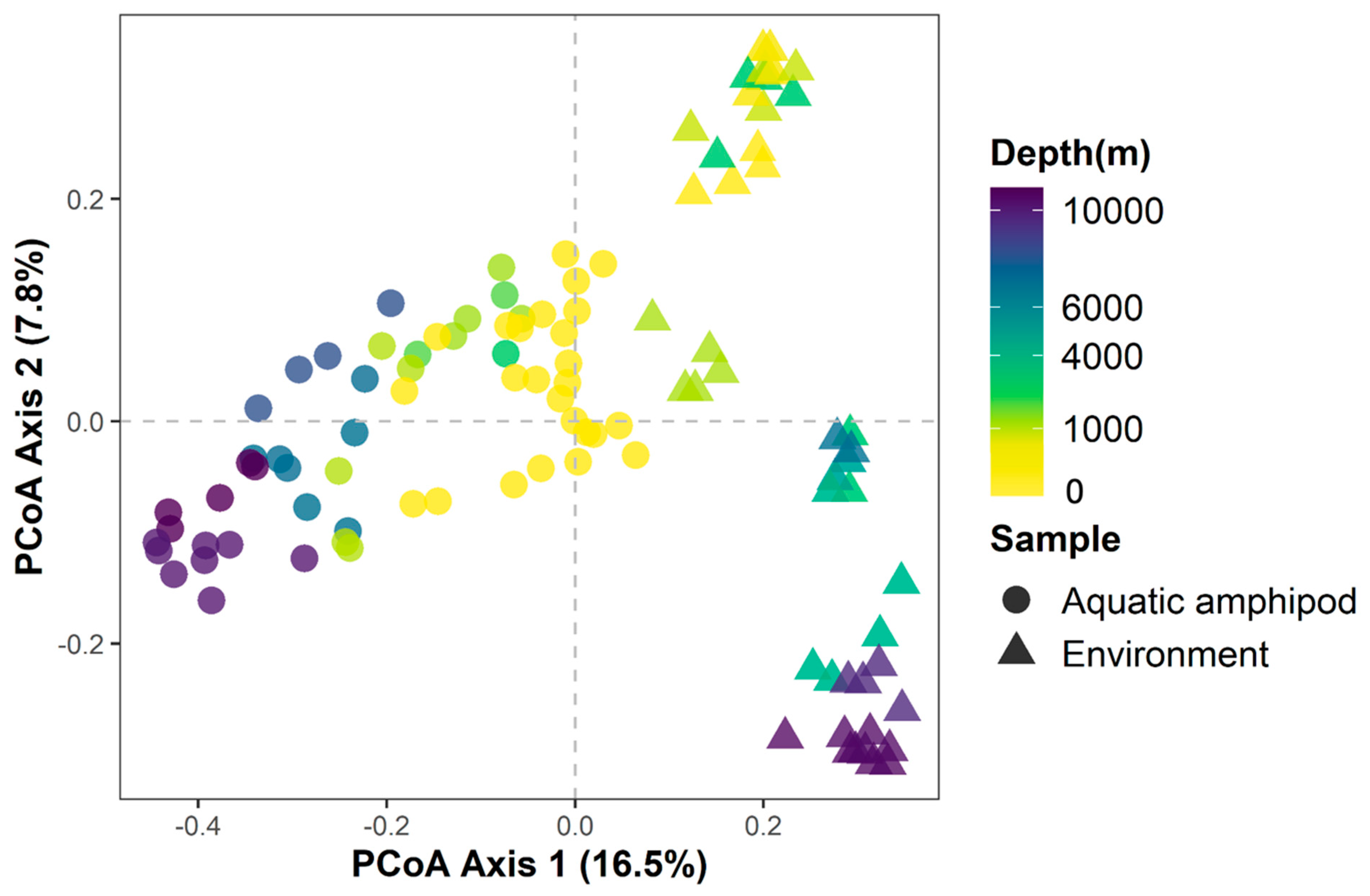
3.4. Relationships between Amphipod Gut and Environmental Bacterial Communities

To further investigate the relationship of bacterial communities between amphipod gut and surrounding seawater, microbial communities from surrounding environmental seawater were obtained and compared with that from amphipod guts by Unweighted UniFrac PCoA (Supplementary Materials Table S1). The results showed that samples from environmental seawater and amphipod guts were clearly separated at Axis1, especially the samples from deeper depths. The linear regression analysis showed that Axis2 was almost linearly related to depth (r = −0.61, p < 0.005) (Figure 4, Supplementary Materials, Figure S5).
The shared bacteria genera between the bathyal or hadal amphipods and seawater were investigated. The results showed that there were 102 common bacteria genera between bathyal amphipods and seawater (occupying more than 60% of the bacteria from bathyal amphipod guts) (Supplementary Materials, Figure S6). While there were 18 common bacteria existing in both hadal amphipods and environmental samples (occupying about 20% of the bacteria from hadal amphipod guts). The total bacteria in the amphipod gut and environment were further examined, which revealed that the dominant microorganisms for supralittoral, intertidal, and subtidal amphipod were distributed across multiple phyla (classes), including Tenerictes, Epsilonproteobacteria, Planctomycetes, and Firmicutes (Supplementary Materials, Figure S7). And the phyla of enriched bacteria obtained in bathyal amphipod gut were classified as Actinobacteria, Firmicutes, Bacteroidetes, Tenerictes, and Betaproteobacteria Gammaproteobacteria (Supplementary Materials, Figure S8). While the enriched bacteria in the gut of hadal amphipods were primarily classified as Tenerictes and Firmicutes.

3.5. Discriminant Taxa for Gut Microbial Communities

The microbial communities were examined for the amphipod guts from five habitats by LefSe implemented with an LDA threshold of 4.0. A total of 71 microbial clades were considered to be differentially abundant, of which there were 18, 17, 12, 11, and 13 distinguished taxa determining supralittoral, intertidal, subtidal, bathypelagic, and hadopelagic clades, respectively. Taxa with a significantly higher abundance in the intertidal group mainly belonged to Gammaproteobacteria (including genera Vibrio and Thiothrix), Alphaproteobacteria (including genera Deviosia, Ruegeria, Shimia, and Octadecabacter), family Flavobacteriaceae (including genera Olleya and Tenacibaculum), and Saprospiraceae. The genera Renibacterium, Lysinibacilus, and Ralstonia were enriched in the subtidal group. Taxa with abundant advantages in supralittoral groups were mainly Firmicutes and Proteobacteria. In addition to Staphylococcus, genera Ruminococcus and Johnsonella, affiliated with the family Lachnospiraceae, were significantly abundant in the supralittoral groups. The genera Spingomonas and Neisseria, both belonging to Betaproteobacteria, were also enriched in supralittoral groups. Hadopelagic amphipods were distinguished by the dominance of microbial genera Macrococcus, Ca. Hepatoplasma, Psychromonas, and Psychrobacter, while bathypelagic amphipods harbored more Aliagarivorans (Figure 5).

4. Discussion

4.1. Taxonomic Features of Amphipod Gut Microbiota

In this study, meta-analysis of the16S rRNA sequence was used to characterize the gut microbial communities of the 73 samples from 16 amphipod species that ranged from coastal to at depths of ten thousand meters of the ocean. Dominant taxa of gut microbiomes from different habitats were investigated. In the present study, Proteobacteria, Bacteroidota, Tenericutes, and Firmicutes comprised a large proportion of the amphipod gut microbiota, in accordance with the results of Cheng et al. (2017). Hirondellea is a genus that has been well documented within hadal depths [26]. Comparison of the gut microbial communities of H. gigas from two hadal trenches, Mariana and Japan Trenches, showed that the genera of Psychromonas, Propionibacterium, and Pseudoalteromonas were the dominant microbes in H. gigas, but with a light difference in relative abundance [27]. Similarly, the gut microbiota of the hadal snailfishes from Mariana has a similar community structure to that from Yap Trenches [28]. These indicated that the geographical isolation affects the gut microbial community, but not by much. Across all samples from littoral to hadal zones, Ca. Hepatoplasma and Streptococcus were more prevalent than other genera in this study. However, Ca. Hepatoplasma was the most enriched genus in the gut microbiota of hadal amphipods represented by A. gigantea, whereas Streptococcus was more abundant in supralittoral individuals. Ca. Hepatoplasma was reported as a symbiont colonizing the midgut glands of terrestrial and marine isopods [29,30]. Hepatopancreatic bacteria, such as the gut symbionts of termites and other soil arthropods that feed on fiber-rich diets, have been speculated to be involved in the decomposition of leaf litter by producing cellulases or phenol oxidases [31,32,33,34]. Hadal amphipods may digest debris, as demonstrated by a previous study that showed that H. gigas from the Challenger Deep contained cellulose with a potential contribution to the digestion of wood debris on the seafloor [16]. In that case, Ca. Hepatoplasma, which dominates the hadal amphipod gut, may be involved in the process of nutrition supplementation [14]. The genera Ca. Hepatoplasma, Psychromonas, and Psychrobacter were differentially abundant in the hadopelagic group [14]. Members of the genus Psychromonas include piezophilic, halophilic, and psychrophilic species and are widespread in marine environments. These members have been detected from the gut of a decaying deep-sea amphipods [35] and observed to be overrepresented in amphipods from the Mariana Trench [14]. The genus Psychrobacter has been isolated from low-temperature marine environments, including the Japan Trench and Antarctic sea ice [36,37], and the internal tissues of marine ascidian and crustacean species [38]. Members of the genus Psychrobacter are also commonly found in fish, poultry, and fermented seafood [39,40,41]. A strain Psychrobacter proteolyticus, isolated from the stomach contents of Antarctic krill Euphausia superba Dana, was found to excrete a cold-adapted metalloprotease [36]. Future verification is needed for the hypothesis that the genus Psychrobacter, a statistically enriched hadopelagic group, may be related to cold adaptation.
Our results showed that amphipods among various habitats had significantly different abundances of distinct gut microbes. Among all the genera discriminating intertidal groups, members of Tenacibaculum and Shimia have been commonly isolated from marine animals [42,43,44,45,46], suggesting their close relationship with creatures in marine habitats. Previous studies reported that Vibrio was a free-living, halophilic, facultative aerobic bacterium in marine environments worldwide. Vibrio is strongly correlated with parasitic infestation and mechanical injuries, which suppress immunity and increase the susceptibility of the host to vibriosis [47,48]. Members of the sulfur-oxidizing bacteria Thiothrix were suggested to be ectosymbionts of freshwater and marine amphipods residing in sulfide-rich environments [49,50,51]. A phylotype of Thiothrix bacteria has been discovered in the groundwater amphipod genus Niphargus, forming symbionts on amphipod appendix hairs and spines [50]. Thus, the amphipoda has unique intestinal flora, which may be partly related to their living environment.

4.2. Alpha Diversities of Marine Amphipod Gut Microbiota

In this study, alpha index, including ASV Count, Faith’s PD, Pielou’s Evenness, and Simpson, were analyzed for the amphipod gut microbial community from diverse habitats. Both in terms of species richness and diversity, amphipod gut microbes in the Hadopelagic zone were the lowest. The diversity of food often affects the diversity of intestinal flora [52]. Deep-sea deposit-feeding animals, such as amphipods, feed on the continual fall of organic material from the upper water column [53]. It was also suggested that bacteria could be a nutrition source for deep-sea creatures [54]. To sum up, the hadal zone is an extreme environment where nutrients are rare [53]. In comparison, coastal amphipods mainly feed on organic matter, including detritus, crustaceans, and macroalgae, which are more enriched in surface seawater than in aphotic deep-sea zones [55,56]. The mass of organic matter varied with depth, and therefore depth strongly determines food availability [57]. In our study, there is a significant difference in the Pielou’s Evenness and Simpson index of the amphipod gut microbial community between the supralittoral and subtidal or intertidal groups (p < 0.05). Finally, the supralittoral amphipod gut microbiome is high in richness and low in evenness. This phenomenon may be due to the fact that the basic food along the supralittoral zone is microscopic plants, chiefly diatoms, bacteria, various other unicellular algae and detritus, which is abundant but single [58]. Generally, high microbial diversity is linked to a strong metabolic ability and stability [59]. The possible reason for this may be due to that a diverse microbiome is better equipped to perform a wider range of functions, including digestion, nutrient absorption, and immune system regulation [60,61]. Especially, the commensal bacteria play a vital role in food digestion by extracting and synthesizing essential nutrients and metabolites, which are critical for maintaining their host health [61]. Interestingly, adult hadal amphipod H. gigas harbored less sediment and bacteria in their digestive tracts than the young, probably because they tended to reduce feeding in order to save energy, optimizing reproductive success [62].

4.3. Factors Associated with Intestinal Microbiota

Our work indicated that the host species, feeding habits, and habitat of amphipods exert significant but partial effects on the host gut microbial communities. They explained around 27%, 9%, and 12% of overall structure variability, respectively. Both host-associated and environmental factors have been shown to modulate microbial communities in many other animal intestines, such as the composition of bacteria in fish gut being influenced by salinity, trophic level, and potentially the host’s phylogeny [63,64]. The study related to two hadal amphipod species showed that host species could be a determining factor of hadal gut microbiota under certain conditions [14]. Actually, these factors are not independent. Organisms living in a certain area often have unique food habits and living species. The reason for this may be that habitats determine the distribution of organic matter, and organic matter resources vary greatly among different habitats. Shallow-sea amphipods derive their nutrition directly from nutrients in surface seawater [55]; however, deep-sea amphipods rely almost entirely on surface vertical sedimentation or possibly also directly on microorganisms as a food source [54,57]. Also, a considerable number of species only live in specific zones with unique feeding habits. In this study, species A. distinctus, A. gigantea, E. magellanicus, Eurythenes sp., Halice sp. MT-2017, H. gigas, and Princaxelia sp., generally living in bathyal and hadal zones, were scavengers. This may be due to the scarcity of food sources in the deep sea and the fact that most of the food that reaches the seafloor is in the form of putrefactive debris.
Generally, marine animals intimately interact with microbes existing in their surrounding environment [65]. Microbes in the surrounding water or sediments are a major source of gut microbes for marine creatures [54,66]. Yet the compositions of the microbial community in the sediments and guts of the scavenger Molpadia musculus from the environment where the sediment was poor in the organic matter were distinctly different, likely due to the need for hosts in the deep sea to develop a specialized gut bacterial community aiding host digestion [67]. Our observation that microbiomes from deep-sea scavenger guts were far distinct from those of environmental samples indicates that deep-sea amphipods might select certain mutualistic microbes, helping hosts digest refractory organic matter and thus boosting host digestion efficiency. Factors including diet, development stage, host phylogeny, and habitat have been shown to be correlated with microbial community structure in the animal gut [9,65,68]. However, because environmental and host-associated factors interact, this makes it difficult to study their relative contribution to variation; for example, environmental factors, such as diet or exposure to contaminants, can influence the expression of host-associated factors [69]. Therefore, developing a standard method to decouple and quantify the relative contributions of variables rather than conducting qualitative studies is vital for studying the mechanisms of gut bacteria assembly.

5. Conclusions

This study revealed that diet, habitat, and host species influenced amphipod gut microbial diversity and composition. The main phyla were Proteobacteria, Firmicutes, Bacteroides, and Actinomycetes in the amphipods from supralittoral to hadopelagic zones, with Firmicutes dominant in the hadal ones. Moreover, the gut microbiome of amphipods from the hadal zone displayed the lowest richness, lowest diversity, and shared few microorganisms with surrounding seawater. The amphipods in different inhabits have different specific gut taxa. Overall, this study provides a preliminary exploration of microbial colonization in the gut of amphipods, serving as a basis for a further investigation of the relationship among gut microbes, phylogenetic factors, and environmental factors.

Supplementary Materials

The following supporting information can be downloaded at: https://www.mdpi.com/article/10.3390/jmse11112197/s1, Figure S1. Distribution of the prevalence for ASVs across all amphipod samples. Figure S2. Rarefaction curves (a) and species accumulation curves (b) of 73 amphipod gut samples. Figure S3. Most prevalent genera among the entire amphipod dataset. Figure S4. The influence of environmental and genetic factors on the amphipod gut microbial community by a clustering analysis. Figure S5. Linear regression relationship between depth and PCoA Axis2 of marine amphipod and environmental microbial communities. Figure S6. Overlapping and non-overlapping counts of genera differentially abundant in deep-sea amphipods and environment. Figure S7. Microbial genera enriched in shallow-water amphipod gut or amphipod and overlapping genera between shallow-water amphipod gut and environment. Figure S8. Microbial genera enriched in bathyal amphipod gut or amphipod and overlapping genera between bathyal amphipod gut and environment. Table S1. Metadata corresponding to the amphipod and environmental samples used in this study. Table S2. Alpha index of all amphipod datasets used in this study. Table S3. Post-hoc Dunn tests assessing alpha diversity across habitats.

Author Contributions

L.H., T.W. and Y.L. designed the project. J.L., Y.L., L.H. and Y.W. collected the samples. T.W. and Y.L. conducted the laboratory work and data analysis. T.W. and Y.L. wrote the manuscript with input from all co-authors. L.H. and Y.W. supervised the project. All authors have read and agreed to the published version of the manuscript.

Funding

This research was supported by the General projects of National Natural Science Foundation of China (Grant NO.42176125), the Scientific research and manufacture projects of Sanya City (Grant NO.2020KS01), and supported by Hainan Provincial Natural Science Foundation of China (Grant NO.322CXTD531).

Institutional Review Board Statement

The Mariana hadal amphipods and the South China Sea bathyal amphipods were trapped using several deep-sea landers. The captured amphipods were dead once collected on deck. The intertidal sample amphipods were collected from depths of 0–3 m from Qingdao and Sanya using plastic barrels. All the amphipods are neither endangered nor protected animals. The animal sampling procedures conducted were carried out in strict accordance with the animal experimentation guidelines and regulations of the Institute of Deep-Sea Science and Engineering.

Informed Consent Statement

Not applicable.

Data Availability Statement

The data supporting the conclusions of this article were deposited into the NCBI Sequence Read Archive (SRA) database (BioProject ID: PRJNA717782). Source code of the statistical programs and data sheets were archived at GitHub (https://github.com/weitaoshu/amphipod-gut-microbiome/tree/main founded on 24 April 2023).

Acknowledgments

The authors are grateful to Ming-xin Lv and Xi-kun Song for collecting specimens from Xiamen.

Conflicts of Interest

The authors declare no competing interests.

References

  1. Väinölä, R.; Witt, J.D.S.; Grabowski, M.; Bradbury, J.H.; Jazdzewski, K.; Sket, B. Global diversity of amphipods (Amphipoda; Crustacea) in freshwater. In Freshwater Animal Diversity Assessment; Balian, E.V., Lévêque, C., Segers, H., Martens, K.V., Väinölä, R., Witt, J.D.S., Grabowski, M., Bradbury, J.H., Jazdzewski, K., Sket, B., Eds.; Developments in Hydrobiology; Springer: Dordrecht, The Netherlands, 2008; pp. 241–255. [Google Scholar]
  2. Jamieson, A.J.; Fujii, T.; Mayor, D.J.; Solan, M.; Priede, I.G. Hadal trenches: The ecology of the deepest places on Earth. Trends Ecol. Evol. 2010, 25, 190–197. [Google Scholar] [CrossRef]
  3. Fujii, T.; Kilgallen, N.M.; Rowden, A.A.; Jamieson, A.J. Deep-sea amphipod community structure across abyssal to hadal depths in the Peru-Chile and Kermadec trenches. Mar. Ecol. Prog. Ser. 2013, 492, 125–138. [Google Scholar] [CrossRef]
  4. Lindeman, D. Natural history of the terrestrial amphipod Cerrorchestia hyloraina Lindeman (Crustacea: Amphipoda; Talitridae) in a Costa Rican cloud forest. J. Nat. Hist. 1991, 25, 623–638. [Google Scholar] [CrossRef]
  5. Glazier, D.S. Amphipoda. In Encyclopedia of Inland Waters; Likens, G.E.G., White, D.S., Eds.; Academic Press: Oxford, UK, 2009; pp. 89–115. [Google Scholar]
  6. Horton, T.; Gofas, S.; Kroh, A.; Poore, G.C.B.; Read, G.; Rosenberg, G.; Stöhr, S.; Bailly, N.; Boury-Esnault, N.; Brandão, S.N.; et al. Improving nomenclatural consistency: A decade of experience in the World Register of Marine Species. Eur. J. Taxon. 2017, 389, 1–24. [Google Scholar] [CrossRef]
  7. Hughes, L.E.; Ahyong, S.T. Collecting and processing amphipods. J. Crustac. Biol. 2016, 36, 584–588. [Google Scholar] [CrossRef]
  8. Wade, S.; Corbin, T.; McDowell, L.M.; Bradbury, J.; Authority, S.A.E.P. Critter Catalogue: A Guide to the Aquatic Invertebrates of South Australian Inland Waters; Environment Protection Authority: Washington, DC, USA, 2004. [Google Scholar]
  9. McFall-Ngai, M.; Hadfield, M.G.; Bosch, T.C.; Carey, H.V.; Domazet-Loso, T.; Douglas, A.E.; Dubilier, N.; Eberl, G.; Fukami, T.; Gilbert, S.F.; et al. Animals in a bacterial world, a new imperative for the life sciences. Proc. Natl. Acad. Sci. USA 2013, 110, 3229–3236. [Google Scholar] [CrossRef] [PubMed]
  10. Douglas, A.E. Multiorganismal insects: Diversity and function of resident microorganisms. Annu. Rev. Entomol. 2015, 60, 17–34. [Google Scholar] [CrossRef]
  11. Turnbaugh, P.J.; Ley, R.E.; Mahowald, M.A.; Magrini, V.; Mardis, E.R.; Gordon, J.I. An obesity-associated gut microbiome with increased capacity for energy harvest. Nature 2006, 444, 1027–1031. [Google Scholar] [CrossRef]
  12. Abdelrhman, K.F.; Bacci, G.; Marras, B.; Nistri, A.; Schintu, M.; Ugolini, A.; Mengoni, A. Exploring the bacterial gut microbiota of supralittoral talitrid amphipods. Res. Microbiol. 2017, 168, 74–84. [Google Scholar] [CrossRef]
  13. Abdelrhman, K.F.; Bacci, G.; Nistri, A.; Mengoni, A.; Ugolini, A. Diet and gut microbiota of two supralittoral amphipods Orchestia montagui and Talitrus saltator living in different microhabitats. Estuar. Coast. Shelf S 2017, 197, 119–125. [Google Scholar] [CrossRef]
  14. Cheng, X.Y.; Wang, Y.; Li, J.Y.; Yan, G.Y.; He, L.S. Comparative analysis of the gut microbial communities between two dominant amphipods from the Challenger Deep, Mariana Trench. Deep Sea Res. Part I 2019, 151, 103081. [Google Scholar] [CrossRef]
  15. Chan, J.; Geng, D.; Pan, B.; Zhang, Q.; Xu, Q. Gut microbial divergence between three hadal amphipod species from the isolated hadal trenches. Microb. Ecol. 2022, 84, 627–637. [Google Scholar] [CrossRef] [PubMed]
  16. Kobayashi, H.; Hatada, Y.; Tsubouchi, T.; Nagahama, T.; Takami, H. The Hadal Amphipod Hirondellea gigas possessing a unique cellulase for digesting wooden debris buried in the deepest seafloor. PLoS ONE 2012, 7, e42727. [Google Scholar] [CrossRef]
  17. Folmer, O.; Black, M.; Hoeh, W.; Lutz, R.; Vrijenhoek, R. DNA primers for amplification of mitochondrial cytochrome c oxidase subunit I from diverse metazoan invertebrates. Mol. Mar. Biol. Biotechnol. 1994, 3, 294–299. [Google Scholar]
  18. Herlemann, D.P.; Labrenz, M.; Jurgens, K.; Bertilsson, S.; Waniek, J.J.; Andersson, A.F. Transitions in bacterial communities along the 2000 km salinity gradient of the Baltic Sea. ISME J. 2011, 5, 1571–1579. [Google Scholar] [CrossRef] [PubMed]
  19. Bolyen, E.; Rideout, J.R.; Dillon, M.R.; Bokulich, N.A.; Abnet, C.C.; Al-Ghalith, G.A.; Alexander, H.; Alm, E.J.; Arumugam, M.; Asnicar, F.; et al. Reproducible, interactive, scalable and extensible microbiome data science using QIIME 2. Nat. Biotechnol. 2019, 37, 852–857. [Google Scholar] [CrossRef] [PubMed]
  20. Arumugam, M.; Raes, J.; Pelletier, E.; Le Paslier, D.; Yamada, T.; Mende, D.R.; Fernandes, G.R.; Tap, J.; Bruls, T.; Batto, J.M.; et al. Enterotypes of the human gut microbiome. Nature 2011, 473, 174–180. [Google Scholar] [CrossRef]
  21. Jari, O.; Blanchet, F.G.; Friendly, M.; Roeland Kindt, P.L.; McGlinn, D.; Minchin, P.R.; O’Hara, R.B.; Simpson, G.L.; Solymos, P.; Stevens, M.H.H.; et al. vegan: Community Ecology Package. R. Package Version 2018; version 2.5–7. Available online: https://cran.r-project.org/web/packages/vegan/index.html (accessed on 11 December 2020).
  22. Paradis, E.; Schliep, K. ape 5.0: An environment for modern phylogenetics and evolutionary analyses in R. Bioinformatics 2018, 35, 526–528. [Google Scholar] [CrossRef]
  23. Segata, N.; Izard, J.; Waldron, L.; Gevers, D.; Miropolsky, L.; Garrett, W.S.; Huttenhower, C. Metagenomic biomarker discovery and explanation. Genome Biol. 2011, 12, R60. [Google Scholar] [CrossRef]
  24. Nishida, A.H.; Ochman, H. Rates of gut microbiome divergence in mammals. Mol. Ecol. 2018, 27, 1884–1897. [Google Scholar] [CrossRef] [PubMed]
  25. Benson, A.K.; Kelly, S.A.; Legge, R.; Ma, F.; Low, S.J.; Kim, J.; Zhang, M.; Oh, P.L.; Nehrenberg, D.; Hua, K.; et al. Individuality in gut microbiota composition is a complex polygenic trait shaped by multiple environmental and host genetic factors. Proc. Natl. Acad. Sci. USA 2010, 107, 18933–18938. [Google Scholar] [CrossRef]
  26. Eustace, R.M.; Kilgallen, N.M.; Lacey, N.C.; Jamieson, A.J. Population Structure of the Hadal Amphipod Hirondellea Gigas (Amphipoda: Lysianassoidea) from the Izu-Bonin Trench. J. Crustac. Biol. 2013, 33, 793–801. [Google Scholar] [CrossRef]
  27. Zhang, W.; Watanabe, H.K.; Ding, W.; Lan, Y.; Tian, R.-M.; Sun, J.; Chen, C.; Cai, L.; Li, Y.; Oguri, K. Gut microbial divergence between two populations of the hadal amphipod Hirondellea gigas. Appl. Environ. Microbiol. 2019, 85, e02032-18. [Google Scholar] [CrossRef]
  28. Lian, C.-A.; Yan, G.-Y.; Huang, J.-M.; Danchin, A.; Wang, Y.; He, L.-S. Genomic characterization of a novel gut symbiont from the hadal snailfish. Front. Microbiol. 2020, 10, 2978. [Google Scholar] [CrossRef] [PubMed]
  29. Leclercq, S.; Dittmer, J.; Bouchon, D.; Cordaux, R. Phylogenomics of “Candidatus Hepatoplasma crinochetorum,” a lineage of mollicutes associated with noninsect arthropods. Genome Biol. Evol. 2014, 6, 407–415. [Google Scholar] [CrossRef] [PubMed]
  30. Wang, Y.; Huang, J.M.; Wang, S.L.; Gao, Z.M.; Zhang, A.Q.; Danchin, A.; He, L.S. Genomic characterization of symbiotic mycoplasmas from the stomach of deep-sea isopod bathynomus sp. Environ. Microbiol. 2016, 18, 2646–2659. [Google Scholar] [CrossRef]
  31. Brune, A.; Resh, V.; Cardé, R. Encyclopedia of Insects; Academic Press: Cambridge, MA, USA, 2003. [Google Scholar]
  32. Fraune, S.; Zimmer, M. Host-specificity of environmentally transmitted Mycoplasma-like isopod symbionts. Environ. Microbiol. 2008, 10, 2497–2504. [Google Scholar] [CrossRef]
  33. Wang, Y.; Stingl, U.; Anton-Erxleben, F.; Geisler, S.; Brune, A.; Zimmer, M. “Candidatus Hepatoplasma crinochetorum,” a new, stalk-forming lineage of Mollicutes colonizing the midgut glands of a terrestrial isopod. Appl. Environ. Microbiol. 2004, 70, 6166–6172. [Google Scholar] [CrossRef]
  34. Breznak, J.A.; Brune, A. Role of microorganisms in the digestion of lignocellulose by termites. Ann. Rev. Entomol. 1994, 39, 453–487. [Google Scholar] [CrossRef]
  35. Lauro, F.M.; Stratton, T.K.; Chastain, R.A.; Ferriera, S.; Johnson, J.; Goldberg, S.M.; Yayanos, A.A.; Bartlett, D.H. Complete Genome Sequence of the Deep-Sea Bacterium Psychromonas Strain CNPT3. Genome Announc. 2013, 1, 10–1128. [Google Scholar] [CrossRef]
  36. Denner, E.B.; Mark, B.; Busse, H.J.; Turkiewicz, M.; Lubitz, W. Psychrobacter proteolyticus sp. nov., a psychrotrophic, halotolerant bacterium isolated from the Antarctic krill Euphausia superba Dana, excreting a cold-adapted metalloprotease. Syst. Appl. Microbiol. 2001, 24, 44–53. [Google Scholar] [CrossRef]
  37. Maruyama, A.; Honda, D.; Yamamoto, H.; Kitamura, K.; Higashihara, T. Phylogenetic analysis of psychrophilic bacteria isolated from the Japan Trench, including a description of the deep-sea species Psychrobacter pacificensis sp. nov. Int. J. Syst. Evol. Microbiol. 2000, 50, 835–846. [Google Scholar] [CrossRef] [PubMed]
  38. Romanenko, L.A.; Tanaka, N.; Frolova, G.M.; Mikhailov, V.V. Psychrobacter fulvigenes sp. nov., isolated from a marine crustacean from the Sea of Japan. Int. J. Syst. Evol. Microbiol. 2009, 59, 1480–1486. [Google Scholar] [CrossRef]
  39. Kämpfer, P.; Albrecht, A.; Buczolits, S.; Busse, H.-J. Psychrobacter faecalis sp. nov., a new species from a bioaerosol originating from pigeon faeces. Syst. Appl. Microbiol. 2002, 25, 31–36. [Google Scholar] [CrossRef] [PubMed]
  40. Gennari, M.; Parini, M.; Volpon, D.; Serio, M. Isolation and characterization by conventional methods and genetic transformation of Psychrobacter and Acinetobacter from fresh and spoiled meat, milk and cheese. Int. J. Food Microbiol. 1992, 15, 61–75. [Google Scholar] [CrossRef] [PubMed]
  41. Juni, E.; Heym, G.A. Psychrobacter immobilis gen. nov., sp. nov.: Genospecies Composed of Gram-Negative, Aerobic, Oxidase-Positive Coccobacilli. Int. J. Syst. Bacteriol. 1986, 36, 388–391. [Google Scholar] [CrossRef]
  42. Wright, A.C.; Hill, R.T.; Johnson, J.A.; Roghman, M.C.; Colwell, R.R.; Morris, J.G., Jr. Distribution of Vibrio vulnificus in the Chesapeake Bay. Appl. Environ. Microbiol. 1996, 62, 717–724. [Google Scholar] [CrossRef]
  43. Kim, Y.O.; Park, I.S.; Park, S.; Nam, B.H.; Park, J.M.; Kim, D.G.; Yoon, J.H. Tenacibaculum haliotis sp. nov., isolated from the gut of an abalone Haliotis discus hannai. Int. J. Syst. Evol. Microbiol. 2017, 67, 3268–3273. [Google Scholar] [CrossRef]
  44. Småge, S.B.; Frisch, K.; Vold, V.; Duesund, H.; Brevik, Ø.J.; Olsen, R.H.; Sjaatil, S.T.; Klevan, A.; Brudeseth, B.; Watanabe, K.; et al. Induction of tenacibaculosis in Atlantic salmon smolts using Tenacibaculum finnmarkense and the evaluation of a whole cell inactivated vaccine. Aquaculture 2018, 495, 858–864. [Google Scholar] [CrossRef]
  45. Hyun, D.W.; Kim, M.S.; Shin, N.R.; Kim, J.Y.; Kim, P.S.; Whon, T.W.; Yun, J.H.; Bae, J.W. Shimia haliotis sp. nov., a bacterium isolated from the gut of an abalone, Haliotis discus hannai. Int. J. Syst. Evol. Microbiol. 2013, 63, 4248–4253. [Google Scholar] [CrossRef]
  46. Pagan-Jimenez, M.; Ruiz-Calderon, J.F.; Dominguez-Bello, M.G.; Garcia-Arraras, J.E. Characterization of the intestinal microbiota of the sea cucumber Holothuria glaberrima. PLoS ONE 2019, 14, e0208011. [Google Scholar] [CrossRef] [PubMed]
  47. El-Bouhy, Z.; El-Nobi, G.; El-Murr, A.; Abd El-Hakim, S. Study on Vibriosis in Mugil Capito in El-Dakahlia and Damitta Governorates, Egypt. Abbassa Int. J. Aquat 2016, 9, 2016. [Google Scholar]
  48. Haenen, O.L.; van Zanten, E.; Jansen, R.; Roozenburg, I.; Engelsma, M.Y.; Dijkstra, A.; Boers, S.A.; Voorbergen-Laarman, M.; Moller, A.V. Vibrio vulnificus outbreaks in Dutch eel farms since 1996: Strain diversity and impact. Dis. Aquat. Organ 2014, 108, 201–209. [Google Scholar] [CrossRef] [PubMed]
  49. Gillan, D.C.; Dubilier, N. Novel epibiotic thiothrix bacterium on a marine amphipod. Appl. Environ. Microbiol. 2004, 70, 3772–3775. [Google Scholar] [CrossRef]
  50. Bauermeister, J.; Ramette, A.; Dattagupta, S. Repeatedly evolved host-specific ectosymbioses between sulfur-oxidizing bacteria and amphipods living in a cave ecosystem. PLoS ONE 2012, 7, e50254. [Google Scholar] [CrossRef]
  51. Dattagupta, S.; Schaperdoth, I.; Montanari, A.; Mariani, S.; Kita, N.; Valley, J.W.; Macalady, J.L. A novel symbiosis between chemoautotrophic bacteria and a freshwater cave amphipod. ISME J. 2009, 3, 935–943. [Google Scholar] [CrossRef]
  52. Senghor, B.; Sokhna, C.; Ruimy, R.; Lagier, J.-C. Gut microbiota diversity according to dietary habits and geographical provenance. Human. Microb. J. 2018, 7, 1–9. [Google Scholar] [CrossRef]
  53. Gerringer, M.E.; Popp, B.N.; Linley, T.D.; Jamieson, A.J.; Drazen, J.C. Comparative feeding ecology of abyssal and hadal fishes through stomach content and amino acid isotope analysis. Deep Sea Res. Part I Oceanogr. Res. Pap. 2017, 121, 110–120. [Google Scholar] [CrossRef]
  54. Eardly, D.F.; Carton, M.W.; Gallagher, J.M.; Patching, J.W. Bacterial abundance and activity in deep-sea sediments from the eastern North Atlantic. Prog. Oceanogr. 2001, 50, 245–259. [Google Scholar] [CrossRef]
  55. Guerra-García, J.M.; Tierno de Figueroa, J.M.; Navarro-Barranco, C.; Ros, M.; Sánchez-Moyano, J.E.; Moreira, J. Dietary analysis of the marine Amphipoda (Crustacea: Peracarida) from the Iberian Peninsula. J. Sea Res. 2014, 85, 508–517. [Google Scholar] [CrossRef]
  56. Navarro-Barranco, C.; Tierno-de-Figueroa, J.M.; Guerra-García, J.M.; Sánchez-Tocino, L.; García-Gómez, J.C. Feeding habits of amphipods (Crustacea: Malacostraca) from shallow soft bottom communities: Comparison between marine caves and open habitats. J. Sea Res. 2013, 78, 1–7. [Google Scholar] [CrossRef]
  57. Luo, M.; Gieskes, J.; Chen, L.; Shi, X.; Chen, D. Provenances, distribution, and accumulation of organic matter in the southern Mariana Trench rim and slope: Implication for carbon cycle and burial in hadal trenches. Mar. Geol. 2017, 386, 98–106. [Google Scholar] [CrossRef]
  58. Gunter, G. Notes on sea beach ecology. Food sources on sandy beaches and localized diatom blooms bordering gulf beaches. Gulf Caribb. Res. 1979, 6, 305–307. [Google Scholar] [CrossRef]
  59. Tap, J.; Furet, J.P.; Bensaada, M.; Philippe, C.; Roth, H.; Rabot, S.; Lakhdari, O.; Lombard, V.; Henrissat, B.; Corthier, G.; et al. Gut microbiota richness promotes its stability upon increased dietary fibre intake in healthy adults. Environ. Microbiol. 2015, 17, 4954–4964. [Google Scholar] [CrossRef] [PubMed]
  60. Leser, T.D.; Mølbak, L. Better living through microbial action: The benefits of the mammalian gastrointestinal microbiota on the host. Environ. Microbiol. 2009, 11, 2194–2206. [Google Scholar] [CrossRef]
  61. Brestoff, J.R.; Artis, D. Commensal bacteria at the interface of host metabolism and the immune system. Nat. Immunol. 2013, 14, 676–684. [Google Scholar] [CrossRef]
  62. Hessler, R.R.; Ingram, C.L.; Aristides Yayanos, A.; Burnett, B.R. Scavenging amphipods from the floor of the Philippine trench. Deep. Sea Res. Part I 1978, 25, 1029–1047. [Google Scholar] [CrossRef]
  63. Ley, R.E.; Lozupone, C.A.; Hamady, M.; Knight, R.; Gordon, J.I. Worlds within worlds: Evolution of the vertebrate gut microbiota. Nat. Rev. Microbiol. 2008, 6, 776–788. [Google Scholar] [CrossRef] [PubMed]
  64. Sullam, K.E.; Essinger, S.D.; Lozupone, C.A.; O’Connor, M.P.; Rosen, G.L.; Knight, R.; Kilham, S.S.; Russell, J.A. Environmental and ecological factors that shape the gut bacterial communities of fish: A meta-analysis. Mol. Ecol. 2012, 21, 3363–3378. [Google Scholar] [CrossRef] [PubMed]
  65. Egerton, S.; Culloty, S.; Whooley, J.; Stanton, C.; Ross, R.P. The Gut Microbiota of Marine Fish. Front. Microbiol. 2018, 9, 873. [Google Scholar] [CrossRef]
  66. Wu, S.; Wang, G.; Angert, E.R.; Wang, W.; Li, W.; Zou, H. Composition, diversity, and origin of the bacterial community in grass carp intestine. PLoS ONE 2012, 7, e30440. [Google Scholar] [CrossRef] [PubMed]
  67. Amaro, T.; Witte, H.; Herndl, G.J.; Cunha, M.R.; Billett, D.S. Deep-sea bacterial communities in sediments and guts of deposit-feeding holothurians in Portuguese canyons (NE Atlantic). Deep Sea Res. Part I 2009, 56, 1834–1843. [Google Scholar] [CrossRef]
  68. Huang, Q.; Sham, R.C.; Deng, Y.; Mao, Y.; Wang, C.; Zhang, T.; Leung, K.M.Y. Diversity of gut microbiomes in marine fishes is shaped by host-related factors. Mol. Ecol. 2020, 29, 5019–5034. [Google Scholar] [CrossRef] [PubMed]
  69. Kers, J.G.; Velkers, F.C.; Fischer, E.A.; Hermes, G.D.; Stegeman, J.A.; Smidt, H. Host and environmental factors affecting the intestinal microbiota in chickens. Front. Microbiol. 2018, 9, 235. [Google Scholar] [CrossRef]
Figure 1. Microbial community composition at the phylum level. (a) The pie chart showing the average relative abundance of the five most abundant phyla and minor phyla. (b) Relative abundance of phyla with average relative abundance >1% in each habitat. (c) Relative abundance at the phylum level for each sample grouped by habitat and diet from which sample derived. The 10 most abundant phyla were shown. The ‘Others’ referred to all phyla other than the top 10.
Jmse 11 02197 g001
Figure 2. Alpha diversity of the amphipod gut microbiota from different marine zonations. Alpha diversity indices, including Simpson index, Faith’s PD, Pielous’ evenness, and ASV counts, were calculated. Each index was represented with mean ± standard errors.
Jmse 11 02197 g002
Figure 3. Principal coordinates analysis (PCoA) depicting the similarity of the amphipod gut microbial communities. Symbols representing communities were colored by diet. The shape represents habitats where hosts resided.
Jmse 11 02197 g003
Figure 4. Unweighted UniFrac PCoA depicting the dissimilarity of bacterial communities from marine aquatic amphipod gut and seawater across depth. The different colors represent the depth of the samples, and the shapes represent the source.
Jmse 11 02197 g004
Figure 5. LEfSe analysis showing differentially abundant microbial clades for each habitat determined using Kruskal–Wallis test (p < 0.05) with LDA score > 4.0. (a) Taxonomic cladogram obtained from LDA Effect Size (LEfSe) analysis among 5 habitats. The evolutionary branches of different bacterial clades, with circles radiating from inside to outside representing taxonomic levels from class to genus. The root of the cladogram denoted the domain Bacteria. The taxonomic levels of the class were labeled, while order and family were abbreviated. Bacterial clades with no significant difference were colored yellow. The size of each node represented their relative abundance. (b) Barplot of LDA effect score for amphipod gut samples from supralittoral, intertidal, subtidal, bathypelagic, and hadopelagic zones. LDA score was used to evaluate the contributions of the microbes to the differences among the groups.
Jmse 11 02197 g005
Table 1. Summary of PERMANOVA results based on unweighted Unifrac distance.
VariableSums of SqsMean SqsF.ModelR2p
Entire amphipod datasetDiet4.620 2.310 13.568 0.199 0.0001
Habitat2.768 1.384 8.129 0.119 0.0001
Site1.883 0.942 5.530 0.081 0.0001
Host family2.055 0.685 4.024 0.088 0.0001
Host species2.196 0.366 2.150 0.095 0.0001
Number of permutations: 999.
Disclaimer/Publisher’s Note: The statements, opinions and data contained in all publications are solely those of the individual author(s) and contributor(s) and not of MDPI and/or the editor(s). MDPI and/or the editor(s) disclaim responsibility for any injury to people or property resulting from any ideas, methods, instructions or products referred to in the content.

