Article

Microbial Biofilms Colonizing Plastic Substrates in the Ross Sea (Antarctica)

by
Carmela Caroppo
1,*,
Maurizio Azzaro
2,
Ombretta Dell’Acqua
3,
Filippo Azzaro
2,
Giovanna Maimone
2,
Alessandro Ciro Rappazzo
2,4,
Francesco Raffa
5 and
Gabriella Caruso
2,*
1
Water Research Institute, National Research Council (CNR-IRSA), Via Roma 3, 74121 Taranto, Italy
2
Institute of Polar Sciences, National Research Council (CNR-ISP), Spianata S. Raineri 86, 98122 Messina, Italy
3
Department of Earth, Environment and Life Sciences, University of Genoa (DISTAV), Corso Europa 26, 16132 Genoa, Italy
4
Department of Environmental Sciences, Informatics and Statistics, University of Venice Ca’ Foscari, Dorsoduro 3246, 30123 Venice, Italy
5
Institute of Geosciences and Georesources, National Research Council (CNR-IGG), Via G. Moruzzi 1, 56124 Pisa, Italy
*
Authors to whom correspondence should be addressed.
J. Mar. Sci. Eng. 2022, 10(11), 1714; https://doi.org/10.3390/jmse10111714
Submission received: 31 August 2022 / Revised: 2 November 2022 / Accepted: 6 November 2022 / Published: 10 November 2022

Abstract

:
Very few studies have investigated marine microbial colonization in polar regions, but climate-changing scenarios stress the importance of these investigations to protect life in such extremely vulnerable ecosystems. In two different coastal sites of the Ross Sea (Road and Tethys Bays, Antarctica) exposed to different stressors, the microbial biofilm colonizing the surface of plastic (polyvinyl chloride, PVC, and polyethylene, PE) panels left submerged in two experiments at different timescales (“short-term”: 3 months, and “long-term”: 9 and 12 months) was studied. The abundance and metabolic enzymatic activities [leucine aminopeptidase (LAP), beta-glucosidase (GLU) and alkaline phosphatase (AP)] of the prokaryotes and the microalgal abundance and species composition were analyzed, in parallel with the main environmental parameters. The prokaryotic community showed higher abundance and metabolic activities on PVC than on PE as opposed to microalgae. A peak in the microfouling prokaryotic abundance and metabolic functions was frequently recorded after 3 months of immersion, corresponding to the late austral summer period. LAP and AP were the most active enzymes, suggesting that microbial metabolic profiles were modulated by labile organic substrates. Our results suggest that the composition and function of microbial biofilm could be considered as sentinels of natural or anthropic-related disturbances.

1. Introduction

Climate change and increased human pressure are recognized as the major threats to extremely vulnerable regions such as the Antarctic ecosystems [1]. Plastic pollution has become an emerging concern also on this continent, once considered to be pristine [2,3], but the ecological consequences of these new pollutants on the ecosystem functioning are, to date, unpredictable.
All submerged substrates, including plastic polymers, provide a suitable surface for colonization by a complex biofilm layer, which is the first step for the formation of a very active three-dimensional structure [4,5,6]. Marine biofilms consist of microbial communities, very different from a phylogenetic and functional point of view, enclosed in a matrix of extracellular polymeric substances (EPS) [7,8]. Bacteria and diatoms are the main components of biofilms, but other microalgae, microscopic fungi, and heterotrophic protists (flagellates and ciliates) characterize these assemblages [9].
In many aquatic ecosystems, environmental variables such as temperature, photosynthetically available radiation, pH, dissolved oxygen and nutrients influence the composition of the microbial biofilm community [8,9]. As the composition of microbial biofilm communities is known to vary in response to environmental variables, and species succession is observed during biofilm development [8,10], the relative dominance of certain biofilm microorganisms undergoes spatial and temporal changes [9]. For example, diatom assemblages show a different composition in relation to the physico-chemical and biological features of the water column over different seasons [11].
Biofilms are complex structures that represent “hotspots” of both structural and functional microbial diversity [10,12], which differs from the microbial assemblages living in the surrounding waters, as molecular techniques recently demonstrated [13,14].
From an ecological point of view, biofilms are beneficial to microbial communities in terms of competition and survival as well as nutrient retention and defense against environmental stress and predation [15]. Moreover, microbial biofilms play a critical role in the settlement of sessile organisms (like invertebrate larvae or propagules) adapted to live attached to substrates [16].
In Antarctic marine ecosystems, current knowledge of marine biodiversity is still far from complete. A detailed review of the micro- and meio-faunal communities colonizing soft and hard substrates was previously provided [17], but microbial benthic colonization has been disregarded and several areas of this continent remain almost unexplored for this feature. Marine fouling in Antarctica has also been addressed in terms of the chemical corrosion of submerged substrates [18]. However, only in the last decade have very few studies [19,20] appeared that focused on biofilm composition and how its microbial community reacts to natural or human-related disturbance. Many reports of the growing occurrence of plastic pollution in these extreme environments–previously considered “pristine”–have also resulted in the recognition of this emerging threat [2,3]. Plastics polymers represent a surface available for the settlement of pioneer microorganisms and a substrate to study the processes involved in biofouling formation; moreover, marine microbial communities (bacteria and microalgae) are major actors in many processes driving the functioning of the ecosystem such as the organic matter decomposition and biogeochemical processes. Therefore, the structural and functional features of microbial biofilm components add basic insights into the colonization dynamics of benthic environments by microbial communities and cast a light on the role of plastic material fragments as potential vectors for the spread of a variety of attached microorganisms.
The Ross Sea, where the Italian research station Mario Zucchelli is located, is a heterogeneous marine ecosystem that includes a mosaic of different habitats [21]. This makes it a suitable area to explore if, and to what extent, microbial communities can represent potential “sentinels” of emerging environmental perturbation, such as that related to plastic pollution, which has begun to threaten Antarctic ecosystems due to its detrimental effects on both environment and biota [3,22]. This consideration further explains why, especially in the Ross Sea—where the colonization process has never been explored from a microbiological point of view—looking at the structural and functional features of microbial communities belonging to the “plastisphere” community (sensu Amaral-Zettler et al. [23]) represents a critical challenge to improve our currently limited knowledge on plastic pollution in polar regions [24]. A global approach to this emerging threat, which includes physical, chemical and biological features, as well as interactions with chemical and climate stressors, is urgently required, to suggest possible directions to counteract it [25,26].
The present research aimed at studying microbial colonization of two plastic materials in two marine sites of Terra Nova Bay subjected to different environmental conditions. As microbial communities are known to be extremely responsive to changing environmental conditions, they could represent a new parameter to assess the effects on marine benthic biota of potential environmental disturbance. Bearing this in mind, the specific objectives of this survey were:
  • To study the prokaryotic and microalgal communities within the microbial biofilms and to assess their spatial patterns in relation to the physico-chemical, trophic (nutrient concentration) and microbiological characteristics of the examined sites
  • To analyze variations on a short- and long-term timescale of the abundance, biomass and functional metabolism of microbial biofilm assemblage components
  • To investigate whether, and to what extent, a differential response of microbial biofilm community (prokaryotes and microalgae) can be envisaged in the taxonomic structure, prokaryotic abundance and metabolism in sites exposed to natural perturbations (such as salinity gradients due to the proximity to a glacier in Tethys Bay) or anthropogenic disturbance (such as the discharge of treated wastewater in Road Bay) compared to control, unperturbed, sites
  • To identify potential associations between a particular typology of pressure (natural/anthropic) and key features (i.e., prokaryotic abundance/metabolic patterns dominant algal species,) and evaluate their role as candidate bioindicators of environmental changes in this extreme environment.

2. Materials and Methods

2.1. Experimental Design

During the XXXIII Italian expedition to Antarctica (November 2017) artificial structures made in stainless steel and mounting plastic panels as substrates for colonization experiments were deployed and fixed at 5 and 20 m depths in the Road Bay and Tethys Bay areas (Figure 1).
The main characteristics of the sampling areas were as follows:
(1)
Road Bay, a small bay near the Mario Zucchelli station research settlement, was chosen as representative of a human-impacted area since its waters receive the treated wastewater coming from the research station;
(2)
Tethys Bay, a large and isolated bay 2 km north of Road Bay, was chosen as representative of a naturally-impacted area, due to the presence of a large glacier (Amorphous Glacier) approaching the shore, as a natural forcing; here, a salinity gradient was expected to occur following climate warming.
Within each area, two stations (one directly impacted and one far from the forcing variable, used as a control) were studied, and their geographic coordinates are reported in Table 1.
Two colonization experiments (short-term and long-term) were performed.
For the long-term experiment, after 12 months of immersion (Time 12, T12), during the following expedition (XXXIV, November 2018), the structures deployed at all the stations were recovered to study the development of prokaryotic and microalgal fouling and to evaluate their evolution in coastal sites differently exposed to natural or anthropogenic forcings. Only in the Road Bay area, a short-term experiment was also carried out, with the deployment of artificial structures in replicate; one set of these was recovered after 3 months of deployment (January 2018, Time 3, T3) and replaced with a new one that was left submerged and recovered 9 months later, in November 2018.
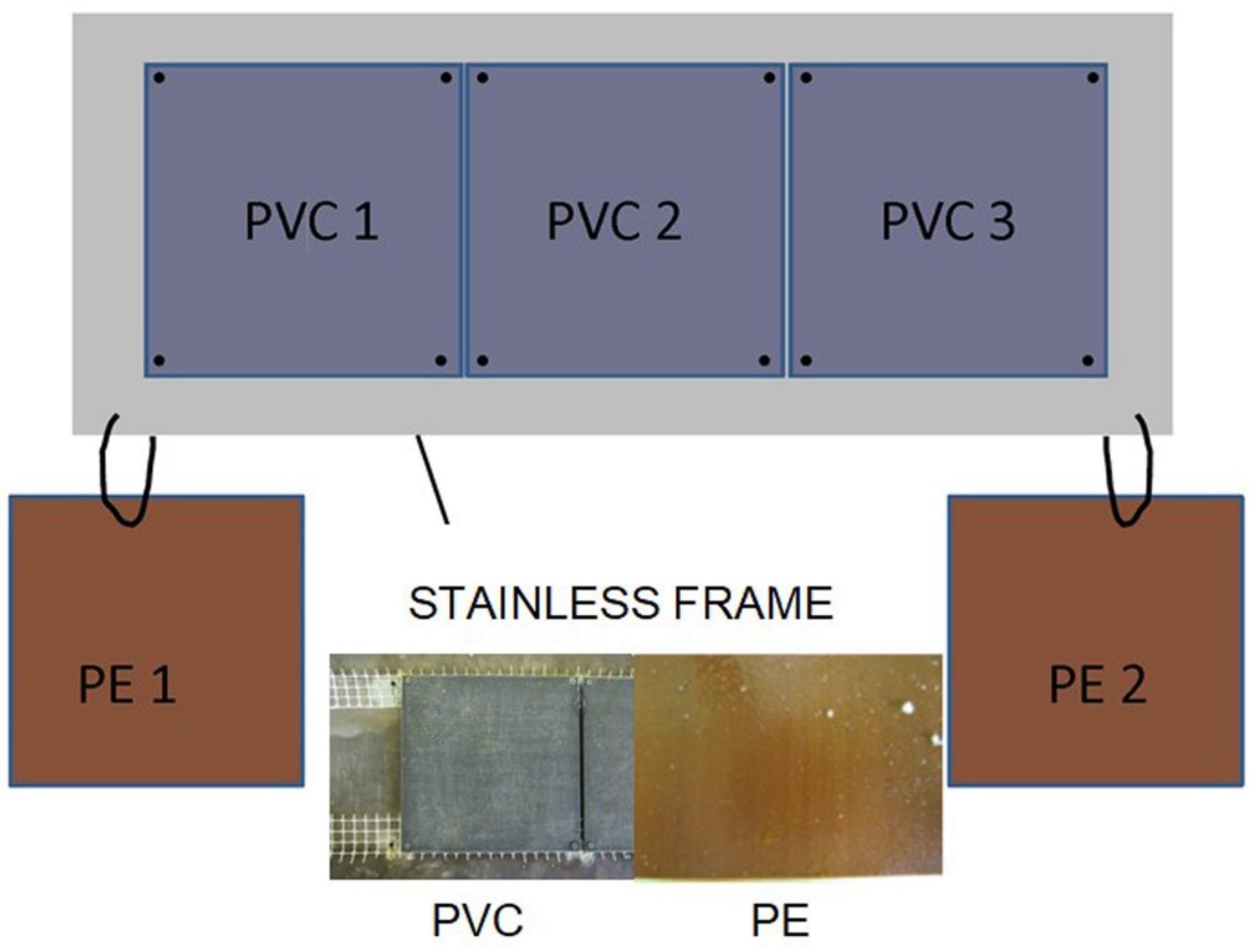
The settlement structures, deployed through holes dug in the pack ice (Figure 2), consisted of three replicate polyvinyl chloride (PVC) panels inserted in a stainless steel frame, plus two replicate polyethylene (PE) panels attached to the structure, but floating (Figure 3). Therefore, each of the structures hosted five panels for the study of microbial biofilms. Before being submerged, the plastic panels were first cleaned with ethanol and then washed with sterilized water. In their final assembly, the panels were covered with a nylon net (1-cm mesh) to prevent the interference of large predators on microbial colonization.
After panel recovery, any biofilm was removed by delicate scraping with a sterile scalpel from a surface equal to 6 × 18 cm of the PVC and PE panels deployed at 5 m and 20 m depths. Removal by scraping is a method commonly adopted to detach aquatic biofilms from the surface where they have developed [27,28,29]. Biofilm biomass was collected in a Petri plate, weighed using an analytical scale (Mettler-Toledo, Milan, Italy), then differently treated for each parameter to be measured.
During the three surveys, water samples were also collected at the 5 and 20 m depths (the depths at which the panels were deployed) using a Niskin bottle, to study the environmental parameters and microbial planktonic communities.

2.2. Environmental Parameters

Vertical profiles of temperature (T), salinity (S), dissolved oxygen (DO) and pH were recorded by a SeaBird 9–11 multiparametric probe (SeaBird Instruments, Inc., Washington, DC, USA) plus CTD-O-Fl profiler equipped with a Scufa (Turner Designs, San Jose, CA, USA) fluorometer to determine chlorophyll a (Chl a) as a proxy of phytoplankton biomass. A Niskin bottle (10 L-volume, SCUBLA S.r.l., Remanzacco, Udine, Italy) was used to collect water samples.
Samples for nutrient [ammonia (NH4), nitrite (NO2), nitrate (NO3) and orthophosphate (PO4)] analyses were filtered using GF⁄F glass-fibre filters and kept at −20 °C. Analytical determinations were carried out in triplicate using a Varian Mod. Cary 50 spectrophotometer (VARIAN Inc., Palo Alto, CA, USA) and following the Strickland & Parsons method [30], and, in the case of NH4, the Aminot & Chaussepied method [31].

2.3. Prokaryotic Community: Abundance, Biomass, Morphological Types and Metabolism

For the determination of the biofilm total prokaryotic abundance (including Bacteria and Archaea), biofilm samples were collected in sterile Falcon tubes, prefixed with pre-filtered formaldehyde (0.2 µm porosity; final concentration 2%), and stored in the dark at 4 °C. Appropriate aliquots (0.1 g) were diluted in sterile seawater (at least 10 times, w/v ratio), sonicated in a Branson sonifier for 2 min and filtered on polycarbonate black membranes (porosity 0.2 µm; GE Water & Process Technologies), then further stained with 4′,6-diamidino-2-phenylindole (DAPI, Sigma, final concentration 10 µg mL−1) [32]. Stained cells were counted under a Zeiss AXIOPLAN 2 Imaging microscope (magnification: Plan-Neofluar 100× objective and 10× ocular; HBO 100 W lamp; filter sets: G365 exciter filter, FT395 chromatic beam splitter, LP420 barrier filter) equipped with an AXIOCAM-HR digital camera (Carl Zeiss, Oberkochen, Germany). Moreover, image acquisition allowed us to distinguish different morphotypes. Cells were classified as cocci if their length and width differed by less than 0.10 μm, coccobacilli if their length and width differed by more than 0.10 μm, and rods if their length was at least double their width; V-shaped, C-shaped and S-shaped cells were defined as vibrios, curved rods and spirillae, respectively, while cells exceeding 4 µm in length were classified as filamentous bacteria. The detailed methodological procedures used for the single-cell volume calculation and the volume-to-biomass conversion factor were reported by La Ferla et al. [33]. For the measurements of total prokaryotic abundance in surrounding waters, seawater samples (45 mL) were collected, stored and analyzed as undiluted samples with the same procedure used for the biofilm analyses.
The fraction of viable prokaryotic cells (living cells) was quantified in the biofilm samples using the Live/Dead BacLight kit (Molecular Probes, Eugene, OR, USA), which uses SYTO® 9 and propidium iodide as reagents (the final concentration of each dye was 6 μM for SYTO 9 stain and 30 μM for propidium iodide). A 1 mL sample of the mixture of the two reagents was added to 1 mL of biofilm sample, with subsequent shaking and incubation at 4 °C in the dark for 1 h, followed by freezing at −20 °C. Samples were then filtered on 0.22 µm Nuclepore polycarbonate black membranes (GE Water and Process Technologies, Feasterville-Trevose, Pennsylvania, PA, USA) and these were observed under an epifluorescence microscope. Cell counts were performed using the specific sets for fluoresceine (BP450-490; FT510; LP520) and rhodamine (BP546/12; FT580; LP590).
For the quantification of actively respiring, CTC+ cells, 1 mL of biofilm sample was added to 0.1 mL of CTC cyanotetrazolium chloride (50 mM) (from the Bac Light Redox Sensor CTC Vitality Kit purchased from Molecular Probes) and incubated for 5 h in the dark at 4 °C. The sample was subsequently fixed and stored in the freezer at −20 °C. As reported above, the samples were then filtered on 0.22 µm Nuclepore polycarbonate black membranes and these were observed under an epifluorescence microscope using the rhodamine-specific filter set. CTC+ cells were visualized as cells with red-fluorescent formazan granules, produced by CTC reduction.
For the estimation of the enzymatic activity rates with respect to the enzymes leucine aminopeptidase (LAP), beta-glucosidase (GLU) and alkaline phosphatase (AP), a multi-concentration method described in previous studies on the metabolism of the plastic-associated prokaryotic community [34,35] was applied, by incubating 5 mL sub-volumes of diluted biofilm (1:50 w/v ratio) with methylumbelliferone (MUF) or methylcoumarin (MCA)-derived fluorogenic substrates (l-leucine-7-amido-4-methylcoumarin hydrochloride, 4-methylumbelliferyl-B-d-glucopyranoside and 4-methylumbelliferyl phosphate, for LAP, GLU and AP, respectively, Sigma-Aldrich, Merck, Milan, Italy). Fluorescence measurements were performed with a Jenway fluorimeter (model 6280, Dunmow, UK) at 365 nm and 440 nm excitation and emission wavelengths for MCA substrates, respectively, and at 365 nm and 455 nm for MUF substrates, respectively. Sample tubes were incubated for 2.5 h in the dark at 4 °C [34]. The enzyme kinetics were calculated through a Lineweaver-Burk equation, plotting the reciprocal of each substrate concentration against the reciprocal of the corresponding reaction velocity. Finally, from the intercept and slope of the equation, the maximum velocity (Vmax) and the half-saturation constant (Km) of the enzymatic reaction were calculated. The turnover times of organic substrates were obtained using the formula [36]:
Turnover time = Km/Vmax
The results of (1) are calculated in days and reported in Table S1. Turnover times increase when Vmax values are low.

2.4. Microalgae: Abundance, Biomass and Composition

In January and November 2018, microalgal biofilms were collected from the panels in the Road Bay area after 3 (T3) and 12 months (T12), respectively. Phytoplankton was collected from the sampling areas during all three surveys (November 2017, January and November 2018).
For the analysis of the microalgal biofilm, biofilm samples were fixed with formalin (final concentration 4%) [37]. The qualitative-quantitative analysis was carried out in triplicate using an inverted microscope AXIOVERT 200 M (Carl Zeiss, Oberkochen, Germany) equipped with phase contrast at 400× and 630× magnification, following the Utermöhl method [38]. A minimum of 200 cells was counted per sample, to ensure a confidence limit of 14% [39].
The water samples for phytoplankton analysis were collected using a Niskin bottle at a depth of 5 m at stations AG, RB, PTS and TB; 500 mL volumes were immediately fixed with neutralized formalin (final concentration 1.6%) and stored at 4 °C. Identification and counting were performed using an inverted microscope, as described for the microalgal biofilms. To complete the qualitative analysis of the phytoplankton community, vertical nets were also carried out using a 40-μm mesh net.
Microalgal cell sizes were measured using an AXIOCAM ICc 5 digital camera (Carl Zeiss, Oberkochen, Germany). Cell biovolumes were evaluated by approximating species shapes to geometrical models [40]. The carbon content was estimated from mean cell biovolumes using the Menden-Deuer & Lessard [41] formula.

2.5. Data Elaboration and Statistical Analyses

Physico-chemical variables, microbial abundance and biomass are reported as the mean value ± s.d.
The statistical significance of the differences found between and within groups of data was tested through one-way analysis of variance (ANOVA) and parametric (Fisher’s LSD test on means for homogenous variances) or non-parametric (Kruskal–Wallis test on medians for non-homogeneous variances) analyses.
Pearson correlations were computed to detect relevant pairwise relationships among biotic and abiotic variables, using PAST software, version 4.0 [42].
The spatio-temporal distribution of the microalgal community was assessed using the PRIMER v.7 software package (Primer-E). The initial matrix comprised all the taxa detected on the panels as well as in the water column (N = 48) [43].
Bi-dimensional representations of the statistical comparisons among samples collected on the panels and in the water were obtained by non-parametric multidimensional scaling (nMDS) performed on Bray–Curtis similarity matrices (Fourth root-transformed data) [43]. Then, to compare samples collected from different matrices (biofilm and water) all the data were standardized. This procedure allows the researcher to “…put each species on an equal footing, with its values summing to 100% across all samples” [43]. To evaluate the differences in both the prokaryotic and microalgal communities among different periods, sites and matrices, a one-way analysis of similarities (ANOSIM) was applied. When a significant Global R-value was yielded by ANOSIM, a one-way similarity percentage procedure (SIMPER routine) was used to obtain the percentage contribution of each variable to the Bray–Curtis similarity between the groups of samples in the different samples.
As regards the microalgal diversity indices, the species richness (S), the Margalef species richness index (d), the Shannon–Wiener diversity index (H′), and Pielou’s species evenness index (J′) were determined using PRIMER v.7 software.

3. Results

3.1. Physical and Chemical Characterization of the Study Areas

The values of the physico-chemical variables recorded in Road Bay and Tethys Bay are shown in Table 2.
In Road Bay, slightly lower temperatures (−1.79 ± 0.05 °C) and higher salinity (35.23 ± 0.03 psu) than in Tethys Bay were detected. The available data of dissolved oxygen (8.22 ± 0.84 mL L−1) and pH (7.88 ± 0.12) displayed significantly higher values in Tethys Bay and Road Bay, respectively, as statistical analyses evidenced (ANOVA, F = 1440.9, p < 10−4). Phytoplankton biomass (Chl a) did not reach high values, with average maxima in Tethys Bay (0.79 ± 0.18 RFU). As regards nutrients, higher average values of N-compounds were detected in Road Bay (NH4: 3.10 ± 1.98 µmol L−1; NO2: 0.20 ± 0.21 µmol L−1; NO3: 22.46 ± 4.77 µmol L−1) while PO4 reached higher concentrations in Tethys Bay (1.81 ± 0.44 µmol L−1). Consequently, the N/P ratio was significantly higher in Road Bay (16.15 ± 4.53) than in Tethys Bay (13.63 ± 2.70) (ANOVA, F = 5.834, p < 0.01).
Significant differences across the different sampling times were detected for temperature (F = 4.279, p < 0.05), salinity (F = 5.354, p < 0.01), PO4 (F = 4.760, p < 0.01) and N/P ratio (F = 11.180, p < 10−4).
Finally, the distribution of all the examined parameters was not affected by depth, although slightly higher values of N-compounds were measured in deeper waters (depth of 20 m).

3.2. Microbial Biofilm Community

3.2.1. Prokaryotic Biofilm Abundance

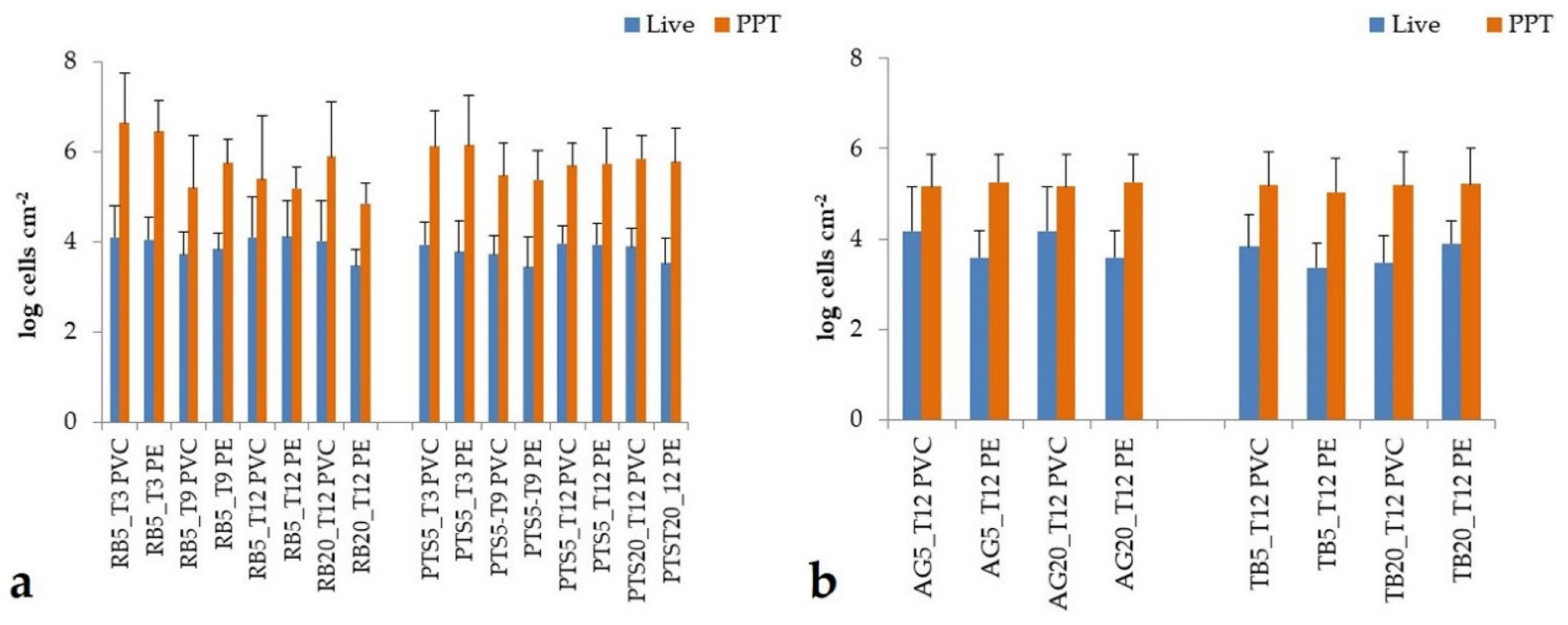
In the Road Bay area (Figure 4a), the abundance of total prokaryotes (PPT) grown as biofilm communities on PVC ranged from 1.57 × 10⁵ to 4.36 × 10⁶ cells cm−2, with higher values at station RB than station PTS and after 3 months of deployment. On PE, biofilm prokaryotes varied in a slightly wider range (6.97 × 10⁴–2.80 × 10⁶ cells mL−1) although their distribution patterns across the different sampling times were similar to those for PVC (i.e., peaking at station RB after 3 months). Prokaryotic biomass ranged between 3.9 and 196.4 mg C cm−2; the highest values were detected in January 2018 on PVC panels.
The fraction of viable cells (Live) on PVC comprised between 5.29 × 10³ and 1.27 × 10⁴ cells cm−2, with a high percentage at station RB after 3 months of immersion, as with PPT. On PE, the living cells ranged within similar values (2.83 × 10³–1.30 × 10⁴ cells cm−2) and with similar temporal trends (peak values at RB after 3 months).
In the Tethys Bay area (Figure 4b), PPT abundance on PVC comprised between 1.43 × 10⁵ and 1.53 × 10⁵ cells cm−2 and showed peaks at station TB after 12 months of immersion. The abundance of PPT on PE was at a similar order of magnitude (1.08 × 10⁵–1.82 × 10⁵ cells cm−2) but with higher values at station AG than TB.
On PVC, the living cells fall in a range from 3.02 × 10³ and 1.51 × 10⁴ cells mL−1 with high concentrations at station AG after 12 months. The living cells on PE varied within lower concentrations (2.36 × 10³–7.93 × 10³ cells cm−2), predominately at station TB after the same immersion period.
Significant variations were recorded by ANOVA for the living cells among sites on PVC (F = 6.18, p < 0.01), as well as between PVC and PE (F = 4.69, p < 0.05).
Regarding the prokaryotic morphotypes (Figure S1), in Tethys Bay, similar contributions of the different cell types between PVC and PE were observed (ANOSIM, Global R = 0.222, p = 0.70%). Cocci accounted for about 50% of PPT, followed by rods and coccobacilli (26% and 19%, respectively). The incidence of vibrios was fairly negligible (2%), while spirillae were generally absent. In addition, in Road Bay in both experiments, no significant differences were observed (ANOSIM, Global R = 0.12, p = 0.80%). Cocci and coccobacilli accounted for similar percentages of PPT (on average 36%). Moreover, in Road Bay, across the different sampling times, coccobacilli increased on PVC while cocci increased on PE. In this area, the incidence of vibrios, spirillae, rods and curved rods was comparable to that found in Tethys Bay. The only exception was the presence, albeit in low percentages, of filamentous forms characterized by straight filaments without visible septae and bacterial chains, on both PVC and PE.

3.2.2. Heterotrophic Microbial Biofilm Metabolism

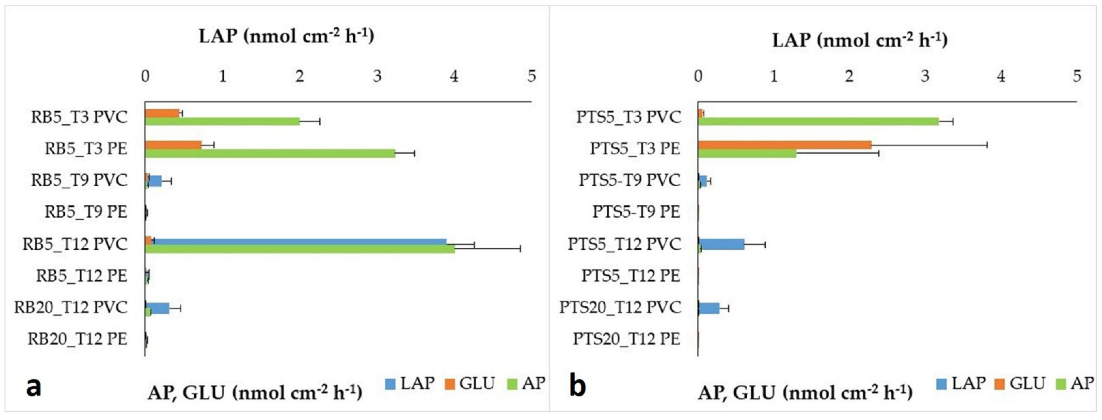
In the Road Bay area (Figure 5), enzymatic activity patterns of microbial biofilm were characterized by high LAP and AP on PVC, while GLU predominated on PE. On PVC, microbial biofilm activity rates ranged from 0.01 to 3.90 nmol cm−2 h−1 for LAP; from 0.01 to 2.54 nmol cm−2 h−1 for AP and from 0.0006 to 0.0875 nmol cm−2 h−1 for GLU. The highest activity rates of LAP and GLU were recorded at RB after 12 months. On PE, LAP activity rates varied from 0.0017 to 0.59 nmol cm−2 h−1 and AP from 0.00013 to 1.04 nmol cm−2 h−1, while GLU from 0.00003 to 1.83 nmol cm−2 h−1.
In the Tethys Bay area (Figure 6), similar levels of GLU were recorded on PVC and PE (0.017–0.058 nmol cm−2 h−1 and 0.00011–0.061 nmol cm−2 h−1, respectively). LAP and AP were the predominant enzymes, and microbial biofilms grown on PVC were more active than those on PE (LAP: 0.27–6.28 nmol cm−2 h−1 and 0.0016–0.232 nmol cm−2 h−1, respectively; AP: 0.03–0.158 nmol cm−2 h−1 and 0.000078–0.193 nmol cm−2 h−1, respectively). All the enzyme activities were higher at station AG than at TB.
Compared to abundance data, metabolic activities showed more variable patterns. However, significant differences were recorded on PVC for LAP only, both among stations (F = 5.13, p < 0.01) and between PVC and PE (F = 5.857, p < 0.01); in the Road Bay area, all the enzyme activities of biofilm grown on PE showed statistically significant differences (F = 65.48, p < 10−4; 65.90 p < 10−4; 65.70, p < 10−4) across the different sampling times.

3.2.3. Microalgal Biofilm Assemblages

The microalgal community showed extremely variable abundance and biomass values on the two plastic materials. Higher densities and biomass were observed on PE panels (2.3 ± 3.2 × 10⁸ cells cm−2; 404.4 ± 556.7 mg C cm−2) compared to those on PVC (1.0 ± 1.6 × 10⁸ cells cm−2; 228.2 ± 406.1 mg C cm−2) (Figure 7). By comparing prokaryotic and microalgal biomass (Figure 7b), the latter dominated over prokaryotes after 3 months of exposure at the PTS station and much more after 12 months at the RB station on the PE panels. Exposure time significantly affected diatom (F = 5.341, p < 0.05) and dinoflagellate abundance (F = 5.949, p < 0.05). In fact, at station RB, the highest algal concentrations and biomass were reached on both plastic materials after 12 months of immersion.
The composition of the microalgal assemblages evidenced the predominance of diatoms (88.0 ± 11.6% of the total community) while “other microalgae” (6.7 ± 7.0%) and dinoflagellates (5.2 ± 10.5%) were minor components of the assemblages. In addition, in terms of biomass, diatoms were the most conspicuous component, representing on average 94.4 ± 6.0% of the total biomass with percentages varying between 85.6% and 99.8%. On the panels, 17 diatoms and 1 dinoflagellate were identified (Table S2).
Concerning the microalgal species abundance distribution, this showed significant differences between the two periods of investigation (ANOSIM, Global R = 0.542, p = 0.1%). After three months of immersion, SIMPER analysis revealed that 76.9% of cumulative similarity was due, in decreasing order of abundance, to Fragilariopsis curta, F. kerguelensis, Chlamydomonas spp., Licmophora cf. gracilis, Thalassiothrix antarctica. After 12 months of immersion, 71.6% of cumulative similarity was justified by F. kerguelensis, Achnanthes antarctica, Prorocentrum sp., Chlamydomonas spp.

3.3. Water Microbial Community

3.3.1. Prokaryotic Abundance

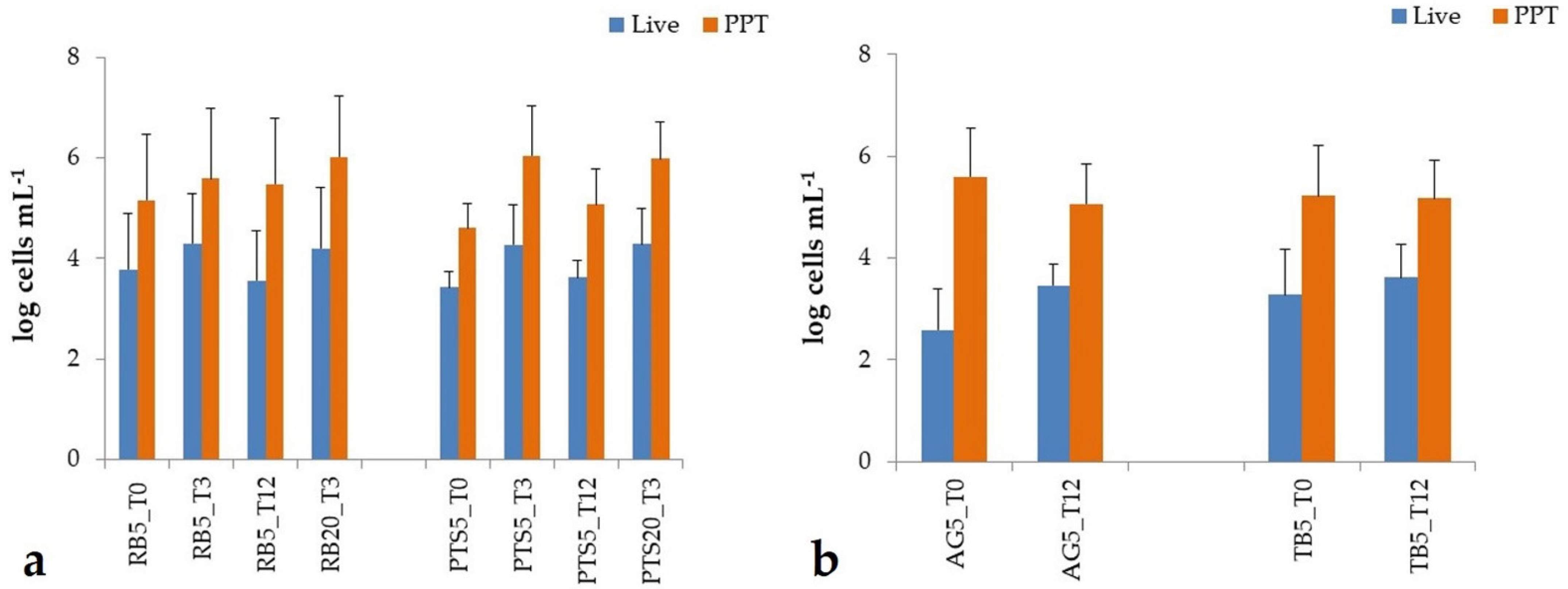
Road Bay was characterized by high PPT abundance (Figure 8a), comprised between 4.05 × 10⁴ and 1.1 × 10⁶ cells mL−1, which were higher at station RB than station PTS and reached a peak during the second sampling in January 2018; similar temporal trends were observed in prokaryotic biomass, which ranged between 0.6 and 28.3 μg C L−1.
As regards the fraction of living cells, their concentrations varied from 2.64 × 10³ to 1.96 × 10⁴ cells mL−1, with peak values in the late austral summer period (January 2018).
In the Tethys Bay area (Figure 8b), a PPT abundance lower than those in the Road Bay area was recorded, not exceeding 105 cells mL−1; PPT counts were higher at AG station (range: 1.15 × 10⁵−3.89 × 10⁵ cells mL−1) than at TB station (range; 1.46 × 10⁵–1.66 × 10⁵ cells mL−1). Living cells were about two orders of magnitude lower than PPT, with an abundance comprised between 3.77 × 10² and 2.83 × 10³ cells mL−1 at station AG compared to TB (1.89 × 10³–4.16 × 10³ cells mL−1).
ANOVA revealed that time variations in the living fraction were significant (F = 24.96, p < 0.01).
With respect to the prokaryotic morphotypes, no significant differences were observed among the cell types at all the stations (ANOSIM, Global R = 0.375, p = 6%); the relative percentages of the morphotypes increased across the different sampling times. Coccal forms were predominant, accounting for 50% of PPT, followed by coccobacilli (13%). Rods and curved rods accounted for a low percentage only (13%), while vibrios and spirilli were characterized by an uneven distribution with an extremely small proportion present within the PPT abundance (<5%).

3.3.2. Heterotrophic Microbial Community Metabolism

An active microbial community with respect to AP was detected in the Road Bay area (Figure 9a); in Tethys Bay (Figure 9b), the microbial community exhibited high LAP and GLU. Within the Road Bay area, RB water hosted a microbial community with higher metabolic levels (range LAP: 0.125–9.52 nmol L−1 h−1; GLU 0.153–5.81 nmol L−1 h−1 and AP: 0.017–7.81 nmol L−1 h−1) than station PTS (LAP: 0.391–6.04 nmol L−1 h−1; GLU: 0.077–3.95 nmol L−1 h−1; AP: 0.007–6.76 nmol L−1 h−1).
In the Tethys Bay area, the enzyme patterns showed higher metabolic patterns at station AG than station TB (range LAP: 0.039–10.58 nmol L−1 h−1 versus 0.299–4.22 nmol L−1 h−1; GLU: 0.186–1.49 nmol L−1 h−1 versus 0.002–0.054 nmol L−1 h−1), except for AP (0.078–0.33 versus 0.001–2.6 nmol L−1 h−1).
Differences among stations were significant for GLU (F = 6048.42, p < 0.001) and AP (F = 24.69, p < 0.01), respectively; in addition, across the different sampling times, significant differences were recorded for LAP and GLU (F = 10.47, p < 0.01 and 9.99, p < 0.01, respectively).

3.3.3. Phytoplankton Abundance and Biomass

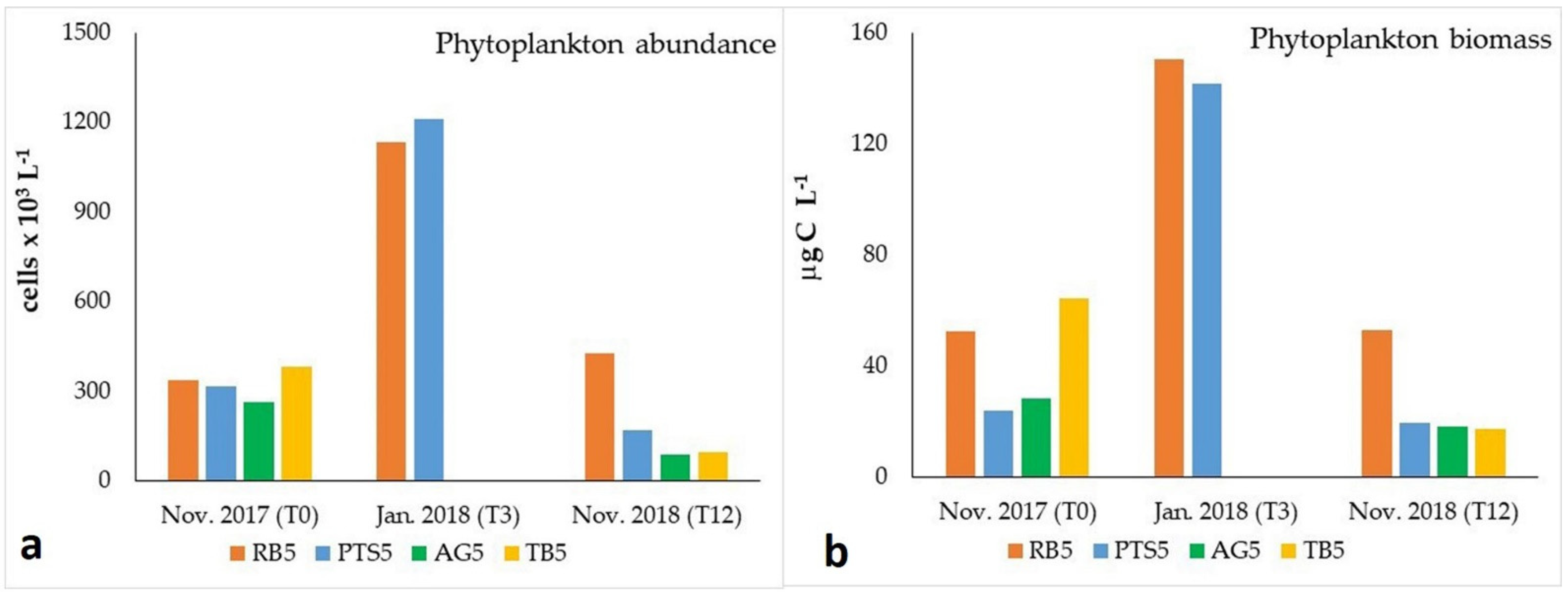
Phytoplankton abundance and biomass were characterized by extremely variable values during the study period, with the highest values detected in January 2018 (Figure 10), even if statistical analyses did not evidence significant differences among the investigated periods. In November 2017, phytoplankton abundance was comparable in all the sampled stations (up to 382.2 cells 103 L−1) while biomass showed slightly higher values at the stations RB and TB (up to 64.4 μg C L−1). In January 2018, considering only the Road Bay area, abundance peaks were observed at the PTS station (1208.5 cells × 103 L−1) while the highest biomass was found at the TB station (150.4 μg C L−1). During November 2018, RB station differed from the other stations for its high values of abundance (424.5 cells × 103 L−1) and biomass (53.0 μg C L−1).
From a qualitative point of view, the only component that showed significant differences in terms of the sampling periods was “other microalgae” (F = 0.014, p < 0.01). Particularly, in November 2017, these microalgae (range: 4.4–85.8% of the total abundance) represented the most conspicuous component of the community and accounted on average for 64.3 ± 16.1% of the total biomass. Diatoms (10.8–42.6%) contributed to 28.3 ± 13.3% /of the total biomass, while dinoflagellates (0.6–24.4%) only to 7.3 ± 11.5%.
In January 2018, in Road Bay, diatoms were the dominant component of the community in terms of abundance (up to 94.0% of the total) and biomass (up to 97.5% of the total). The contribution of dinoflagellates and the “other microalgae” was negligible.
In November 2018, the community was well diversified, and diatoms were again the most important phytoplankton group with a percentage abundance ranging between 20.2% and 78.9% and an average contribution to the total biomass of 65.7 ± 16.5%. Dinoflagellates (abundance range: 16.3–55.8%) and “other microalgae” (abundance range: 4.8–33.1%) contributed to the 30.4 ± 15.9% and 3.9 ± 2.7% of the total biomass, respectively.
A total of 35 taxa including 20 diatoms, 11 dinoflagellates and 4 species classified in the group “other microalgae” were identified (Table S2).
Concerning the phytoplankton species distribution, it showed significant differences between 0, 3 and 12 months, as nMDS (ANOSIM, Global R = 0.99, p = 0.1%) confirmed by the non-overlapping among the assemblages of the three periods in the nMDS plot (useful stress value) (Figure S2). Moreover, significant differences were observed between the stations (Road Bay and Tethys Bay), by considering both PVC and PE panels for total phytoplankton, diatoms and dinoflagellates abundance and biomass (p < 0.01). Moreover, “other microalgae” were significantly different on the PE panels (p < 0.01).
SIMPER analysis revealed that 70.8% of cumulative similarity in November 2017 was due, in decreasing order of abundance, to Phaeocystis antarctica, Chlamydomonas spp., Thalassiosira gracilis, Chaetoceros spp. and the Pseudo-nitzschia delicatissima group. In January 2018, 77.1% of cumulative similarity was justified by Fragilariopsis curta, F. kerguelensis, Phaeocystis antarctica, Chlamydomonas spp. And the Pseudo-nitzschia delicatissima group. Finally, in November 2018, 77.1% of cumulative similarity was due to Prorocentrum spp., Chlamydomonas spp., the Pseudo-nitzschia delicatissima group, Navicula spp. and F. kerguelensis. The highest average dissimilarity was detected between January and November 2018 (70.8%).

3.4. Comparison between the Plastisphere and the Surrounding Planktonic Communities

MDS analysis of prokaryotic abundance (PPT, Live, Dead, Live + Dead cells, as standardized values) in Road Bay (Figure S3) underlined that in January 2018 (B period) the microbial community clustered separately from the community observed at the initial and late periods (A, November 2017; C, November 2018). Moreover, such seasonal quantitative differences between B (late summer) and the other A and C sampling times (early summer) were significant for the plastisphere only. ANOSIM confirmed the significance of the time differences (Global R = 0.342, p = 0.012%; pairwise comparisons of B versus A, R = 0.786, p = 0.036% and B versus C, R = 0.260, p = 0.041%).
Conversely, all the water samples (R = Road Bay, P = Punta Stocchino, W = Water) generally clustered together regardless of their sampling periods, suggesting that interannual variations in pelagic abundance values were not statistically different.
In contrast with the variable “time”, the plastisphere community was quantitatively different from the pelagic community (ANOSIM Global R = 0.244, p = 0.035% among the matrices), although the plastic type (PVC and PE) did not play a critical role in shaping the prokaryotic community abundance. In addition, prokaryotic communities recorded at stations RB and PTS stations clustered separately.
Prokaryotic abundance analyzed by MDS (PPT, Live, Dead, Live + Dead cells, as standardized values) revealed that in Tethys Bay (Figure S4) the variable “time” was a factor discriminating the community between the two samplings, just as was observed in Road Bay. At station AG, waters at the start of the experiment showed a prokaryotic community significantly different from all the other samples. With respect to the matrix, PVC and PE-associated communities clustered separately, suggesting that the plastic-type affected their abundance patterns. Moreover, at station TB a close similarity was observed between the two colonized materials, while a marked quantitative difference (as suggested by the very high distance) was observed between PVC- and PE-associated communities developed at station AG, perhaps due to the environmental conditions characterizing this site.
MDS analysis carried out on the metabolic profiles of the microbial community in Road Bay (Figure S5) showed that reflecting what was observed in the prokaryotic abundance, in January 2019 (B sampling) microbial metabolic levels were substantially different from those recorded in the A and C samplings. In contrast with this trend, similarities between the second and third samplings were found for the pelagic community. The variable “station” did not affect the microbial metabolism, with RB and PTS microbial communities often clustering together. In addition, PVC- and PE-associated microbial communities did not show distinct metabolic patterns.
Microbial metabolic patterns observed in Tethys Bay analyzed by MDS (Figure S6) did not evidence significant differences related to the variable “period”; microbial communities of stations TB and AG also showed close similarities in their metabolic activities. PVC- and PE-associated microbial communities differed reciprocally at both stations, particularly those colonizing station TB.
In Road Bay, by comparing phytoplankton and microalgal biofilm assemblages in January and November 2018, some differences emerged (percentage of dissimilarity = 63.0%), as shown in Figure S7. In January 2018, the two communities showed similar features, but in the following sampling, they displayed marked differences. ANOSIM confirmed the significant differences depending on the variable “time” (Global R = 0.531, p = 0.1%) and “substrate” (water and PVC and PE panels) (Global R = 0.605, p = 0.1%). SIMPER analysis demonstrated that the taxa responsible for the differences were the Pseudo-nitzschia delicatissima group, Prorocentrum spp. and Gymnodinum spp. which were more abundant in the water column. On the contrary, Fragiliaropsis curta, F. kerguelensis, Achnantes antarctica, Licmophora gracilis and Thalassiothrix antarctica developed on the panels.
Finally, in terms of the diversity indices, the microalgal community showed lower values of the diversity indices with respect to the phytoplankton (Table S3). Particularly lower were the d and H’ values observed in the communities of the PE panels with respect to those measured in the plankton.

3.5. Statistical Analysis of Data: Pearson Correlation

The outputs of Pearson correlation analysis performed per each area (Road and Tethys Bays) for biofilm (PVC and PE) and water communities separately are reported in Figure S8. Within the microbial biofilm community colonizing the PVC panels fixed in the Road Bay area, statistically, significant direct relationships were detected between T and the autotrophic (total microalgae, diatoms and “other microalgae” abundance and biomass) and total prokaryotic components (PPT), while the same populations correlated negatively with S, NO3 and PO4 which were consumed for their growth. Opposite relationships were found for dinoflagellates, whose abundance is negatively related to T and positively to S and the same nutrients. PPT was negatively related to dinoflagellates; CTC+ cells were also related to the biofilm weight. GLU activity levels correlated to PPT, total microalgae, diatoms and phytoplankton biomass (as fluorescence); they were affected positively by T and negatively by S, NO3 and PO4, suggesting that both prokaryotic and microalgal communities were involved in the production of this enzyme. Microbial biofilm grown on PE at RB showed a significant association between microalgae and diatoms. As with that recorded for PVC-associated biofilm, PPT correlated directly with T and inversely with S and NO3, while dinoflagellates were inversely related to T and directly to S, NO3 and PO4. Great synergy in metabolic processes was suggested by the positive relationships linking LAP with GLU and AP. In the surrounding waters, a greater number of significant Pearson correlations was found in the Road Bay area compared to the Tethys Bay area. In Road Bay waters, a close direct relationship was observed between the autotrophic and heterotrophic components, as shown by the positive correlations found between total phytoplankton, diatoms, PPT and its living fraction. As with that observed for PVC biofilm, microbial glycolytic activity was also positively affected by T, total phytoplankton and diatoms. S and PO4 exerted an inverse influence on GLU, in agreement with the highest microbial metabolic level detected at station RB impacted by wastewater inputs. LAP and AP correlated reciprocally, suggesting synergy between carbon and phosphorus fluxes.
In the Tethys Bay area, microbial biofilm community colonizing PVC panels was characterized by similar relationships linking PPT with T, dissolved oxygen, NH4 and NO3; conversely, biofilm weight, GLU and AP were directly related to S and PO4. GLU correlated with Live + Dead cells, while there was a lack of significant relationships for CTC+ cells. On PE panels, biofilm weight was directly affected by T, NH4 and NO3; direct relationships were also detected between living and actively metabolizing CTC+ cells. All enzyme activities were directly affected by S, Chl a (phytoplankton biomass) and PO4 and correlated to each other, suggesting that the organic matter absorbed on this plastic polymer was easily decomposed by the microbial biofilm community. In Tethys Bay waters, the direct correlation of the total prokaryotic abundance with NH4 and PO4 indicated that these nutrients modulated prokaryotic growth.

4. Discussion

In polar regions, there is increasing evidence of the occurrence of plastic pollution; this issue needs to be more deeply investigated regarding microbial colonization since plastic items provide a surface suitable for the attachment of microorganisms that colonize many types of these materials. Several studies have regarded the biogeography of microbial biofilms and the differences between their composition and that of surrounding seawaters [2,12,13,44,45,46,47], and more recently Amaral-Zettler et al. [23] have reviewed the ecology of the plastisphere as a novel anthropogenic habitat. Nevertheless, many knowledge gaps on the microbial abundance, diversity and metabolism of the plastisphere community in polar regions persist, apart from a few studies [19,20].
Exploring how microorganisms interact with plastic debris and the factors that affect microbial colonization is extremely important. Recently, increased attention has been addressed to the microbial community assemblage (i.e., autotrophs, heterotrophs and predators) colonizing plastic debris, as documented by a recent review [16].
This study focused on microbial colonization of Antarctic environments, exploring the biofilms developing on plastic materials with regard to the composition and function of their biological components (i.e., total prokaryotes and their living, dead and actively metabolizing fraction abundance, as well as their enzymatic profiles and microalgal abundance, biomass and taxonomy).
Heterotrophic bacteria, together with microalgae (diatoms and cyanobacteria), protozoa and macrophyte spores are the main components of biofilms developed on soft or hard substrates [48,49,50,51,52,53]. Heterotrophic microorganisms provide important nutrients acting as organic matter decomposers and mineralizers [8]. Within the biofilm matrix, microorganisms are able to structurally self-organize themselves and their activities in response to external conditions and the various biofilm members [54]. Another important component of biofilm is microalgae which play a key role as a pioneer assemblage, favoring the settlement of macroalgae and invertebrates [55,56] and representing an important carbon source for benthic and pelagic trophic webs [50]. In the sections below, the dynamics of the prokaryotic and microalgal components of the plastisphere community were considered, focusing also on the temporal succession of their biomasses within the microbial biofilm formation. The effects of the plastic material (PVC versus PE) on the plastisphere structural and functional diversity were considered, paying attention also to the microbial biofilm abundance and metabolism in comparison with the microbial community present in the surrounding ambient seawater.

4.1. Spatial and Temporal Patterns of Microbial Biofilms

In our study, the prokaryotic abundance present in the biofilms was quantified in terms of both total and viable cells by epifluorescence microscopy after DAPI and Live/Dead staining, respectively. Potential advantages and limitations are associated with these methods [57,58]: microscopy methods are important tools to determine biofilm viability and spatial organization; they offer potential advantages including prompt data acquisition (within 1 h), the spatial resolution of living/dead cells and identification of viable but non-culturable cells within the biofilm. However, the main disadvantages of these methods are related to the need for trained personnel, specific instrumentation and non-specific binding of propidium iodide with the production of artefacts. Since none of the reported approaches is fully suitable to quantitatively determine the viable cells within the biofilm matrix, a combination of methods is the only way to get a more accurate estimation of microbial abundance [59]. Although non-disruptive methods—such as the Fluorescent In Situ Hybridization combined with Confocal Laser Scanning Microscopy (FISH-CLSM)–have been applied to quantify microbial abundance in marine biofilms [60], collection by scraping and sonication, together with observation by epifluorescence microscopy have been used as suitable tools to study microbial biofilms without sophisticated instruments not available at all laboratories [28,61,62,63].
In the present research, the simultaneous application of different (microscopy and biochemistry) methods enabled the obtaining of complementary information on the abundance and functional diversity of the plastisphere community on PVC and PE. As the methods applied were the same for all collected biofilm samples, comparisons of the microbial abundance among different stations and between the two different polymers—which were the objectives of this investigation—were carried out.
Spatial variability in biofilm composition was studied in this survey, to detect the main forcings affecting the growth of microbial assemblages colonizing plastic surfaces.
In proximity to the research station, local contamination caused by improper waste disposal was previously recorded. Fecal pollution indicators were detected near the outfall of the sewage disposal plant, which increased its activity with human presence in the summer season [64]. Furthermore, the high concentration of pollutants including plastics in sediments documented the chemical impact of the human presence in this area of the Ross Sea [3,22].
In our study, significantly higher (p < 0.01) bacterial and microalgal abundance, biomass and metabolic activity levels were observed in the microbial biofilms colonizing the impact stations RB and AG compared to the respective control stations, as reported in Section 3.2. This suggested that the colonization process in the two Ross Sea sites examined in our survey was affected by their different biogeographical features in terms of trophic characteristics. At station RB the sewage wastes enriched the organic matter of the water column, while at station AG, the detritus released from ice melting stimulated microbial growth. Seasonal and geographical factors were reported to influence the composition of microbial biofilm on plastics [13,45,65]. Biofilm formation and composition were found to undergo variations over space, depending on environmental and biological parameters including nutrient availability and grazing predation [66,67,68,69,70]. Moreover, the properties of the substrate surface such as microstructure and roughness [9,71] affected the complexity of biofilm micro-topography. In addition, the levels of pollution were found to shape the community structure of microbial biofilm; in the only study to date available on microbial colonization in benthic Antarctic environments [19], microbial community associated with glass surfaces was found to vary in relation to the different levels of contamination.
Metabolic activities performed by microbial biofilms at Road Bay suggested that a higher nutritional versatility characterized the microorganisms colonizing the benthic environment of this site with respect to the free-living communities of the same area, as well as in that of the Tethys Bay area. From a global perspective study [10], the high functional potential and taxonomic diversity were reported to drive niche differentiation between biofilm and planktonic communities. To date, information on the enzymatic activities of biofilms has been reported by a few studies only [61,62,63,72,73,74], moreover, none of these have been performed in Antarctic regions, making our investigation a pioneering study on the metabolic activity of the plastisphere.
Spatial diversification in the structure and the nutritional abilities of the bacterial communities depending on locally different environmental conditions was also reported in the composition of heterotrophic bacteria inhabiting two different stations of Terra Nova Bay (Mergellina and Santa Maria Novella) [75], as well as in the phenotypical traits of biofilm bacterial strains isolated from Road and Tethys Bays [76]; this suggested that microbial functional characteristics were adapted to local conditions.
In this survey, the kinetic approach used for enzyme activity measurements allowed us to estimate the turnover time of organic substrates; this provides information on the velocity at which microorganisms were able to mobilize potential nutrients from the organic matter pool. Comparatively shorter turnover times were obtained at station RB compared to the other sites, suggesting a faster decomposition of organic polymers in relation to the high enzyme activity rates exhibited by the microbial community both colonizing the panels and in the surrounding ambient seawater. Conversely, turnover times increased at station PTS, where maximum values were recorded for AP due to very low Vmax values shown by this enzyme. Similar high turnover times were found in the Tethys Bay area, especially at station TB, for AP and GLU suggesting that the turnover of organic phosphates and polysaccharides mediated by microbes was significantly slowed down or delayed in this site compared to the other stations.
Regarding the time variability of microbial biofilm composition, within a few hours of their release in marine waters, microplastic debris is colonized by biofilm-forming bacteria that favor the settlement of other marine organisms (e.g., fungi, algae, and protozoa) [65,70,77]. Bacterial and microalgal biomass estimated in our study showed that three months after deployment (T3 sampling) bacteria accounted for 45.2% of the total biomass, while microalgae for the remaining 54.8%. After twelve months (T12 sampling), the relative contribution of the bacterial component decreased to a percentage of 30.8% of the total biomass, while microalgae become predominant (69.2%), confirming that succession was occurring in the microbial biofilm community components. In contrast with our results, other studies [71] suggested that at the first stages of colonization, microalgae, represented mainly by diatoms, are dominant with respect to the other organisms, but afterwards, they are replaced by bacteria (autotrophic and heterotrophic), probably because of copepod grazing [77]. Moreover, the microalgal biomass values observed at Road Bay were four orders of magnitude higher than those observed in items immersed in a temperate marine environment (Woods Hole, MA, USA), considering the same immersion time (3 months) and different materials (polyethylene, polypropylene, polystyrene and glass) [78].
In this study, a comparison between short- and long-term microbial compositions allowed us to determine whether biofouling in the Antarctic was characterized by changes in the species assemblage and variations in the species dominance occurred. Significant differences were detected across the different sampling times in most of the microbial variables by ANOVA; comparison between samplings evidenced significant variability of the microbial community between 3 and 12 months of immersion, while 9 and 12 months were reciprocally similar, confirming that biofilm assemblage varied significantly over time. This result was consistent with the similarity in microbial communities detected in mature biofilms by previous studies; indeed, irrespective of the colonized substrate (low-and high-density PE, PVC and polypropylene), Pinto et al. [79] recorded common bacterial colonizers at late incubation stages (more than one month of immersion), while plastic-type specific bacterial taxa were more abundant at early colonization steps. Overall, these findings confirmed that the diversity of microbes attached to plastic debris varied with the duration of exposure.
Previous research aimed to study the composition and biomass of mixed algal and bacterial biofilms developed on polystyrene Petri dishes in two seasonal periods (summer and winter) demonstrated differences related to environmental conditions (temperature and salinity) [80]. In our study, the increase in microalgal abundance and biomass seemed to be linked more to the maturity phase of the biofilm than to the environmental variables.
Mejdandžić et al. [81] investigated the succession and settling of microalgae on plexiglass plates immersed in the northeast Adriatic Sea (Mediterranean Sea) for one month. Even if the timescale is different, the authors demonstrated that, in the beginning, pioneer planktonic diatom species colonized the substrates, and then a more specialized community developed, as in our study.

4.2. Within the Plastisphere, Differences between PVC and PE

Among the factors affecting biofilm formation, in addition to environmental factors and matrix, polymers’ type has been recognized to play a major role [12,45,46,47]. In our study, the PVC-associated plastisphere was characterized by higher abundance and metabolic levels compared to PE; the differences between the two substrates were quantified, on average, six and twenty-four times for enzymatic activities in Road Bay and Tethys Bay, respectively.
ANOVA outputs revealed that in PVC-attached microbial biofilm, significant differences were observed among the sampling stations for living and dead components of the prokaryotic community, as well as for the abundance of total microalgae, diatoms, dinoflagellates and leucine aminopeptidase activity. Conversely, within PE-attached microbial biofilm, spatial differences among the sampling stations were recorded for microalgae abundance only. The living cells on PVC exhibited more significant differences in relation to the variable “sampling stations” than the “polymer type” (F 6.18 versus 4.69). Moreover, differences observed in relation to the polymer type were more significant than those recorded among the sampling stations for leucine aminopeptidase activity rates (F 5.85 versus 5.16), suggesting an uncoupling between the abundance and the metabolic activity of the microbial biofilm components.
Briand et al. [82] studied the community composition of the microbial biofilm developed on PVC and four antifouling coatings at two marine French stations. Both surface type and environmental variables such as temperature and salinity or organic matter were found to be the major drivers affecting microbial composition (mostly dominated by gamma and alpha-Proteobacteria and Bacteroidetes, together with small diatoms belonging to Amphora and Navicula spp.), while seasons were not a relevant factor.
The importance of microhabitat created by the material composition on the structure and composition of marine biofilm has been underlined by a recent study [71]. In an experiment performed on marine infrastructures made of concrete with different roughness degrees inoculated with microalgal biofilm and immersed in sterilized seawater for six days under a controlled photoperiod, microalgal biomass growing on PVC detached in water after 10 days, suggesting that the biofilm developing on PVC material was unstable due to smooth surface of this material compared to concrete structures. These results agree with our data, which demonstrated the preference for PE panels over PVC panels by microalgae, mainly in the mature stages of biofilm development.
In the North Pacific Gyre, higher bacterial abundance was found on PE compared to PP [83]. Different bacterial and eukaryotic communities were found on PE and PP collected from the North Atlantic [24], and from the North Pacific [14].

4.3. Diversity of the Plastisphere versus Surrounding Water and Factors Affecting Colonization

Relevant questions in the ecology of the plastisphere assemblages concern the specificity of plastic-associated microbial communities (that is, those communities specifically enriched in association with plastic debris compared to those inhabiting the surrounding ambient seawater) as well as the presence of “core” communities. The latter includes those microorganisms that are common members of the plastisphere, independent of the nature of the polymeric substrates [47,84].
Whether specific plastisphere assemblages exist, however, is still an open question. Although the microalgal plastisphere assemblages differed from the phytoplankton communities of the ambient seawater, this does not necessarily mean that the surface-attached microalgal assemblages were specific to the plastics. At sea, microbes, as documented by previous studies [85,86,87], colonize any submerged surface. Moreover, microbes also colonize many particles in seawater [74,88]. Some microbial groups that are common colonizers on various submerged surfaces are also common members of the plastisphere; these include the marine Rhodobacteraceae group of the Alphaproteobacteria (i.e., the marine Roseobacter clade), the Alteromonadaceae and Vibrionaceae groups of the Gammaproteobacteria, and Bacteroidetes (mainly the Flavobacteria group) [74]. Regarding the origin of plastic-associated communities, although plastisphere microbial communities may not be exclusively specific to plastics, several variables, such as the presence of organic aggregates, or microbes from the sediments (including both prokaryotes and microalgae) through the sediment resuspension process, may provide a source of surface-associated prokaryotes and microalgae on plastic surfaces. It is well known that many sediment-dwelling microbes are surface-associated organisms that attach to sediment grains and live in biofilms [74,89]. Thus, many plastisphere microalgae may come from surface-associated benthic microalgae residing in the sediment environment. Unfortunately, this study did not investigate the species composition of the plastic biofilm, marine particle-associated and seawater free-living bacterial communities, nor the species composition of the microalgal communities in the sedimentary domain; therefore, speculations on these issues must be performed based on previously published reports [74].
In our study, the composition of the microalgal community colonizing PVC and PE panels differed significantly from that found in the waters, supporting the idea that a specific algal community was colonizing plastic substrates. Significant differences were also observed in the metabolic levels of the plastic-associated assemblage compared to that measured in the waters.
Differences between the plastisphere and the surrounding waters were not only structural but also functional, since they regarded not only the abundance but also the metabolic abilities of the plastic-associated microbial community. The highest metabolic levels measured in the microbial community colonizing plastics suggested that, within the biofilm structure, microbes were stimulated to synthesize enzymes to sustain the metabolic needs of biofilm components; this was consistent with the role of biofilm lifestyle as a strategy adopted by microorganisms to assimilate particulate substrates (see the review by Silvadon et al. [90]). Indeed, particulate aggregates could be degraded by extracellular hydrolysis performed by heterotrophic bacteria forming biofilms on polymeric natural or human-derived organic compounds; therefore, particle-degrading biofilms could play a key ecological role being the major drivers of carbon recycling and pollution removal. In this context, the functional diversity of enzymes active within the biofilm matrix could ensure the breakdown of complex organic substrates.
As with our results, significant differences between biofilm communities colonizing PVC and free-living or particle-attached communities were reported by Catao et al. [91] in three coastal polluted sites from the North-Western Mediterranean Sea, the Atlantic and Indian Oceans with different environmental conditions.
The microbial communities attached to plastics displayed unique characteristics compared to the surrounding waters, as was also observed by other studies [13,14,24,45,46,92,93,94,95,96]; moreover, under extreme environmental conditions microorganisms were found to survive on plastic debris better than in the environment [14,94]. According to Miao et al. [72], plastisphere composition derived from a selection of microorganisms, while Oberbeckmann et al. [46] confirmed that plastic substrates provided a surface for microbial colonization only, without involving a selective process on the plastisphere.
Significant differences between the abundance and taxonomic diversity of microalgae growing on fiberglass and glass coupons and those of the phytoplankton communities in a monsoon-influenced tropical estuary (Dona Paula Bay, India) were observed. Centric diatoms, in contrast with the biofilm community, which was dominated by pennate diatoms (Navicula, Amphora, Nitzschia, Pleurosigma and Thalassionema), prevailed within the planktonic community [11].
As regards the phytoplankton communities investigated in our study, the detected abundance values were lower than those reported by Saggiomo et al. [97] in the Ross Sea during the summer season. While conversely, considering the same period, abundance and biomass values were higher with respect to those monitored in the Southern Ocean [98]. This could be attributed to the fact that the Ross Sea is one of the most productive areas in the Antarctic [99,100].
Phytoplankton blooms are well known, with the predominance of diatoms and the haptophytes Phaeocystis antarctica, which can contribute to up to 75% of the primary production [101,102]. However, over the last few years, the structure and dynamics of the phytoplankton community seem to differ from the traditional models [103,104].
Our data showed a quite different phytoplankton concentrations, biomass and composition during the three investigation periods. During November 2017 (late spring cruise), the dominance of Chlamydomonas and other nano-sized phytoflagellates (<20 µm) was an unusual event in the Ross Sea; the first record of this species has recently been described by Saggiomo et al. [105]. In particular, the presence of Chlamydomonas is typical of freshwater habitats [106,107] and has been associated with the freshening of the Ross Sea waters [108]. In the following year (November 2018), diatoms were the main component of the community but dinoflagellates, which usually are regarded as playing minor importance in the Antarctic food web [109,110], are the second most important contributors to total phytoplankton abundance and biomass. Recently Bolinesi et al. [104] reported a high abundance of dinoflagellates in the Ross Sea and attributed their increase to the changed phytoplankton features in Antarctica. Finally, our data from the summer cruise (January 2018) showed a phytoplankton bloom condition mostly sustained by the Bacillariophyceae, according to classical models. Generally, in terms of diatom species composition, the higher abundance of pennate diatoms with respect to the centric diatoms that we detected agreed with data collected by previous authors [99,102].
Among the detected diatom species, the Fragilariopsis genus, and F. curta, in particular, are the most relevant diatoms in the Antarctic [109,111]. Moreover, the presence of Pseudonitzschia spp., Dactyliosolen tenuijunctus, Chaetoceros spp., Thalassiosira gracilis and Eucampia antarctica in the Antarctic Sea is also well documented [105].

5. Conclusions

A large fraction of microbial diversity within marine biofilm remains largely undiscovered, especially in cold environments such as the Antarctic aquatic ecosystems, therefore understanding the relationships between phototrophic and heterotrophic biomass of microbial biofilms is of primary importance to evaluate their colonization patterns and biogeochemical role within ecosystem functioning from a comprehensive point of view. This is even more valid for Antarctic benthic environments, in view of their hard accessibility, and the original dataset collected in this study from four sites showing different trophic conditions underlines the added value of our research on marine biofilm communities in the Ross Sea.
Biofilm formation on two different materials was studied, to detect whether plastic colonization was affected by the site or the type of the substrate and identify the main forcings affecting the growth of microbial assemblages colonizing plastic surfaces. Our initial hypothesis was that the abundance, structure (as prokaryotes and microalgae) and metabolic rates of microbial communities could represent candidate sentinels to detect potential impacts in Antarctic waters. The obtained results can be summarized as follows:
  • The abundance and metabolic ability of the prokaryotic biofilm community colonizing the benthic domain of stations of Road and Tethys Bays differed significantly, with high microbial abundance and activity levels recorded at the Road Bay and Amorphous Glacier stations. At Road Bay, this finding underlined that human activities—although sewage waste underwent specific treatments—stimulated microbial growth and metabolism. Also in Tethys Bay, microbial biofilms colonizing PVC at the less haline site, Amorphous Glacier, exhibited the highest abundance of prokaryotic living cells and levels of LAP activity probably favored by the detritus released from ice melting.
  • Significant differences in the microbial (prokaryotes and microalgae) abundance and composition were recorded at successive sampling times, especially after 3 months of immersion. As the samplings covered different seasonal periods (i.e., 3 months: late summer; 9 months: autumn-winter, 12 months: early summer), differences in microbial abundance, biomass and functional diversity suggested that microbial biofilm community was differently modulated by seasonally changing environmental variables.
  • Prokaryotic communities were found to colonize PVC panels with high abundance and metabolic activity rates, while microalgal communities developed more on the PE panels. The good ability of microbes to colonize the plastic substrates suggested that the biofilm lifestyle compared to the behavior of single planktonic organisms provides a protected ecological niche and a strategy functional to overcome the hard environmental conditions of the Antarctic seabed.
  • Among the parameters assayed in this study, the enzymes leucine aminopeptidase and alkaline phosphatase, as well as the taxonomic composition of the microalgal communities, were the most responsive variables to environmental changing conditions, suggesting their role as potential candidate sentinels for early detection of environmental natural or anthropic-related disturbances. Moreover, the huge microalgal biomass detected on the plastic panels, quantitatively higher than other coastal ecosystems, demonstrated a better adaptation of the sessile organism communities than the planktonic ones to the cold Antarctic conditions.
Finally, since the recruitment of marine invertebrates is closely related to the pioneer microbial colonization of surfaces, further investigation of the colonization on other materials, as well as of the recruitment and settlement of eukaryotic and high trophic level organisms at the studied sites, should facilitate comprehension of the ecological implications of the current observations on microbial community abundance and functions.

Supplementary Materials

The following supporting information can be downloaded at: https://www.mdpi.com/article/10.3390/jmse10111714/s1, Figure S1: Abundance of the different morphotypes recorded in the microbial biofilms collected from Polyvinyl Chloride (PVC) and Polyethylene (PE) panels submerged for 9 or 12 months in the Road Bay area (RB, Road Bay; PTS, Punta Stocchino) and for 12 months in the Tethys Bay area (TB, Tethys Bay; AG, Amorphous Glacier); Figure S2:Non-metric multidimensional scaling (nMDS) ordination plot of the phytoplanktonic species abundance collected in the three periods of sampling. The groups identified by the green line are obtained by overlaying the cluster analysis performed on the same matrix at a similarity level of 50%. AB = start of the experiment, B = after 3 months of immersion and C = after 12 months of immersion; Figure S3: Outputs of multi-dimensional scaling analysis performed on the prokaryotic abundance measured at Road Bay. A, November 2017; B and C, January 2018 and November 2018, respectively. W, water; PVC, Polyvinyl Chloride; PE, Polyethylene; Figure S4: Outputs of multi-dimensional scaling analysis performed on the prokaryotic abundance measured at Tethys Bay. A, November 2017; C, November 2018. W, water; PVC, Polyvinyl Chloride; PE, Polyethylene; Figure S5: Outputs of multi-dimensional analysis performed on the enzymatic activity rates measured at Road Bay. A, November 2017; B and C, January 2018 and November 2018, respectively. W, water; PVC, Polyvinyl Chloride; PE, Polyethylene; Figure S6: Outputs of multi-dimensional scaling analysis performed on the enzymatic activity rates measured at Tethys Bay. A, November 2017; C, November 2018. W, water; PVC, Polyvinyl Chloride; PE, Polyethylene; Figure S7: Non-metric multi-dimensional scaling (nMDS) ordination plot of the planktonic (W) and microphytobenthic (P) species abundance collected after 3 and 12 months from the start of the experiment (B and C). The groups identified by the green line are obtained by overlaying the cluster analysis performed on the same matrix at a similarity level of 50%; Figure S8: Pearson correlation plots computed in Road Bay (on the left) and Tethys Bay (on the right) for the following matrices: (a) PVC; (b) PE; (c) water. Values significant at p < 0.05 are shown as squared boxes. Table S1. Turnover time mediated by microbial enzymes leucine aminopeptidase (LAP), beta-glucosidase (GLU) and alkaline phosphatase (AP), expressed in days. PVC = polyvinyl chloride, PE = polyethylene. Table S2. List and range of abundance (cells × 103 L−1) of the microalgal taxa detected on the panels and in the water column of the Ross Sea; Table S3. Diversity indices of the microalgal communities found in the water and associated with PVC and PE panels.

Author Contributions

Conceptualization, C.C. and G.C.; methodology, C.C., G.M., M.A., O.D., F.A., A.C.R., F.R. and A.C.R.; formal analysis, C.C. and G.C.; investigation, M.A. and O.D.; resources, G.C.; data curation, all the authors; writing—original draft preparation, all the authors; writing—review and editing, all the authors; funding acquisition, G.C. All authors have read and agreed to the published version of the manuscript.

Funding

This research was funded by the Italian Ministry of University and Research (MIUR) within the framework of the National Antarctic Research Programme (PNRA), grant number PNRA16_00105“Microbial colonization of Antarctic benthic environments: response of microbial abundance, diversity, activities and larval settlement to natural and anthropogenic disturbances and search for secondary metabolites (ANT-Biofilm)”.

Institutional Review Board Statement

Not applicable.

Informed Consent Statement

Not applicable.

Data Availability Statement

Data are available upon request.

Acknowledgments

The Authors are grateful to the ENEA for its logistical support during the experimental activities performed in Antarctica and for the delivery of the samples in Italy. Special thanks are also due to Gerard Pichon (Barbier Group, Sainte-Sigolène, France), who provided the polyethylene coupons used in the Antarctic experiments. The authors wish to thank also the three anonymous referees that allowed the improvement of the manuscript quality.

Conflicts of Interest

The authors declare no conflict of interest. The funders had no role in the design of the study; in the collection, analyses, or interpretation of data; in the writing of the manuscript; or in the decision to publish the results.

References

  1. Convey, P.; Peck, L.S. Antarctic environmental change and biological responses. Sci. Adv. 2019, 11, eaaz0888. [Google Scholar] [CrossRef] [Green Version]
  2. Corsi, I.; Bergami, E.; Caruso, G. Plastics in polar regions. Environ. Int. 2021, 149, 106203. [Google Scholar] [CrossRef] [PubMed]
  3. Caruso, G.; Bergami, E.; Singh, N.; Corsi, I. Plastic Occurrence, Sources, and Impacts in Antarctic Environment and Biota. Water Biol. Secur. 2022, 1, 100034. [Google Scholar] [CrossRef]
  4. Davey, M.; O’Toole, G. Microbial biofilms: From ecology to molecular genetics. Microbiol. Mol. Biol. Rev. 2000, 64, 847–867. [Google Scholar] [CrossRef] [PubMed] [Green Version]
  5. Huggett, M.; Nedved, B.; Hadfield, M. Effects of initial surface wettability on biofilm formation and subsequent settlement of Hydroides elegans. Biofoulingg 2009, 25, 387–399. [Google Scholar] [CrossRef] [PubMed]
  6. Molino, P.J.; Childs, S.; Hubbard, M.R.; Carey, J.M.; Burgman, M.A.; Wetherbee, R. Development of the primary bacterial microfouling layer on antifouling and fouling release coatings in temperate and tropical environments in Eastern Australia. Biofouling 2009, 25, 149–162. [Google Scholar] [CrossRef]
  7. Dobretsov, S.; Thomason, J.C. The development of marine biofilms on two commercial non-biocidal coatings: A comparison between silicone and fluoropolymer technologies. Biofouling 2011, 27, 869–880. [Google Scholar] [CrossRef]
  8. Anderson, R.O. Marine and estuarine natural microbial biofilms: Ecological and biogeochemical dimensions. AIMS Microbiol. 2016, 2, 304–331. [Google Scholar] [CrossRef]
  9. Salta, M.; Wharton, J.A.; Blache, Y.; Stokes, K.R.; Briand, J.F. Marine biofilms on artificial surfaces: Structure and dynamics. Environ. Microbiol. 2013, 15, 2879–2893. [Google Scholar] [CrossRef]
  10. Zhang, W.; Ding, W.; Li, Y.-X.; Tam, C.; Bougouffa, S.; Wang, R.; Pei, B.; Chiang, H.; Leung, P.; Lu, Y.; et al. Marine biofilms constitute a bank of hidden microbial diversity and functional potential. Nat. Commun. 2019, 10, 517. [Google Scholar] [CrossRef]
  11. Patil, J.; Anil, A. Biofilm diatom community structure: Influence of temporal and substratum variability. Biofouling 2005, 21, 189–206. [Google Scholar] [CrossRef] [PubMed]
  12. Caruso, G. Microbial Colonization in Marine Environments: Overview of Current Knowledge and Emerging Research Topics. J. Mar. Sci. Eng. 2020, 8, 78. [Google Scholar] [CrossRef] [Green Version]
  13. Amaral-Zettler, L.A.; Zettler, E.R.; Slikas, B.; Boyd, G.D.; Melvin, D.W.; Morrall, C.E.; Proskurowski, G.; Mincer, T.J. The biogeography of the Plastisphere: Implications for policy. Front. Ecol. Environ. 2015, 13, 541–546. [Google Scholar] [CrossRef] [Green Version]
  14. Bryant, J.A.; Clemente, T.M.; Viviani, D.A.; Fong, A.A.; Thomas, K.A.; Kemp, P.; Karl, D.M.; White, A.E.; DeLong, E.F. Diversity and activity of communities inhabiting plastic debris in the North Pacific Gyre. mSystems 2016, 1, e00024-16. [Google Scholar] [CrossRef] [Green Version]
  15. Donlan, R.M. Biofilms: Microbial life on surfaces. Emerg. Infect. Dis. 2002, 8, 881–890. [Google Scholar] [CrossRef]
  16. Dey, S.; Rout, A.K.; Behera, B.K.; Ghosh, K. Plastisphere community assemblage of aquatic environment: Plastic-microbe interaction, role in degradation and characterization technologies. Environ. Microbiome 2022, 17, 32. [Google Scholar] [CrossRef]
  17. Barnes, D.K.A.; Conlan, K.E. Disturbance, colonization and development of Antarctic benthic communities. Philos. Trans. R. Soc. B 2007, 362, 11–38. [Google Scholar] [CrossRef]
  18. Alabiso, G.; Montini, U.; Milillo, M. One year corrosion test of four stainless steel grades at Ny-Alesund (Svalbard Islands): June 2004–June 2005. In CNR-IAMC Technical Report; National Research Council: Taranto, Italy, 2005. [Google Scholar]
  19. Webster, N.S.; Negri, A.P. Site-specific variation in Antarctic marine biofilms established on artificial surfaces. Environ. Microbiol. 2006, 8, 1177–1190. [Google Scholar] [CrossRef]
  20. Lee, Y.M.; Cho, K.H.; Hwang, K.; Kim, E.H.; Kim, M.; Hong, S.G.; Lee, H.K. Succession of bacterial community structure during the early stage of biofilm development in the Antarctic marine environment. Korean J. Microbiol. 2016, 52, 49–58. [Google Scholar] [CrossRef] [Green Version]
  21. Smith, W.O., Jr.; Ainley, D.G.; Arrigo, K.R.; Dinniman, M.S. The Oceanography and Ecology of the Ross Sea. Ann. Rev. Mar. Sci. 2014, 6, 469–487. [Google Scholar] [CrossRef]
  22. Lo Giudice, A.; Caruso, G.; Rizzo, C.; Papale, M.; Azzaro, M. Bacterial communities versus anthropogenic disturbances in the Antarctic coastal marine environment. Environ. Sustain. 2019, 2, 297–310. [Google Scholar] [CrossRef]
  23. Amaral-Zettler, L.A.; Zettler, E.R.; Mincer, T.J. Ecology of the plastisphere. Nat. Rev. Microbiol. 2020, 18, 139–151. [Google Scholar] [CrossRef] [PubMed]
  24. Zettler, E.R.; Mincer, T.J.; Amaral-Zettler, L.A. Life in the “Plastisphere”: Microbial Communities on Plastic Marine Debris. Environ. Sci. Technol. 2013, 47, 7137–7146. [Google Scholar] [CrossRef] [PubMed]
  25. Dąbrowska, A. A roadmap for a Plastisphere. Mar. Pollut. Bull. 2021, 167, 112322. [Google Scholar] [CrossRef]
  26. Rowlands, E.; Galloway, T.; Manno, C. A Polar outlook: Potential interactions of micro- and nano-plastic with other anthropogenic stressors. Sci. Total Environ. 2021, 754, 142379. [Google Scholar] [CrossRef]
  27. Schwartz, T.; Hoffmann, S.; Obst, U. Formation and bacterial composition of young, natural biofilms obtained from public bank-filtered drinking water systems. Wat. Res. 1998, 32, 2787–2797. [Google Scholar] [CrossRef]
  28. Romaní, A.M.; Borrego, C.M.; Díaz-Villanueva, V.; Freixa, A.; Gich, F.; Ylla, I. Shifts in microbial community structure and function in light- and dark-grown biofilms driven by warming. Environ. Microbiol. 2014, 16, 2550–2567. [Google Scholar] [CrossRef]
  29. Pastor, A.; Freixa, A.; Skovsholt, L.J.; Wu, N.; Romaní, A.M.; Riis, T. Microbial Organic Matter Utilization in High-Arctic Streams: Key Enzymatic Controls. Microb. Ecol. 2019, 78, 539–554. [Google Scholar] [CrossRef]
  30. Strickland, J.D.H.; Parsons, T.R. A Practical Handbook of Seawater Analysis, 2nd ed.; Fisheries Research Board of Canada Bulletin 157: Ottawa, ON, Canada, 1972; pp. 1–310. [Google Scholar]
  31. Aminot, A.; Chaussepied, M. Manuel des Analyses Chimiques en Milieu Marin; Jouve, Ed.; Centre National Pour l’Exploitation des Océans: Brest, France, 1983; pp. 1–395. [Google Scholar]
  32. Porter, K.G.; Feig, Y.S. The use of DAPI for identifying and counting aquatic microflora. Limnol. Oceanogr. 1980, 25, 943–948. [Google Scholar] [CrossRef]
  33. La Ferla, R.; Maimone, G.; Azzaro, M.; Conversano, F.; Brunet, C.; Cabral, A.S.; Paranhos, R. Vertical distribution of the prokaryotic cell size in the Mediterranean Sea. Helgol. Mar. Res. 2012, 66, 635–650. [Google Scholar] [CrossRef]
  34. Cappello, S.; Caruso, G.; Bergami, E.; Macrì, A.; Venuti, V.; Majolino, D.; Corsi, I. New insights into the structure and function of the prokaryotic communities colonizing plastic debris collected in King George Island (Antarctica): Preliminary observations from two plastic fragments. J. Hazard. Mater. 2021, 414, 125586. [Google Scholar] [CrossRef] [PubMed]
  35. Caruso, G.; Pedà, C.; Cappello, S.; Leonardi, M.; La Ferla, R.; Lo Giudice, A.; Maricchiolo, G.; Rizzo, C.; Maimone, G.; Rappazzo, A.C.; et al. Effects of microplastics on trophic parameters, abundance and metabolic activities of seawater and fish gut bacteria in mesocosm conditions. Environ. Sci. Pollut. Res. 2018, 25, 30067–30083. [Google Scholar] [CrossRef] [PubMed]
  36. Nedoma, J.; Garcia, J.C.; Comerma, M.; Simek, K.; Armengol, J. Extracellular phosphatases in a Mediterranean reservoir: Seasonal, spatial and kinetic heterogeneity. Freshwater Biol. 2006, 51, 1264–1276. [Google Scholar] [CrossRef]
  37. Totti, C.; De Stefano, M.; Facca, C.; Ghirardelli, L.A. Microfitobenthos. In Manuale di Metodologie di Campionamento e Studio del Benthos Marino Mediterraneo; Gambi, M.C., Dappiano, M., Eds.; Società Italiana di Biologia Marina: Genova, Italy, 2003; Volume 10, pp. 263–284. [Google Scholar]
  38. Edler, L.; Elbrächter, M. The Utermöhl method for quantitative phytoplankton analysis. In Microscopic and Methods for Quantitative Phytoplankton Analysis; Karlson, B., Cusack, C., Bresnan, E., Eds.; UNESCO IOC Manuals and Guides: Paris, France, 2010; Volume 55, pp. 13–20. [Google Scholar]
  39. Andersen, P.; Throndsen, J. Estimating cell numbers. In Manual on Harmful Marine Microalgae. Monographs on Oceanographic Methodology No. 11; Hallegraeff, G.M., Anderson, D.M., Cembella, A.D., Eds.; UNESCO Publishing: Paris, France, 2004; pp. 99–130. [Google Scholar]
  40. Hillebrand, H.; Durselen, C.D.; Kirschtel, D.; Pollingher, U.; Zohary, T. Biovolume calculation for pelagic and benthic microalgae. J. Phycol. 1999, 35, 403–424. [Google Scholar] [CrossRef]
  41. Menden-Deuer, S.; Lessard, E.J. Carbon to volume relationships for dinoflagellates, diatoms, and other protist plankton. Limnol. Oceanogr. 2000, 45, 569–579. [Google Scholar] [CrossRef] [Green Version]
  42. Hammer, Ø.; Harper, D.A.T.; Ryan, P.D. PAST: Paleontological Statistics Software package for education and data analysis. Paleontol. Electron. 2001, 4, 9. [Google Scholar]
  43. Clarke, K.R.; Gorley, R.N.; Somerfield, P.J.; Warwick, R.M. Change in Marine Communities: An Approach to Statistical Analysis and Interpretation, 3rd ed.; PRIMER-E Ltd.: Plymouth, UK, 2014. [Google Scholar]
  44. Dussud, C.; Hudec, C.; George, M.; Fabre, P.; Higgs, P.; Bruzaud, S.; Delort, A.-M.; Eyheraguibel, B.; Meistertzheim, A.-L.; Jacquin, J.; et al. Colonization of Non-biodegradable and Biodegradable Plastics by Marine Microorganisms. Front. Microbiol. 2018, 9, 1571. [Google Scholar] [CrossRef]
  45. Oberbeckmann, S.; Loeder, M.G.; Gerdts, G.; Osborn, A.M. Spatial and seasonal variation in diversity and structure of microbial biofilms on marine plastics in Northern European waters. FEMS Microbiol. Ecol. 2014, 90, 478–492. [Google Scholar] [CrossRef]
  46. Oberbeckmann, S.; Osborn, A.M.; Duhaime, M.B. Microbes on a bottle: Substrate, season and geography influence community composition of microbes colonizing marine plastic debris. PLoS ONE 2016, 11, e0159289. [Google Scholar] [CrossRef] [Green Version]
  47. Oberbeckmann, S.; Kreikemeyer, B.; Labrenz, M. Environmental factors support the formation of specific bacterial assemblages on microplastics. Front. Microbiol. 2018, 8, 2709. [Google Scholar] [CrossRef] [Green Version]
  48. Anderson, M.J. Variations in biofilms colonizing artificial surfaces: Seasonal effects and effects of grazers. J. Mar. Biol. Assoc. 1995, 75, 705–714. [Google Scholar] [CrossRef]
  49. Nagarkar, S.; Williams, G.A.; Subramanian, G.; Saha, S.K. Cyanobacteria-dominated biofilms: A high quality food resource for intertidal grazers. Hydrobiologia 2004, 512, 89–95. [Google Scholar] [CrossRef]
  50. Bulleri, F. Experimental evaluation of early patterns of colonisation of space on rocky shores and seawalls. Mar. Environ. Res. 2005, 60, 355–374. [Google Scholar] [CrossRef] [PubMed]
  51. Leite, L.G.; Ciotti, A.M.; Christofoletti, R.A. Abundance of biofilm on intertidal rocky shores: Can trampling by humans be a negative influence? Mar. Environ. Res. 2012, 79, 111–115. [Google Scholar] [CrossRef]
  52. Orvain, F.; de Crignis, M.; Guizien, K.; Lefebvre, S.; Mallet, C.; Takahashi, E.; Dupuy, C. Tidal and seasonal effects on the short-term temporal patterns of bacteria, microphytobenthos and exopolymers in natural intertidal biofilms (Brouage, France). J. Sea Res. 2014, 92, 6–18. [Google Scholar] [CrossRef]
  53. Morelle, J.; Maire, O.; Richard, A.; Slimani, A.; Orvain, F. Contrasted impact of two macrofaunal species (Hediste diversicolor and Scrobicularia plana) on microphytobenthos spatial distribution and photosynthetic activity at microscale. Mar. Environ. Res. 2021, 163, 105228. [Google Scholar] [CrossRef]
  54. Tolker-Nielsen, T.; Molin, S. Spatial organization of microbial biofilm communities. Microb. Ecol. 2000, 40, 75–84. [Google Scholar] [CrossRef]
  55. Huang, R.; Boney, A.D. Growth interactions between littoral diatoms and juvenile marine algae. J. Exp. Mar. Biol. Ecol. 1984, 81, 21–45. [Google Scholar] [CrossRef]
  56. Hung, O.S.; Thiyagarajan, V.; Zhang, R.; Wu, R.S.S.; Qian, P.Y. Attachment of Balanus amphitrite larvae to biofilms originating from contrasting environments. Mar. Ecol. Prog. Ser. 2007, 333, 229–242. [Google Scholar] [CrossRef] [Green Version]
  57. Azeredo, J.; Azevedo, N.F.; Briandet, R.; Cerca, N.; Coenye, T.; Costa, A.R.; Desvaux, M.; Di Bonaventura, G.; Hébraud, M.; Jaglic, Z.; et al. Critical review on biofilm methods. Crit. Rev. Microbiol. 2017, 43, 313–351. [Google Scholar] [CrossRef] [Green Version]
  58. Wilson, C.; Lukowicz, R.; Merchant, S.; Valquier-Flynn, H.; Caballero, J.; Sandoval, J.; Okuom, M.; Huber, C.; Brooks, T.D.; Wilson, E.; et al. Quantitative and Qualitative Assessment Methods for Biofilm Growth: A Mini-review. Res. Rev. J. Eng. Technol. 2017, 6. Available online: https://www.rroij.com/open-access/quantitative-and-qualitative-assessment-methods-for-biofilm-growth-a-minireview-.pdf (accessed on 22 February 2021).
  59. Rosenberg, M.; Azevedo, N.F.; Ivask, A. Propidium iodide staining underestimates viability of adherent bacterial cells. Sci. Rep. 2019, 9, 6483. [Google Scholar] [CrossRef] [PubMed]
  60. Dang, H.; Lovell, C.R. Numerical dominance and phylotype diversity of marine Rhodobacter species during early colonization of submerged surfaces in coastal marine waters as determined by 16S ribosomal DNA sequence analysis and fluorescence in situ hybridization. Appl. Environ. Microbiol. 2002, 68, 496–504. [Google Scholar] [CrossRef] [PubMed] [Green Version]
  61. Romaní, A.; Sabater, S. Influence of Algal Biomass on Extracellular Enzyme Activity in River Biofilms. Microb. Ecol. 2000, 40, 16–24. [Google Scholar] [CrossRef] [PubMed]
  62. Romaní, A.M.; Giorgi, A.; Acuña, V.; Sabater, S. The influence of substratum type and nutrient supply on biofilm organic matter utilization in streams. Limnol. Oceanogr. 2004, 49, 1713–1721. [Google Scholar] [CrossRef] [Green Version]
  63. Misic, C.; Covazzi Harriague, A. Development of marine biofilm on plastic: Ecological features in different seasons, temperatures, and light regimes. Hydrobiologia 2019, 835, 129–145. [Google Scholar] [CrossRef]
  64. Bruni, V.; Maugeri, T.L.; Monticelli, L. Faecal Pollution Indicators in the Terra Nova Bay (Ross Sea, Antarctica). Mar. Pollut. Bull. 1997, 34, 908–912. [Google Scholar] [CrossRef]
  65. Lobelle, D.; Cunliffe, M. Early microbial biofilm formation on marine plastic debris. Mar. Pollut. Bull. 2011, 62, 197–200. [Google Scholar] [CrossRef]
  66. Hillebrand, H.; Worm, B.; Lotze, H.K. Marine microbenthic community structure regulated by nitrogen loading and grazing pressure. Mar. Ecol. Prog. Ser. 2000, 204, 27–38. [Google Scholar] [CrossRef]
  67. Jenkins, S.R.; Arenas, F.; Arrontes, J.; Bussell, J.; Castro, J.; Coleman, R.A.; Hawkins, S.J.; Kay, S.; Martinez, B.; Oliveros, J.; et al. European-scale analysis of seasonal variability in limpet grazing activity and microalgal abundance. Mar. Ecol. Prog. Ser. 2001, 211, 193–203. [Google Scholar] [CrossRef]
  68. Thompson, R.C.; Norton, T.A.; Hawkins, S.J. Physical stress and biological control regulate the producer–consumer balance in intertidal biofilms. Ecology 2004, 85, 1372–1382. [Google Scholar] [CrossRef]
  69. Thompson, R.C.; Moschella, P.S.; Jenkins, S.R.; Norton, T.A.; Hawkins, S.J. Differences in photosynthetic marine biofilms between sheltered and moderately exposed rocky shores. Mar. Ecol. Prog. Ser. 2005, 296, 53–63. [Google Scholar] [CrossRef]
  70. Xu, X.; Wang, S.; Gao, F.; Li, J.; Zheng, L.; Sun, C.; He, C.; Wang, Z.; Qu, L. Marine microplastic-associated bacterial community succession in response to geography, exposure time, and plastic type in China’s coastal seawaters. Mar. Pollut. Bull. 2019, 145, 278–286. [Google Scholar] [CrossRef] [PubMed]
  71. Vivier, B.; Claquin, P.; Lelong, C.; Lesage, Q.; Peccate, M.; Hamel, B.; Georges, M.; Bourguiba, A.; Sebaibi, N.; Boutouil, M.; et al. Influence of infrastructure material composition and microtopography on marine biofilm growth and photobiology. Biofouling 2021, 37, 740–756. [Google Scholar] [CrossRef]
  72. Miao, L.; Wang, P.; Hou, J.; Yao, Y.; Liu, Z.; Liu, S.; Li, T. Distinct community structure and microbial functions of biofilms colonizing microplastics. Sci. Total Environ. 2019, 650, 2395–2402. [Google Scholar] [CrossRef]
  73. Odobel, C.; Dussud, C.; Philip, L.; Derippe, G.; Lauters, M.; Eyheraguibel, B.; Burgaud, G.; Ter Halle, A.; Meistertzheim, A.-L.; Bruzaud, S.; et al. Bacterial Abundance, Diversity and Activity During Long-Term Colonization of Non-biodegradable and Biodegradable Plastics in Seawater. Front. Microbiol. 2021, 12, 734782. [Google Scholar] [CrossRef]
  74. Dang, H.; Lovell, C.R. Microbial surface colonization and biofilm development in marine environments. Microbiol. Mol. Biol. Rev. 2016, 80, 91–138. [Google Scholar] [CrossRef] [Green Version]
  75. Maugeri, T.L.; Gugliandolo, C.; Bruni, V. Heterotrophic bacteria in the Ross Sea (Terra Nova Bay, Antarctica). New Microbiol. 1996, 19, 67–76. [Google Scholar]
  76. Caruso, G.; Dell’Acqua, O.; Caruso, R.; Azzaro, M. Phenotypic characterization of bacterial isolates from marine waters and plastisphere communities of the Ross Sea (Antarctica). J. Clin. Microbiol. Biochem. Technol. 2022, 8, 001–009. [Google Scholar] [CrossRef]
  77. Casabianca, S.; Bellingeri, A.; Capellacci, S.; Sbrana, A.; Russo, T.; Corsi, I.; Penna, A. Ecological implications beyond the ecotoxicity of plastic debris on marine phytoplankton assemblage structure and functioning. Environ. Pollut. 2021, 290, 118101. [Google Scholar] [CrossRef]
  78. Zhao, S.; Zettler, E.R.; Amaral-Zettler, L.A.; Mincer, T.J. Microbial carrying capacity and carbon biomass of plastic marine debris. ISME J. 2021, 15, 67–77. [Google Scholar] [CrossRef] [PubMed]
  79. Pinto, M.; Langer, T.M.; Hüer, T.; Hofmann, T.; Herndl, G.J. The composition of bacterial communities associated with plastic biofilms differs between different polymers and stages of biofilm succession. PLoS ONE 2019, 14, e0217165. [Google Scholar] [CrossRef] [PubMed] [Green Version]
  80. Chiu, J.M.Y.; Thiyagarajan, V.; Tsoi, M.M.Y.; Qian, P.Y. Qualitative and quantitative changes in marine biofilms as a function of temperature and salinity in summer and winter. Biofilms 2005, 2, 183–195. [Google Scholar] [CrossRef]
  81. Mejdandžić, M.; Ivanković, T.; Pfannkuchen, M.; Godrijan, J.M.; Pfannkuchen, D.M.; Hrenović, J.; Ljubešić, Z. Colonization of diatoms and bacteria on artificial substrates in the northeastern coastal Adriatic Sea. Acta Bot. Croat. 2015, 74, 407–422. [Google Scholar] [CrossRef] [Green Version]
  82. Briand, J.-F.; Barani, A.; Garnieri, C.; Réhel, K.; Urvois, F.; LePoupon, C.; Bouchez, A.; Debroas, D.; Bressy, C. Spatio-temporal variations of marine biofilm communities colonizing artificial substrata including antifouling coatings in contrasted French coastal environments. Microb. Ecol. 2017, 74, 585–598. [Google Scholar] [CrossRef]
  83. Carson, H.S.; Nerheim, M.S.; Carroll, K.A.; Eriksen, M. The plastic-associated microorganisms of the North Pacific Gyre. Mar. Pollut. Bull. 2013, 75, 126–132. [Google Scholar] [CrossRef]
  84. Oberbeckmann, S.; Labrenz, M. Marine Microbial Assemblages on Microplastics: Diversity, Adaptation, and Role in Degradation. Ann. Rev. Mar. Sci. 2020, 12, 209–232. [Google Scholar] [CrossRef] [Green Version]
  85. Dang, H.; Lovell, C.R. Seasonal dynamics of particle-associated and free-living marine Proteobacteria in a salt marsh tidal creek as determined using fluorescence in situ hybridization. Environ. Microbiol. 2002, 4, 287–295. [Google Scholar] [CrossRef]
  86. Dang, H.; Lovell, C.R. Bacterial primary colonization and early succession on surfaces in marine waters as determined by amplified rRNA gene restriction analysis and sequence analysis of 16S rRNA genes. Appl. Environ. Microbiol. 2000, 66, 467–475. [Google Scholar] [CrossRef] [Green Version]
  87. Dang, H.; Li, T.; Chen, M.; Huang, G. Cross-ocean distribution of Rhodobacterales bacteria as primary surface colonizers in temperate coastal marine waters. Appl. Environ. Microbiol. 2008, 74, 52–60. [Google Scholar] [CrossRef] [Green Version]
  88. Dang, H.; Chen, R.; Wang, L.; Shao, S.; Dai, L.; Ye, Y.; Guo, L.; Huang, G.; Klotz, M.G. Molecular characterization of putative biocorroding microbiota with a novel niche detection of Epsilon- and Zetaproteobacteria in Pacific Ocean coastal seawaters. Environ. Microbiol. 2011, 13, 3059–3074. [Google Scholar] [CrossRef] [PubMed]
  89. Flemming, H.C.; Wuertz, S. Bacteria and archaea on earth and their abundance in biofilms. Nat. Rev. Microbiol. 2019, 17, 247–260. [Google Scholar] [CrossRef] [PubMed]
  90. Silvadon, P.; Barnier, C.; Urlos, L.; Grimaud, R. Biofilm formation as a microbial strategy to assimilate particulate substrates. Environ. Microbiol. Rep. 2019, 11, 749–764. [Google Scholar] [CrossRef]
  91. Catão, C.P.E.; Pollet, T.; Garnier, C.; Barry-Martinet, R.; Rehel, K.; Linossier, I.; Tunin-Ley, A.; Turquet, J.; Briand, J.F. Temperate and tropical coastal waters share relatively similar microbial biofilm communities while free-living or particle-attached communities are distinct. Mol. Ecol. 2021, 30, 2891–2904. [Google Scholar] [CrossRef]
  92. Kirstein, I.V.; Wichels, A.; Krohne, G.; Gerdts, G. Mature biofilm communities on synthetic polymers in seawater—Specific or general? Mar. Environ. Res. 2018, 142, 147–154. [Google Scholar] [CrossRef] [PubMed]
  93. Catão, E.C.P.; Pollet, T.; Misson, B.; Garnier, C.; Ghiglione, J.-F.; Barry-Martinet, R.; Maintenay, M.; Bressy, C.; Briand, J.-F. Shear stress as a major driver of marine biofilm communities in the NW Mediterranean Sea. Front. Microbiol. 2019, 10, 1768. [Google Scholar] [CrossRef] [PubMed]
  94. Laganà, P.; Caruso, G.; Corsi, I.; Bergami, E.; Venuti, V.; Majolino, D.; La Ferla, R.; Azzaro, M.; Cappello, S. Do plastics serve as a possible vector for the spread of antibiotic resistance? First insights from bacteria associated to a polystyrene piece from King George Island (Antarctica). Int. J. Hyg. Environ. Health 2019, 222, 89–100. [Google Scholar] [CrossRef]
  95. Mammo, F.K.; Amoah, I.D.; Gani, K.M.; Pillay, L.; Ratha, S.K.; Bux, F.; Kumari, S. Microplastics in the environment: Interactions with microbes and chemical contaminants. Sci. Total Environ. 2020, 743, 140518. [Google Scholar] [CrossRef]
  96. Harrison, J.P.; Hoellein, T.J.; Sapp, M.; Tagg, A.S.; Ju-Nam, Y.; Ojeda, J.J. Microplastic-Associated Biofilms: A Comparison of Freshwater and Marine Environments. In Handbook of Environmental Chemistry; Barcelò, D., Kostianoy, A.G., Eds.; Springer: Cham, Switzerland, 2018; Volume 58, pp. 181–201. [Google Scholar] [CrossRef]
  97. Saggiomo, M.; Escalera, L.; Bolinesi, F.; Rivaro, P.; Saggiomo, V.; Mangoni, O. Diatom diversity during two austral summers in the Ross Sea (Antarctica). Mar. Micropaleontol. 2021, 165, 101993. [Google Scholar] [CrossRef]
  98. Armand, L.K.; Cornet-Barthaux, V.; Mosseri, J.; Quéguiner, B. Late summer diatom biomass and community structure on and around the naturally iron-fertilised Kerguelen Plateau in the Southern Ocean. Deep-Sea Res. II Top. Stud. Oceanogr. 2008, 55, 653–676. [Google Scholar] [CrossRef]
  99. Arrigo, K.R.; Robinson, D.H.; Worthen, D.L.; Dunbar, R.B.; DiTullio, G.R.; VanWoert, M.; Lizotte, M.P. Phytoplankton community structure and the drawdown of nutrients and CO2 in the Southern Ocean. Science 1999, 283, 365–367. [Google Scholar] [CrossRef] [Green Version]
  100. Arrigo, K.R.; van Dijken, G.L.; Bushinsky, S. Primary production in the Southern Ocean, 1997–2006. J. Geophys. Res. 2008, 113, C08004. [Google Scholar] [CrossRef]
  101. Tréguer, P.; Nelson, D.M.; van Bennekom, A.J.; DeMaster, D.J.; Leynaert, A.; Quéguiner, B. The silica balance in the world ocean: A reestimate. Science 1995, 268, 375–379. [Google Scholar] [CrossRef]
  102. Smith, W.O.; Ainley, D.G.; Cattaneo-Vietti, R. Trophic interactions within the Ross Sea continental shelf ecosystem. Philos. Trans. R. Soc. Ser. B Biol. Sci. 2007, 362, 95–111. [Google Scholar] [CrossRef] [PubMed]
  103. Mangoni, O.; Saggiomo, M.; Bolinesi, F.; Castellano, M.; Povero, P.; Saggiomo, V.; DiTullio, G.D. Phaeocystis antarctica unusual summer bloom in stratified antarctic coastal waters (Terra Nova Bay, Ross Sea). Mar. Environ. Res. 2019, 151, 104733. [Google Scholar] [CrossRef]
  104. Bolinesi, F.; Saggiomo, M.; Ardini, F.; Castagno, P.; Cordone, A.; Fusco, G.; Rivaro, P.; Saggiomo, V.; Mangoni, O. Spatial-related community structure and dynamics in phytoplankton of the Ross Sea, Antarctica. Front. Mar. Sci. 2020, 7, 1092. [Google Scholar] [CrossRef]
  105. Saggiomo, M.; Escalera, L.; Saggiomo, V.; Bolinesi, F.; Mangoni, O. Phytoplankton blooms below the Antarctic landfast ice during the melt season between late spring and early summer. J. Phycol. 2021, 57, 541–550. [Google Scholar] [CrossRef]
  106. Tittel, J.; Bissinger, V.; Zippel, B.; Gaedke, U.; Bell, E.; Lorke, A.; Kamjunke, N. Mixotrophs combine resource use to outcompete specialists: Implications for aquatic food webs. Proc. Natl. Acad. Sci. USA 2003, 100, 12776–12781. [Google Scholar] [CrossRef] [Green Version]
  107. Eddie, B.; Krembs, C.; Neuer, S. Characterization and growth response to temperature and salinity of psychrophilic, halotolerant Chlamydomonas sp. ARC isolated from Chukchi Sea ice. Mar. Ecol. Prog. Ser. 2008, 354, 107–117. [Google Scholar] [CrossRef]
  108. Jacobs, S.; Giulivi, C.; Mele, P. Freshening of the Ross Sea during the late 20th century. Science 2002, 297, 386–389. [Google Scholar] [CrossRef] [Green Version]
  109. Andreoli, C.; Tolomio, C.; Moro, I.; Radice, M.; Moschin, E.; Bellato, S. Diatoms and dinoflagellates in Terra Nova Bay (Ross Sea-Antarctica) during austral summer 1990. Polar Biol. 1995, 15, 465–475. [Google Scholar] [CrossRef]
  110. Mangoni, O.; Saggiomo, V.; Bolinesi, F.; Margiotta, F.; Budillon, G.; Cotroneo, Y.; Misic, C.; Rivaro, P.; Saggiomo, M. Phytoplankton blooms during austral summer in the Ross Sea, Antarctica: Driving factors and trophic implications. PLoS ONE 2017, 12, e0176033. [Google Scholar] [CrossRef]
  111. Nuccio, C.; Innamorati, M.; Lazzara, L.; Mori, G.; Massi, L. Spatial and temporal distribution of phytoplankton assemblages in the Ross Sea. In Ross Sea Ecology; Guglielmo, L., Ianora, A., Eds.; Springer: Berlin/Heidelberg, Germany, 2000; pp. 231–245. [Google Scholar]
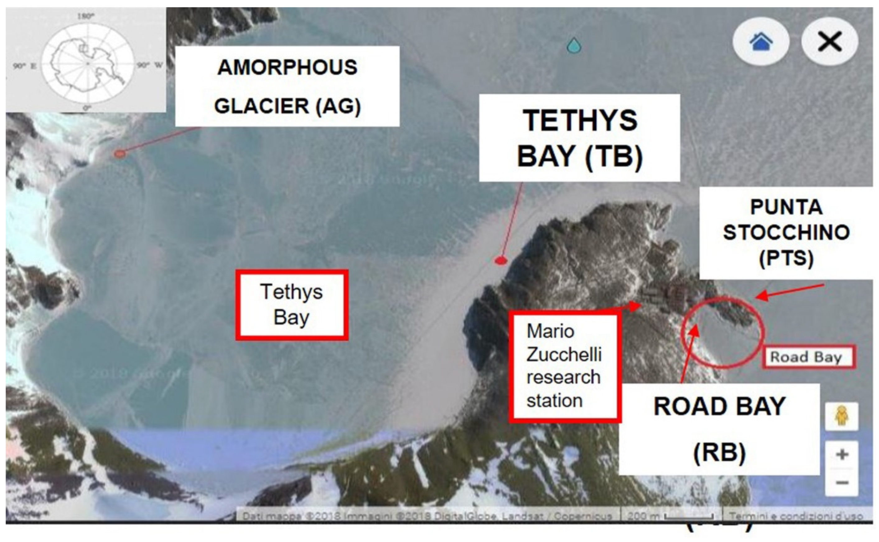
Figure 1. Location of the sampling stations (control and impact) in Tethys Bay and Road Bay (Ross Sea, Antarctica).
Jmse 10 01714 g001
Figure 2. Artificial structures on which the three polyvinyl chloride (PVC) panels (covered with a net for protection) and two polyethylene (PE) sheets were installed.
Jmse 10 01714 g002
Figure 3. Scheme of the structure of the artificial settlement panels in the two study areas.
Jmse 10 01714 g003
Figure 4. Mean values + standard deviations of total prokaryotic (PPT) and living (Live) cell abundance measured in the microbial biofilm developed on plastic panels submerged for 3, 9 and 12 months (T3, T9 and T12, respectively) at 5 and 20 m. (a) Road Bay area; (b) Tethys Bay area. RB, Road Bay (impact station, on the left); PTS, Punta Stocchino (control station, on the right); AG, Amorphous Glacier (impact station, on the left); TB, Tethys Bay (control station, on the right); PVC: Polyvinyl chloride; PE: Polyethylene.
Jmse 10 01714 g004
Figure 5. Mean values + standard deviations of enzymatic activity rates (Leucine aminopeptidase, LAP; beta-glucosidase, GLU; alkaline phosphatase, AP) measured in the microbial biofilm developed on plastic materials submerged in the Road Bay area at 5 m and 20 m for 3, 9 and 12 months (T3, T9 and T12, respectively). (a), RB, Road Bay (impact station); (b), PTS, Punta Stocchino (control station). PVC: Polyvinyl chloride; PE: Polyethylene.
Jmse 10 01714 g005
Figure 6. Mean values + standard deviations of enzymatic activity rates (Leucine aminopeptidase, LAP; beta-glucosidase, GLU; alkaline phosphatase, AP) measured in the microbial biofilm developed on plastic materials submerged in the Tethys Bay area at 5 m and 20 m for 12 months (T12). (a) AG, Amorphous Glacier (impact station); (b), TB, Tethys Bay (control station). PVC: Polyvinyl chloride; PE: Polyethylene.
Jmse 10 01714 g006
Figure 7. Mean values + standard deviations of (a) the microalgal biofilm abundance and (b) biofilm biomass (B) of prokaryotes (P) and microalgae (M) observed on plastic materials submerged in the Road Bay area at 5 m for 3 and 12 months (T3 and T12, respectively). Road Bay (impact station); PTS, Punta Stocchino (control station). PVC: Polyvinyl chloride; PE: Polyethylene.
Jmse 10 01714 g007
Figure 8. Mean values + standard deviations of total prokaryotic (PPT) and living (Live) cells abundance in seawater samples collected at 5 and 20 m at the start of the experiment (Time 0, November 2017), in January 2018 (Time 3, short-term experiment) and at the end (Time 12, November 2018) of the long-term experiment. (a), Road Bay area; (b) Tethys Bay area. RB, Road Bay (impact station, on the left); PTS, Punta Stocchino (control station, on the right); AG, Amorphous Glacier (impact station, on the left). TB, Tethys Bay (control station, on the right); PVC: Polyvinyl chloride; PE: Polyethylene.
Jmse 10 01714 g008
Figure 9. Mean values + standard deviations of the enzymatic activity rates (Leucine aminopeptidase, LAP; beta-glucosidase, GLU; alkaline phosphatase, AP) measured in seawater samples collected at 5 m and 20 m at the start of the experiment (Time 0, November 2017), in January 2018 (Time 3, short-term experiment) and at the end (Time 12, November 2018) of the long-term experiment. (a), Road Bay area; (b), Tethys Bay area. RB, Road Bay (impact station, on the left); PTS, Punta Stocchino (control station, on the right); AG, Amorphous Glacier (impact station, on the left). TB, Tethys Bay (control station, on the right); PVC: Polyvinyl chloride; PE: Polyethylene.
Jmse 10 01714 g009
Figure 10. Phytoplankton abundance (a) and biomass (b) measured in seawater samples collected at 5 m in November 2017, January and November 2018 (Times 0, 3 and 12, respectively). (a) Road Bay area; (b) Tethys Bay area. RB, Road Bay (impact station); PTS, Punta Stocchino (control station); AG, Amorphous Glacier (impact station); TB, Tethys Bay (control station).
Jmse 10 01714 g010
Table 1. Geographical coordinates of the study sites and characteristics of the biofilm samples collected in the Ross Sea (Antarctica).
Station TypologyLatitudeLongitudeImmersion Time (Months)Station Acronym
ROAD BAY
IMPACT, −5 m74°41′743″ S164°07′125″ E3, 9, 12RB5_(3, 9, 12)
IMPACT, −20 m74°41′784″ S164°07′219″ E3, 9, 12RB20_(3, 9, 12)
CONTROL, −5 m74°41′651″ S164°07′303″ E3, 9, 12PTS5_(3, 9, 12)
CONTROL, −20 m74°41′623″ S164°07′343″ E3, 9, 12PTS20_(3, 9, 12)
TETHYS BAY
IMPACT, −5 m74°41′234″ S164°02′135″ E12AG5_12
IMPACT, −20 m74°41′242″ S164°02′186″ E12AG20_12
CONTROL, −5 m74°41′417″ S164°06′303″ E12TB5_12
CONTROL, −20 m74°41′407″ S164°06′311″ E12TB20_12
Table 2. Physico-chemical parameters measured in Road and Tethys Bays at initial time (Time 0) and after 3 (Time 3) and 12 months (Time 12) of the experiment (n.d., not determined).
DateMonthsTSO2Chl apHNH4NO2NO3PO4N/P
°CPsumL L−1RFU µmol L−1µmol L−1µmol L−1µmol L−1
ROAD BAY
RB 522 November 20170−1.8335.198.00.628.021.960.0723.741.8513.95
RB 2022 November 20170−1.8335.208.80.587.741.810.1315.551.869.40
RB 526 January 201830.9034.797.90.998.111.330.096.850.5216.00
RB 2026 January 201830.8634.858.41.058.190.860.188.480.5317.81
RB 517 November 201812−1.7335.24n.d.0.66n.d.1.520.0626.051.3919.87
RB 2017 November 201812−1.7435.25n.d.0.61n.d.3.520.1226.331.4520.67
PTS-523 November 20170−1.8235.227.20.537.934.610.4615.511.6812.28
PTS 2023 November 20170−1.8035.226.90.587.833.530.6020.121.6714.52
PTS 526 January 18201830.9134.848.40.728.101.660.0810.760.7516.72
PTS 2026 January 1830.8734.848.41.058.091.220.1010.440.6318.64
PTS 525 November 201812−1.8635.27n.d.0.66n.d.0.940.0825.771.6815.94
PTS 2025 November 201812−1.7335.25n.d.0.63n.d.6.930.0626.631.4922.56
TETHYS BAY
AG 530 November 20170−1.6835.218.00.927.704.050.1020.672.4110.32
AG 2030 November 20170−1.7735.207.50.927.681.700.0921.301.9711.72
AG 513 November 201812−1.8235.267.50.64n.d.0.940.1021.871.5215.07
AG 2013 November 201812−1.8235.26n.d.0.61n.d.1.100.0421.861.5215.13
TB 526 November 20170−1.7235.148.11.008.121.630.1113.441.3511.24
TB 2026 November 20170−1.7235.198.51.007.752.050.1226.221.7616.13
TB 59 November 201812−1.7534.96n.d.0.63n.d.1.430.0424.031.4417.71
TB 209 November 201812−1.7435.249.7300.62n.d.2.670.0726.532.511.71
Publisher’s Note: MDPI stays neutral with regard to jurisdictional claims in published maps and institutional affiliations.