Article

Copper Induced DNA Damage in the Gills of the Mussel Mytilus trossulus and Reversibility after Depuration

by
Valentina Vladimirovna Slobodskova
,
Victor Pavlovich Chelomin
,
Sergey Petrovich Kukla
and
Andrey Alexandrovich Mazur
*
Il’ichev Pacific Oceanological Institute, Far Eastern Branch, Russian Academy of Sciences, 690041 Vladivostok, Russia
*
Author to whom correspondence should be addressed.
J. Mar. Sci. Eng. 2022, 10(11), 1570; https://doi.org/10.3390/jmse10111570
Submission received: 20 September 2022 / Revised: 17 October 2022 / Accepted: 19 October 2022 / Published: 23 October 2022
(This article belongs to the Special Issue Chemical Contamination on Coastal Ecosystems)

Abstract

:
The pollution of coastal water areas by heavy metals is constantly growing; therefore, the study of the mechanisms of impact of these toxicants on the organisms of hydrobionts is a topical direction of toxicology. Particularly pertinent are questions about the state of the reparation system in the aquatic organisms, which make it possible to assess the resistance, survival of hydrobionts, and the probability of remote consequences under the impact of heavy metals. Therefore, in this work, we investigated genome integrity and DNA repair ability in the gill cells of Mytilus trossulus, under conditions of copper (Cu2+) accumulation, and in the process of tissue depuration from this metal. Although the biochemical detoxification system was functioning, it is noted that destructive processes developed, including the accumulation of lipid peroxidation products (MDA) and DNA damage. It was also found that in all of the experimental groups of Mytilus trossulus, in the process of depuration from Cu2+ for 24 h, the levels of DNA damage and MDA content were markedly reduced, and further reduction was less intense.

1. Introduction

Under the conditions of continuously growing pollution in the marine environment, with heavy metals (HM) having a broad spectrum of effects on various organism systems, the relevance of the in-depth study of the mechanisms of relations between these toxicants and living organisms remains. Studies of the genotoxic manifestations of HM in hydrobionts capable of accumulating and retaining highly toxic metals in their tissues for a long time are of particular relevance. In addition, if the mechanisms of HM accumulation and detoxification in the cells of these organisms are currently relatively well studied [1,2,3,4,5], then the processes of interaction of metals with biochemical systems affecting genome stability are not given due attention. There is concern that the intracellular pools of metals can, through direct or indirect mechanisms, have a destructive effect on the genetic apparatus of the cell, despite the systems of detoxification and deposition. Due to the exceptional role of the genome, all types of destructive rearrangements and modifications that this supramolecular entity undergoes are of great interest, regardless of whether they lead to minor reversible processes, mutations, or death [6,7]. Chronologically, the intensification of genome destruction processes in cells can be the initial stage in the development of a whole set of negative biochemical events, up to the lethal outcome.
Copper is a typical representative of HM; it belongs to essential elements, and is a co-factor in many enzymes, as well as a variety of other functional proteins. However, numerous studies have shown that at elevated concentrations, copper ions can cause a variety of damages of a toxic nature in marine organisms [5,8,9,10,11,12]. According to several authors, the toxic activity of copper ions as a Fenton metal is mainly determined by the ability to participate in redox reactions and induce the formation of reactive oxygen and nitrogen forms, causing oxidative damage to various biostructures, including DNA molecules [8,9].
The most important manifestation of copper toxicity at the molecular level is genotoxicity, which has previously been demonstrated in the tissues of marine invertebrates, including the polychaete Alitta virens [10], corals Montastraea franksi [13], members of the bivalves Mytilus edulis [11], Mizuhopecten yessoensis [12], Scapharca inaequivalvis [14], Mytilus galloprovincialis [15] and Mytilus trossulus [16]. At the same time, it should be noted that most researchers attempting to characterize the genotoxic properties of copper have focused on measuring the degree of damage to the DNA molecule, without paying due attention to the genome repair system [5,9,14]. By using the repair system, the body seeks to minimize the damaging effects of a toxicant to prevent them from escalating to large-scale changes of an irreversible nature. In this regard, information on the state of the repair system in aquatic animals resisting the effects of adverse environmental factors is of great interest, not only in terms of resistance and survival of hydrobionts, but also in terms of estimating the probability of remote consequences of this effect.
Paying attention to the importance of genotoxicity in solving the problems of assessing and predicting the consequences of the accumulation of toxic metals in this research, we performed experiments with a focus on determining the genome integrity and DNA repair ability in gill cells of mussel M. trossulus, under conditions of copper (Cu2+) accumulation, and during tissue cleaning from this metal.

2. Material and Methods

2.1. Description of the Experiment

The mature mussel (Mytilus trossulus) (shell length 4.5–5 cm) was selected in Alekseev Bay (Peter the Great Bay). Before the experiment, the mussels were acclimatized to laboratory conditions by keeping them for 2 days in aerated 140-L aquariums with seawater, purified using a three-fraction gravel filter. During the entire period of the experiment, including the acclimatization period, the animals were not fed; the water was changed every day; and the tanks were kept at a relatively stable temperature (16 °C), salinity (32 psu) and oxygen saturation (100%). Water parameters were measured using an Edge HI 2040-02 device (Hanna Instruments, Germany). After the period of acclimatization, animals were divided into 2 groups of 180 individuals each: control and experimental. Three parallel aquariums of 60 specimens were used for each group. All procedures in the present work, as well as the shellfish disposal methods, were approved by the Commission on Bioethics at the V.I. Il’ichev Pacific Oceanological Institute, Far Eastern Branch of Russian Academy of Science (protocol № 16 and date of approval 15 April 2021), Vladivostok, Russia.
CuSO4-5H2O (LenReactiv, Saint Petersburg, Russia CAS-no 120194) was added to the aquariums, in which the experimental group of mussels were kept at a final copper concentration of 20 µg/L. The selected concentration of copper is widely used in experimental practices as it causes accumulation of the metal in the tissues of aquatic organisms without a pronounced acute toxic effect [4,17]. On the 2nd, 4th, and 7th days of copper exposure, 30 mussels were selected for analysis, and 90 specimens were transplanted to aquariums with pure seawater for washing. To estimate the reparative ability of M. trossulus gill cells, the mussels were selected after 24, 96, and 168 h of being kept in clean seawater. The control group of mussels was kept under the same conditions, but without the addition of copper. During the experiment, the mussels were not fed. No M. trossulus mortality occurred during the experiment.
The gills of M. trossulus were used for biochemical studies.
Fifteen specimens of mollusk were taken from each aquarium to determine the level of LPO and the degree of DNA damage, and 15 specimens were taken to determine the concentration of copper.

2.2. Comet Assay

To determine the degree of DNA molecule damage, an alkaline variant of the comet assay, adapted to marine organisms [18] and described by the above-mentioned authors [19,20,21], was used.
The gills were gently extracted from the mussels and washed several times with 4 °C isotonic solution (500 mM NaCl, 12.5 mM KCl, 5 mM EDTA-Na2, and 20 mM Tris-HCl, pH 7.4). After washing and removing mucus, the gills were carefully cut with scissors in 4–5 mL of CMFS buffer. The cell suspension was filtered through a sieve with a cell diameter of 40 mm from large tissue fragments. The cells in the filtrate were precipitated by centrifugation and resuspended in isotonic solution at a concentration of 105 cells/mL. Next, in 10–20 min of incubation, the isolated cells were used in the comet assay.
Fifty μL of cell suspensions were added to 100 μL of 1% fusible agarose (MP Biomedicals, Eschwege, Germany) in 0.04 M phosphate buffer (pH 7.4) at 37 °C, resuspended, and then placed on a slide previously coated with 1% agarose solution and covered by a coverslip. The sample was placed in a refrigerator for 3 min to solidify the agarose.
After the removal of the coverslip, the slide was transferred to a lysis solution (2.5 M NaCl; 0.1 M EDTA-Na2, 1% Triton X-100; 10% DMSO; 0.02 M Tris, pH 10) for 1 h in a place protected from light at 4 °C. At the end of lysis, the slides were washed with distilled water at a temperature of 4 °C and placed in an electrophoresis buffer (300 mM NaOH, 1 mM EDTA-Na2) and incubated for 40 min. Electrophoresis was performed at 2 V/cm for 15 min, followed by a neutralization step (0.4 M Tris-HCl, pH 7.4). Slides were stained with SYBR Green before imaging.
In the control and experimental groups, comets were counted for each individual (N = 15) in triplicate (n = 45) containing at least 50 comets. DNA comets were visualized and recorded using a fluorescence microscope Axio Imager A1 (Carl Zeiss, Oberkochen, Germany) equipped with an AxioCam MRc digital camera (Carl Zeiss, Oberkochen, Germany). For digital image processing, the CaspLab computer program v 1.2.2. (CASPLab, Wroclaw, Poland) https://casplab.com (accessed on 5 October 2022) was used to calculate various comet parameters indicating the degree of cellular DNA damage. For each comet, the proportion of DNA in the comet tail (% DNA in the tail) was determined.

2.3. Malonic Dialdehyde

The tissues were homogenized in a homogenizer (SILENTCRUSHER S; Heidolph Instruments GmbH & Co. KG Walpersdorfer Str. 12D-91126 Schwabach) in 0.1 M phosphate buffer, pH = 7.5. To prevent lipid peroxidation during the determination of MDA, an alcoholic solution of butylhydroxytoluene (Merck KGaA, Darmstadt, Germany. CAS-no 128-37-0) was added to the samples, to a final concentration of 5 mM. The content of malondialdehyde was determined by color reaction with 2-thiobarbituric acid (TBA, Merck KGaA, Darmstadt, Germany. CAS-no 504-17-6).
30% trichloroacetic acid (Germany, AppliChem GmbH Ottoweg 4 D-64291 Darmstadt. CAS-no 76-03-9) and 0.75% TBA solution were added sequentially to the tissue homogenate. The mixture was thoroughly mixed and heated for 20 min in a water bath (Memmert WNB 7, Memmert GmbH + Co. KG, Aeussere Rittersbacher Strasse 38 D-91126 Schwabach) at a temperature of 950C. After cooling, sediments were separated from the samples by centrifugation at 3000 rpm for 20 min. The measurements were carried out at a wavelength of 580 nm and 532 nm, then the difference in the readings of the optical density was found. To calculate the MDA content, the molar extinction coefficient was used—1.56 × 10⁵/cm/M. The relative content of MDA was expressed in nmol per g wet weight of the tissue. The measurements were carried out on a Shimadzu UV-2550 spectrophotometer.

2.4. Copper Concentration Measurement

For the quantitative analysis of the copper content in tissues and individual fractions, the atomic absorption method was the most widely used in analytical practice [22,23].
The biological material was first dried in a thermostat to constant weight at 85 °C, and carefully ground samples (100.0–200.0 mg) were transferred into solution by wet ashing in a mixture of concentrated acids (HNO3 (Germany, AppliChem GmbH Ottoweg 4 D-64291 Darmstadt. CAS no -7697-37-2):HClO4 (Germany, AppliChem GmbH Ottoweg 4 D-64291 Darmstadt. CAS no7601-90-3), 2:1 v/v).
The mineralization began in glass flasks at 180 °C for 10 h and stopped after the samples became colorless. The dried and discolored residue was redissolved in 0.1 N HCl (Germany, AppliChem GmbH Ottoweg 4 D-64291 Darmstadt. CAS no 7647-01-0) and used for metal analysis. The amount of Cu in the mollusk tissues was determined by atomic absorption spectrophotometry on a Shimadzu AA-7000 spectrophotometer (Japan) in version.

2.5. Statistical Analysis

Experimental results were processed using MS Excel and Statistica 10. Assumptions of normality and homogeneity were assessed using Levene’s and Shapiro-Wilk’s tests, respectively. Data on the DNA content in the comet tail and fertilization did not reach normality, and nonparametric Kruskal-Wallis ANOVA paired tests were conducted. A difference of p < 0.05 was considered statistically significant.

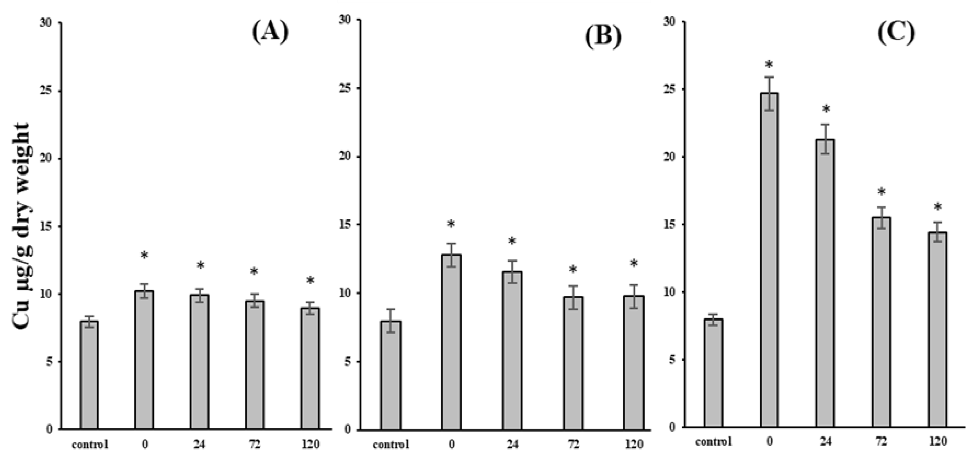
3. Results and Discussion

Currently, in ecotoxicology there is a firmly established point of view that dangerous consequences of an ecological character depend not only on the pollution level, toxicity degree, and the persistence of chemical compounds, but are also conditioned by the possibility of polluting substances accumulation by living organisms. As a model organism in ecotoxicological studies of this direction, representatives of Mytilidae, which demonstrate the ability to concentrate various toxic HM, and in particular, copper [1,4,8,16,24], are widely used. This was demonstrated in a series of our experiments using the far eastern representative of this family of M. trossulus, the results of which are presented in Figure 1.
From the analysis of these data, it follows that even at a relatively low concentration of copper (Cu2+) in the medium, which according to numerous researchers is sublethal and does not cause noticeable physiological disturbances [4,5,17], this species of marine mollusks accumulate the metal in the gills. It should be emphasized that, at a given concentration of copper ion in the external environment, the mussels preserved the linear character of metal accumulation during the experimental period (7 days). It was also found that by the end of the experiments, the content of copper in the gills of mussels increased almost 3-fold compared with the control mollusks, and reached approximately 25 µg/g dry weight of tissue (Figure 1A–C). This metal concentration in the gill cells is well-below the critical bioaccumulation threshold that causes acute toxicity in M. edulis [5,25], and lies within the theoretically calculated minimum level required to meet the minimum metabolic requirements of copper in mussels [26].
Nevertheless, the exposure of mussels to sublethal concentrations is accompanied by the development of oxidative stress processes already in the first stages of copper accumulation. This is evidenced by the reliable accumulation of products of lipid peroxidation (LPO)—malondialdehyde (MDA) (Figure 2A–C) and the increased level of DNA destruction (Figure 3A–C). In the latter case, based on the results of the comet analysis, it should be particularly noted that as copper accumulated in the gills, the proportion of cells with a high degree of genome damage (comet classes 3 and 4), characteristic of apoptotic cells, increased (Figure 4). In this respect, our results are consistent with the popular view that the genotoxic activity of copper is determined by the ability of this metal to induce reactive oxygen species (ROS) that initiate oxidative destruction of DNA molecule [13,27,28,29]. An enzymatic system of ROS generation has been identified in the tissues of bivalves, despite being facultative anaerobes [30,31,32,33]. Intracellular copper can contribute to ROS formation, not only through direct interaction with O2, but also by catalyzing the interaction of highly reactive oxygen species amongst themselves, according to Haber-Weiss reactions, resulting in the formation of an even more toxic -·OH compound than the original products [31], which induces LPO and oxidative DNA damage [9,12,14,16,34].
Based on the two-component model of accumulation of essential trace elements, such as copper [35], it is reasonable to assume that the formation of LPO products (MDA) and enhancement of DNA destruction revealed in our experiments occur as a result of an increase in the “metabolically available” pool copper (“metabolically available” pool of metal).
From the physicochemical point of view, this pool is formed in the initial stages of metal penetration into the cell as a result of binding and formation of various persistent complexes with intracellular biostructures, which leads to its “retention” and slows its removal from the cell. Among these biostructures, there are not only Cu-dependent macromolecules that ensure the functioning of physiological and biochemical systems, but also numerous functional structures whose activity can be damaged by interaction with copper. It is believed that a threat to the normal course of biochemical processes exists until the synthesis of specific proteins-metallothioneins (MT proteins) that bind this relatively labile pool of metal with a very high affinity is activated. The general point of view on the role of MT proteins is that they perform the function of detoxification because they form highly stable complexes with metals, as a result of which the metal ends up in a thermodynamic “trap” [35,36]. In addition to MT proteins, metal-containing granules formed during lysosome transformation are involved in metal detoxification and deposition in mussels [1,2,37].
These notions are evident in our subsequent experiments in the purification of mussels from accumulated copper. When experimental clams with relatively low levels of accumulated metal were transferred to clean water, we observed rapid Cu depuration of the gills to the level of control clams within 5 days (Figure 1A,B). This trend is in agreement with similar earlier experiments, showing that mussels exhibit a high rate of copper excretion during the first days of clearance [2,3]. This indicates that the accumulated copper in these mussels was part of the “metabolically available” copper, which is formed by relatively easily dissociated complexes, and therefore available for rapid excretion. A different picture is observed in experimental mollusks that accumulated higher levels of Cu (Figure 1C). Despite the “leaching” of copper from the gills of mussels kept in pure seawater, by the end of the 5-day experiments, the pools of copper in the tissues were retained almost twice as high as that of the control mollusks. Comparing these results with the literature, there is every reason to believe that this “residual” or weakly exchangeable pool (Cu) is represented by a metal bound to MT proteins or mineralized granules, clearance from which is very slow [1,2].
The above reasoning is, to a certain extent, confirmed by the restoration of biochemical disorders in the experimental mussels associated with the induction of oxidative stress during the purification of the metabolically available copper pool. It is noteworthy that the leaching of copper from the gill cells of all experimental groups of mollusks was accompanied by a sharp decrease in the level of lipid oxidative destruction products—MDA, whose content by the end of the experimental period was even lower than that of the control animals (Figure 2A–C). It is logical to assume that the imbalance between pro- and anti- oxidant processes arising at this stage of metal accumulation is smoothed under cleaning conditions by the activation of compensatory mechanisms and biotransformation systems.
In addition, the results obtained in this work, shown in Figure 3, allow us to state that during the purification of mussel gill cells from accumulated metal, there is a tendency to restore the damaged genome. The restoration of genome integrity in the process of tissue purification from copper is the result of the functioning of an efficient biochemical system of DNA repair in mussels.
The ability to repair DNA damage is an integral part of all living organisms and, according to researchers, is of paramount (crucial, most important) importance in the preservation of life processes, representing the main line of defense against various factors, including pollutants that exhibit genotoxic activity [38,39]. The presence of plastic and sufficiently efficient DNA repair system in hydrobionts, and in particular mussels [34,40], is an essential condition of existence and allows mollusks to tolerate abrupt fluctuations in various ecological and anthropogenic environmental factors characteristic of the littoral zone. Considering that in a normal-functioning cell, DNA is constantly damaged under the influence of various endogenous and exogenous factors, the existing basic level of damage can be considered as a dynamic balance between the intensity of DNA damage and the activity of the repair system components [29,40,41,42]. Based on the above, the fact of damage accumulation in the mussel genome revealed in the experiments means that the rate of Cu-induced destructive changes in DNA exceeds the activity of components responsible for the reparation of these damages. The presented results (Figure 3) show that the more mussels accumulated metal, the more this imbalance increased.
Although different enzyme systems specific to each type of damage are involved in the genome repair process, we consider it reasonable to rely on the integral activity of the repair system when assessing the rates of DNA damage repair in experimental mollusks. The nature of the dynamics of genome integrity repair in experimental mussels with different levels of Cu-induced DNA damage, shown in Figure 3, indicate that differences in repair rates over a 24-h period are particularly striking. According to simple calculations (see materials and methods), during this period approximately 50% of Cu-induced damage is repaired in group I clams; in group II, almost 70%; while in group III, no more than 33%.
In the search for explanations, we should consider that in the absence of a baseline level of reparation system activity, we cannot assess the direction of changes in DNA repair rate in group I of the mussels, with a minimal level of Cu-induced genome damage; whereas the increase in the reparation rate in group II of mussels with nearly 23% DNA damage level can be considered as a consequence of a general nonspecific reaction that can be explained by threshold dependent repair theory [43,44,45]. According to this theory, enzymes of the DNA repair system are activated and, consequently, the repair rate increases when the level of genotoxicant accumulation exceeds the threshold level, below which the repair system functions at the basic level. Following the logic of this theory, we can assume that the activity of the DNA repair system in mussels is sharply stimulated when the accumulation of genotoxic metal (Cu) reaches a level in the range of 15–25 µg/g dry weight.
In contrast, the mussels of group III repaired no more than one third (33%) of Cu-induced DNA damage within 24 h, indicating a sharp decrease in reparation system activity. Analysis of DNA damage repair dynamics shows that in experimental mollusks (groups I and II) with DNA damage levels of 14 and 23%, respectively, there occurred a rapid restoration of DNA integrity to the initial (baseline) level, typical of mollusks from control groups. In contrast, in mussels with Cu-induced DNA damage levels of about 30% (group III mussels exposed to Cu for 7 days), the recovery process was not only very slow, but did not reach the level of control mollusks, even within 5 days of purification (Figure 3). In other words, in this group of experimental mollusks, despite the long recovery period, weakly reparable and/or inaccessible Cu-induced DNA damage remained in the genome. The above-mentioned experimental data give grounds to put forward several mechanisms of decreased activity of the repair system and formation of “unrepaired” DNA.
First, Cu-induced prooxidative processes could result in the formation and gradual accumulation of hard-to-repair damage in the DNA molecule [46,47].
Second, the DNA damage repair system is represented by enzymes whose activity is inhibited by various HM, including copper [29,40,42,48].
Third, during Cu accumulation and detoxification, several energy-dependent processes are stimulated in the cell, including not only the synthesis of MT-proteins [1,2], but also the enhancement of the total protein exchange. Moreover, even after the cessation of metal intake from the external environment during the purification period, MT protein biosynthesis remains at a high level for some time, providing detoxification of accumulated metal [49,50]. The DNA damage repair system also consumes large amounts of energy. Comparing these data, it is logical to assume that when detoxification processes are stimulated, competition for the distribution of energy resources may cause a decrease in the activity of DNA repair system components, even under purification conditions.
When considering the mechanisms of DNA damage repair in gill cells of experimental mollusks, we cannot exclude the possibility of repair through the removal of cells with a severely damaged genome. The validity of this assumption is indicated by the character of distribution of individual classes of comets according to the level of DNA damage (Figure 4). In all of the experimental groups of mollusks, in the process of purification from Cu in 24 h the fraction of cells forming comet classes three and four with a strongly damaged genome noticeably decreased.
Whatever the specific mechanisms are, it is very important to emphasize that the formation of hard-to-repair Cu-induced lesions in the genome of mussel gill cells is, in our opinion, not only diagnostic, but also prognostic in terms of the risk of long-term consequences of pronounced ecotoxicological significance.
Overall, the results of our research have shown that during the accumulation of essential copper in the gill cells of marine mollusks (by the example of M. trossulus), despite the functioning of the biochemical detoxification system, destructive processes, including accumulation of lipid peroxidation products (LPO) and DNA damage, develop. It has also been established that the destruction of the genome is not only initiated and enhanced in the gill cells of mussels during the accumulation of metals, but is also maintained at an elevated level during the depuration of an excess amount of metals. It deserves special attention that against the background of cytogenetic stability disruption caused by metal with genotoxic properties, failures in the activity of the DNA repair system were noted, which led to the formation of “unrepaired” genome damage.
The integral assessment of the DNA repair system functioning in mussels under conditions of copper accumulation with subsequent purification from metal should be considered as the initial stage of research, based on which there is an opportunity to accurately assess and predict the state of specific ecosystem species responding to changes in the environment in which toxic chemical compounds are accumulated.

Author Contributions

Conceptualization, V.P.C. and V.V.S.; methodology, V.V.S., S.P.K. and A.A.M.; soft-ware, S.P.K.; validation, A.A.M. and V.V.S.; formal analysis, S.P.K.; investigation, A.A.M., V.V.S. and S.P.K.; resources, V.P.C.; data curation, V.P.C..; writing—original draft preparation, V.V.S. and V.P.C.; writing—review and editing, V.P.C., A.A.M. and V.V.S.; visualization, S.P.K.; supervision, V.P.C.; project administration, V.P.C.; funding acquisition, V.P.C. All authors have read and agreed to the published version of the manuscript.

Funding

This research was supported by the state assignment for research work of V.I. Il’ichev Pacific Oceanological Institute, FEB RAS (No. 121-21500052-9).

Institutional Review Board Statement

Not applicable.

Informed Consent Statement

Not applicable.

Data Availability Statement

Not applicable.

Acknowledgments

The authors would like to thank a leading engineer of the POI FEB RAS L.F. Kolosova, for the determination of Cu content.

Conflicts of Interest

The authors declare no conflict of interest.

References

  1. Viarengo, A. Biochemical effects of trace metals. Mar. Pollut. Bull. 1985, 16, 153–158. [Google Scholar] [CrossRef]
  2. Viarengo, A. Heavy metals in marine invertebrates: Mechanisms of regulation and toxicity at the cellular level. Crit. Rev. Aquat. Sci. 1989, 1, 295–317. [Google Scholar]
  3. Han, B.C.; Jeng, W.L.; Tsai, Y.N.; Jeng, M.S. Depuration of copper and zinc by green oysters and blue mussels of Taiwan. Environ. Pollut. 1993, 82, 93–97. [Google Scholar] [CrossRef]
  4. Grout, J.A.; Levings, C.D. Effects of acid mine drainage from an abandoned copper mine, brilannia mines, howe sound, British Columbia, Canada, on transplanted blue mussels (Mytilus edulis). Mar. Environ. Res. 2001, 51, 265–288. [Google Scholar] [CrossRef]
  5. Rosen, G.; Rivera-Duarte, I.; Bart Chadwick, D.; Ryan, A.; Santore, R.C.; Paquin, P.R. Critical tissue copper residues for marine bivalve (Mytilus galloprovincialis) and echinoderm (Strongylocentrotus purpuratus) embryonic development: Conceptual, regulatory and environmental implications. Mar. Environ. Res. 2008, 66, 327–336. [Google Scholar] [CrossRef]
  6. Nyberg, K.A.; Michelson, R.J.; Putnam, C.W.; Weinert, A. Toward maintaining the genome: DNA damage and replication checkpoints. Annu. Rev. Genet. 2002, 36, 617–656. [Google Scholar] [CrossRef] [Green Version]
  7. Cooke, M.S.; Evans, M.D.; Dizdaroglu, M.; Lunec, J. Oxidative DNA damage: Mechanisms, mutation, and disease. FASEB J. 2003, 17, 1195–1214. [Google Scholar] [CrossRef] [Green Version]
  8. Chelomin, V.P.; Belcheva, N.N.; Zakhartsev, M. Biochemical mechanisms of adaptation to cadmium and copper ions in the mussel Mytilus trossulus. Russ. J. Mar. Bio. 1998, 24, 330–336. [Google Scholar]
  9. Gomes, T.; Pereiraa, C.G.; Cardosoa, C.; Pinheiro, J.P.; Cancio, I.; Bebianno, M.J. Accumulation and toxicity of copper oxide nanoparticles in the digestive gland of Mytilus galloprovincialis. Aquat. Toxicol. 2012, 118–119, 72–79. [Google Scholar] [CrossRef]
  10. Watson, G.J.; Pini, J.M.; Richir, J. Chronic exposure to copper and zinc induces DNA damage in the polychaete Alitta virens and the implications for future toxicity of coastal sites. Environ. Pollut. 2018, 243, 1498–1508. [Google Scholar] [CrossRef] [Green Version]
  11. Al-Subiai, S.N.; Moody, A.J.; Mustafa, S.A.; Jha, A.N. A multiple biomarker approach to investigate the effects of copper on the marine bivalve mollusc, Mytilus edulis. Ecotoxicol. Environ. Saf. 2011, 74, 1913–1920. [Google Scholar] [CrossRef] [PubMed]
  12. Istomina, A.; Chelomin, V.; Kukla, S.; Zvyagintsev, A.; Karpenko, A.; Slinko, E.; Dovzhenko, N.; Slobodskova, V.; Kolosova, L. Copper effect on the biomarker state of the Mizuhopecten yessoensis tissues in the prespawning period. Environ. Toxicol. Pharmacol. 2019, 70, 103189. [Google Scholar] [CrossRef] [PubMed]
  13. Schwarz, J.A.; Mitchelmore, C.L.; Jones, R.; O’Dea, A.; Seymour, S. Exposure to copper induces oxidative and stress responses and DNA damage in the coral Montastraea franksi. Comp. Biochem. Physiol. C Toxicol. Pharmacol. 2013, 157, 272–279. [Google Scholar] [CrossRef] [PubMed]
  14. Gabbianelli, R.; Lupidi, G.; Villarini, M.; Falcioni, G. DNA Damage induced by copper on erythrocytes of gilthead sea bream Sparus aurata and mollusk Scapharca inaequivalvis. Arch. Environ. Contam. Toxicol. 2003, 45, 350–356. [Google Scholar] [CrossRef]
  15. Gomes, T.; Araújo, O.; Pereira, R.; Almeida, A.C.; Cravo, A.; Bebianno, M.J. Genotoxicity of copper oxide and silver nanoparticles in the mussel Mytilus galloprovincialis. Mar. Environ. Res. 2013, 84, 51–59. [Google Scholar] [CrossRef]
  16. Chelomin, V.P.; Slobodskova, V.V.; Zakhartsev, M.K.; Kukla, S.P. Genotoxic potential of copper oxide nanoparticles in the bivalve mollusk Mytilus trossulus. J. Ocean Univ. China 2017, 16, 339–345. [Google Scholar] [CrossRef]
  17. Amiard-Triquet, C.; Berthet, B.; Metayer, C.; Amiard, J.C. Contribution to the ecotoxicological study of cadmium, copper and zinc in the mussel Mytilus edulis. Mar. Biol. 1986, 92, 7–13. [Google Scholar] [CrossRef]
  18. Mitchelmore, C.L.; Birmelin, C.; Livingstone, D.R.; Chipman, J.K. Detection of DNA strand breaks in isolated mussels (Mytilus edulis) digestive gland cells using the “comet” assay. Ecotoxicol. Environ. Saf. 1998, 41, 51–58. [Google Scholar] [CrossRef] [PubMed]
  19. Slobodskova, V.V.; Kukla, S.P.; Chelomin, V.P. An analysis of the quality of the marine environment based on determina-tion of the genotoxicity of DNA in the gill cells of the Yesso Scallop Mizuhopecten yessoensis (Jay, 1856). Russ. J. Mar. Biol. 2015, 41, 495–498. [Google Scholar] [CrossRef]
  20. Mazur, A.A.; Chelomin, V.P.; Zhuravel, E.V.; Kukla, S.P.; Slobodskova, V.V.; Dovzhenko, N.V. Genotoxicity of Polystyrene (PS) Microspheres in Short-Term Exposure to Gametes of the Sand Dollar Scaphechinus mirabilis (Agassiz, 1864) (Echinodermata, Echinoidea). J. Mar. Sci. Eng. 2021, 9, 1088. [Google Scholar] [CrossRef]
  21. Chelomin, V.P.; Mazur, A.A.; Slobodskova, V.V.; Kukla, S.P.; Dovzhenko, N.V. Genotoxic Properties of Polystyrene (PS) Microspheres in the Filter-Feeder Mollusk Mytilus trossulus (Gould, 1850). J. Mar. Sci. Eng. 2022, 10, 273. [Google Scholar] [CrossRef]
  22. Julshamn, K.; Andersen, K.J. Subcellular distribution of major and minor elements in unexposed molluscs in western norway-I. The distribution and binding of cadmium, zinc and copper in the liver and the digestive system of the oyster Ostrea edulis. Comp. Biochem. Physiol. Part A Mol. Integr. Physiol. 1983, 75, 9–12. [Google Scholar] [CrossRef]
  23. Tabakaeva, O.V.; Tabakaev, A.V.; Piekoszewski, W. Nutritional composition and total collagen content of two commercially important edible bivalve molluscs from the Sea of Japan coast. J. Food Sci. Technol. 2018, 55, 4877–4886. [Google Scholar] [CrossRef] [PubMed] [Green Version]
  24. Brooks, S.J.; Farmena, E.; Heier, L.S.; Blanco-Rayónd, E.; Izagirre, U. Differences in copper bioaccumulation and biological responses in three Mytilus species. Aquat. Toxicol. 2015, 160, 1–12. [Google Scholar] [CrossRef] [PubMed] [Green Version]
  25. Widdows, J.; Johnson, D. Physiological energetics of Mytilus edulis: Scope for growth. Mar. Ecol. 1988, 46, 113–121. [Google Scholar] [CrossRef]
  26. White, S.L.; Rainbow, P.S. On the metabolic requirements for copper and zinc in molluscs and crustaceans. Mar. Environ. Res. 1985, 16, 215–229. [Google Scholar] [CrossRef]
  27. Shukla, A.K.; Pragya, P.; Chowdhuri, K.D. A modified alkaline Comet assay for in vivo detection of oxidative DNA damage in Drosophila melanogaster. Mutat. Res. 2011, 726, 222–226. [Google Scholar] [CrossRef]
  28. Trevisan, R.; Mello, D.F.; Fisher, A.S.; Schuwerack, P.M.; Dafre, A.L.; Moody, A.J. Selenium in water enhances antioxidant defenses and protects against copper-induced DNA damage in the blue mussel Mytilus edulis. Aquat. Toxicol. 2011, 101, 64–71. [Google Scholar] [CrossRef]
  29. Sandrini, J.Z.; Bianchini, A.; Trindade, G.S.; Nery, L.E.M.; Marins, L.F.F. Reactive oxygen species generation and expression of DNA repair-related genes after copper exposure in zebrafish (Danio rerio) ZFL cells. Aquat. Toxicol. 2009, 95, 285–291. [Google Scholar] [CrossRef]
  30. Musgrave, M.E.; Gould, S.P.; Ablett, R.F. Enzymatic lipid peroxidation in the gonadal and hepatopancreatic microsomal fraction of cultivated mussels (Mytilus edulis L.). J. Food Sci. 1987, 52, 609–612. [Google Scholar] [CrossRef]
  31. Winston, G.W.; Livingston, D.R.; Lips, F. Oxygen reduction metabolism by the digestive gland of the common marine mussel, Mytilus edulis L. J. Exp. Zool. 1990, 255, 296–308. [Google Scholar] [CrossRef]
  32. Chelomin, V.P.; Belcheva, N.N. The effect of heavy metals onprocesses of lipid peroxidation in microsomal membranes fromthe hepatopancreas of bivalve mollusks Mizuhopecten yessoensis. Comp. Biochem. Physiol. 1992, 103, 419–422. [Google Scholar] [CrossRef]
  33. Livingston, D.R.; Pipe, R.K. Mussels and environmental contaminants: Molecular and cellular aspects The Mussel Mytilus. Ecology, Physiology, Genetics and Culture. Dev. Aquac. Fish. Sci. 1992, 25, 425–464. [Google Scholar]
  34. Emmanouil, C.; Green, R.M.; Willey, F.R.; Chipman, J.K. Oxidative damage in gill of Mytilus edulis from Merseyside, UK, and reversibility after depuration. Environ. Pollut. 2008, 151, 663–668. [Google Scholar] [CrossRef] [PubMed]
  35. Rainbow, P.S. Trace metal concentrations in aquatic invertebrates: Why and so what? Environ. Pollut. 2002, 120, 497–507. [Google Scholar] [CrossRef]
  36. Mason, A.Z.; Jenkins, K.D. Metal detoxification in aquatic organisms. In Metal Speciation and Bioavailability in Aquatic Systems; Tessier, A., Turner, D.R., Eds.; John Wiley & Sons: Chichester, UK, 1995; pp. 479–608. [Google Scholar]
  37. Soto, M.; Ireland, M.P.; Marigomez, I. The contribution of metal/shell-weight index in target-tissues to metal body burden in sentinel marine molluscs. 2. Mytilus galloprovincialis. Sci. Total Environ. 1997, 198, 149–160. [Google Scholar] [CrossRef]
  38. Glei, M.; Hovhannisyan, G.; Pool-Zobel, B.L. Use of Comet-FISH in the study of DNA damage and repair: Review. Mutat. Res. 2009, 681, 33–43. [Google Scholar] [CrossRef]
  39. Kienzler, A.; Bony, S.; Devaux, A. DNA repair activity in fish and interest in ecotoxicology: A review. Aquat. Toxicol. 2013, 134–135, 47–56. [Google Scholar] [CrossRef]
  40. Pruski, A.M.; Dixon, D.R. Effects of cadmium on nuclear integrity and DNA repair efficiency in the gill cells of Mytilus edulis L. Aquat. Toxicol. 2002, 57, 127–137. [Google Scholar] [CrossRef]
  41. El-Bibany, A.H.; Bodnar, A.G.; Reinardy, H.C. Comparative DNA damage and repair in echinoderm coelomocytes exposed to genotoxicants. PLoS ONE 2014, 17, e107815. [Google Scholar] [CrossRef]
  42. Whiteside, J.R.; Box, C.L.; McMillan, T.J.; Allinson, S.L. Cadmium and copper inhibit both DNA repair activities of polynucleotide kinase. DNA Repair. 2010, 9, 83–89. [Google Scholar] [CrossRef] [PubMed]
  43. Ching, E.W.K.; Siu, W.H.L.; Lam, P.K.S.; Xu, L.; Zhang, Y.; Richardson, B.J.; Wu, R.S.S. DNA adduct formation and DNA strand breaks in green-lipped mussels (Perna viridis) exposed to Benzo[a]pyrene: Dose-and time-dependent relationships. Mar. Pollut. Bull. 2001, 42, 603–610. [Google Scholar] [CrossRef]
  44. Siu, W.H.L.; Hung, C.L.H.; Wong, H.L.; Richardson, B.J.; Lam, P.K.S. Exposure and time dependent DNA strand breakage in hepatopancreas of green-lipped mussels (Perna viridis) exposed to Aroclor 1254, and mixtures of B[a]P and Aroclor 1254. Mar. Pollut. Bull. 2003, 46, 1285–1293. [Google Scholar] [CrossRef]
  45. Siu, W.H.L.; Cao, J.; Jack, R.W.; Wu, R.S.S.; Richardson, B.J.; Xu, L.; Lam, P.K.S. Application of the comet and micronucleus assays to the detection of B[a]P genotoxicity in haemocytes of the green-lipped mussel (Perna viridis). Aquat. Toxicol. 2004, 66, 381–392. [Google Scholar] [CrossRef]
  46. Hook, S.E.; Lee, R.F. Interactive effects of UV, benzo[α] pyrene, and cadmium on DNA damage and repair in embryos of the grass shrimp Paleomonetes pugio. Mar. Environ. Res. 2004, 58, 735–739. [Google Scholar] [CrossRef]
  47. Michel, C.; Vincent-Hubert, F. DNA oxidation and DNA repair in gills of zebra mussels exposed to cadmium and benzo(a)pyrene. Ecotoxicology 2015, 24, 2009–2016. [Google Scholar] [CrossRef]
  48. Tang, S.; Wu, Y.; Ryan, C.N.; Yu, S.; Qin, G.; Edwards, D.S.; Mayer, G.D. Distinct expression profiles of stress defense and DNA repair genes in Daphnia pulex exposed to cadmium, zinc, and quantum dots. Chemosphere 2015, 120, 92–99. [Google Scholar] [CrossRef]
  49. Roesijadi, G.; Vestling, M.M.; Murphy, C.M.; Klerks, P.L.; Fenselau, C.C. Structure and time-dependent behavior of acetylated and non-acetylated forms of a molluscan metallothionein. Biochim. Biophys. Acta 1991, 1074, 230–236. [Google Scholar] [CrossRef]
  50. Carpene, E. Metallothionein in marine molluscs. In Ecotoxicology of Metals in Invertebrates; Dallinger, R., Rainbow, P.S.L., Eds.; Lewis Publishers: Devon, UK, 1993; pp. 55–72. [Google Scholar]
Figure 1. Copper content in the gills of M. trossulus under copper (Cu2+) accumulation conditions and after depuration. (AC)—exposure to Cu 48, 96, 168 h, respectively; 0, 24, 72, 120 h—depuration (mean ± standard deviation). * Difference from the control is significant (p < 0.05).
Jmse 10 01570 g001
Figure 2. MDA content in the gills of M. trossulus under copper (Cu2+) accumulation conditions and after depuration. (AC)—exposure to Cu 48, 96, 168 h, respectively; 0, 24, 72, 120 h—depuration (mean ±standard deviation). * Difference from the control is significant (p < 0.05).
Jmse 10 01570 g002
Figure 3. DNA damage in the gills of M. trossulus. 0, 24, 72, 120 h—depuration. * Difference from the control is significant (p < 0.05). Mean ± 1,96 • SE confidence interval for the population mean with a probability of 95%.
Jmse 10 01570 g003
Figure 4. Distribution of individual classes of comets according to the level of DNA damage.
Jmse 10 01570 g004
Publisher’s Note: MDPI stays neutral with regard to jurisdictional claims in published maps and institutional affiliations.

