Article

Study on the Tissue Heterogeneity and Micromechanical Properties of Maize Kernel

1
School of Mechanical Engineering, Shandong University of Technology, Zibo 255000, China
2
Changchun Ruiguang Science & Technology Co., Ltd., Changchun 130025, China
3
School of Agricultural Engineering and Food Science, Shandong University of Technology, Zibo 255000, China
*
Author to whom correspondence should be addressed.
Agriculture 2025, 15(6), 636; https://doi.org/10.3390/agriculture15060636
Submission received: 18 February 2025 / Revised: 10 March 2025 / Accepted: 14 March 2025 / Published: 17 March 2025
(This article belongs to the Section Agricultural Technology)

Abstract

:
This study measures and analyzes the heterogeneity and mechanical properties of maize kernels at the microscopic scale. Through microscopic tissue analysis and mechanical property tests, it was found that there are significant differences in the mechanical properties of different tissues in maize kernels. The starch granules in the horny endosperm are regular polyhedra, closely arranged, with a high number of proteins tightly filling the gaps between starch granules. The structural characteristics of the horny endosperm give it a high maximum rupture force and elastic modulus, with a maximum rupture force of 128 N and an elastic modulus of 353 MPa. The starch granules in the farinaceous endosperm are spherical and loosely and irregularly arranged, leading to more gaps between the starch granules. As a result, the maximum rupture force and elastic modulus of the farinaceous endosperm are relatively lower. The maximum rupture force of the farinaceous endosperm is 38 N, and the elastic modulus is 136 MPa. Compression tests were conducted on maize kernels, and scanning was performed using a Micro CT system. The results showed that the farinaceous endosperm deforms and breaks more easily, with most damage beginning in the farinaceous endosperm and then extending further. The micromechanics discrete element analysis of the loading process of the farinaceous endosperm was carried out further. It was found that the deformation of the farinaceous endosperm occurs in four stages: initial, crack initiation, crack propagation, and fracture. When the farinaceous endosperm is loaded to 132 N, internal cracks begin to initiate and gradually propagate. At 292 N, the internal particles of the farinaceous endosperm start to break, followed by a drop in load and eventual fracture. During the loading process, significant differences in the velocity field of the farinaceous endosperm were observed.

1. Introduction

As an important crop, corn is cultivated in many parts of the world and its yield ranks first among food crops [1,2]. Corn is also an important source of raw materials for animal husbandry, chemical industry, and other industries [3,4]. Growing populations and the use of animal-derived foods and chemical feedstocks have led to increased demand for corn grains [5,6]. Maize kernels are prone to breakage during harvest and threshing, which not only lowers quality but also increases the likelihood of mold development, adversely affecting subsequent storage [7,8,9]. Therefore, to reduce the breakage rate of maize kernels during harvest and threshing, a more in-depth study of the mechanical properties of maize kernels is necessary [10,11]. Li Xinping et al. [12,13,14] conducted impact experiments on maize kernels with varying moisture content and found that as moisture content increased, the impact resistance of the maize kernels decreased. The ventral surface had the highest impact resistance, followed by the lateral surface, and the dorsal surface had the weakest resistance. Cai Chaojie et al. [15] conducted squeezing and rubbing threshing experiments on the ear of maize. They found that the minimum breakage force of the kernels was positively correlated with the breakage rate. Liu Jiayuan et al. [16] studied the breakage resistance of maize kernels during mechanical harvest and its influencing factors. They found that as moisture content decreased, kernel hardness increased, and the breakage rate was negatively correlated with hardness. Zhang Yongli et al. [17] conducted shear breakage experiments on the dorsal, ventral, and lateral surfaces of maize kernels. The results indicated significant differences in shear failure loads between the different parts of the maize kernel. In summary, both domestic and international scholars have conducted extensive and in-depth experiments and studies on the overall mechanical properties of maize kernels. In numerical analysis, Wang et al. [18] used finite element simulation of three-way force on maize kernels combined with compression experiments and found that the minimum breakage force was required when the kernels were upright, while the maximum force was required when they were placed flat. Guangwan W., et al. [19] applied forces in three directions of corn kernels by the finite element simulation. And the results showed that the maximum principal stress occurred in the middle part of the top of the seed coat and cuticle when the top surface was loaded; the maximum principal stress occurred in the middle part of the seed coat and cuticle and embryo when the side was loaded; and the maximum principal stress occurred in the seed coat and cuticle and silty layer when the abdominal surface was loaded. Tao T., et al. [20] conducted a simulation analysis on the forces of different parts of corn grain under impact load, and the results showed that different parts of corn grain were impacted with different forces. When the same load was applied, the middle section of corn grain was impacted with the largest force. Han T., et al. [21] studied the evolution of impact damage of corn kernels under different impact velocities and orientations and introduced the microscopic impact behavior of corn kernels by the finite element method. The results showed that the fracture property was the comprehensive effect of compressive stress and tensile stress. In both experimental studies and finite element analysis, most of the corn is regarded as a whole, ignoring the problem of internal organizational anisotropy. In order to fully explain the mechanical properties of maize, it is necessary to further explore the coordinated influence of various tissues. At present, some studies have been carried out on the mechanical properties of corn grain microstructure. Peng G., et al. [22] investigated the hardness of keratinous endosperm and silty endosperm of corn grains. The results showed that keratinous endosperm had a relatively dense starchy protein network structure, and the hardness of keratinous endosperm was significantly higher than that of silty endosperm. Mengmeng Q., et al. [23] studied the relationship between corn kernel component content and mechanical properties, and microstructure observations showed that high protein content increased hardness, breaking force, breaking energy, and elasticity, while high starch content increased viscosity. Singh, S.S., et al. [24] studied the effect of different proportions of keratin endosperm on the mechanical properties of corn kernels, and microscopic studies showed that the fracture resistance of the kernels was mainly affected by the grain structure. Bolong W., et al. [25] measured key mechanical parameters for different tissues of maize kernels and pointed out that the mechanical damage caused by external forces during maize kernel processing is closely related to the mechanical properties of its internal tissues. It follows that maize kernels are a complex multi-body system. Each tissue exhibits unique mechanical properties, which intertwine to shape the overall mechanical performance of the maize kernel [26,27,28]. However, research on the mechanical properties of the different internal tissues of maize kernels remains insufficient at present. It is necessary to further focus on the mechanical properties of the various tissues inside the maize kernel and to explore the mechanisms of crack initiation and propagation at a smaller scale.
This study measures and analyzes the mechanical properties of maize kernels at the mesoscopic scale. Firstly, to further explore the impact of the microstructure of different tissues in maize kernels on their mechanical properties, the mechanical properties of the maize kernel tissues were measured. Scanning electron microscopy (SEM) was used to observe and analyze the microstructure of the horny endosperm and farinaceous endosperm in the maize kernel. Then, a universal testing machine was used to test the mechanical properties of the different tissues and analyze the relationship between the microstructure and the mechanical properties of the tissues. Next, the whole compression test of corn grains was carried out, and the Micro CT system was used for slice scanning to observe the damage location and evolution process during the compression process of corn grains. Finally, PFC2D (5.0) particle flow software was used to establish a numerical model of the partial particle flow of corn endosperm on the mesoscopic scale, and the mesoscopic damage evolution of endosperm was simulated by model loading in order to reveal the internal relationship between the microstructure and mechanical properties from the mesoscopic level.

2. Experimental Design

2.1. Experimental Materials

In this study, the Zhengdan 958 maize variety was selected, with a half-dent type and a thousand-kernel weight of 312 g, harvested in 2022 in the Jinan area of Shandong. To ensure the accuracy and reliability of the experiment, maize kernels with consistent appearance and size, and no obvious damage, were selected. The internal structure of the maize kernel, as shown in Figure 1, consists of the seed coat, endosperm, and embryo. The seed coat thickness is about 30 μm, accounting for 5–7% of the total kernel weight. The endosperm of the maize kernel provides the necessary nutrients and energy for the growth and development of the maize seed, accounting for about 80% of the total kernel weight. The endosperm is further divided into horny endosperm and farinaceous endosperm. The embryo is a starting point for maize growth and development.
It is concluded that the internal structure of maize kernels is complex, with significant differences in composition, physical structure, morphology, and other aspects among the tissues. This results in high heterogeneity within the maize kernel, leading to noticeable differences in the mechanical properties of the various tissues. When the maize kernel is subjected to external loading, the damage behavior of different tissues may vary significantly.

2.2. Experimental Equipment and Methods

2.2.1. Microstructure Detection

The microstructure of the maize kernel endosperm was obtained using a scanning electron microscope. Before imaging with the scanning electron microscope, the maize kernel samples were pretreated. Firstly, the samples were cut along the short axis of the kernel with a blade to expose the internal endosperm tissue. The sample surface was cleaned with high-purity alcohol and then blow-dried. The maize kernel samples were then placed in a high-vacuum ion sputtering coater for gold coating treatment.

2.2.2. Measurement of Mechanical Properties of Various Tissues in Maize Kernel

The mechanical properties of the various tissues in maize kernels were measured using an Instron 5543 universal testing machine. A cylindrical flat-head probe of the universal testing machine was used to load the horny endosperm, farinaceous endosperm, and embryo of the maize kernel, and data from the loading process were recorded. The specific methods are as follows:
(a)
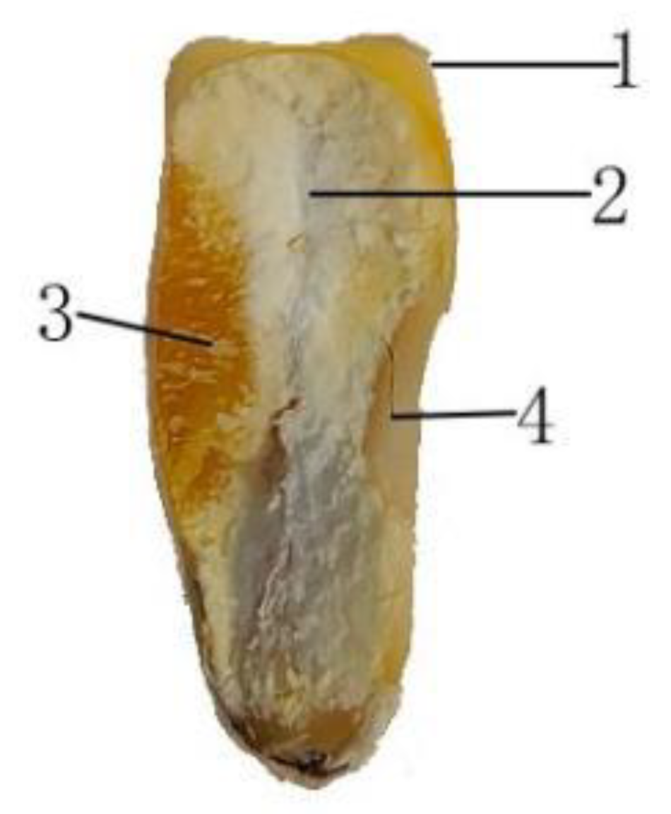
Sample preparation: The sample was cut along the short axis of the maize kernel with a blade to expose the internal horny endosperm, farinaceous endosperm, and embryo tissues. The cross-sections were polished using sandpaper of varying coarseness.
(b)
Sample placement: To ensure stable loading, the cut surface of the sample was adjusted to be parallel to the working plane of the round probe, and the sample was fixed to the stage using adhesive.
(c)
Loading: The round probe was pressed into the maize kernel tissues at a rate of 1 mm/min. The test was terminated when the probe reached a depth of 1.2 mm, and the probe was returned to the initial position.
To ensure the accuracy and rigor of the test results, each sample was used for only one test. Ten independent repeated tests were conducted under each test condition, and the average of the repeated tests was taken as the final result.

2.2.3. Maize Kernel Compression Test

A compression test was conducted on the entire maize kernel using an MTS static universal material testing machine. The kernel was placed horizontally, with the lower loading plate fixed and the upper loading plate moving downward at a constant speed. The loading speed was set to 1.5 mm/min, and the test was terminated when a rapid decrease in force was detected on the loading plate.

2.2.4. Maize Kernel Tomographic Scanning

Sixty repeated compression tests were conducted on the maize kernel. During the compression tests, the maize kernels were scanned using a Hiscan XM Micro CT system (Suzhou Hiscan Information Thechnology Co., Ltd., Suzhou, China) with the following scanning conditions: 80 kV, 100 µA, single exposure time of 50 ms, scanning resolution of 25 µm, and scanning angular intervals of 0.5 degrees. After scanning, the data were reconstructed using Hiscan Reconstruct software (V3.0) and analyzed using Hiscan Analyzer software (V3.0).

3. Results and Discussion

3.1. Microstructure of Maize Kernels

The microstructure of the horny and farinaceous endosperm of the maize kernel was observed using scanning electron microscopy, as shown in Figure 2. It can be seen that both the horny and farinaceous endosperms are primarily composed of starch granules, which are dense and abundant. In addition, a small amount of proteins and other components are present in the endosperm, distributed in the gaps between the starch granules. These components serve a binding function, maintaining the overall structure of the endosperm.
Figure 2e,f mark the starch granules, pores, and protein structures in the floury and horny endosperms, respectively. In the farinaceous endosperm, starch granules are spherical in shape, and the granules are loosely and irregularly arranged. Proteins are distributed in the gaps between the starch granules and have an irregular shape. Although the proteins partially fill the spaces between the starch granules, there are still unfilled areas, leading to the presence of pores. In the horny endosperm, starch is also present in granule form, but these granules are polyhedral, arranged more densely and regularly. In the horny endosperm, proteins are also distributed in the gaps between the starch granules, with an irregular shape. A comparison reveals that the structure of the horny endosperm is more compact and organized than that of the farinaceous endosperm.

3.2. Mechanical Properties of Maize Kernel Tissues

In order to further investigate the difference in mechanical properties of different tissues, the maximum rupture force, rupture shape variable, and elastic modulus of keratinous endosperm, silty endosperm, and embryo were measured. In order to ensure the reliability and repeatability of the experiment, the mean value, standard deviation, and confidence interval of the data were calculated.
The mean value, standard deviation, and 95% confidence interval of the mechanical parameters of each tissue were calculated. The results are shown in Table 1, Table 2 and Table 3 below.
Based on the statistical results, the horny endosperm was taken and analyzed as an example. The maximum rupture force of horny endosperm has an average of 127.24, with a standard deviation of 5.0525. It can be indicated that the standard deviations of maximum rupture force are significantly far less than its mean values, differing by several orders of magnitude, which demonstrates small data variability. The 95% confidence interval for this value is [123.6259, 130.8541], meaning that the average maximum rupture force of the horny endosperm falls within this range with a 95% chance. The average rupture deformation for the horny endosperm is 0.893, with a standard deviation of 0.0078, also showing minimal variability. The 95% confidence interval for this value is [0.8874, 0.8986], indicating that the average rupture deformation for the horny endosperm falls within this range with a 95% chance. The average modulus of elasticity for the horny endosperm is 356.11, with a standard deviation of 7.2263, and the 95% confidence interval is [350.9409, 361.2790]. It can be observed that the standard deviations of maximum rupture force, rupture deformation, and modulus of elasticity are significantly far less than their respective mean values, differing by several orders of magnitude, which demonstrates minimal data variability. Similar analyses for the farinaceous endosperm and embryo demonstrated the same conclusion, thus the data are deemed reliable.
It can be seen that the experimental data are relatively reasonable and accurate, and the average value is selected for analysis. The maximum rupture force, rupture strain, and elastic modulus for the horny endosperm, farinaceous endosperm, and embryo of the maize kernel are shown in Figure 3.
In Figure 3, it is evident that there are significant differences in the mechanical properties of the different tissues of maize kernels. Specifically, the horny endosperm has significantly higher maximum rupture force and elastic modulus than the farinaceous endosperm and embryo, while the farinaceous endosperm is slightly higher than the embryo in these indicators. The maximum rupture force of the horny endosperm is 128 N, while that of the farinaceous endosperm is 38 N. The elastic modulus of the horny endosperm is 353 MPa, while that of the farinaceous endosperm is 136 MPa. In terms of rupture strain, the difference between the horny endosperm and the embryo is small, with both being slightly higher than the farinaceous endosperm. For the same strain, materials with a higher elastic modulus can withstand larger loads. When the maize kernel is subjected to an external load, the farinaceous endosperm, with a smaller rupture strain and elastic modulus, reaches its load limit first, leading to the formation of microscopic cracks. These cracks expand and propagate under sustained external load, ultimately causing the maize kernel to fracture. The differences in the mechanical properties among the different tissues of the maize kernel are mainly due to variations in their composition and the microstructural features of the tissues.
Analyzing the microstructures of the floury and horny endosperms (Figure 2), the starch granules in the horny endosperm are regularly polyhedral, densely packed, with a large amount of protein filling the gaps between the starch granules. This compact and orderly structure gives the horny endosperm a solid texture, higher hardness, and density, allowing it to exhibit higher maximum rupture force and elastic modulus when resisting external forces. Therefore, the horny endosperm shows better structural stability and load-bearing capacity on a macroscopic scale. In contrast, the starch granules in the farinaceous endosperm are spherical in shape, and loosely and irregularly arranged, resulting in more gaps between the starch granules. This loose tissue structure makes the farinaceous endosperm texture less dense. As a result, under the same external force, its maximum rupture force and elastic modulus are lower, making it more prone to deformation and rupture.

3.3. Analysis of Maize Kernel Damage and Fracture Performance

During the 60 compression tests on maize kernels, the Hiscan XM Micro CT system was used to scan the kernels, and the initial locations of the cracks in the kernels were statistically analyzed. The results are shown in Figure 4. It can be observed that the initial cracks in the maize kernels mainly originate in the farinaceous endosperm, accounting for 70% of the total, followed by the horny endosperm, which accounts for 20%, and the embryo, which accounts for less than 10%.
Figure 5 shows the damage process of a maize kernel under repeated compression. By observing the compression process, it was found that the starting point of the kernel damage is located in the farinaceous endosperm region in the middle of the kernel. Subsequently, under continuous loading, the damage accumulates, and cracks in the farinaceous endosperm continue to expand and extend. When the maize kernel is considered as a whole and subjected to external loads, the smaller fracture strain and elastic modulus of the farinaceous endosperm, compared to the horny endosperm, make it more likely to reach its bearing limit first, which leads to the formation of microscopic cracks. This is the main reason that the initial cracks in the maize kernel primarily occur in the farinaceous endosperm. After the compression test, the final CT images of the corn kernel are obtained. The part of the corn kernel is shown in Figure 6. It can be seen that under repeated compression loads, the cracks in the kernel continuously expand outward from the inner farinaceous endosperm to the horny endosperm and embryo until the kernel ultimately breaks.

4. Finite Element Simulation and Analysis

Through the mechanical property tests of various tissues of maize in the previous part, combined with SEM and the tracking of Hiscan XM Micro CT systems, it was found that during the loading process for maize kernels, microscopic cracks first arose from the powdered endosperm portion. And with the accumulation of damages, the cracks gradually expanded, which ultimately led to the destruction of the kernels. In order to further investigate the generation and development of cracks in the powdered endosperm, this part takes the powdered endosperm as the research object. Based on the particle characteristics of the powdered endosperm, a particle flow numerical model of the powdered endosperm portion of maize kernel was established on a mesoscale using the particle flow software PFC2D. The microscopic damage evolution of the powdered endosperm was simulated and analyzed by loading the model.

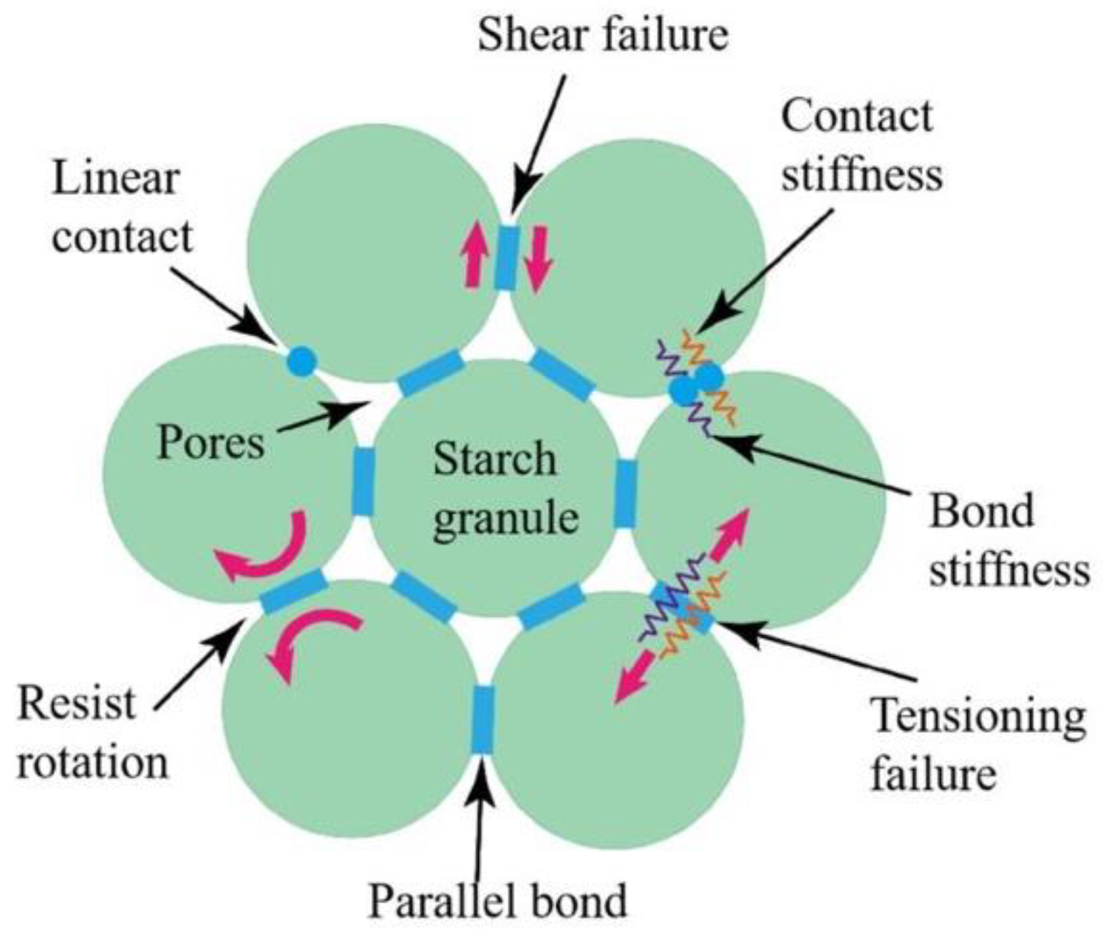
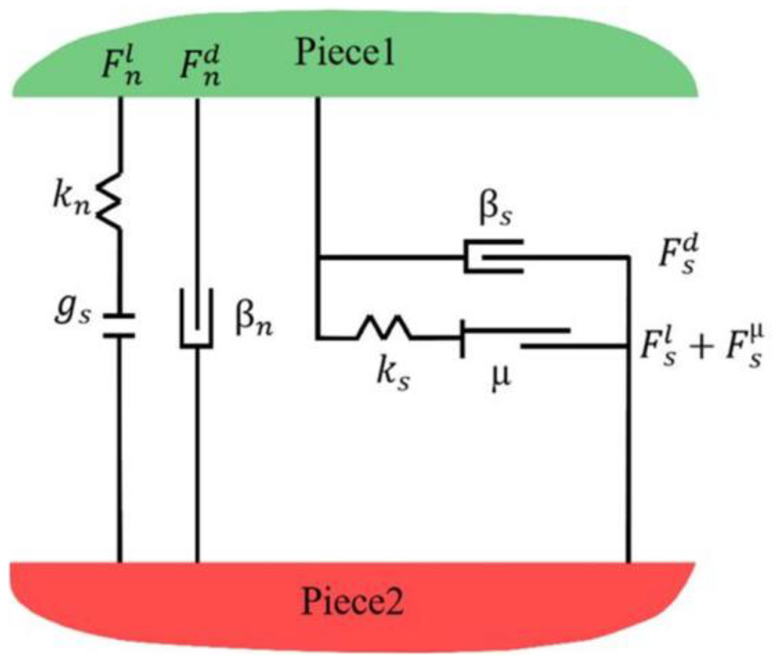
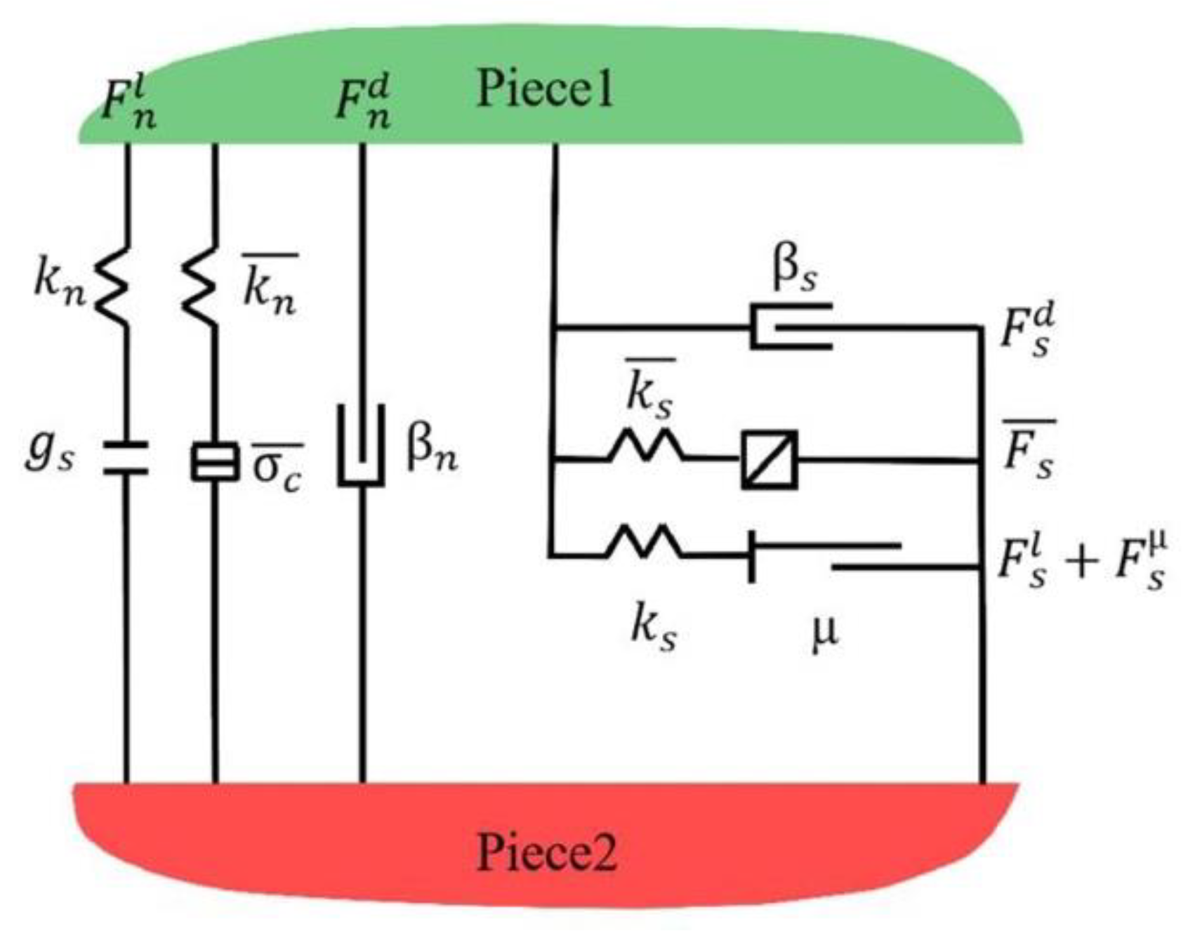
4.1. Contact Model Setup

In the maize kernel’s farinaceous endosperm structure, starch granules serve as the basic building units, bonded together by substances such as proteins, forming a network structure with gaps. When force is applied to the farinaceous endosperm, the stress on the starch granules at the microscopic level is shown in Figure 7.
To fully reflect the interaction characteristics between starch granules, the linear contact model and the linear parallel bonding model are prioritized when constructing the particle flow model of the farinaceous endosperm. These two models can better simulate the bonding and sliding behavior between the starch granules.
If only the rigid contact between starch granules is considered, without taking into account the bonding effect of proteins and other substances on the starch granules, the contact between starch granules can be considered as linear contact. The principle of the linear contact model is shown in Figure 8. K n is the normal contact stiffness; K s is the tangential contact stiffness; β n and β s represent the damping forces in the normal and tangential directions, respectively; μ is the friction coefficient in the shear direction; and g s   is the surface contact gap. The linear model includes linear components and damping components that act in parallel with each other. These two components act in the normal and tangential directions, respectively. The linear component provides linear elastic behavior, which only functions under compression and cannot withstand tension, while the damping component provides viscous behavior. Clearly, considering only the linear interactions between particles is insufficient; the bonding behavior between starch granules should also be included. Therefore, a parallel bonding force is added to the linear model. The contact principle of the linear parallel bonding model is shown in Figure 9, where the parallel bonding and linear components act in parallel. The presence of parallel bonding links generates forces and moments due to the relative motion of the two particles at the contact point. The linear parallel bonding contact model matches the actual mechanical properties of the maize kernel’s farinaceous endosperm, thus the linear parallel bonding model was selected.

4.2. Structural Parameter Setup

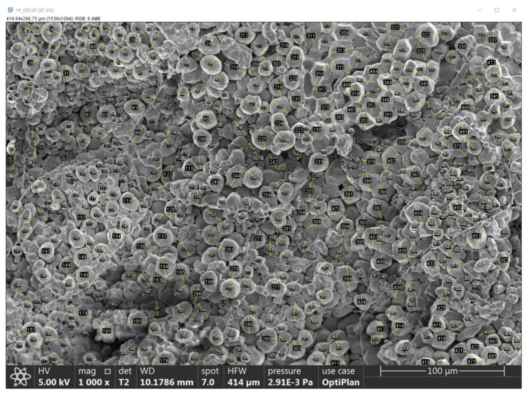
4.2.1. Porosity Statistics

The porosity of powdered endosperm was analyzed and counted using Image-Pro Plus (IPP) software (6.0). Firstly, the SEM images of powdered endosperm with a magnification of 1000 times were imported into IPP and pre-processed. The contrast between the pore region and the seed tissue was highlighted by contrast ratio adjustment, binarization, and other settings. Subsequently, the pre-processed images were subjected to particle segmentation and identification, and the pore and non-porous regions were accurately distinguished by setting segmentation parameters including particle size and shape. Finally, the number of pores and the area of each pore were measured by IPP, and a summation operation was performed on the areas of all pores to obtain the total pore area. The percentage of the area of pore regions was calculated to derive the porosity. The porosity was calculated using the following formula:
D = S p S T × 100 %
To ensure the accuracy of the results, the statistics were repeated several times and the average value was taken as the final measurement of porosity. The number of pores, pore area, and porosity of the powdered endosperm were obtained by averaging multiple measurements, and the final data are shown in Table 4.

4.2.2. Parameter Characterization

Starch granule sizes in the powdered endosperm scanning electron microscopy images were determined using ImageJ software (1.8.0). Firstly, the starch granules were labeled according to the scale on the image and then the diameter of each starch granule was measured by the tool built into the software. The powdered endosperm with completed diameter labeling is shown in Figure 10.
By extracting the porosity and measuring the particle size of the farinaceous endosperm, it was found that the porosity of the farinaceous endosperm is 5.71%. The diameter of starch particles is generally distributed in the range of 2~22 μm. And the diameter is mainly concentrated in the range of 4~16 μm, accounting for nearly 90%, and the proportion distribution is relatively uniform in this interval. In the modeling process, the starch granules were classified into six groups based on their size, with the particle grading parameters shown in Table 5. The model was calibrated using the mechanical property parameters measured from the tissue mechanics experiments, as listed in Table 6.

4.3. Establishment of the Particle Flow Model

The boundaries of the particle flow numerical model for the farinaceous endosperm were set to a height of 1400 μm and a width of 700 μm. Within the selected computational area, several particles were generated based on preset parameters, with each particle representing a starch granule. The generated model is shown in Figure 11. The model generated 16,370 particles, with a total of 40,327 force chains established between the particles. The four red lines around the model represent the four loading plates, and the connecting lines between the particles represent the force chains, with the line thickness indicating the magnitude of the force experienced by the force chains. The model is based on the grouping of particle diameters, as shown in Figure 12.
Boundary conditions need to be imposed on the model after particle generation. And the processing steps are, in order, applying a perimeter pressure, adding a glue joint, unloading, and removing the side-loading plates. The purpose of applying a perimeter pressure is to bring the loose particles into close contact with each other. The way of applying circumferential pressure is to carry out servo control on the four loading plates and set the target stress of 10 KPa for the four loading plates. And each loading plate can independently detect the size of its own stress value. When the current stress value is smaller than the target stress, the loading plate will move toward the internal direction of the model and squeeze the internal particles to increase the force. And vice versa, it will move towards the outside until the stress size of the four loading plates is in line with the target stress. After applying the circumferential pressure, the particles are tightly stacked but not bonded to each other. The particles are just simple linear contact, at this time the particles cannot resist the rotation and tensile force. It is necessary to add glue to the particles, that is, to add a parallel bonding bond between the particles. After the contact relationship between the particles is established, the whole model can be unloaded slowly to reduce the force between the particles inside.

4.4. Simulation Results and Analysis

4.4.1. Force and Deformation During the Loading Process

Force is transmitted through the force chains in the particle flow numerical model. The entire loading process of the farinaceous endosperm deformation can be divided into four stages: initial, crack initiation, crack propagation, and fracture. The four stages are shown in Figure 13a–d.
In the initial stage, as shown in Figure 13a, the force applied by the loading plates is small, and the deformation of the farinaceous endosperm is relatively minor. The number and thickness distribution of the force chains inside the farinaceous endosperm are relatively uniform, with a net-like shape and no apparent directional pattern. At this point, the force chains are mainly in a compressed form, and there are no obvious tensile force chains yet. As the loading plates continue to apply force, cracks begin to appear inside the farinaceous endosperm, and the force chains gradually transition from the initial stage to the development stage, as shown in Figure 13b. In this stage, the crack starts from the inside, with the load at 132 N. The force chains begin to show obvious directionality, which aligns with the direction of the force applied by the loading plates. The shape of the force chains gradually changes to a root-like structure. As the loading increases, the directionality of the force chains becomes more pronounced, and the tensile force chains between the farinaceous endosperm particles gradually emerge. As loading continues, the model enters the crack propagation stage, as shown in Figure 13c. In this stage, cracks propagate inside the model. At this point, the load on the farinaceous endosperm reaches its maximum value of approximately 292 N, and the farinaceous endosperm is close to its bearing limit. The connections between the farinaceous endosperm particles begin to fail, leading to large-scale cracks. In the crack propagation regions, the distribution of the force chains is relatively concentrated, indicating that these areas are the key locations where stress is concentrated. After reaching the bearing limit of the farinaceous endosperm, continued loading causes the farinaceous endosperm to fracture, as shown in Figure 13d. In this stage, the force on the endosperm particles near the cracks exceeds the connection strength between the particles, leading to extensive fracture of the particle connections, while the load on the farinaceous endosperm begins to decrease rapidly. This process reveals the damage mechanism and mechanical response of the internal organizational structure of the farinaceous endosperm in maize kernels under external loading.
The process of damage accumulation of the powdered endosperm during the whole loading process is consistent with the change tracked and photographed by the Hiscan XM Micro CT system in the test of Section 2. And as can be seen in comparison with Figure 6, the direction of crack development is also consistent.

4.4.2. Speed Field Analysis During the Loading Process

During loading, the distribution of the speed field of the farinaceous endosperm particles is visually represented by arrows, as shown in Figure 14. The red arrows clearly reflect the main direction and trend of the speed field. To better understand the damage behavior of the farinaceous endosperm model during the loading process, the locations where major cracks initiate are marked with black dashed lines in the figure. The four states in Figure 14 correspond to the four different stages in Figure 13: initial, crack initiation, crack propagation, and fracture. Under different loading conditions, the speed field of the endosperm particles shows significant differences.
Figure 14a shows the initial stage of loading, during which the applied loading force on the endosperm is small, resulting in relatively small forces on the internal particles of the endosperm. During this stage, the direction of the speed field of the endosperm particles appears random with no obvious directionality, reflecting that a stable force chain structure has not yet formed between the particles. As the loading force gradually increases, the endosperm enters the crack initiation stage, as shown in Figure 14b, where the forces on the internal particles of the endosperm gradually increase. In this stage, the direction of the speed field of the endosperm particles begins to align with the main stress direction. The speed field direction of the endosperm particles near the loading plates is consistent with the loading direction of the plates, showing clear directionality. However, the endosperm particles near the center of the model are subjected to forces in opposite directions, showing a tendency to move toward both sides. This phenomenon indicates that a stable force chain structure is beginning to form between the endosperm particles, effectively transmitting and distributing the external load. As the loading force continues to increase and reaches the bearing limit of the connections between endosperm particles, the connections between the particles begin to break, and the model enters the crack propagation stage, as shown in Figure 14c. In this stage, the direction of the speed field of the endosperm particles deviates from the main stress direction, and the motion directions of the particles begin to exhibit a crisscross pattern. This crisscross pattern generates stress concentrations at the locations where the motion directions of the particles intersect, leading to further crack propagation. This phenomenon reveals the damage mechanism of the internal structure and the dynamic process of crack initiation in the endosperm particles once the bearing limit is reached. When the applied load exceeds the bearing limit of the farinaceous endosperm, as shown in Figure 14d, the connections between the endosperm particles begin to break down significantly. At this point, there is a considerable deviation between the direction of the particle velocity field and the principal stress direction, and the phenomenon of interlaced particle movement also increases further. Crack propagation occurs at the intersections of the crisscrossing particle motion directions, accelerating the destruction of the endosperm.

5. Conclusions

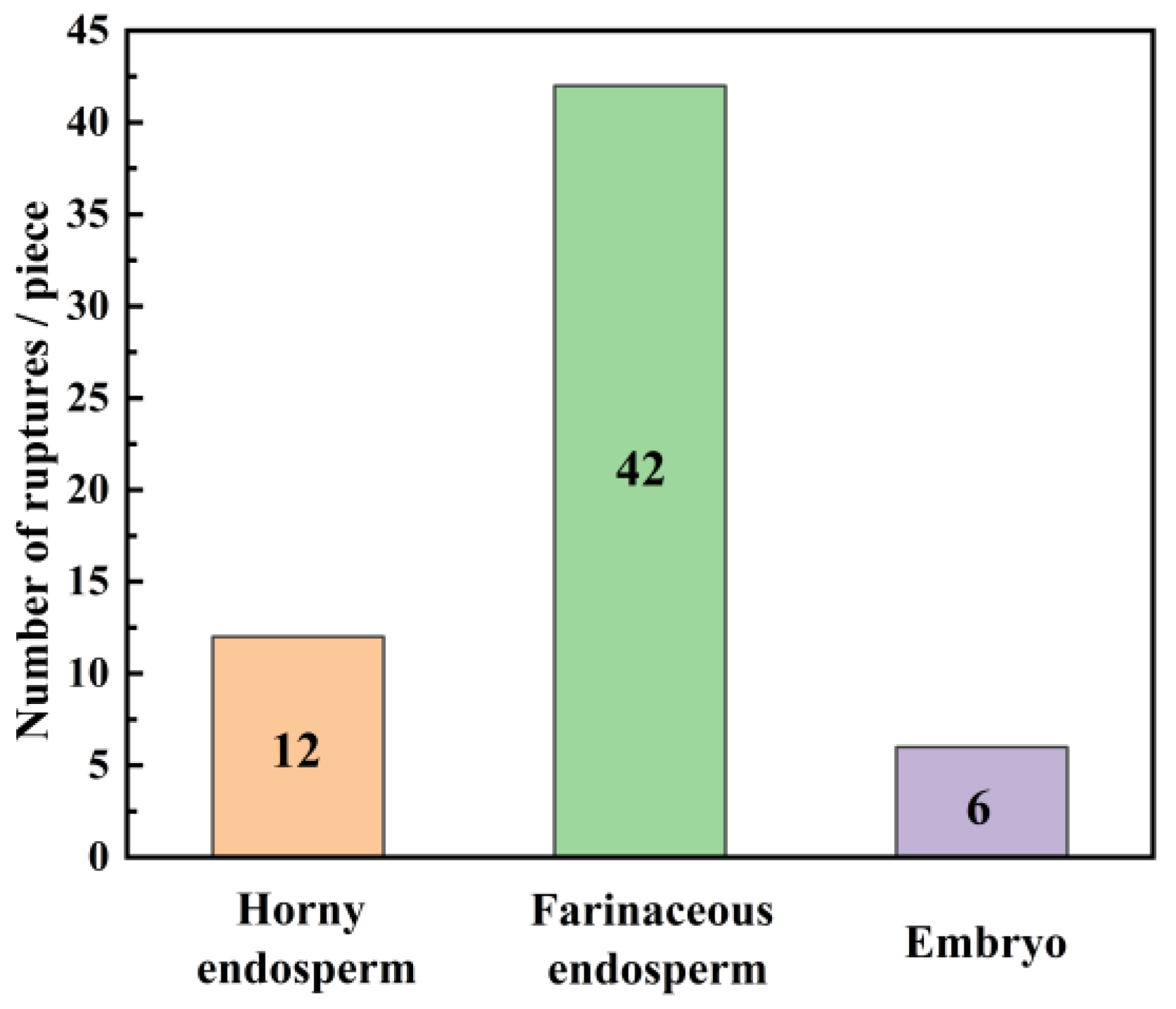
  • There is a significant difference in the mechanical properties of different tissues in maize kernels. The starch particles in the horny endosperm are of a regular polyhedral shape, tightly arranged, resulting in higher maximum rupture force (128 N) and elasticity modulus (353 MPa). Therefore, the horny endosperm exhibits good structural stability and bearing capacity at the macroscopic level. Starch particles in the powdery endosperm are spheroid, arranged loosely and irregularly with more gaps, which makes the maximum rupture force (38 N) and elastic modulus (136 MPa) of the powdery endosperm relatively small. The powdery endosperm is more prone to deformation and rupture. Under compression loading, the initial damage in maize kernels mainly occurs in the farinaceous endosperm.
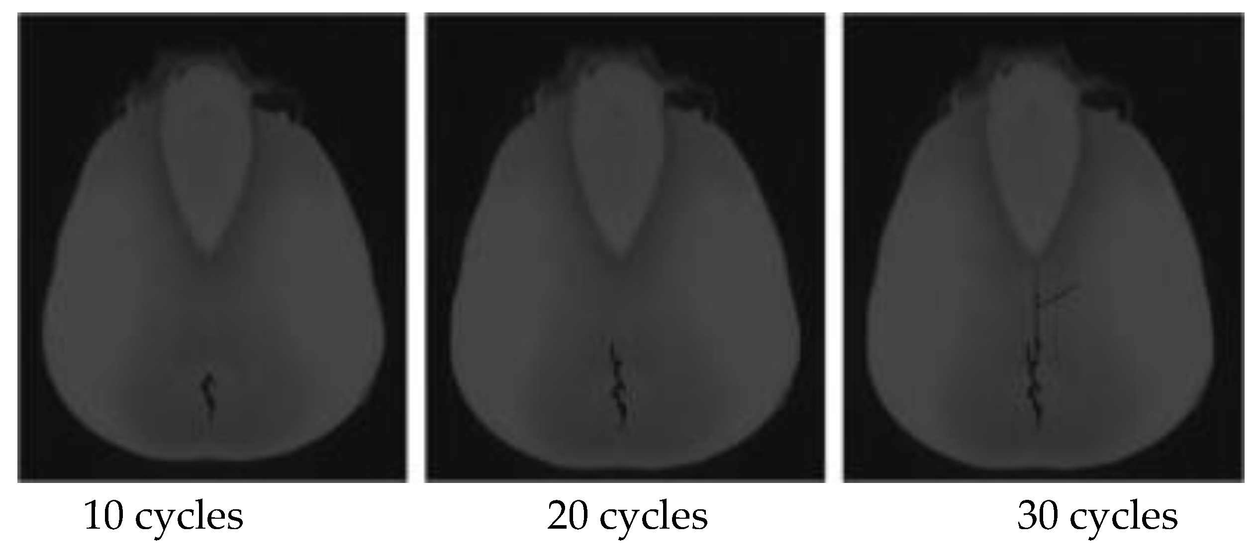
  • By further particle flow simulation of the loading process of powdered endosperm, it is concluded that the damage evolution of the powdered endosperm during the whole loading process is divided into four stages, firstly, cracks start to sprout from the inner endosperm, and then gradually expand, and the powdered endosperm starts to rupture when the maximum force reaches 803 N. The process of damage accumulation of the powdered endosperm during the simulation is consistent with the process of change in maize after loading by test. And the direction of crack development is also consistent.
Based on the microstructure characteristics of corn grains, this study explored the evolution process of the microscopic damage of corn grains after loading. Firstly, it was a cross-scale complementary study of the macroscopic crushing of corn grains under compression loading. Secondly, it explains the conditions of germination and evolution of microscopic damage after macroscopic loading of seed, which can determine the maximum load range in the threshing process, and can be used as a theoretical basis for determining the load of the threshing system.

Author Contributions

Conceptualization, D.G. and G.Z.; methodology, B.W. and Y.L.; software, J.B. and P.X.; formal analysis, Z.S. and R.L.; resources, B.W.; data curation, Z.S. and J.B.; writing—original draft, Z.S. and B.W.; writing—review and editing, B.W.; funding acquisition, B.W. All authors have read and agreed to the published version of the manuscript.

Funding

This work was supported by the Key Research and Development Program of Shandong (2023TSGC0536), the Key Research and Development Program of Ningxia (2023BCF01052), the Key Research and Development Program of Rizhao (2023ZDYF010114), and the National Key Research and Development Program of China (2021YFD2000502).

Data Availability Statement

The original contributions presented in this study are included in the article. Further inquiries can be directed to the corresponding author.

Conflicts of Interest

Author Rui Li was employed by the company Changchun Ruiguang Science & Technology Co., Ltd. The remaining authors declare that the research was conducted in the absence of any commercial or financial relationships that could be construed as a potential conflict of interest.

References

  1. Yadava, P.; Abhishek, A.; Singh, R.; Singh, I.; Kaul, T.; Pattanayak, A.; Agrawal, P.K. Advances in Maize Transformation Technologies and Development of Transgenic Maize. Front. Plant Sci. 2017, 7, 1949. [Google Scholar] [CrossRef] [PubMed]
  2. Li, X.; Zhang, W.; Xu, S.; Du, Z.; Ma, Y.; Ma, F.; Liu, J. Low-Damage Corn Threshing Technology and Corn Threshing Devices: A Review of Recent Developments. Agriculture 2023, 13, 1006. [Google Scholar] [CrossRef]
  3. Shi, M.L.; Zhu, G.Q.; Chen, G.Y.; Xue, L.; Hu, J.R.; Yin, Z.T.; Chen, G.Q. Medicinal value of maize and its industrialization development and application. Chin. Food Nutr. 2004, 12, 23–25. [Google Scholar]
  4. Ariyanti, D.; Rimantho, D.; Leonardus, M.; Ardyani, T.; Fiviyanti, S.; Sarwana, W.; Hanifah, Y.; Agustian, E.; Meliana, Y.; Susparini, N.T.; et al. Valorization of corn cob waste for furfural production. A circular economy approach. Biomass Bioenergy 2025, 194, 0961–9534. [Google Scholar] [CrossRef]
  5. Chen, S.; Xu, Z.; Li, X.; Yu, J.; Cai, M.; Jin, M. Integrated bioethanol production from mixtures of corn and corn stove. Bioresour. Technol. 2018, 258, 18–25. [Google Scholar] [CrossRef]
  6. Cao, H.; Yan, L.; Song, X.; Liu, L.; Xu, S.; Hu, S.; Zhang, S. Determining the nutritional values of new corn varieties on pigs and broilers. Anim. Nutr. Metab. 2024, 11, 1358773. [Google Scholar] [CrossRef]
  7. Cui, Z.K.; Zhang, H.; Zhou, J.; Di, Z.F.; Wang, X.J. Design and experiment of intelligent corn grain harvester. J. Chin. Agric. Mech. 2019, 40, 26–30. [Google Scholar]
  8. Ke, Y.; Chunxia, J.; Wei, Z.; Enke, L.; Guangqian, Z.; Dongmei, Z. Study on the Effects of Different Treatment on Mechanical Harvest Broken Rate of Maize Grains. Crops 2024, 40, 138–143. [Google Scholar]
  9. Wang, F.; Alimu, M.; Zhang, J.; Li, Q.; Xu, L. Design and Experiment of Pre-screening Cleaning Device for Combined Screen Surface of Corn Grain Harvester. Trans. Chin. Soc. Agric. Mach. 2024, 55, 135–147+166. [Google Scholar]
  10. Cui, T.; Fan, C.L.; Zhang, D.X.; Yang, L.; Li, Y.B.; Zhao, H.H. Research Progress of Maize Mechanized Harvesting Technology. Trans. Chin. Soc. Agric. Mach. 2019, 50, 1–13. [Google Scholar]
  11. Geng, D.; Wang, Q.; Li, H.; He, Q.; Yue, D.; Ma, J.; Wang, Y.; Xu, H. Online detection technology for broken corn kernels based on deep learning. Trans. Chin. Soc. Agric. Eng. 2023, 39, 270–278. [Google Scholar]
  12. Li, X.; Ma, F.; Gao, L. Drop impact test of corn seed. Trans. Chin. Soc. Agric. Eng. 2009, 25, 113–116. [Google Scholar]
  13. Li, X.; Gao, L.; Ma, F. Experimental study on threshing characteristics of maize seeds. J. Agric. Mech. Res. 2007, 156–158. [Google Scholar]
  14. Li, X.; Gao, L.; Ma, F. Experimental study on impact damage of maize seed. J. Shenyang Agric. Univ. 2007, 38, 5. [Google Scholar]
  15. Cai, C.J.; Chen, Z.; Han, Z.D.; Liu, G.M.; Zhang, Z.L.; Hao, J.F. Study on Relationship of Biomechanical Characteristics of Corn Seed and Threshing Performance. J. Agric. Mech. Res. 2017, 39, 192–196. [Google Scholar]
  16. Liu, J.; Liu, Q.; Chen, X.; Deng, Z.; Yuan, J.; Kong, F. Diversity and influencing factors of kernel breakage tolerance of different maize cultivars. J. China Agric. Univ. 2021, 26, 207–220. [Google Scholar]
  17. Zhang, Y.; Gao, L.; Liu, H.; Li, X. Experimental Study on Corn Kernel Shear Crash. J. Agric. Mech. Res. 2007, 136–138. [Google Scholar]
  18. Wang, B.; Gao, M.; Geng, D.; Zhou, S. Study on damage mechanism and crack growth of the corn grain. J. Fail. Anal. Prev. 2022, 22, 1526–1534. [Google Scholar]
  19. Wang, G. Finite Element Analysis of compression mechanical properties of Corn grains. Chin. J. Agric. Mech. 2016, 37, 117–120. [Google Scholar]
  20. Tao, T.; Wei, X. Finite element analysis of corn seed under impact load. J. Agric. Mech. Res. 2019, 41, 35–39. [Google Scholar]
  21. Tang, H.; Zhu, G.; Sun, Z.; Xu, C.; Wang, J. Impact damage evolution rules of maize kernel based on FEM. Biosyst. Eng. 2024, 247, 162–174. [Google Scholar] [CrossRef]
  22. Gao, P. Study on Mechanical Properties of Corn Grains and Their Relationship with Grain Disintegration in Storage. Master’s Thesis, Henan University of Technology, Zhengzhou, China, 2024. [Google Scholar]
  23. Qiao, M.; Xia, G.; Cui, T.; Xu, Y.; Gao, X.; Su, Y.; Li, Y.; Fan, H. Effect of moisture, protein, starch, soluble sugar contents and microstructure on mechanical properties of maize kernels. Food Chem. 2022, 379, 13214. [Google Scholar] [CrossRef] [PubMed]
  24. Singh, S.S.; Finner, M.F.; Rohatgi, P.K.; Buelow, F.H.; Schaller, M. Structure and mechanical properties of corn kernels: A hybrid composite material. Mater. Sci. 1991, 26, 274–284. [Google Scholar] [CrossRef]
  25. Wang, B.; Wang, J. Mechanical properties of maize kernel horny endosperm, floury endosperm and germ. Int. J. Food Prop. 2019, 22, 863–877. [Google Scholar] [CrossRef]
  26. Yi, Z.; Zhang, H. Starch Granule Size, Pasting Properties and Textural Properties of Horny Endosperm and Silty Endosperm in Maize. J. Chin. Cereals Oils Assoc. 2014, 29, 27–32. [Google Scholar]
  27. Yin, X.; Ming, B.; Hou, J.; Ming, B.; Zhang, Y.; Guo, X.; Gao, S.; Wang, K.; Hou, P.; Li, S. Effects of various grain positions of ear on the internal Structural parameters of maize grain using X-ray μCT. Trans. Chin. Soc. Agric. Eng. 2021, 37, 8–14. [Google Scholar]
  28. Xu, A. Development and Characteristics of Corn Keratin and Silty Endosperm Starch. Master’s Thesis, Yangzhou University, Yangzhou, China, 2020. [Google Scholar]
Figure 1. Internal composition and structure of maize kernels. 1. Seed coat; 2. farinaceous endosperm; 3. horny endosperm; and 4. embryo.
Agriculture 15 00636 g001
Figure 2. Microscopic structure of starchy endosperm and horny endosperm.
Agriculture 15 00636 g002
Figure 3. Mechanical properties of various tissues in maize kernels.
Agriculture 15 00636 g003
Figure 4. Statistical analysis of initial damage locations in maize kernels.
Agriculture 15 00636 g004
Figure 5. The internal damage of corn grains accumulated under repeated compression.
Agriculture 15 00636 g005
Figure 6. Image of maize kernels at the end of compression.
Agriculture 15 00636 g006
Figure 7. Principle diagram of contact between starch granules.
Agriculture 15 00636 g007
Figure 8. Schematic diagram of the linear contact model.
Agriculture 15 00636 g008
Figure 9. Schematic diagram of the linear parallel bonding contact model.
Agriculture 15 00636 g009
Figure 10. Marking of diameter size of starch granules in powdery endosperm.
Agriculture 15 00636 g010
Figure 11. Discrete element numerical model of the farinaceous endosperm.
Agriculture 15 00636 g011
Figure 12. Particle grouping.
Agriculture 15 00636 g012
Figure 13. Force and deformation during the loading process. (a) initial stage; (b) crack initiation; (c) crack propagation; (d) fracture.
Agriculture 15 00636 g013
Figure 14. Particle velocity field distribution at different loading stages. (a) initial stage; (b) crack initiation; (c) crack propagation; (d) fracture.
Agriculture 15 00636 g014
Table 1. Mechanical parameters of horny endosperm.
NumberMaximum Rupture Force/NRupture Deformation/mmModulus of Elasticity/MPa
1121.370.883345.91
2123.690.889346.93
3135.120.884349.79
4131.950.894350.45
5129.080.888360.17
6127.240.895363.45
7125.440.899366.37
8121.390.893358.12
9133.760.910360.43
10123.360.895359.48
Mean127.240.893356.11
SD5.05250.00787.2263
Upper confidence limits of 95%123.62590.8874350.9409
Lower confidence limits of 95%130.85410.8986361.2790
Table 2. Mechanical parameters of silty endosperm.
NumberMaximum Rupture Force/NRupture Deformation/mmModulus of Elasticity/MPa
130.340.825115.36
231.820.827116.47
332.840.829116.52
432.910.835115.29
534.560.837116.92
636.450.840117.66
736.120.845116.67
833.310.843123.8
930.680.846121.31
1030.670.823122.7
Mean32.970.835118.27
SD2.19770.00853.1243
Upper confidence limits of 95%31.39790.8289116.0351
Lower confidence limits of 95%34.54210.8411120.5049
Table 3. Embryo mechanical parameters.
NumberMaximum Rupture force/NRupture Deformation/mmModulus of Elasticity/MPa
117.930.89230.16
217.910.88929.27
317.890.91729.97
419.590.91230.19
520.360.92631.45
621.650.96334.65
719.360.81434.37
818.490.89635.43
921.380.92336.02
1020.840.93830.99
Mean19.540.90732.25
SD1.464730.03962.5711
Upper confidence limits of 95%18.49230.878630.4108
Lower confidence limits of 95%20.58770.935434.0892
Table 4. Pore information of powdery endosperm.
PartPore NumberPore Area/μm2Total Area/μm2Porosity/%
Powdery endosperm981176,342.23,093,656.35.70
Table 5. Particle gradation of starch granules.
GroupParticle Size (mm)Proportions (%)
12~42.3
24~826.7
38~1231.7
412~1631.1
516~185.8
618~222.4
Table 6. Simulation parameter setting.
ParameterValue
Rmax/μm2
Rmin/μm22
ρ/kg·m−31300
porosity/%5.71
kn/N·m−10.85 × 108
kratio1.5
fric0.41
pb_coh/MPa0.65
pb_ten/MPa0.65
Disclaimer/Publisher’s Note: The statements, opinions and data contained in all publications are solely those of the individual author(s) and contributor(s) and not of MDPI and/or the editor(s). MDPI and/or the editor(s) disclaim responsibility for any injury to people or property resulting from any ideas, methods, instructions or products referred to in the content.

