Review

Techniques for In Vitro Fertilisation of Vitrified Cattle Oocytes: Challenges and New Developments

by
Mahlatsana Ramaesela Ledwaba
1,2,
Hester Adri O’Neill
2,
Mamonene Angelinah Thema
1,
Ayanda Maqhashu
2 and
Masindi Lottus Mphaphathi
1,*
1
Agricultural Research Council, Animal Production, Germplasm Conservation and Reproductive Biotechnologies, Private Bag X2, Pretoria 0062, South Africa
2
Department of Animal, Wildlife and Grassland Sciences, University of the Free State, P.O. Box 339, Bloemfontein 9300, South Africa
*
Author to whom correspondence should be addressed.
Agriculture 2025, 15(4), 363; https://doi.org/10.3390/agriculture15040363
Submission received: 17 December 2024 / Revised: 27 January 2025 / Accepted: 1 February 2025 / Published: 8 February 2025
(This article belongs to the Section Farm Animal Production)

Abstract

:
Cryopreservation is currently an essential technique in reproductive technologies that is used for the extended preservation of genetic material. Vitrification has become the industry’s standard cryopreservation technique for cattle oocytes and embryos. The current results of this technology, however, are still not good in terms of viability, fertilisation capacity, embryo development, or pregnancy. The oocytes’ susceptibility to freezing is associated with significant changes in the structures, functioning of the oocytes, and cryoinjury, which is harmful to the survival of cells and their subsequent growth. The effectiveness of producing embryos with in vitro techniques utilising vitrified cattle oocytes rarely exceeds 30–40%. A significant number of vitrified oocytes do not successfully develop into the embryo stage following in vitro fertilisation and culture. This review focuses on issues related to oocyte cryopreservation, ways to overcome them, and how to enhance the vitrified oocyte fertilisation process.

1. Introduction

Cryopreservation is a crucial procedure that keeps oocytes and embryos viable for later use and genome banking for valuable animal species [1]. Cryopreservation makes it possible to use preserved oocytes and embryos to generate a larger number of progeny from a valuable individual for decades following storage [2]. The ability to cryopreserve cattle oocytes could help expand in vitro programs that produce embryos by enabling the long-term conservation and widespread distribution of female genetics. Long-term efforts have been made to enhance the process of vitrifying unfertilised bovine oocytes [3,4]. Nevertheless, compared to embryos obtained from fresh oocytes, the developing rates of bovine embryos derived from vitrified oocytes continue to be lower. Even though oocyte cryopreservation has existed for a while and seems to be a reliable technique for assisted reproduction, there is currently no appropriate technique that would prevent damage during freezing and thawing [5]. Oocytes are cells approximately 120 microns in diameter, surrounded by a robust membrane known as the zona pellucida. Oocytes are commonly identified as the biggest cells in the human body [6]. Surface and volume have a significant impact on how cryobiological activities turn out. Thus, a great deal of theoretical and practical information is needed to freeze and thaw unfertilised oocytes [7]. Therefore, this review will discuss issues related to oocyte cryopreservation, as well as various techniques of in vitro fertilisation (IVF), including standard IVF, intracytoplasmic sperm injection (ICSI), physiological intracytoplasmic sperm injection (PICSI), and parthenogenetic activation.

2. Problems Currently Associated with Fertilisation of Vitrified Cattle Oocytes

Cryopreservation of cattle oocytes has proven to be exceedingly challenging [8]. Vitrification was introduced in the mid-1980s as an alternate cryopreservation technique to conventional slow-freezing methods. Nonetheless, modest improvement has been achieved despite intense research over the past two decades, as the capacity of cryopreserved oocytes to complete embryonic development remains significantly low in numerous animal species [9]. This may be attributed to a greater concentration of phospholipids in the oocytes of animals compared to humans [10]. Oocytes are highly vulnerable to injury during cooling and cryopreservation. Additionally, oocytes have comparatively reduced membrane permeability to water and cryoprotectants (CPAs) [11]. Therefore, the latest results of this technology regarding viability, fertilisation capacity, embryo development, and pregnancy are unsatisfactory [1]. According to Mandawala et al. [12], vitrification is a commonly utilised approach for the cryopreservation of cattle oocytes and embryos due to its low risk of harm due to the absence of ice crystal formation compared to solid-surface vitrification.
It is now evident that the cellular and molecular alterations brought about by cryopreservation procedures invariably result in poor embryo development and fertilisation rate [13]. Oocyte cryopreservation induces excessive intracellular reactive oxygen species (ROS) production in oocytes, resulting in oxidative stress [14,15] and impairing proteins, DNA [16], lipids [17], and other macromolecules [18], ultimately leading to diminished developmental potential of oocytes following parthenogenetic activation or in vitro fertilisation (IVF). Furthermore, ROS impacts phospholipids, enzymes, and the carbon–carbon double bonds of membrane receptor-associated polyunsaturated fatty acids (PUFAs), generating cytotoxic chemicals that compromise the structural integrity and functionality of biofilms, ultimately resulting in cell death [19]. As per Khazaei and Aghaz [20], the degree of ROS during in vitro maturation (IVM) reduces the ability of oocytes to develop to the blastocyst stage, and oocytes from cattle undergo higher levels of ROS after vitrification (Figure 1) [21]. Due to the significant role that ROS plays in the apoptotic process, there has been a close correlation between the increased rate of apoptosis and the increased ROS level of vitrified oocytes [21].
Exposure to CPAs and extremely rapid chilling rates has been demonstrated to cause harmful morphological and molecular alterations to the oocyte and its organelles, ultimately leading to diminished fertilisation rates and impaired developmental capacity [23]. The increased sensitivity of oocytes to freezing is linked to the complicated nature of their structures and functions. Cryoinjury occurs at various cellular levels, negatively impacting cell viability and subsequent development [1]. According to Dujíčková et al. [5], a vitrification-ready oocyte is generally defined as a fully developed Graafian follicle that is ready for ovulation and has an oocyte surrounded by a layer of cumulus cells called the cumulus oophorus. The quality of the oocyte (Table 1) is crucial in determining embryonic developmental potential, rendering this specialised cell relevant in studies focused on improving assisted reproductive technology (ART) outcomes [24].

2.1. Survival Rate of Immature and Matured Oocytes Following Vitrification

The process of oocyte vitrification in cattle continues to encounter difficulties since it diminishes the success of embryonic development outcomes [28]. The survival and developmental ability of oocytes following vitrification are influenced by various parameters, including the meiotic stage [29]. A study by Mphaphathi et al. [30] stated that numerous reproductive centres have developed cryopreservation techniques for various species. Nonetheless, the survival rate of gametes or tissues has diminished with time due to reduced temperatures and altered metabolic reactions of the gametes under cryopreservation. There is a disagreement among studies about the ideal stage for freezing cattle oocytes. Previous research has shown that cow oocytes at the MII stage have a higher survival rate after cryopreservation compared to those at the germinal vesicle (GV) stage [31]. Alternatively, other research has shown that the process of vitrification for cattle oocytes is more beneficial when performed at the GV stage or during germinal vesicle breakdown (GVBD). In contrast, Chaves et al. [32] found that cattle immature oocytes (Figure 2A) exhibited greater tolerance to cryoprotectant treatment in comparison to mature oocytes (Figure 2B). There is ongoing disagreement regarding the vitrification of mature vs. immature oocytes and their subsequent survivability post-vitrification. Exposure of MII stage oocytes to sub-room temperature conditions results in permanent meiotic disruption, spindle damage, and chromosomal scattering [33]. Exposure of GV oocytes to low temperatures disrupts the migration of cortical granules and induces hardness of the zona pellucida [34]. The susceptibility of oocytes to intracellular CPAs is evidenced by cytoskeletal abnormalities and an increase in intracellular calcium levels, which trigger oocyte activation and zona hardness [34]. Despite the ability to circumvent zona hardening with ICSI, numerous hurdles persist in attaining effective oocyte cryopreservation.
The cryopreservation survival rate of oocytes utilising the slow freezing technique varied between 74% and 90% [30]. Oocytes are susceptible to ice recrystallisation events during storage and thawing [35,36]. Sperm and oocytes are susceptible to metabolic damage from oxidative stress during the thawing process. In such abnormal circumstances, the cell death mechanism of oocytes may be triggered, leading to apoptosis of the oocytes [37]. Nevertheless, upon exposure to cooling stress, immature cattle oocytes may attempt to adjust to the extremely low-temperature environment [33]. Recent studies have found that freezing oocytes during the MII stage could lead to changes in epigenetic modifications, specifically in histone acetylation and methylation, as well as DNA methylation in preimplantation embryos. Furthermore, the process of oocyte vitrification has the potential to impact the expression of genes, including those that are epigenetically connected and potentially imprinted [38].

2.2. The Effect of Morphological Structure of Vitrified Oocytes During Fertilisation

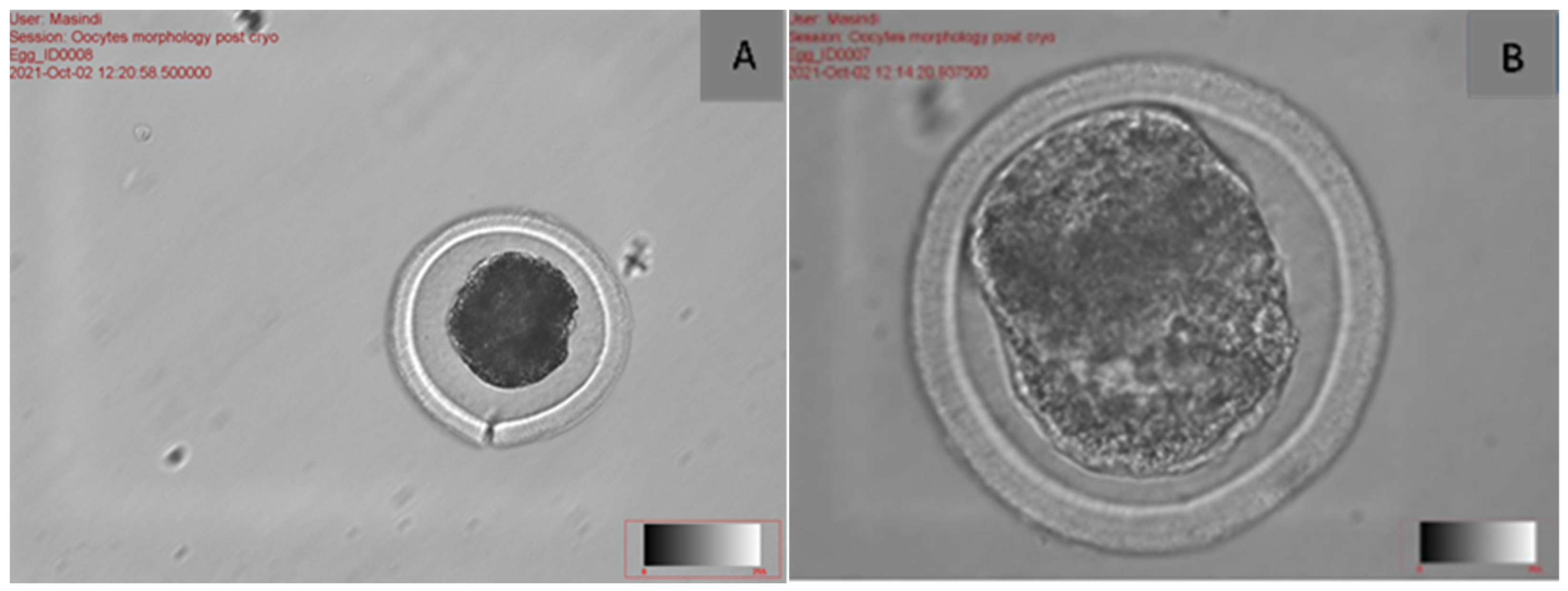
Traditionally, oocyte selection mostly relies on morphological criteria. A high-quality metaphase II (MII) oocyte is characterised by a distinctly granular cytoplasm, a minimal perivitelline space, and a transparent zona pellucida [39]. Oocyte morphological abnormalities (Figure 3) are frequently observed following vitrification [40]. Conventional indicators of oocyte quality rely on evaluating the physical characteristics of the cumulus–oocyte complex, such as the size and shape of the oocytes, as well as the presence of certain markers in the cytoplasm and outside the cytoplasm. Additionally, the meiotic spindle is also considered a measure of oocyte quality [41]. The main criteria for assessing oocyte morphology include cumulus–oocyte complex scoring, zona pellucida scoring, deficiencies in the perivitelline space (e.g., dilatation or granularity), polar body scoring, cytoplasmic scoring, and vacuolisation [42]. Vitrified oocytes may display various abnormalities associated with a decrease in competence [43], such as cytoskeleton damage, elevated levels of Ca2+ ions in the cytoplasm, and the hardening or cracking of the zona pellucida (Figure 3A) [44]. Additionally, abnormalities may be observed in the plasma membrane, meiotic spindle, and other components [45]. According to Menéndez-Blanco et al. [8], the effectiveness of vitrification and fertilisation depends on the quality of the oocyte and its nuclear stage.
A study by Fernández-Reyez et al. [46] showed that vitrification of germinal vesicle (GV) oocytes leads to reduced rates of maturation, fertilisation, blastocyst development, and viability. Evidence from multiple studies indicates that oocyte viability decreases after in vitro culture (IVC) during vitrification [47,48]. These outcomes support the idea that during vitrification, proteins undergo secondary structural rearrangements, as seen by an increase in the content of b-sheets at the expense of a-helices and changes in the lipid and carbohydrate architecture of the zona pellucida [44].

The Effect of Meiotic Spindle on Vitrified Cattle Oocytes

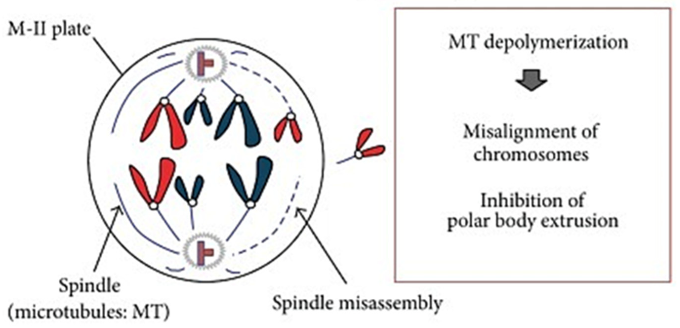
The spindle is a crucial barrel-shaped structure of cells, predominantly made up of microtubules and centrosomes, required for the accurate segregation of homologous chromosomes during meiosis I or two sets of chromatids during meiosis II in germ cell division [27]. The process of chromosomal segregation is highly susceptible, even to little alterations in the timing or biochemistry of spindle formation [49]. Vitrification adversely impacts the structure and function of oocyte mitochondria [23]. Electron micrographs of cattle and pig oocytes demonstrated that vitrification and warming resulted in mitochondrial enlargement, electron-dense cristae, and compromised inner and outer membranes [50]. There is a significant cryoinjury that is frequently detrimental to the viability of oocytes: damage to the meiotic spindle. This structure, which completes meiosis, is essential for healthy embryo development after fertilisation. Aneuploidy, aberrant fertilisation, and stunted embryo development can all arise from spindle microtubule destabilisation [51]. The meiotic spindle plays a crucial role in ensuring the accurate alignment of chromosomes in the oocyte and their proper separation during meiosis (Figure 4). Microtubules are essential elements of the meiotic spindle’s cytoskeleton, and their proper formation is necessary for the spindle to be well-organized and functioning [52]. The location and refraction of the meiotic spindle are frequently utilised as indicators to assess the quality of oocytes.
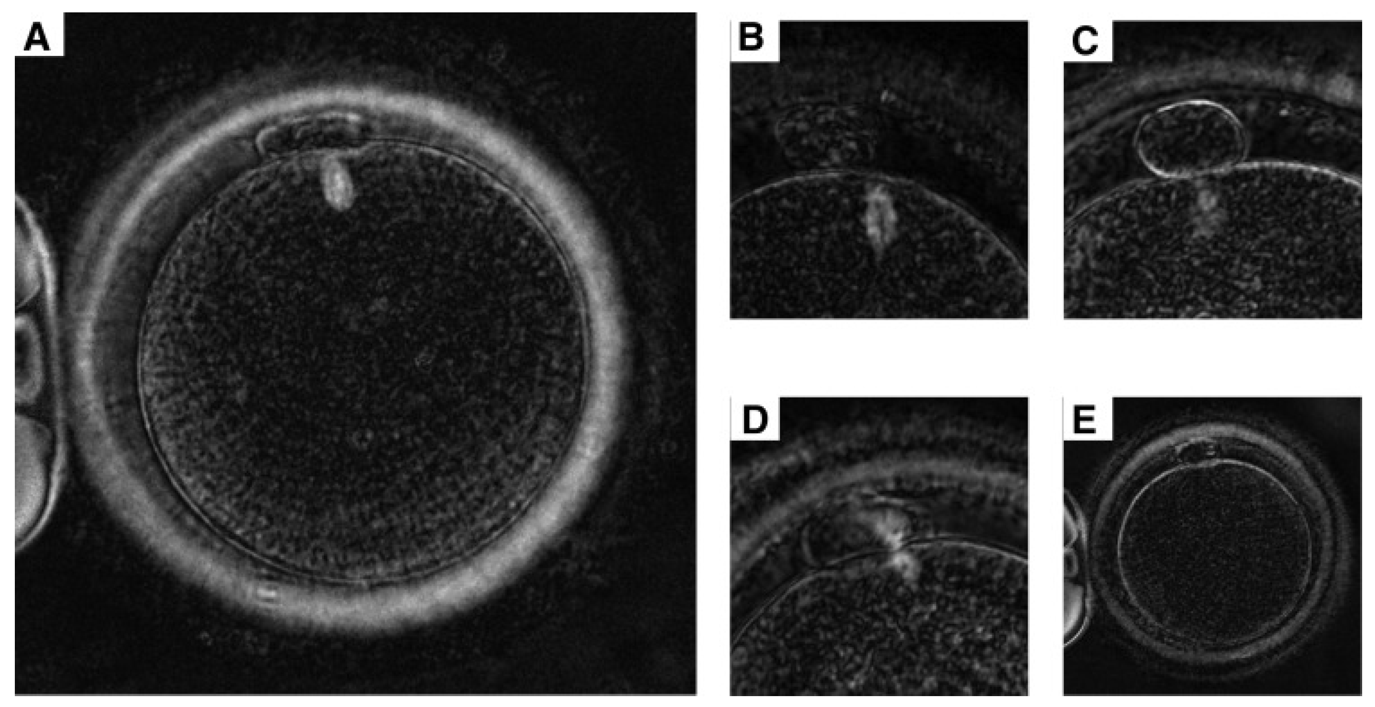
One potential measure of oocyte health is the positive detection of the normal spindle (Figure 5A) in MII oocytes. The meiotic spindle of mammalian oocytes is susceptible to variations in environmental conditions, particularly temperature and pH [53]; however, it demonstrates the ability to regenerate after cryopreservation [54]. Nevertheless, the timeframe for spindle recovery is finite. Exposing oocytes to unfavourable conditions, such as CPAs and low temperatures, has been demonstrated to cause disruption of the microtubules [1]. The disruption of microtubules can cause unstable microtubules which can cause dysmorphic spindle (Figure 5B), which will result in chromosome misalignment. The significance of maintaining the structural integrity (Figure 5C) of the meiotic spindle was clearly demonstrated in the process of oocyte cryopreservation [49], whereby the spindle checkpoint mechanism makes sure that before anaphase begins, chromosomes are correctly connected to the spindle microtubules. Following cryopreservation, oocytes exhibit reduced developmental ability, which may be attributed to harm to the meiotic apparatus, such as disorganisation of the spindle and loss of microtubules, along with other structural changes [32]. Furthermore, the utilisation of spindle retardance as a means of measuring spindle birefringence has been proposed as a reliable method for assessing both the structural density and the degree of alignment and orientation of the spindle. Spindle retardance has the potential to be utilised for the selection of oocytes with enhanced embryonic developmental capacity. It has been suggested as a quality indicator in human oocytes [52].
Various approaches have been suggested to mitigate the impact of vitrification on mitochondrial function. A post-warming incubation period, a widely utilised method to promote the reassembly of the meiotic spindle following cryopreservation, has also been shown to aid in the recovery of mitochondrial function [56]. Oocytes with normal spindle morphology are far more likely to yield euploid embryos than those with translucent (Figure 5D) or absent meiotic spindles (Figure 5E). According to Girka et al. [51], the stabilisation of microtubules using paclitaxel or a comparable compound would preserve the meiotic spindle in its polymerised form, even in adverse environmental conditions. The stabilisation observed may lead to the preservation of the meiotic spindle in its typical structure during vitrification, potentially enhancing oocyte survival following warming.

2.3. The Effect of Cumulus Cells During Cryopreservation and Fertilisation

The cells that directly surround the oocyte are referred to as cumulus cells. These cells accompany the oocyte during its development from an immature to a completely mature ovulated state, playing a crucial function in sustaining the oocyte, both in vivo and in vitro [27]. The disparities in morphological and functional characteristics between oocytes and cumulus cells (CCs) may result in reduced vitrification efficiency for cumulus–oocyte complexes (COCs) [57]. The visual characteristics of the cumulus–oocyte complex provide information about the oocyte’s level of development and excellence. The presence of radiating corona cells surrounded by an expanded mass of cumulus cells typically indicates a mature oocyte [41]. Oocytes undergoing maturation are encased by several layers of cumulus cells (CCs), which facilitate the transport of nutrients and signalling molecules, notably via gap junctions that are crucial for oocyte maturation [58,59,60]. Elevated levels of penetrating and non-penetrating CPAs, along with ultra-rapid cooling and thawing rates, are employed in the vitrification of oocytes to achieve the glassy state and reduce gamete damage. The presence of multiple dense layers of cumulus cells surrounding an immature oocyte, on one hand, impedes its equilibration with a cryoprotective solution; consequently, this may prolong the duration required [57]. These variables fall into two categories: oocyte factors, which include cumulus cell presence and oocyte developmental stage (mature or germinal vesicle), and technical factors, which include various procedures, CPAs, and equipment utilised [61].

3. How to Improve Fertilisation on Vitrified Cattle Oocytes?

3.1. The Use of Traditional In Vitro Fertilisation on Vitrified Cattle Oocytes

According to Neri et al. [62], fertilisation is the process that happens when the egg and sperm, the two parental gametes, fuse. Mammalian sperm and oocytes combine in the oviduct, initiating a sequence of events that culminate in fertilisation and the creation of an embryo. The zygote develops as the outcome of a series of crucial events that are triggered by fertilisation. Sperm that swim freely and are capacitated must first identify and bind to the zona pellucida of the ovulated egg after cumulus penetration [63].
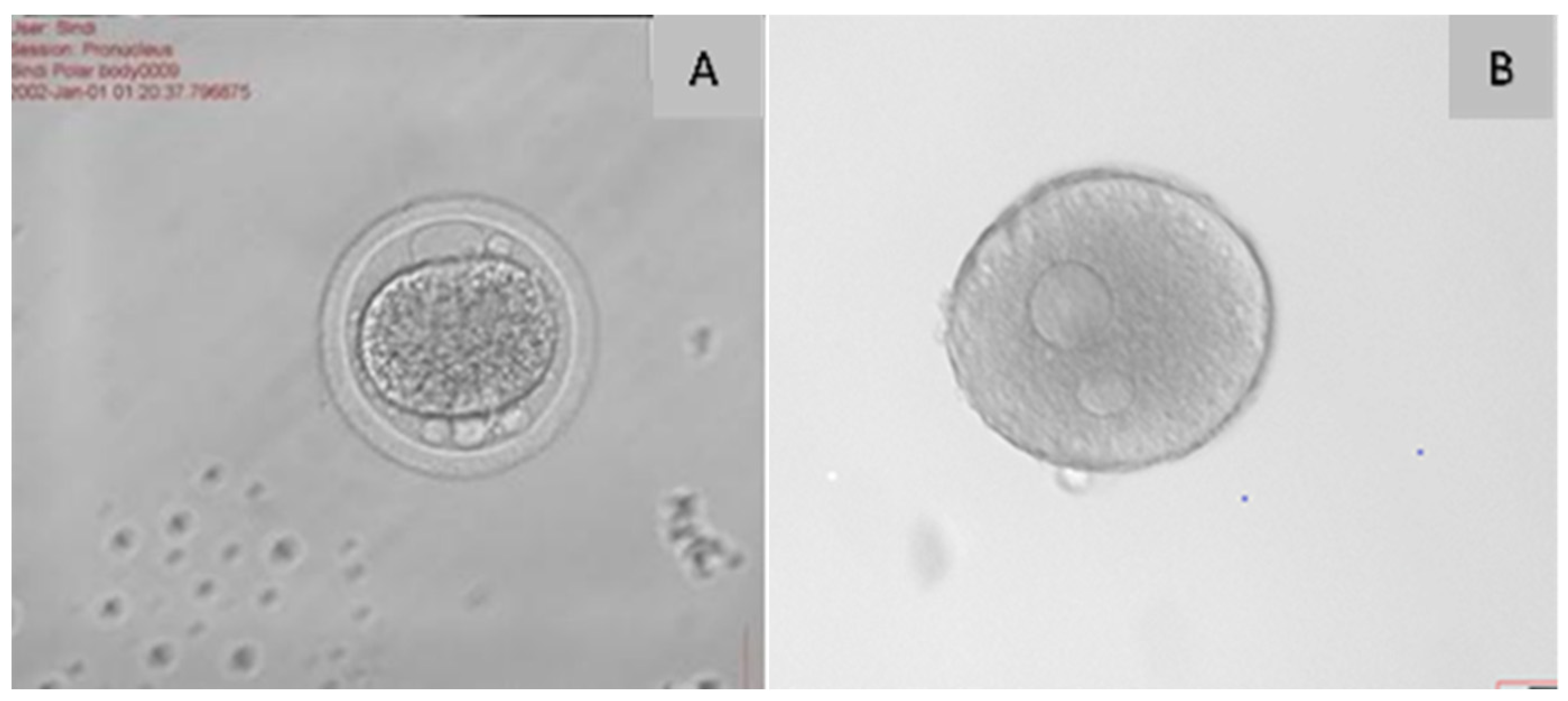
The mature oocyte becomes active in the formation of the embryo during fertilisation. Depending on the species, a variety of molecular changes occur during oocyte activation. According to Neri et al. [62], it is typically caused by the male gamete attaching to the oolemma, which causes the release of intracellular Ca2+ [Ca2+]i within the ooplasm during fertilisation. Liu et al. [64] state that inappropriate sperm–oocyte penetration and interaction are the main causes of fertilisation failure in traditional IVF resulting in Inconsistencies in pronuclear alignment or creation, either dysmorphism (Figure 6A) or asynchronous pronuclear development. Furthermore, a frequent consequence of vitrification, particularly with the application of Dimethyl Sulfoxide (DMSO) and ethylene glycol as CPAs, is the elevation of cytoplasmic calcium levels, which artificially triggers the zona block to polyspermy mechanism [65]. This injury, referred to as zona hardening, typically results in diminished fertilisation rates of vitrified oocytes in bovines [9]. Although fertilised zygotes typically have two pronuclei (Figure 6B), IVF labs regularly observe aberrant fertilisation patterns. The ovum is stationary during secondary meiosis and is surrounded by a ZP and a cumulus layer before fertilisation. The extracellular zinc partition (ZP), which is composed of four glycoproteins (ZP1, ZP2, ZP3, and ZP4), is only crossed by sperm that go through the acrosome reaction. Oocytes are encased in extracellular ZP. The continuation of this process requires control of ionic signalling [33].

3.2. The Use of Intracytoplasmic Sperm Injection on Vitrified Cattle Oocytes

Intracytoplasmic sperm injection (ICSI) is an enhanced reproductive technique involving the injection of sperm into the ooplasm of an oocyte inhibited in metaphase II (MII) [67]. To avoid the zona pellucida and oolemma penetration by the sperm itself, ICSI, in which a single sperm is directly injected into the ooplasm, may, therefore, be able to solve these issues. Previous research has revealed that applying ICSI to vitrified oocytes is a more suitable method for producing embryos than traditional IVF [68]. Nonetheless, compared to IVF embryos, the developmental rates of bovine ICSI embryos created with fresh oocytes are lower [69]. There has been extensive discussion over the impact of the meiotic spindle machinery on the viability of oocytes.
The oocyte is a highly specialised cell that undergoes numerous maturational changes in order to prepare for fertilisation and embryonic development. The alterations encompass nuclear and cytoplasmic occurrences [70]. Furthermore, the microtubules of the meiotic spindle have been found to suffer damage when manipulated, in addition to disintegration caused by changes in temperature and pH. Previous studies have shown that the rate of blastocyst development in ICSI-fertilised vitrified oocytes was lower than that of IVF-fertilized conventionally fertilised oocytes [71]. As sperm injection alone cannot start (Ca2Í)i oscillations and other aforementioned events, most ICSI-treated oocytes in the bovine species do not activate; this is in contrast to observations made in other species, including mice and humans [72]. This is the reason why ICSI-treated bovine oocytes need outside activation cues to start the development of embryos. Therefore, the development of innovative sperm selection criteria is the foundation of various programs aimed at increasing ICSI efficiency.

3.3. The Use of Physiological Intracytoplasmic Sperm Injection on Vitrified Cattle Oocytes

A variety of techniques have been established for assisted reproduction to enhance the probability of pregnancy. Intracytoplasmic sperm injection, commonly referred to as conventional ICSI, is one of the most frequently employed techniques [73]. This technique involves subjective sperm selection, as the embryologist determines which sperm are optimal based on low-resolution observation, thereby negating the natural selection process. The risk of congenital defects and miscarriages is heightened, as it is unfeasible to ascertain whether the selected sperm possesses nuclear abnormalities or exhibits DNA fragmentation [74]. Consequently, the physiological ICSI technique emerged (PICSI—physiologically selected intracytoplasmic sperm injection).
Physiological intracytoplasmic sperm injection (Figure 7) is a method used to choose sperm for ICSI treatment. It entails exposing sperm to hyaluronic acid (HA), an organic substance present in the body [75]. This technique relies on the presence of a particular receptor in mature sperm heads that enables binding to hyaluronic acid (HA), the principal component of the cumulus oophorus; in contrast, immature sperm lack this binding capability [76]. This method relies on research indicating that mature and structurally intact sperm adhere to hyaluronic acid, which is prevalent in the extracellular matrix of the cumulus oophorus, and are, thus, presumed to possess greater fertilisation potential [76]. Sperm that can bind to HA are found via PICSI, and these sperm are chosen for use in therapy. This is critical because the quality of the sperm has a direct impact on future embryo development and implantation. Because of this, sperm selection with HA may be crucial in boosting embryo development and fertilisation rates in both fresh and vitrified immature oocytes [71]. The improvement in embryo development is achieved through the PICSI mechanism, whereby only mature and competent sperm possess ligand receptors unique to hyaluronan, which facilitates fertilisation [77]. Adherence of sperm to an HA gel during HA selection enhances fertilisation by stimulating the acrosome reaction and natural sperm–granulosa cell identification [78]. The hemi-zona binding test and hyaluronic acid (HA) yield equivalent outcomes regarding reproductive potential when selecting sperm for fertilisation [79].
Several commercial products utilise binding capacity to hyaluronic acid as a measure of sperm integrity. Two variants of HA-ICSI are the physiologic ICSI (PICSI) dish© and the SpermSlow© techniques. Moreover, a growing body of reports has elucidated the practical advantages of these products, such as HA-coated dishes (PICSI® dish) and HA-containing media (SpermSlow™ and SpermCatch™) [81]. The PICSI dish contains hyaluronan samples that function as a binding agent, attracting the most vigorous sperm. It entails the systematic selection of the most viable sperm for fertilisation before its introduction into the ovum [82]. SpermSlow employs a viscous medium containing hyaluronic acid to enhance sperm selection [76]. While there is a possibility that hyaluronic acid (HA) molecules may accompany the selected sperm within the ICSI pipette, it is important to recognise that HA is a naturally occurring substance present in cervical mucus, cumulus cells, and follicular fluid [82]. To date, there are no recorded adverse effects on fertilisation or embryo development associated with the use of HA-selected sperm in clinical IVF settings [77]. Although HA-ICSI has been clinically employed for more than ten years, it has not been extensively researched on both fresh and vitrified cattle oocytes.

3.4. The Use of Parthenogenetic Activation on Vitrified Cattle Oocytes

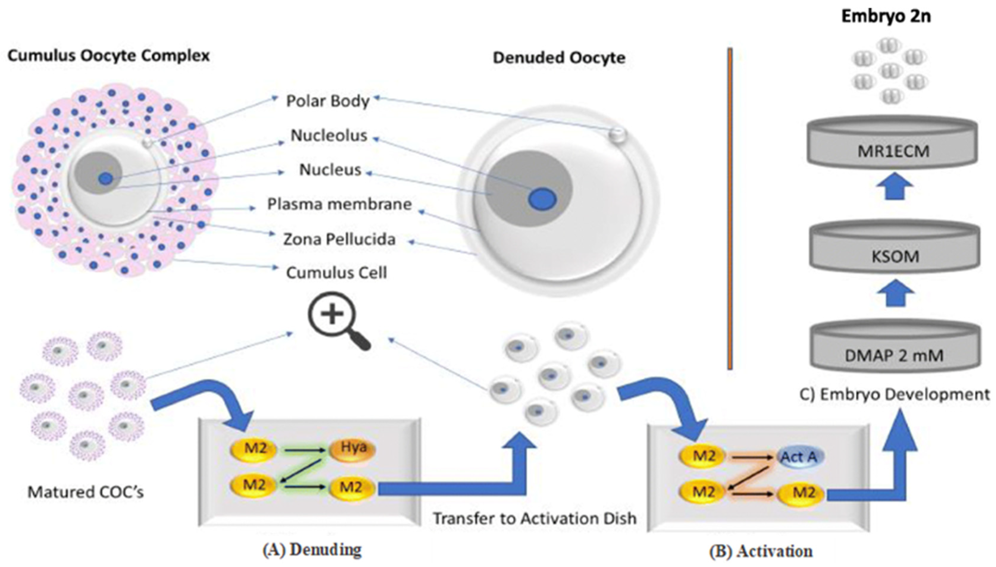
Parthenogenesis is a type of asexual reproduction occurring in females, wherein embryos develop and grow without male fertilisation. Many studies have made use of parthenogenesis as a model to examine the morphological and biochemical processes that take place in the initial phases of embryonic development [83]. The parthenogenetically activated oocytes (Figure 8) contain a maternal genome and can develop into haploid, diploid, or polyploid embryos. This enables the analysis of the potential roles of all genes associated with imprinting processes, as well as the influence of the paternal genome during early embryonic development, by comparing them with fertilised embryos [84]. Bovine oocyte activation has typically been achieved by chemical or physical (electrical) stimuli, employing various treatments to cause Ca2+ oscillations in the ooplasm. However, these alone are usually insufficient to maintain Ca2+ oscillations and oocyte activation, so they are often combined with other substances, such as protein phosphorylation inhibitors or MPF activity inhibitors [67].
Pronuclear formation and cortical granule exocytosis are two activation-related events that have been found to occur when oocytes are exposed to an electrical pulse [86]. However, compared to the nonactivated group, the cleavage and blastocyst rates are higher when the oocytes are electrically activated during an ICSI procedure [87]. Parthenogenetic activation (Figure 9) is employed in fertilisation modelling, somatic cell nuclear transfer (SCNT) (cloning), and stem cell research [88]. According to He et al. [89], the activation involved in the formation of haploid embryonic stem cells (PGhaESCs) can be employed to elucidate the roles of recessive or X-linked genes, generate genetically modified organisms, or evaluate the condition of oocyte activation for procedures such as somatic cell nuclear transfer or ICSI [90,91]. Together with cycloheximide, an inhibitor of protein synthesis, MPF is inhibited by selectively phosphorylating CDK1Thr14-Tyr15 [92].
Parthenogenesis may commence early embryonic development in mammals; yet, its failure in this class presents fundamental and unresolved issues concerning the role of fertilisation in reproductive physiology and embryonic development [84]. If oocytes lack a paternal genome, external or artificial activation can be used to create parthenotes, which are useful research tools even though they have a limited capacity for development [93]. Consequently, this remains a significant reason for the renewed interest in parthenogenetic research.

4. How to Avoid Cryo Injuries During Vitrification of Cattle Oocytes?

Cryodamage refers to the damage incurred during cryopreservation, characterised by a sequence of events resulting from impaired cellular functions following the freeze–thaw cycle [94]. The pronounced sensitivity of oocytes to cryopreservation can be attributed to their distinct morphological and functional traits, including cell size, cytoplasmic water volume, cytoskeletal arrangement, organelle distribution, and chromatin organisation stage [95]. In the process of vitrification, the cryoprotectant infiltrates the oocyte, leading to the replacement of intracellular water, resulting in dehydration, followed by rehydration upon thawing. Consequently, it is essential to take into account the varying membrane permeabilities of the oocyte throughout its maturation stages [5].

4.1. The Use of Cryoprotectants in Oocytes Cryopreservation

Oxidative stresses induced by ROS (superoxide anion, hydroxyl radical, hydrogen peroxide, and lipid peroxides) contribute to the occurrence of oocyte cryoinjury [96]. Oocyte cryoinjuries can happen during every stage of the cryopreservation process, which includes the addition of cryoprotective agents, cooling, freezing, and thawing or warming. Vitrification seems to be the most effective method for preserving oocytes and minimising cryodamage [97]. Vitrification is a complex procedure that entails the gradual exposure of oocytes to higher concentrations of permeable CPAs, frequently in conjunction with non-permeable CPAs, to inhibit both intracellular and extracellular ice crystallisation, prior to their exposure to ultrarapid cooling in liquid nitrogen [98].
Oocyte warming, on the other hand, exposes oocytes to gradually decreasing levels of non-permeable CPAs to manage hypoosmotic shock during the rehydration process [98]. Consequently, oocytes ought to be subjected to those agents for the minimal duration necessary to ensure reliable packaging. Given that both permeating agents (PAs) and non-permeating agents (NPAs) utilise the same vitrification mechanism, it is possible to incorporate NPAs into the solution, facilitating effective cryobanking while utilising reduced concentrations of PAs [99]. Sucrose, a non-permeating cryoprotectant, plays a crucial role in dehydration and vitrification, which subsequently mitigates the toxicity of ethylene glycol by lowering its concentration [30]. Therefore, a deeper comprehension of the mechanisms underlying CPA toxicity and strategies to mitigate it could pave the way for significant advancements in cryobiology.

4.2. The Use of Proteins and Antioxidants in Oocyte Cryopreservation

Among them, α-tocopherol, melatonin, l-proline, and resveratrol have been documented to safeguard mammalian oocytes from injuries associated with vitrification [100]. In cattle, it was previously reported that short-term recovery culture with α-tocopherol prior to IVF enhanced the blastocyst yield from vitrified-warmed mature oocytes [101]. The addition of melatonin in the vitrification solution enhanced the embryonic development of post-warm bovine mature oocytes [21]. Somfai and Hirao [102] studied the vitrification of immature bovine oocytes in protein-free media. During their research, they developed a vitrification protocol for the cryopreservation of immature bovine oocytes using protein-free media, which resulted in high blastocyst quality without evident toxic effects.
To minimise the oxidative damage caused by cryopreservation to oocytes, enzymatic and non-enzymatic antioxidants are commonly utilised in the vitrification, warming, and/or culture media to alleviate oxidative stress [103]. A variety of antioxidants such as melatonin, resveratrol, L-carnitine, quercetin, vitamin E, astaxanthin, proline, and coenzyme Q10 have shown positive effects on the maturation and development of oocytes [104]. The data gathered from previous studies offer promising support that oxidative damage in vitrified-warmed oocytes can be alleviated, significantly enhancing their in vitro developmental potential [15,105]. Exogenous antioxidants appear to be crucial in reducing cryo-oxidative damage, particularly given that endogenous enzymatic antioxidants alone are inadequate to combat the ROS produced during cryopreservation [106]. Furthermore, glutathione (GSH), cysteine, and cysteamine serve as essential endogenous non-enzymatic antioxidants in oocytes. GSH produced in oocytes has the ability to influence the sulfur–oxygen reduction state of cells and facilitate cytoplasmic maturation [104]. GSH has the capability to safeguard the spindle’s morphology and function from oxidative stress-related damage during the IVM of bovine oocytes, and it enhances the developmental potential of sheep embryos [107].

4.3. Cooling and Warming Rates

The cooling rate and warming rate are significant factors that contribute to variations in the survival of bovine oocytes following vitrification [5]. In order to prevent ice crystals from growing during the transition state, warming is carried out extremely quickly [108]. Survival was predominantly influenced by the warming rate, with the sole exception of the lowest cooling rate of 37 °C/min. The subsequent step that needs to be undertaken in the cryopreservation process involves determining the appropriate cooling and thawing rates. Given that the effectiveness of cryoprotection relies on preventing intracellular ice formation, it is essential to closely examine the dynamics of water movement across membranes throughout this process [109]. To reduce the likelihood of intracellular ice formation, it is essential for water to exit the cell as the temperature is lowered. While many authors in their reports focused solely on cooling rate values, the studies referenced above indicated that the warming rate was also a significant parameter for successful vitrification, particularly regarding blastocyst rates [5]. Consequently, the warming rate should be factored in when assessing the success of vitrification.

5. How Can the Cryopreservation Method Be Improved to Enhance the Fertilisation Efficiency?

The growing demand for ART and advancements in cryopreservation methods are transforming the therapeutic landscape in fertility treatment [110]. Recent advancements in ART protocols and procedures have been driven by changes in practice, alongside the introduction of new cryopreservation methods, such as vitrification, which serve as an alternative to the conventional slow-freezing technique [111]. Due to advancements in ART and, notably, in the efficacy of cryopreservation techniques, the trend of freezing oocytes has seen a significant rise in recent years.
Vitrification and slow freezing are the two methodologies employed for the long-term preservation of oocytes. Each of these strategies possesses distinct advantages and disadvantages. Comprehending these factors is crucial for making an informed conclusion regarding the optimal choice for oocyte cryopreservation [109]. Oocytes are less likely to sustain mechanical injury when vitrification is used instead of gradual freezing because it can stop intracellular and extracellular ice crystals from forming [112]. Moreover, vitrification is established from the principles of slow freezing. Vitrification presents numerous benefits compared to conventional freezing techniques: (1) It decreases the duration involved in the cryopreservation process; (2) It employs a high-concentration cryoprotectant that can lessen the exposure time to the cryoprotectant; (3) It reduces penetration damage [113]. Therefore, vitrification can be the same method used for the cryopreservation of oocytes.

6. Future Considerations

The most effective technique for fertilising and creating blastocysts from vitrified GV oocytes is the PICSI procedure [71]. There are several benefits to this process over traditional IVF for oocytes: (1) polyspermy is avoided during PICSI; (2) each oocyte uses only one selected sperm; and (3) sperm selection permits the injection of a high-quality sperm. When compared to other meiotic or developmental stages, cryopreserved GV oocytes have the benefit of being more readily collected than MII oocytes, which facilitates the creation of a large number of blastocysts for embryo transfer [71]. Moreover, ovarian stimulation is not necessary to generate GV oocytes. Prepubescent females can provide them, but IVM is necessary.
Cryopreservation has the potential to greatly influence fertilisation capabilities. The main objective of cryopreservation is to maintain the structural and functional integrity of oocytes throughout the freezing and thawing processes. The integrity of sperm chromatin is regarded as a crucial factor in male fertility [114]. During fertilisation, sperm with superior quality and good chromatin undergo natural selection. In ART, particularly in ICSI, sperm with DNA fragmentation are inherently eliminated from selection, which can adversely impact fertilisation rates, embryo quality, and implantation success [115]. Thus, the rate of fertilisation of developed cattle oocytes depends critically on the sperm genome and epigenome health.
To summarise, the technical result (efficacy) of employing traditional IVF or ICSI for oocyte in-semination in the absence of the male factor must be didactically divided into two factors. More research is required to examine the clinical results of ART cycles following conventional IVF or ICSI, taking into account all begun cycles and total fertilisation failure, to assess the efficacy of the procedures. Previous research assessing the clinical results of traditional IVF and ICSI failed to take into account the increased pregnancy rates following frozen–thawed embryo transfers with the use of vitrification. According to Giacobbe et al. [116], the rationale behind the increased utilisation of ICSI is its potential to increase the number of embryos transferred during a single stimulation cycle by reducing total fertilisation failure and increasing fertilisation rates.

7. Conclusions

In conclusion, with respect to oocytes, ICSI can be useful in the fertilisation of oocytes, with alterations preventing the normal process of fertilisation, such as cryopreserved oocytes, in vitro matured oocytes, and oocytes obtained from prepubertal females. More technical developments are required to increase the reliability of fertilisation and embryo formation through various IVF techniques using vitrified oocytes. These advancements include optimising morphological structure, IVM, oocyte vitrification, and sperm treatment.

Author Contributions

Conceptualisation, M.R.L., H.A.O., M.A.T., A.M. and M.L.M.; investigation, M.R.L. and M.A.T. resources, M.L.M., M.A.T., A.M. and H.A.O.; writing—original draft preparation, M.R.L.; writing—review and editing, M.L.M., M.A.T., A.M. and H.A.O.; funding acquisition, M.L.M. All authors have read and agreed to the published version of the manuscript.

Funding

The running cost of the project was funded by the Agricultural Research Council (ARC; P02000016) and the Department of Agriculture Land Reform and Rural Development (DALRRD; P02000253), South Africa.

Data Availability Statement

No new data were created or analysed in this study. Data sharing is not applicable to this article.

Acknowledgments

The Agricultural Research Council (ARC) is acknowledged for funding the running costs and providing the facility for the project; the Department of Agriculture Land Reform and Rural Development (DALRRD), South Africa, is acknowledged for funding the running costs of this research, and the National Research Foundation (NRF), South Africa, and the AgriSeta, South Africa, for providing abursary award to the first author.

Conflicts of Interest

The authors declare no conflicts of interest.

References

  1. Tharasanit, T.; Thuwanut, P. Oocyte cryopreservation in domestic animals and humans: Principles, techniques and updated outcomes. Animals 2021, 11, 2949. [Google Scholar] [CrossRef] [PubMed]
  2. Makarevich, A.V.; Olexiková, L.; Főldešiová, M.; Pivko, J.; Kubovičová, E. Ooplasm cryopreservation: Ovarian fragments versus oocytes alone. Slovak J. Anim. Sci. 2018, 51, 156–160. [Google Scholar]
  3. Díez, C.; Munoz, M.; Caamano, J.N.; Gomez, E. Cryopreservation of the bovine oocyte: Current status and perspectives. Reprod. Domest. Anim. 2012, 47, 76–83. [Google Scholar] [CrossRef]
  4. Hwang, I.S.; Hochi, S. Recent progress in cryopreservation of bovine oocytes. BioMed Res. Int. 2014, 2014, 570647. [Google Scholar] [CrossRef]
  5. Dujíčková, L.; Makarevich, A.V.; Olexiková, L.; Kubovičová, E.; Strejček, F. Methodological approaches for vitrification of bovine oocytes. Zygote 2021, 29, 1–11. [Google Scholar] [CrossRef]
  6. Houda, A.; Jankowski, P.M.; Romeo, M.; Eid, H.M. Female Fertility Preservation: Different Interventions and Procedures. In InCryopreservation-Applications and Challenges; IntechOpen: Rijeka, Croatia, 2022. [Google Scholar]
  7. Gandhi, G.; Ramesh, S.; Khatoon, A. Vitrification of oocytes: General considerations. In Vitrification in Assisted Reproduction: A User’s Manual; Springer: New Delhi, India, 2015; pp. 17–30. [Google Scholar] [CrossRef]
  8. Menéndez-Blanco, I.; Soto-Heras, S.; Catalá, M.G.; Piras, A.R.; Izquierdo, D.; Paramio, M.T. Effect of vitrification of in vitro matured prepubertal goat oocytes on embryo development after parthenogenic activation and intracytoplasmic sperm injection. Cryobiology 2020, 93, 56–61. [Google Scholar] [CrossRef] [PubMed]
  9. Wiesak, T.; Wasielak, M.; Złotkowska, A.; Milewski, R. Effect of vitrification on the zona pellucida hardening and follistatin and cathepsin B genes expression and developmental competence of in vitro matured bovine oocytes. Cryobiology 2017, 76, 18–23. [Google Scholar] [CrossRef]
  10. Jung, J.; Shin, H.; Bang, S.; Mok, H.J.; Suh, C.S.; Kim, K.P.; Lim, H.J. Analysis of the phospholipid profile of metaphase II mouse oocytes undergoing vitrification. PLoS ONE 2014, 9, e102620. [Google Scholar] [CrossRef] [PubMed]
  11. García-Martínez, T.; Mogas, T.; Mullen, S.F.; Martínez-Rodero, I.; Gulieva, R.E.; Higgins, A.Z. Effect of cryoprotectant concentration on bovine oocyte permeability and comparison of two membrane permeability modelling approaches. Sci. Rep. 2021, 11, 15387. [Google Scholar] [CrossRef] [PubMed]
  12. Mandawala, A.A.; Harvey, S.C.; Roy, T.K.; Fowler, K.E. Cryopreservation of animal oocytes and embryos: Current progress and future prospects. Theriogenology 2016, 86, 1637–1644. [Google Scholar] [CrossRef] [PubMed]
  13. Chian, R.C.; Wang, Y.; Li, Y.R. Oocyte vitrification: Advances, progress and future goals. J. Assist. Reprod. Genet. 2014, 31, 411–420. [Google Scholar] [CrossRef] [PubMed]
  14. Pan, B.; Qazi, I.H.; Guo, S.; Yang, J.; Qin, J.; Lv, T.; Zang, S.; Zhang, Y.; Zeng, C.; Meng, Q.; et al. Melatonin improves the first cleavage of parthenogenetic embryos from vitrified–warmed mouse oocytes potentially by promoting cell cycle progression. J. Anim. Sci. Biotechnol. 2021, 12, 1–7. [Google Scholar] [CrossRef]
  15. Guo, S.; Yang, J.; Qin, J.; Qazi, I.H.; Pan, B.; Zang, S.; Lv, T.; Deng, S.; Fang, Y.; Zhou, G. Melatonin promotes in vitro maturation of vitrified-warmed mouse germinal vesicle oocytes, potentially by reducing oxidative stress through the Nrf2 pathway. Animals 2021, 11, 2324. [Google Scholar] [CrossRef] [PubMed]
  16. Somfai, T.; Haraguchi, S.; Dang-Nguyen, T.Q.; Kaneko, H.; Kikuchi, K. Vitrification of porcine immature oocytes and zygotes results in different levels of DNA damage which reflects developmental competence to the blastocyst stage. PLoS ONE 2023, 18, e0282959. [Google Scholar] [CrossRef] [PubMed]
  17. Berteli, T.S.; Vireque, A.A.; Da Luz, C.M.; Borges, E.D.; Ferreira, C.R.; Navarro, P.A. Equilibration solution composition and extended exposure to equilibration phase affect embryo development and lipid profile of mouse oocytes. Reprod. BioMedicine Online 2022, 44, 961–975. [Google Scholar] [CrossRef]
  18. Angel-Velez, D.; Meese, T.; Hedia, M.; Fernandez-Montoro, A.; De Coster, T.; Pascottini, O.B.; Van Nieuwerburgh, F.; Govaere, J.; Van Soom, A.; Pavani, K.; et al. Transcriptomics reveal molecular differences in equine oocytes vitrified before and after In Vitro maturation. Int. J. Mol. Sci. 2023, 24, 6915. [Google Scholar] [CrossRef]
  19. Feng, H.; Stockwell, B.R. Unsolved mysteries: How does lipid peroxidation cause ferroptosis? PLoS Biol. 2018, 16, e2006203. [Google Scholar] [CrossRef] [PubMed]
  20. Khazaei, M.; Aghaz, F. Reactive oxygen species generation and use of antioxidants during in vitro maturation of oocytes. Int. J. Fertil. Steril. 2017, 11, 63. [Google Scholar] [CrossRef] [PubMed]
  21. Zhao, X.M.; Hao, H.S.; Du, W.H.; Zhao, S.J.; Wang, H.Y.; Wang, N.; Wang, D.; Liu, Y.; Qin, T.; Zhu, H.B. Melatonin inhibits apoptosis and improves the developmental potential of vitrified bovine oocytes. J. Pineal Res. 2016, 60, 132–141. [Google Scholar] [CrossRef] [PubMed]
  22. Soto-Heras, S.; Paramio, M.T. Impact of oxidative stress on oocyte competence for in vitro embryo production programs. Res. Vet. Sci. 2020, 132, 342–350. [Google Scholar] [CrossRef]
  23. Gutierrez-Castillo, E.; Diaz, F.A.; Talbot, S.A.; Bondioli, K.R. Effect of bovine oocyte vitrification with EGTA and post-warming recovery with resveratrol on meiotic spindle, mitochondrial function, reactive oxygen species, and developmental competence. Theriogenology 2023, 196, 59–67. [Google Scholar] [CrossRef]
  24. Lemseffer, Y.; Terret, M.; Campillo, C.; Labrune, E. Methods for assessing oocyte quality: A review of literature. Biomedicines 2022, 10, 2184. [Google Scholar] [CrossRef] [PubMed]
  25. Hoshino, Y. Updating the markers for oocyte quality evaluation: Intracellular temperature as a new index. Reprod. Med. Biol. 2018, 17, 434–441. [Google Scholar] [CrossRef]
  26. Ozturk, S. Selection of competent oocytes by morphological criteria for assisted reproductive technologies. Mol. Reprod. Dev. 2020, 87, 1021–1036. [Google Scholar] [CrossRef] [PubMed]
  27. Moghadam, A.R.; Moghadam, M.T.; Hemadi, M.; Saki, G. Oocyte quality and aging. JBRA Assist. Reprod. 2022, 26, 105. [Google Scholar] [CrossRef]
  28. Gutierrez-Castillo, E.J. Effect of Vitrification on Epigenetic Modifications and the Meiotic Spindle of Bovine Oocytes. Master’s Thesis, Louisiana State University and Agricultural & Mechanical College, Baton Rouge, LA, USA, 2018. [Google Scholar]
  29. Mo, X.H.; Fu, X.W.; Yuan, D.S.; Wu, G.Q.; Jia, B.Y.; Cheng, K.R.; Du, M.; Zhou, Y.H.; Yue, M.X.; Hou, Y.P.; et al. Effect of meiotic status, cumulus cells and cytoskeleton stabilizer on the developmental competence of ovine oocytes following vitrification. Small Rumin. Res. 2014, 117, 151–157. [Google Scholar] [CrossRef]
  30. Mphaphathi, M.L. Factors Affecting Cryopreservation of Immature and Matured Cattle Oocytes. Ph.D. Thesis, Department of Animal, Wildlife and Grassland Sciences, Faculty of Natural and Agricultural Sciences, University of the Free State, Bloemfontein, South Africa, 2022. [Google Scholar]
  31. Purohit, G.N.; Meena, H.; Solanki, K. Effects of vitrification on immature and in vitro matured, denuded and cumulus compact goat oocytes and their subsequent fertilization. J. Reprod. Infertil. 2012, 13, 53. [Google Scholar] [PubMed]
  32. Chaves, D.F.; Corbin, E.; Almanana, C.; Locatelli, Y.; Souza-Fabjan, M.G.; Bhat, M.H.; Freitas, V.J.F.; Mermillod, P. Vitrification of immature and in vitro matured bovine cumulus–oocyte complexes: Effects on oocyte structure and embryo development. Livest. Sci. 2017, 199, 50–60. [Google Scholar] [CrossRef]
  33. Zhang, F.; Zhang, Z.Y.; Cai, M.D.; Li, X.X.; Li, Y.H.; Lei, Y.; Yu, X.L. Effect of vitrification temperature and cryoprotectant concentrations on the mRNA transcriptome of bovine mature oocytes after vitrifying at immature stage. Theriogenology 2020, 148, 225–235. [Google Scholar] [CrossRef]
  34. Canesin, H.S.; Brom-de-Luna, J.G.; Choi, Y.H.; Ortiz, I.; Diaw, M.; Hinrichs, K. Blastocyst development after intracytoplasmic sperm injection of equine oocytes vitrified at the germinal-vesicle stage. Cryobiology 2017, 75, 52–59. [Google Scholar] [CrossRef]
  35. De Munck, N.; Vajta, G.; Rienzi, L. Oocyte cryopreservation technique. In Preventing Age Related Fertility Loss; Springer: Cham, Switzerland; Gewerbestrasse, Switzerland, 2018; pp. 87–101. [Google Scholar] [CrossRef]
  36. Baust, J.M.; Corwin, W.; Snyder, K.K.; Van Buskirk, R.; Baust, J.G. Cryopreservation: Evolution of molecular based strategies. In Biobanking and Cryopreservation of Stem Cells; Springer: Cham, Switzerland, 2016; pp. 13–29. [Google Scholar] [CrossRef]
  37. Gebel, J.; Tuppi, M.; Sänger, N.; Schumacher, B.; Dötsch, V. DNA damaged induced cell death in oocytes. Molecules 2020, 25, 5714. [Google Scholar] [CrossRef] [PubMed]
  38. Chen, H.; Zhang, L.; Deng, T.; Zou, P.; Wang, Y.; Quan, F.; Zhang, Y. Effects of oocyte vitrification on epigenetic status in early bovine embryos. Theriogenology 2016, 86, 868–878. [Google Scholar] [CrossRef]
  39. Coello, A.; Sanchez, E.; Vallejo, B.; Meseguer, M.; Remohí, J.; Cobo, A. Effect of oocyte morphology on post-warming survival and embryo development in vitrified autologous oocytes. Reprod. BioMedicine Online 2019, 38, 313–320. [Google Scholar] [CrossRef]
  40. Tejera, A.; Herrero, J.; de Los Santos, M.J.; Garrido, N.; Ramsing, N.; Meseguer, M. Oxygen consumption is a quality marker for human oocyte competence conditioned by ovarian stimulation regimens. Fertil. Steril. 2011, 96, 618–623.e2. [Google Scholar] [CrossRef]
  41. Minasi, M.G.; Anagnostopoulou, C.; Boitrelle, F.; Vogiatzi, P.; Sallam, H.; Saleh, R.; Colpi, G.; Agarwal, A. Oocytes evaluation and in-vitro fertilization/intra cytoplasmic sperm injection outcomes. Panminerva Medica 2023, 27, 179–187. [Google Scholar] [CrossRef]
  42. Hassa, H.; Aydın, Y.; Taplamacıoğlu, F. The role of perivitelline space abnormalities of oocytes in the developmental potential of embryos. J. Turk. Ger. Gynecol. Assoc. 2014, 15, 161. [Google Scholar] [CrossRef] [PubMed]
  43. Gutnisky, C.; Morado, S.; Gadze, T.; Donato, A.; Alvarez, G.; Dalvit, G.; Cetica, P. Morphological, biochemical and functional studies to evaluate bovine oocyte vitrification. Theriogenology 2020, 143, 18–26. [Google Scholar] [CrossRef]
  44. Rusciano, G.; De Canditiis, C.; Zito, G.; Rubessa, M.; Roca, M.S.; Carotenuto, R.; Sasso, A.; Gasparrini, B. Raman-microscopy investigation of vitrification-induced structural damages in mature bovine oocytes. PLoS ONE 2017, 12, e0177677. [Google Scholar] [CrossRef]
  45. Arav, A.; Natan, Y. The near future of vitrification of oocytes and embryos: Looking into past experience and planning into the future. Transfus. Med. Hemotherapy 2019, 46, 182–187. [Google Scholar] [CrossRef] [PubMed]
  46. Fernández-Reyez, F.; Ducolomb, Y.; Romo, S.; Casas, E.; Salazar, Z.; Betancourt, M. Viability, maturation and embryo development in vitro of immature porcine and ovine oocytes vitrified in different devices. Cryobiology 2012, 64, 261–266. [Google Scholar] [CrossRef] [PubMed]
  47. Casillas, F.; Teteltitla-Silvestre, M.; Ducolomb, Y.; Lemus, A.E.; Salazar, Z.; Casas, E.; Betancourt, M. Co-culture with granulosa cells improve the in vitro maturation ability of porcine immature oocytes vitrified with cryolock. Cryobiology 2014, 69, 299–304. [Google Scholar] [CrossRef] [PubMed]
  48. Casillas, F.; Ducolomb, Y.; Lemus, A.E.; Cuello, C.; Betancourt, M. Porcine embryo production following in vitro fertilization and intracytoplasmic sperm injection from vitrified immature oocytes matured with a granulosa cell co-culture system. Cryobiology 2015, 71, 299–305. [Google Scholar] [CrossRef]
  49. Wasielak-Politowska, M.; Kordowitzki, P. Chromosome segregation in the oocyte: What goes wrong during aging. Int. J. Mol. Sci. 2022, 23, 2880. [Google Scholar] [CrossRef] [PubMed]
  50. Dai, J.; Wu, C.; Muneri, C.W.; Niu, Y.; Zhang, S.; Rui, R.; Zhang, D. Changes in mitochondrial function in porcine vitrified MII-stage oocytes and their impacts on apoptosis and developmental ability. Cryobiology 2015, 71, 291–298. [Google Scholar] [CrossRef]
  51. Girka, E.; Gatenby, L.; Gutierrez, E.J.; Bondioli, K.R. The effects of microtubule stabilizing and recovery agents on vitrified bovine oocytes. Theriogenology 2022, 182, 9–16. [Google Scholar] [CrossRef] [PubMed]
  52. Caamaño, J.N.; Díez, C.; Trigal, B.; Muñoz, M.; Morató, R.; Martín, D.; Carrocera, S.; Mogas, T.; Gomez, E. Assessment of Meiotic Spindle Configuration and Post-Warming Bovine Oocyte Viability Using Polarized Light Microscopy. Reprod. Domest. Anim. 2013, 48, 470–476. [Google Scholar] [CrossRef] [PubMed]
  53. Swearman, H.; Koustas, G.; Knight, E.; Liperis, G.; Grupen, C.; Sjoblom, C. pH: The silent variable significantly impacting meiotic spindle assembly in mouse oocytes. Reprod Biomed Online 2018, 37, 279–290. [Google Scholar] [CrossRef]
  54. Serra, E.; Gadau, S.D.; Berlinguer, F.; Naitana, S.; Succu, S. Morphological features and microtubular changes in vitrified ovine oocytes. Theriogenology 2020, 148, 216–224. [Google Scholar] [CrossRef] [PubMed]
  55. Tilia, L.; Venetis, C.; Kilani, S.; Cooke, S.; Chapman, M. Is oocyte meiotic spindle morphology associated with embryo ploidy? A prospective cohort study. Fertil. Steril. 2016, 105, 1085–1092. [Google Scholar] [CrossRef] [PubMed]
  56. Gutierrez-Castillo, E.; Diaz, F.A.; Talbot, S.A.; Bondioli, K.R. Recovery of spindle morphology and mitochondrial function through extended culture after vitrification-warming of bovine oocytes. Theriogenology 2022, 189, 192–198. [Google Scholar] [CrossRef]
  57. Yurchuk, T.; Likszo, P.; Witek, K.; Petrushko, M.; Skarzynski, D.J. New Approach to the Cryopreservation of GV Oocytes and Cumulus Cells through the Lens of Preserving the Intercellular Gap Junctions Based on the Bovine Model. Int. J. Mol. Sci. 2024, 25, 6074. [Google Scholar] [CrossRef]
  58. Xie, J.; Xu, X.; Liu, S. Intercellular communication in the cumulus–oocyte complex during folliculogenesis: A review. Front. Cell Dev. Biol. 2023, 11, 1087612. [Google Scholar] [CrossRef] [PubMed]
  59. Virant-Klun, I.; Bauer, C.; Ståhlberg, A.; Kubista, M.; Skutella, T. Human oocyte maturation in vitro is improved by co-culture with cumulus cells from mature oocytes. Reprod. Biomed. Online 2018, 36, 508–523. [Google Scholar] [CrossRef] [PubMed]
  60. Jia, B.Y.; Xiang, D.C.; Zhang, B.; Quan, G.B.; Shao, Q.Y.; Hong, Q.H.; Wu, G.Q. Quality of vitrified porcine immature oocytes is improved by coculture with fresh oocytes during in vitro maturation. Mol. Reprod. Dev. 2019, 86, 1615–1627. [Google Scholar] [CrossRef] [PubMed]
  61. Ortiz-Escribano, N.; Smits, K.; Piepers, S.; Van den Abbeel, E.; Woelders, H.; Van Soom, A. Role of cumulus cells during vitrification and fertilization of mature bovine oocytes: Effects on survival, fertilization, and blastocyst development. Theriogenology 2016, 86, 635–641. [Google Scholar] [CrossRef] [PubMed]
  62. Neri, Q.V.; Lee, B.; Rosenwaks, Z.; Machaca, K.; Palermo, G.D. Understanding fertilization through intracytoplasmic sperm injection (ICSI). Cell Calcium 2014, 55, 24–37. [Google Scholar] [CrossRef]
  63. Hamze, J.G.; Canha-Gouveia, A.; Algarra, B.; Gómez-Torres, M.J.; Olivares, M.C.; Romar, R.; Jiménez-Movilla, M. Mammalian spermatozoa and cumulus cells bind to a 3D model generated by recombinant zona pellucida protein-coated beads. Sci. Rep. 2019, 9, 17989. [Google Scholar] [CrossRef]
  64. Liu, Z.; Zhu, L.; Wang, J.; Luo, G.; Xi, Q.; Zhou, X.; Li, Z.; Yang, X.; Duan, J.; Jin, L.; et al. Novel homozygous mutations in PATL2 lead to female infertility with oocyte maturation arrest. J. Assist. Reprod. Genet. 2020, 37, 841–847. [Google Scholar] [CrossRef]
  65. Wiltshire, A.; Schaal, R.; Wang, F.; Tsou, T.; McKerrow, W.; Keefe, D. Vitrification with dimethyl sulfoxide induces transcriptomic alteration of gene and transposable element expression in immature human oocytes. Genes 2023, 14, 1232. [Google Scholar] [CrossRef]
  66. Sithole, S.M.; Mphaphathi, M.L.; Sebopela, M.D.; Nedambale, T.L. Comparison of in vitro Maturation Media on Cattle Oocytes after in vitro Embryo Production. Am. J. Anim. Vet. Sci. 2023, 18, 27–39. [Google Scholar] [CrossRef]
  67. Fuentes, F.; Munoz, E.; Contreras, M.J.; Arias, M.E.; Felmer, R. Bovine ICSI: Limiting factors, strategies to improve its efficiency and alternative approaches. Zygote 2022, 30, 749–767. [Google Scholar] [CrossRef] [PubMed]
  68. Mezzalira, J.C.; Ohlweiler, L.U.; Klein, N.; dos Santos Brum, D.; Leivas, F.G.; Mezzalira, A. Intracytoplasmic Sperm Injection after Vitrification of Immature Oocytes in Follicular Fluid Increases Bovine Embryo Production. Acta Sci. Vet. 2017, 45, 1–7. [Google Scholar] [CrossRef]
  69. Unnikrishnan, V.; Kastelic, J.; Thundathil, J. Intracytoplasmic sperm injection in cattle. Genes 2021, 12, 198. [Google Scholar] [CrossRef]
  70. Ozcan, P.; Varli, B.; Findikli, N.; Basar, M.; Oral, E. Oocyte quality evaluation and cryopreservation. In Management of Infertility; Academic Press: New York, NY, USA, 2023; pp. 211–222. [Google Scholar] [CrossRef]
  71. Casillas, F.; Betancourt, M.; Cuello, C.; Ducolomb, Y.; López, A.; Juárez-Rojas, L.; Retana-Márquez, S. An efficiency comparison of different in vitro fertilization methods: IVF, ICSI, and PICSI for embryo development to the blastocyst stage from vitrified porcine immature oocytes. Porc. Health Manag. 2018, 4, 1–12. [Google Scholar] [CrossRef] [PubMed]
  72. Salamone, D.F.; Canel, N.G.; Rodríguez, M.B. Intracytoplasmic sperm injection in domestic and wild mammals. Reproduction 2017, 154, F111–F124. [Google Scholar] [CrossRef]
  73. Avalos-Durán, G.; Cañedo-Del Ángel, A.M.; Rivero-Murillo, J.; Zambrano-Guerrero, J.E.; Carballo-Mondragón, E.; Checa-Vizcaíno, M.Á. Physiological ICSI (PICSI) vs. conventional ICSI in couples with male factor: A systematic review. JBRA Assist. Reprod. 2018, 22, 139. [Google Scholar] [CrossRef] [PubMed]
  74. Castillo-Baso, J.; García-Villafaña, G. Routine Sperm Selection Using the Physiological ICSI Technique. Ph.D. Thesis, Subspecialty: Autonomous University of Baja California, Mexicali, Mexico, 2012. [Google Scholar]
  75. Mokánszki, A.; Tóthné, E.V.; Bodnár, B.; Tándor, Z.; Molnár, Z.; Jakab, A.; Ujfalusi, A.; Oláh, É. Is sperm hyaluronic acid binding ability predictive for clinical success of intracytoplasmic sperm injection: PICSI vs. ICSI? Syst. Biol. Reprod. Med. 2014, 60, 348–354. [Google Scholar] [CrossRef] [PubMed]
  76. Novoselsky Persky, M.; Hershko-Klement, A.; Solnica, A.; Bdolah, Y.; Hurwitz, A.; Ketzin, E.l.; Gilad, M.; Nefesh, I.; Esh-Broder, E. Conventional ICSI vs. physiological selection of spermatozoa for ICSI (Picsi) Sibling Oocytes. Andrology 2021, 9, 873–877. [Google Scholar] [CrossRef]
  77. Miller, D.; Pavitt, S.; Sharma, V.; Forbes, G.; Hooper, R.; Bhattacharya, S.; Kirkman-Brown, J.; Coomarasamy, A.; Lewis, S.; Cutting, R.; et al. Physiological, hyaluronan-selected intracytoplasmic sperm injection for infertility treatment (HABSelect): A parallel, two-group, randomised trial. Lancet 2019, 393, 416–422. [Google Scholar] [CrossRef]
  78. Worrilow, K.C.; Eid, S.; Woodhouse, D.; Perloe, M.; Smith, S.; Witmyer, J.; Ivani, K.; Khoury, C.; Ball, G.D.; Elliot, T.; et al. Use of hyaluronan in the selection of sperm for intracytoplasmic sperm injection (ICSI): Significant improvement in clinical outcomes-multicenter, double-blinded and randomized controlled trial. Hum. Reprod. 2013, 28, 306–314. [Google Scholar] [CrossRef]
  79. Le, M.T.; Nguyen, H.T.T.; Van Nguyen, T.; Nguyen, T.T.T.; Dang, H.N.T.; Dang, T.C.; Nguyen, Q.H.V. Physiological intracytoplasmic sperm injection does not improve the quality of embryos: A cross-sectional investigation on sibling oocytes. Clin. Exp. Reprod. Med. 2023, 50, 123. [Google Scholar] [CrossRef] [PubMed]
  80. Blanca Paraíso, M.D.; Ferrer, C.M.; Ramírez, J.M.; Espinel, L.Q.; Gómez, M.B. What Is PICSI or Physiological ICSI in IVF? 2024. Available online: https://www.invitra.com/en/picsi-physiological-icsi/#picsi-procedure (accessed on 17 September 2024).
  81. Liu, Y.; Feenan, K.; Chapple, V.; Roberts, P.; Matson, P. Intracytoplasmic sperm injection using hyaluronic acid or polyvinylpyrrolidone: A time-lapse sibling oocyte study. Hum. Fertil. 2019, 22, 39–45. [Google Scholar] [CrossRef] [PubMed]
  82. Rao, S. PICSI—Physiological Intracytoplasmic Sperm Injection. 2023. Available online: https://www.esco-medical.com/news/picsi-physiological-intracytoplasmic-sperm-injection (accessed on 9 December 2024).
  83. Valencia, C.; Pérez-García, F.; Aguila, L.; Felmer, R.; Arias, M.E. Combined Exogenous Activation of Bovine Oocytes: Effects on Maturation-Promoting Factor, Mitogen-Activated Protein Kinases, and Embryonic Competence. Int. J. Mol. Sci. 2023, 24, 15794. [Google Scholar] [CrossRef]
  84. Kharche, S.D.; Birade, H.S. Parthenogenesis and activation of mammalian oocytes for in vitro embryo production: A review. Adv. Biosci. Biotechnol. 2013, 2, 28406. [Google Scholar]
  85. Ailia, M.J.; Jin, Y.K.; Kim, H.K.; Jang, G. Development of in-vitro maturation protocol for rat oocytes; under simple culture vs co-culture with cumulus cell monolayer and its developmental potential via Parthenogenetic/artificial activation. BMC Vet. Res. 2021, 17, 1–7. [Google Scholar] [CrossRef]
  86. Piñeiro-Silva, C.; Gadea, J. Optimizing oocyte electroporation for genetic modification of porcine embryos: Evaluation of the parthenogenetic activation. Theriogenology 2024, 218, 126–136. [Google Scholar] [CrossRef] [PubMed]
  87. Yoo, J.G.; Hur, C.G.; Park, M.R.; Park, J.Y.; Hwang, K.C.; Kim, J.H.; Kim, J.H.; Cho, S.K. Electrical activation enhances pre-implantation embryo development following sperm injection into in vitro matured pig oocytes. J. Vet. Med. Sci. 2012, 74, 429–434. [Google Scholar] [CrossRef]
  88. Taşkın, A.; Coşkun, N.; Kocabay, A. Effects of different parthenogenetic activation periods on mouse embryo development and quality. Kocatepe Vet. J. 2020, 13, 383–387. [Google Scholar] [CrossRef]
  89. He, W.; Chen, J.; Gao, S. Mammalian haploid stem cells: Establishment, engineering and applications. Cell. Mol. Life Sci. 2019, 76, 2349–2367. [Google Scholar] [CrossRef] [PubMed]
  90. Felmer, R.; Arias, M.E. Activation treatment of recipient oocytes affects the subsequent development and ploidy of bovine parthenogenetic and somatic cell nuclear transfer (SCNT) embryos. Mol. Reprod. Dev. 2015, 82, 441–449. [Google Scholar] [CrossRef] [PubMed]
  91. Arias, M.E.; Sánchez, R.; Felmer, R. Effect of anisomycin, a protein synthesis inhibitor, on the in vitro developmental potential, ploidy and embryo quality of bovine ICSI embryos. Zygote 2016, 24, 724–732. [Google Scholar] [CrossRef] [PubMed]
  92. Valencia, C.; Pérez, F.A.; Matus, C.; Felmer, R.; Arias, M.E. Activation of bovine oocytes by protein synthesis inhibitors: New findings on the role of MPF/MAPKs. Biol. Reprod. 2021, 104, 1126–1138. [Google Scholar] [CrossRef] [PubMed]
  93. Suvá, M.; Canel, N.G.; Salamone, D.F. Effect of single and combined treatments with MPF or MAPK inhibitors on parthenogenetic haploid activation of bovine oocytes. Reprod. Biol. 2019, 19, 386–393. [Google Scholar] [CrossRef]
  94. Teng, J. Improving Cryopreservation of Bovine Oocytes and Embryos. Bachelor’s Thesis, College of Agriculture and Life Sciences, Animal Science of Cornell University, Ithaca, NY, USA, 2020. [Google Scholar]
  95. Sprícigo, J.F.; Morais, K.; Ferreira, A.R.; Machado, G.M.; Gomes, A.C.; Rumpf, R.; Franco, M.M.; Dode, M.A. Vitrification of bovine oocytes at different meiotic stages using the Cryotop method: Assessment of morphological, molecular and functional patterns. Cryobiology 2014, 69, 256–265. [Google Scholar] [CrossRef]
  96. Gutnisky, C.; Morado, S.; Dalvit, G.C.; Thompson, J.G.; Cetica, P.D. Glycolytic pathway activity: Effect on IVM and oxidative metabolism of bovine oocytes. Reprod. Fertil. Dev. 2013, 25, 1026–1035. [Google Scholar] [CrossRef]
  97. Arav, A. Cryopreservation of oocytes and embryos. Theriogenology 2014, 81, 96–102. [Google Scholar] [CrossRef] [PubMed]
  98. Marques, C.C.; Santos-Silva, C.; Rodrigues, C.; Matos, J.E.; Moura, T.; Baptista, M.C.; Horta, A.E.; Bessa, R.J.; Alves, S.P.; Soveral, G.; et al. Bovine oocyte membrane permeability and cryosurvival: Effects of different cryoprotectants and calcium in the vitrification media. Cryobiology 2018, 81, 4–11. [Google Scholar] [CrossRef]
  99. Kojayan, G.; Whaley, D.; Alexander, M.; Rodriguez, S.; Lee, S.; Lakey, J.R. Improved cryopreservation yield of pancreatic islets using combination of lower dose permeable cryoprotective agents. Cryobiology 2019, 88, 23–28. [Google Scholar] [CrossRef] [PubMed]
  100. Chinen, S.; Yamanaka, T.; Hirabayashi, M.; Hochi, S. Rescue of vitrified-warmed bovine mature oocytes by short-term recovery culture with resveratrol. Cryobiology 2020, 97, 185–190. [Google Scholar] [CrossRef] [PubMed]
  101. Yashiro, I.; Tagiri, M.; Ogawa, H.; Tashima, K.; Takashima, S.; Hara, H.; Hirabayashi, M.; Hochi, S. High Revivability of Vitrified-Warmed Bovine Mature Oocytes After Recovery Culture with Alpha-Tocopherol. Ph.D. Thesis, Shinshu University Library, Matsumoto, Japan, 2015. [Google Scholar]
  102. Somfai, T.; Hirao, Y. Vitrification of immature bovine oocytes in protein-free media: The impact of the cryoprotectant treatment protocol, base medium, and ovary storage. Theriogenology 2021, 172, 47–54. [Google Scholar] [CrossRef] [PubMed]
  103. Zhang, Z.; Mu, Y.; Ding, D.; Zou, W.; Li, X.; Chen, B.; Leung, P.C.; Chang, H.M.; Zhu, Q.; Wang, K.; et al. Melatonin improves the effect of cryopreservation on human oocytes by suppressing oxidative stress and maintaining the permeability of the oolemma. J. Pineal Res. 2021, 70, e12707. [Google Scholar] [CrossRef] [PubMed]
  104. Xu, H.; Jia, C.; Cheng, W.; Zhang, T.; Tao, R.; Ma, Y.; Si, L.; Xu, Y.; Li, J. The Effect of L-Carnitine Additive During In Vitro Maturation on the Vitrification of Pig Oocytes. Cell. Reprogr. 2020, 22, 198–207. [Google Scholar] [CrossRef] [PubMed]
  105. Yang, J.; Guo, S.; Pan, B.; Qazi, I.H.; Qin, J.; Zang, S.; Han, H.; Meng, Q.; Zhou, G. Melatonin promotes in vitro maturation of vitrified-warmed mouse GV oocytes potentially by modulating MAD2 protein expression of SAC component through MTRs. Cryobiology 2021, 102, 82–91. [Google Scholar] [CrossRef]
  106. Cao, B.; Qin, J.; Pan, B.; Qazi, I.H.; Ye, J.; Fang, Y.; Zhou, G. Oxidative stress and oocyte cryopreservation: Recent advances in mitigation strategies involving antioxidants. Cells 2022, 11, 3573. [Google Scholar] [CrossRef]
  107. García-Martínez, T.; Vendrell-Flotats, M.; Martínez-Rodero, I.; Ordóñez-León, E.A.; Álvarez-Rodríguez, M.; López-Béjar, M.; Yeste, M.; Mogas, T. Glutathione Ethyl Ester Protects In Vitro-Maturing Bovine Oocytes against Oxidative Stress Induced by Subsequent Vitrification/Warming. Int. J. Mol. Sci 2020, 21, 7547. [Google Scholar] [CrossRef] [PubMed]
  108. Iussig, B.; Maggiulli, R.; Fabozzi, G.; Bertelle, S.; Vaiarelli, A.; Cimadomo, D.; Ubaldi, F.M.; Rienzi, L. A brief history of oocyte cryopreservation: Arguments and facts. Acta Obstet. Et Gynecol. Scand. 2019, 98, 550–558. [Google Scholar] [CrossRef] [PubMed]
  109. Whaley, D.; Damyar, K.; Witek, R.P.; Mendoza, A.; Alexander, M.; Lakey, J.R. Cryopreservation: An overview of principles and cell-specific considerations. Cell Transplant. 2021, 30, 0963689721999617. [Google Scholar] [CrossRef] [PubMed]
  110. Bosch, E.; De Vos, M.; Humaidan, P. The future of cryopreservation in assisted reproductive technologies. Front. Endocrinol. 2020, 11, 67. [Google Scholar] [CrossRef] [PubMed]
  111. Banker, M.; Kotdawala, A.; Gupta, R. The impact of vitrification in artificial reproductive technology programmes. Eur. Med. J. 2017, 2, 82–89. [Google Scholar] [CrossRef]
  112. Marques, L.S.; Fossati, A.A.; Rodrigues, R.B.; Da Rosa, H.T.; Izaguirry, A.P.; Ramalho, J.B.; Moreira, J.C.; Santos, F.W.; Zhang, T.; Streit, D.P., Jr. Slow freezing versus vitrification for the cryopreservation of zebrafish (Danio rerio) ovarian tissue. Sci. Rep. 2019, 9, 15353. [Google Scholar] [CrossRef] [PubMed]
  113. Chen, H.; Zhang, L.; Meng, L.; Liang, L.; Zhang, C. Advantages of vitrification preservation in assisted reproduction and potential influences on imprinted genes. Clin. Epigenetics 2022, 14, 141. [Google Scholar] [CrossRef] [PubMed]
  114. Zeqiraj, A.; Beadini, S.; Beadini, N.; Aliu, H.; Gashi, Z.; Elezaj, S.; Bexheti, S.; Shabani, A. Male infertility and sperm DNA fragmentation. Open Access Maced. J. Med. Sci. 2018, 6, 1342. [Google Scholar] [CrossRef] [PubMed]
  115. Zini, A. Are sperm chromatin and DNA defects relevant in the clinic? Syst. Biol. Reprod. Med. 2011, 57, 78–85. [Google Scholar] [CrossRef] [PubMed]
  116. Giacobbe, M.; Conatti, M.; Gomes, A.; Bonetti, T.C.; Monteleone, P.A. Effectivity of conventional in vitro fertilization (IVF) and intracytoplasmic sperm injection (ICSI) when male factor is absent: A perspective point of view. JBRA Assist. Reprod. 2022, 26, 123. [Google Scholar] [CrossRef]
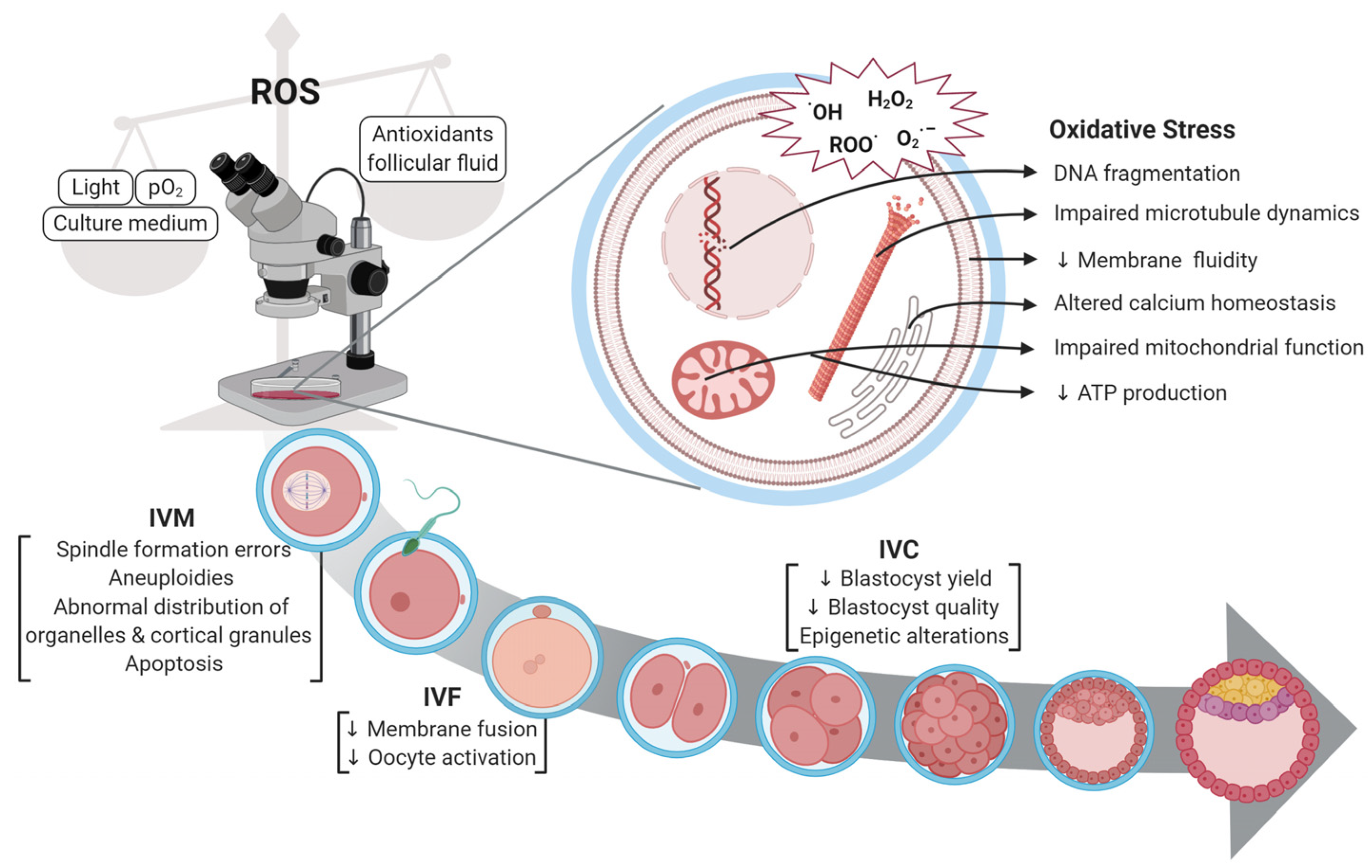
Figure 1. Oxidative stress in in vitro embryo formation and its impact on oocyte developmental capability [22].
Agriculture 15 00363 g001
Figure 2. Survival rate of cattle oocytes following thawing or warming and then maturing in vitro. (A) Immature oocyte at 10× magnification. (B) Mature oocyte at 60× magnification.
Agriculture 15 00363 g002
Figure 3. Oocytes that were cryopreserved and have undergone varying morphologies after being thawed or warmed up and then maturing in vitro. (A) Zona cracked at 10× magnification, and (B) Oocyte changed shape at 60× magnification [30].
Agriculture 15 00363 g003
Figure 4. Hypotheses concerning cryoinjuries in mammalian oocytes. The disorganisation of microtubules results in the depolymerisation of tubulin proteins, causing improper assembly of meiotic spindles, which subsequently leads to chromosome misalignment and the suppression of second polar body extrusion [4].
Agriculture 15 00363 g004
Figure 5. Images showcasing the morphology of oocyte spindles, captured through polarised light microscopy, illustrating the different types of meiotic spindles in oocytes. (A) normal; (B); dysmorphic; (C) telophase; (D) translucent; (E) no visible spindle [55].
Agriculture 15 00363 g005
Figure 6. (A) Presumptive zygote dysmorphism: multiple polar bodies [66]. (B) non-vitrified presuemptive zygote with two pronucleius [30].
Agriculture 15 00363 g006
Figure 7. Schematic illustration showing the procedure of PICSI [80].
Agriculture 15 00363 g007
Figure 8. Schematic diagram of parthenogenetic activation and procedure until embryo development (2 cells). The main phases involved denuding, activation using either ionomycin or ethanol 7%, and development in growth media till 2-cell embryos [85].
Agriculture 15 00363 g008
Figure 9. Schematic illustration of in vitro parthenogenetic activation of cattle oocytes using ionomycin and 6-DMAP. Created in BioRender.com.
Agriculture 15 00363 g009
Table 1. The criteria showing the quality of oocyte.
CriterionFunctionReference
Cumulus–oocyte complexCumulus cells are essential for oocyte quality; they sustain energy production in the cumulus–oocyte complex and may safeguard the oocyte against ROS during cryopreservation.[25]
Polar bodyThe polar globule is a tiny cell with 23 chromosomes that is released during the oocyte’s maturation. The intact, properly shaped, and smooth surface of the polar body increases the rate of fertilisation and the quality of the embryo.[24,26]
Zona pellucidaThe zona pellucida is a glycoprotein layer that covers oocytes and their surrounding cells, serving to safeguard the mammalian ovum. A robust zona pellucida or a substantial inner layer of the zona pellucida correlates with enhanced oocyte development and embryo quality.[26,27]
Perivitelline spaceThe perivitelline space (PVS) reveals that the acellular region between the plasma membrane of the oocyte (oolema) and the zona pellucida (ZP) has a hyaluronan-rich extracellular matrix that is not discernible under optical microscopy.[27]
CytoplasmCytoplasmic and nuclear factors are critical determinants of oocyte quality, directly affecting the developmental competence of embryos in ART. Nuclear maturity signifies the continuation of meiosis and the advancement to metaphase II, the stage at which it is interrupted during ovulation.[27]
Disclaimer/Publisher’s Note: The statements, opinions and data contained in all publications are solely those of the individual author(s) and contributor(s) and not of MDPI and/or the editor(s). MDPI and/or the editor(s) disclaim responsibility for any injury to people or property resulting from any ideas, methods, instructions or products referred to in the content.