Article

Exogenous Catalase Supplementation Alleviates Fusarium graminearum Mycotoxins-Induced Oxidative Stress in Weaned Piglets

Guangdong Provincial Key Laboratory of Animal Nutrition Control, College of Animal Science, South China Agricultural University, No. 483 of Wushan Road, Guangzhou 510642, China
*
Author to whom correspondence should be addressed.
These authors contributed equally to this work.
Agriculture 2025, 15(17), 1892; https://doi.org/10.3390/agriculture15171892
Submission received: 31 July 2025 / Revised: 29 August 2025 / Accepted: 3 September 2025 / Published: 5 September 2025
(This article belongs to the Section Farm Animal Production)

Abstract

The objective of this study was to investigate the impact of exogenous catalase (CAT) on antioxidant properties, as well as on hepatic and intestinal health, in piglets exposed to Fusarium graminearum mycotoxins (FGM). Forty female weaned piglets were divided into five groups (eight replicates per group). The pre-feeding period was 3 days, followed by a 28-day experimental period. The piglets in the control (CON) group were fed a diet without FGM contamination, while those in the FGM-exposed (TOX) group were fed a diet with FGM contamination. The LCAT, MCAT, and HCAT groups received an FGM-contaminated diet supplemented with 100, 200, and 400 U/kg of CAT, respectively. The results indicated that 400 U/kg CAT supplementation inhibited (p < 0.05, linear p < 0.05, quadratic p < 0.05) the decreases in average daily gain and average daily feed intake of piglets exposed to FGM. Moreover, all doses of supplemental CAT suppressed (p < 0.05) the increases in diarrhea rate and diarrhea index of FGM-exposed piglets. Additionally, supplemental CAT reversed (p < 0.05, linear and quadratic p < 0.05 in ileal tissue, quadratic p < 0.05 in ileal chyme) the decrease in ileal tissue and increase in ileal chyme of reactive oxygen species of piglets exposed to FGM. Supplemental CAT also enhanced the activities of ileal CAT (p < 0.05, quadratic p < 0.05) coupled with hepatic superoxide dismutase and CAT (p < 0.05, linear p < 0.05, quadratic p < 0.05) and elevated (p < 0.05) the expression of ileal and hepatic antioxidation-related genes of FGM-exposed piglets. Furthermore, the CAT supplementation increased (p < 0.05) the expression of Occludin and Claudin-1 in the ileum and colon of piglets exposed to FGM. The FGM-induced increase in the genus Staphylococcus and decrease in the genus Lactobacillus in the ileum of piglets were inhibited (p < 0.05) by supplemental 400 U/kg CAT, which also modulated the metabolite profiles involved in the glycerophospholipid metabolism pathway in hepatic portal vein blood. Exogenous CAT mitigates oxidative stress induced by FGM, along with improving intestinal and hepatic health of piglets, which can be associated with its ability to enhance intestinal microbiota and regulate hepatic glycerophospholipid metabolism, aside from its direct ability to scavenge oxygen radicals. The appropriate amount of supplemental CAT was 400 U/kg.

1. Introduction

Fusarium graminearum, the most common pathogenic fungus infecting cereal crops, is capable of producing mycotoxins like deoxynivalenol (DON) and zearalenone (ZEA) during its growth and reproduction [1]. These mycotoxins exist in both free and modified forms [2]. Besides impairing the growth performance of animals, Fusarium graminearum mycotoxins (FGM) can accumulate in various organs and tissues of animals to endanger human health [3]. Due to fungal infection of cereal crops and the use of moldy grains and forage as ingredients in animal feed, FGM can appear in the feed chain and even exceed the limit range of the standard for feeds [3,4].
FGM-exposed animals may experience either acute or chronic poisoning, which is reflected in the decrease in feed intake and reduction in growth and nutrient absorption, accompanied by immune dysfunction [4,5]. Pigs have a high absorption rate and high bioavailability of FGM such as DON, alongside an absence of detoxifying gut microorganisms and a prolonged clearance time [6]. Thus, pigs are more susceptible to the toxicity of FGM. FGM are toxic to several organs (e.g., intestine, liver, and kidney), with oxidative stress being a key mechanism contributing to their toxicity [7]. Specifically, FGM results in dysfunction of the mitochondria and an elevation in the generation of reactive oxygen species (ROS) along with the reduction in antioxidants within the cells, which disrupts the body’s redox balance [7]. Therefore, inhibiting induced oxidative stress is an effective strategy for reducing the toxic effects of FGM on pigs.
Antioxidant enzymes are essential in supporting the antioxidant system’s ability to neutralize various free radicals produced in the body [8]. Hydrogen peroxide (H2O2) is a type of ROS that can induce oxidative stress by generating other ROS, such as hydroxyl radical, through the Fenton reaction [9]. Among the antioxidant enzymes, catalase (CAT) can neutralize H2O2 by decomposing it into molecular oxygen and H2O [10]. Additionally, CAT can function in its peroxidatic mode to break down small substrates like methanol and ethanol, which can help reduce oxidative stress [11]. A previous study has demonstrated that the supplementation of CAT in the diet improved antioxidant properties, reduced peroxidation products, and simultaneously enhanced growth and health performance in pigs [12].
The intestinal microbiota is an emerging target for FGM, and changes in intestinal microbiota caused by FGM exposure have been found in pigs, broilers, and mice [13]. Through fecal microbiota transplantation and antibiotic treatment, it has been proved that DON and aflatoxin B1 (AFB1) exert their toxicity on the intestine, liver, and spleen of mice through intestinal microbiota, as manifested by promoted accumulation of hepatic ROS and pyroptosis in the liver and spleen [14,15]. Our previous study discovered that exogenous CAT reduces oxidative stress and intestinal injury in broilers exposed to DON, potentially due to its capacity to enhance intestinal microbiota, in addition to its direct effects in scavenging oxygen radicals [16]. However, the influence of CAT on antioxidant properties, intestinal microbiota, and blood metabolite profiles in FGM-exposed piglets is still unclear. Therefore, this study is intended to explore the potential effects of exogenous CAT on reducing oxidative stress, intestinal microbiota dysbiosis, and changes in metabolite profiles in piglets exposed to FGM. The results of this research may provide valuable insights into how exogenous CAT can be utilized in piglet farming, offering a practical strategy to prevent mycotoxin contamination.

2. Materials and Methods

2.1. Animals, Diet, and Experimental Design

The Animal Care and Use Committee of the South China Agricultural University (China) approved the experimental animal protocols for this study (Ethics Approval Number: 2024F227/15 March 2024).
Forty female weaned piglets (aged 28 ± 2 days; body weight [BW] = 7.90 ± 0.68 kg; Duroc × Landrace × Large White; weaned at 25 days of age, followed by a 3-day adaptation period to minimize weaning stress) were randomly allotted to five treatments in a completely block design with 8 replicates per treatment and 1 piglet per replicate. Piglets that received a basal diet served as the control (CON) group, while those receiving an FGM-contaminated diet formed the FGM-exposed (TOX) group. The other three groups received an FGM-contaminated diet supplemented with CAT at 100 U/kg (LCAT group), 200 U/kg (MCAT group), or 400 U/kg (HCAT group). The experiment lasted for a total of 28 days. The basal diet (Table 1) was formulated to satisfy the specific nutritional needs of piglets, following the Chinese National Feeding Standard (GB/T 39235-2020) [17]. Briefly, values of metabolizable energy and amino acid nutrient levels were calculated with reference to the Chinese National Feeding Standard (GB/T 39235-2020) [17]. Nitrogen (N) was quantified using an automated Kjeldahl nitrogen analyzer (Foss Kjeltec 8400, Horsens, Denmark), according to the standard GB/T 6432-2018 [18], and the crude protein content was obtained by N × 6.25. The levels of calcium and phosphorus in the diet were evaluated based on the ammonium metavanadate colorimetric and ethylene diamine tetraacetic acid methods, as specified in the standards GB/T 6436-2018 [19] and GB/T 6437-2018 [20]. In addition, the dietary levels of organic matter, neutral detergent fiber, and acid detergent fiber were analyzed according to the standards GB/T 6438-2007 [21], GB/T 20806-2006 [22], and NY/T 1459-2022 [23], respectively. The basal diet was supplemented with FGM-enriched rice to fortify the diet with FGM. FGM-enriching rices were fabricated following the methods outlined in our previous study [16], with minor modifications.
A good correlation between the results obtained by high-performance liquid chromatography and the ELISA kit for the determination of mycotoxin content in feed indicated that the ELISA results were reliable [24]. Furthermore, our previous study [16] and that of Marquis et al. [25] have shown that the contents of modified DON in diets contaminated with mycotoxins were low, with free-form DON being the dominant form. Therefore, the contents of four primary FGM in the diet were quantified using the respective ELISA kit (Romer Labs, Getzersdorf, Austria), which are exhibited in Table S1. The estimated levels of DON and ZEA in the FGM-contaminated diet were 1.87 mg/kg and 1.91 mg/kg, respectively. The preparation of CAT was sourced from Vetland Bio-Technology Co., Ltd. (Shenyang, China), and the actual activity of the supplement was determined to be 520 U/g. All piglets had unrestricted access to water and food. The diarrhea severity of each piglet was visually evaluated every day based on the pig’s fecal score assessment (Table S2).
At the end of the experiment, all piglets underwent an overnight fasting period. Blood samples from the hepatic portal vein were collected in vacutainers after anesthesia by injecting sodium pentobarbital (40 mg/kg BW). The serum samples were then collected following centrifugation (3500× g for 10 min) and preserved at −80 °C. Subsequently, the visceral organs, including the liver, spleen, thymus, ileum, and colon, were separated. The central sections of the ileum, colon, and liver were gently rinsed with phosphate-buffered saline, then quickly frozen and preserved at −80 °C. Furthermore, the chyme from the middle section of the ileum was immediately collected, frozen, and kept at −80 °C for future microbial analysis.

2.2. Assessment of Growth Performance, Diarrhea Index and Organ Indexes

Piglets were weighed at days 1 and 28, and the feed intake per pen was recorded to calculate average daily gain (ADG), average daily feed intake (ADFI), and feed conversion ratio (FCR). The evaluation of diarrhea was evaluated using both diarrhea incidence and diarrhea index (a fecal score of 2 or 3 is classified as clinical diarrhea). The calculation of diarrhea incidence and diarrhea index followed the methods outlined by [26].
The harvested organs, including the liver, thymus, and spleen, were weighed to determine their organ indexes, which were determined by the ratio of organ weight (g) to BW (kg).

2.3. Detection of Oxidative Status

The activities of superoxide dismutase (SOD; Product No. A001-3-2) and CAT (Product No. A007-1-1), along with the content of H2O2 (Product No. A064-1-1) in the ileum and liver of piglets, were measured using the appropriate assay kits (Jiancheng Bioengineering Institute, Nanjing, China). The protein contents in the ileum and liver were measured using a BCA assay with the Pierce BCA Protein Assay Kit (Product No. 23225; Thermo Fisher Scientific, Waltham, MA, USA).
Following the kit’s guidelines (Product No. JL-T3037; JONLANBIO, Shanghai, China), the ileal tissue, ileal chyme, and liver were homogenized in extracting buffer. Subsequently, the homogenate was then centrifuged at 12,000× g for 10 min at 4 °C to collect the supernatant. Then the ROS production was assessed by treating the supernatant with 2′,7′-dichlorohydro fluorescein diacetate. The samples were incubated at 37 °C for 30 min, and fluorescence intensity was measured using a Synergy 2 Bio Tek microplate reader (Agilent, Santa Clara, CA, USA).

2.4. Analysis of Gene Expression

In short, total RNA from the ileum, colon, and liver was extracted and then reverse transcribed to cDNA following the methods outlined in our previous study [16]. Then the gene expression was analyzed via real-time PCR on a CFX96 Touch Real-Time PCR system (Bio-Rad Laboratories, Hercules, CA, USA) with the 2×Polarsignal qPCR Mix (MIKX Co., Ltd., Shenzhen, China). The primer information of β-actin (used as a reference gene) and the target genes is presented in Table S3. The genes’ relative mRNA expression levels were calculated according to the 2−△△Ct method.

2.5. Analysis of Ileal Microbiota and Metabolome Analysis of Hepatic Portal Vein Blood

Bacterial genomic DNA was extracted from ileum digesta and then analyzed for microbiota following the methods in our previous study [16]. The serum collected from the hepatic portal vein was used for metabolomics analysis according to the methods of [27]. Then Spearman correlation analysis was performed to evaluate the relationships between the bacterial composition and metabolite profiles of hepatic portal vein blood.

2.6. Statistical Analysis

Data analysis was conducted utilizing the one-way ANOVA in the SPSS 21.0 software (SPSS Inc., Chicago, IL, USA). Differences among groups were assessed using Duncan’s multiple comparisons. Orthogonal polynomial contrasts facilitated the estimation of both linear and quadratic effects of the increasing supplemental CAT levels. The chi-square test was applied to analyze the diarrhea rate and diarrhea index. Results are presented as mean ± SEM, with statistical significance set at p < 0.05, while a p-value between 0.05 and 0.10 was interpreted as a trend toward significance.

3. Results

3.1. Effects of Exogenous CAT on Growth Performance and Organ Indexes of FGM-Exposed Piglets

As presented in Table 2, the TOX group exhibited a decreasing trend (p < 0.10) of FBW compared with the CON group, while the supplementation of CAT tended to (p < 0.10, linear p < 0.05, quadratic p < 0.05) increase FBW compared with the TOX group. The supplementation of 400 U/kg CAT increased (p < 0.05, linear p < 0.05, quadratic p < 0.05) both ADG and ADFI of piglets exposed to FGM. Supplemental 200 U/kg and 400 U/kg CAT inhibited (p < 0.05) the increase in diarrhea rate of FGM-exposed piglets. In addition, all doses of CAT supplementation mitigated (p < 0.05) the increase in diarrhea index due to FGM exposure. Supplemental CAT had no impact on FCR of piglets affected by FGM exposure. The liver index increased (p < 0.05) linearly with increasing CAT level in the diet (Table 3).

3.2. Effects of Exogenous CAT on the Oxidative Status of the Intestine and Liver of FGM-Exposed Piglets

As presented in Table 4, the relative ROS level of the TOX group showed a decrease (p < 0.05) in ileal tissue and an increase (p < 0.05) in ileal chyme, relative to the CON group, whereas all doses of CAT supplementation reversed (p < 0.05, linear and quadratic p < 0.05 in ileal tissue, quadratic p < 0.05 in ileal chyme) those effects. The relative ROS level in the liver and the content of H2O2 in the ileum and liver did not differ (p > 0.05) among groups. Regarding the antioxidant enzymes, compared with the TOX group, 200 U/kg and 400 U/kg CAT supplementation increased (p < 0.05, linear p < 0.05, quadratic p < 0.05) the hepatic SOD and CAT activities. The activity of ileal CAT in the TOX group was lower (p < 0.05) than that in the CON group while supplemental 200 U/kg CAT elevated (p < 0.05, quadratic p < 0.05) the activity of CAT.

3.3. Effects of Exogenous CAT on the Relative mRNA Expression of Antioxidation-Related Genes of FGM-Exposed Piglets

As depicted in Figure 1, in contrast to the CON group, the TOX group exhibited decreases (p < 0.05) in the expression of NAD(P)H oxidoreductase 1 (NQO1), SOD1, SOD2, CAT, heme oxygenase 1 (HO-1), Kelch-like ECH-associated protein 1(Keap1) and nuclear factor erythroid-2-related factor (Nrf2) in the ileum of piglets. Supplemental 100 U/kg CAT elevated (p < 0.05) ileal NQO1, HO-1, and Nrf2 expression, which was the group with the highest expression level among the three intervention groups. However, FGM did not affect (p > 0.05) the expression of hepatic antioxidation-related genes, relative to the CON group. Compared with the TOX group, 100 U/kg CAT supplementation elevated (p < 0.05) all these antioxidation-related genes in the liver, while 200 U/kg CAT supplementation elevated (p < 0.05) hepatic NQO1, SOD2, CAT, HO-1, Keap1, and Nrf2 expression.

3.4. Effects of Exogenous CAT on the Relative mRNA Expression of Tight Junction Proteins of FGM-Exposed Piglets

The expression of ileal Occludin and Claudin-1 was lower (p < 0.05) in the TOX group in contrast to the CON group (Figure 2A), whereas ileal Occludin expression in the LCAT group was higher (p < 0.05) than that in the TOX, MCAT, and HCAT groups. In addition, ileal Claudin-1 expression in all doses of CAT supplementation groups was higher (p < 0.05) compared with the TOX group. Similarly, the expression of colonic Occludin was lower (p < 0.001) in the TOX group compared with the CON group (Figure 2B), while colonic Occludin expression in the MCAT group was higher (p = 0.001) than that in the TOX group. Colonic Claudin-1 expression was higher (p < 0.05) in all doses of CAT supplementation groups than that in the TOX group.

3.5. Effects of Exogenous CAT on the Ileal Microbiota of FGM-Exposed Piglets

Based on the results of growth performance, the piglets of the CON, TOX, and HCAT groups were selected for ileal microbiota analysis. No difference (p > 0.05) was observed in the α-diversity indices and β-diversity of ileal microbiota among the groups (Figure 3).
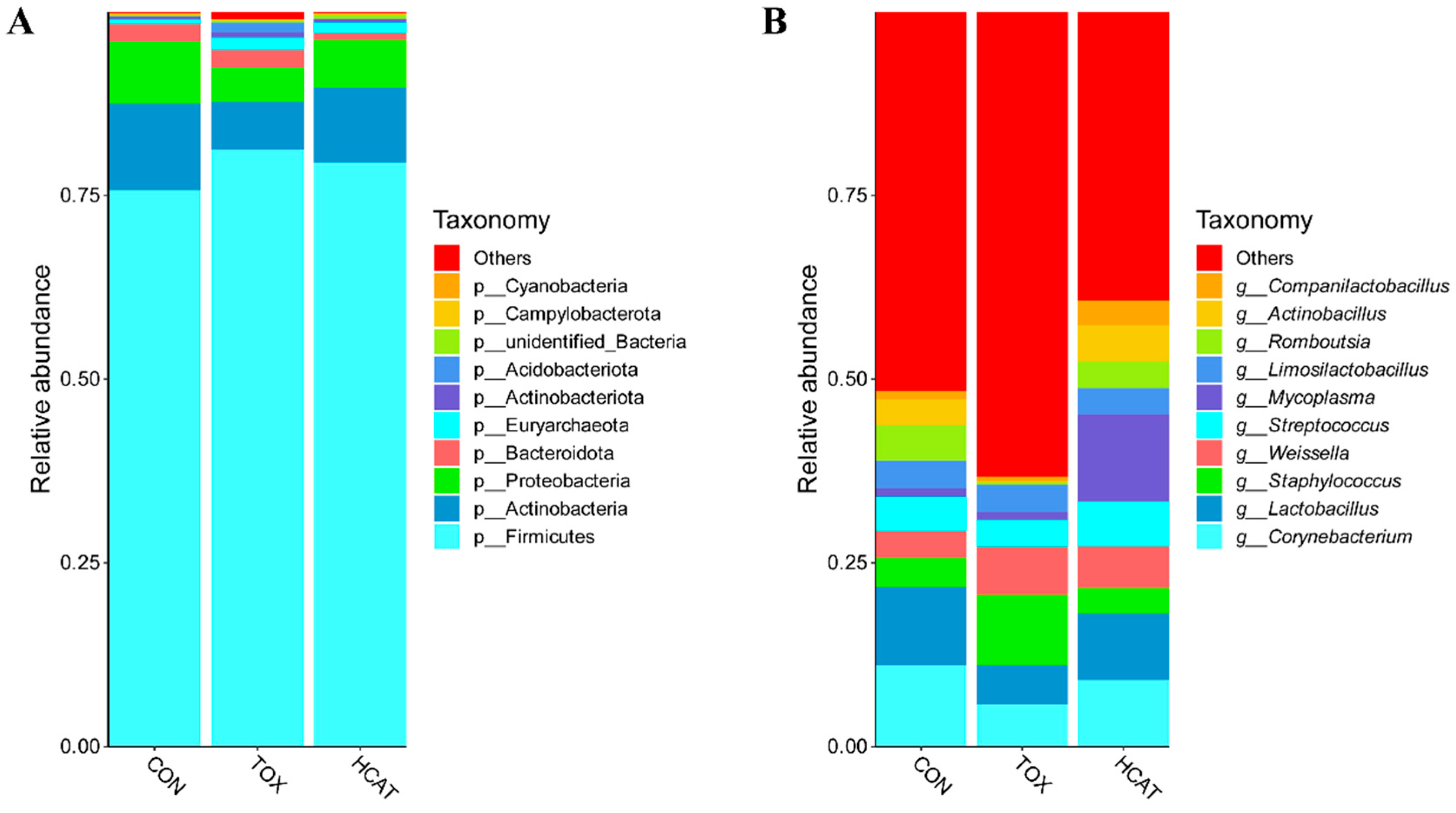
The ileum of piglets predominantly hosted the phyla Firmicutes, Actinobacteria, and Proteobacteria (Figure 4A), with Firmicutes being the most prevalent. At the genus level, the CON group had a higher relative abundance of Staphylococcus with a lower relative abundance of Lactobacillus than those in the TOX group (Figure 4B). On the contrary, the relative abundance of Staphylococcus was lower, whilst Lactobacillus was higher in the HCAT group as compared with the TOX group.
Bacterial richness (p < 0.05, LDA > 2.0) was identified by LDA combined effect size measurements (LEfSe) analysis. As depicted in Figure 5, specific bacterial members, including the order Clostridia, the family Peptostreptococcaceae, and the species Glaesserella parasuis and Enterococcus faecalis, were found to be more abundant in the CON group. Comparatively, the genus Methanobrevibacter was enriched in the TOX group, and the species Streptococcus pluranimalium was enriched in the HCAT group.

3.6. Effects of Exogenous CAT on the Metabolome of Hepatic Portal Vein Blood of FGM-Exposed Piglets

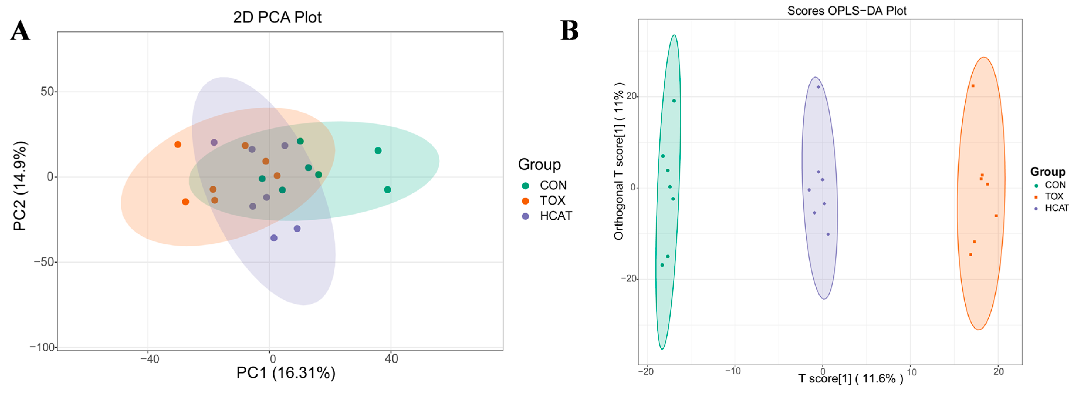
Based on the results of growth performance, the hepatic portal vein blood of piglets from the CON, TOX, and HCAT groups was selected for metabolomic analysis. Principal component analysis (PCA) and orthogonal partial least squares-discriminant analysis (OPLS-DA) were conducted on samples from the CON, TOX, and HCAT groups to assess the differences in metabolite profiles among groups. As shown in Figure 6, the samples from three groups were clustered separately, while samples from the HCAT group were closer to those from the CON group than those from the TOX group.
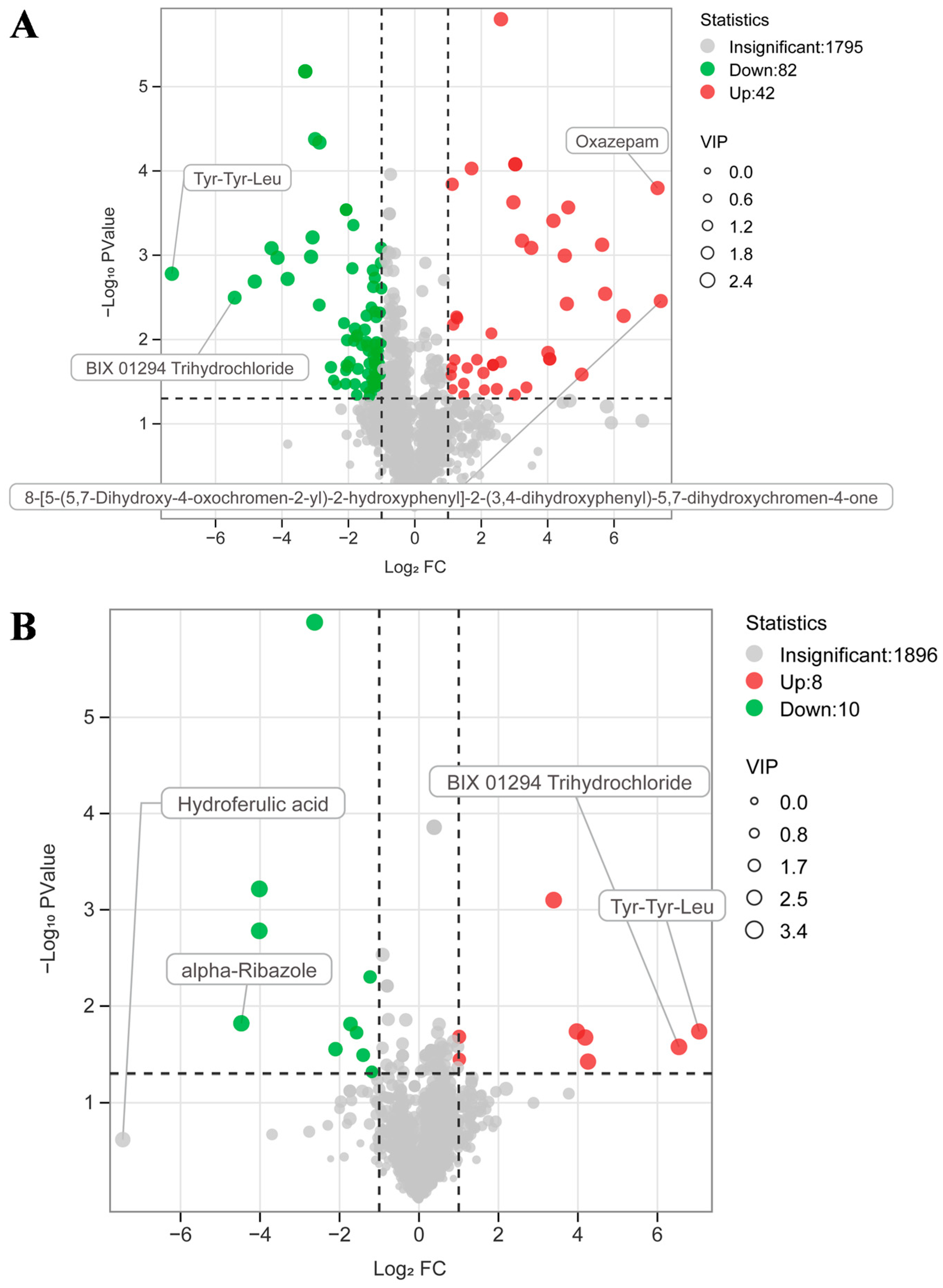
A volcano plot displays the relative differences in metabolite content between two groups and the statistically significant differences. As presented in Figure 7, the TOX group showed an increase (p < 0.05) in 42 metabolites and a decrease (p < 0.05) in 82 metabolites when contrasted with the CON group. However, the HCAT group upregulated (p < 0.05) 8 metabolites and downregulated (p < 0.05) 10 metabolites, relative to the CON group. Among them, as shown in Table S4, the levels of 6 metabolites were lower (p < 0.05) in the TOX group than those in the CON group and higher (p < 0.05) in the HCAT group than those in the TOX group. The TOX group exhibited a higher level of allolithocholate (p = 0.022) when compared to the CON group, whereas the HCAT group showed a lower (p = 0.019) in comparison to the TOX group.
As illustrated in Figure 8, in the TOX–CON comparison, the differential metabolites were primarily enriched in the choline metabolism in cancer as well as glycerophospholipid metabolism. Similarly, in the HCAT–TOX comparison, the differential metabolites were predominantly enriched in glycerophospholipid metabolism.

3.7. Effects of Exogenous CAT on Correlations Between Ileal Microbiota and Metabolome of FGM-Exposed Piglets

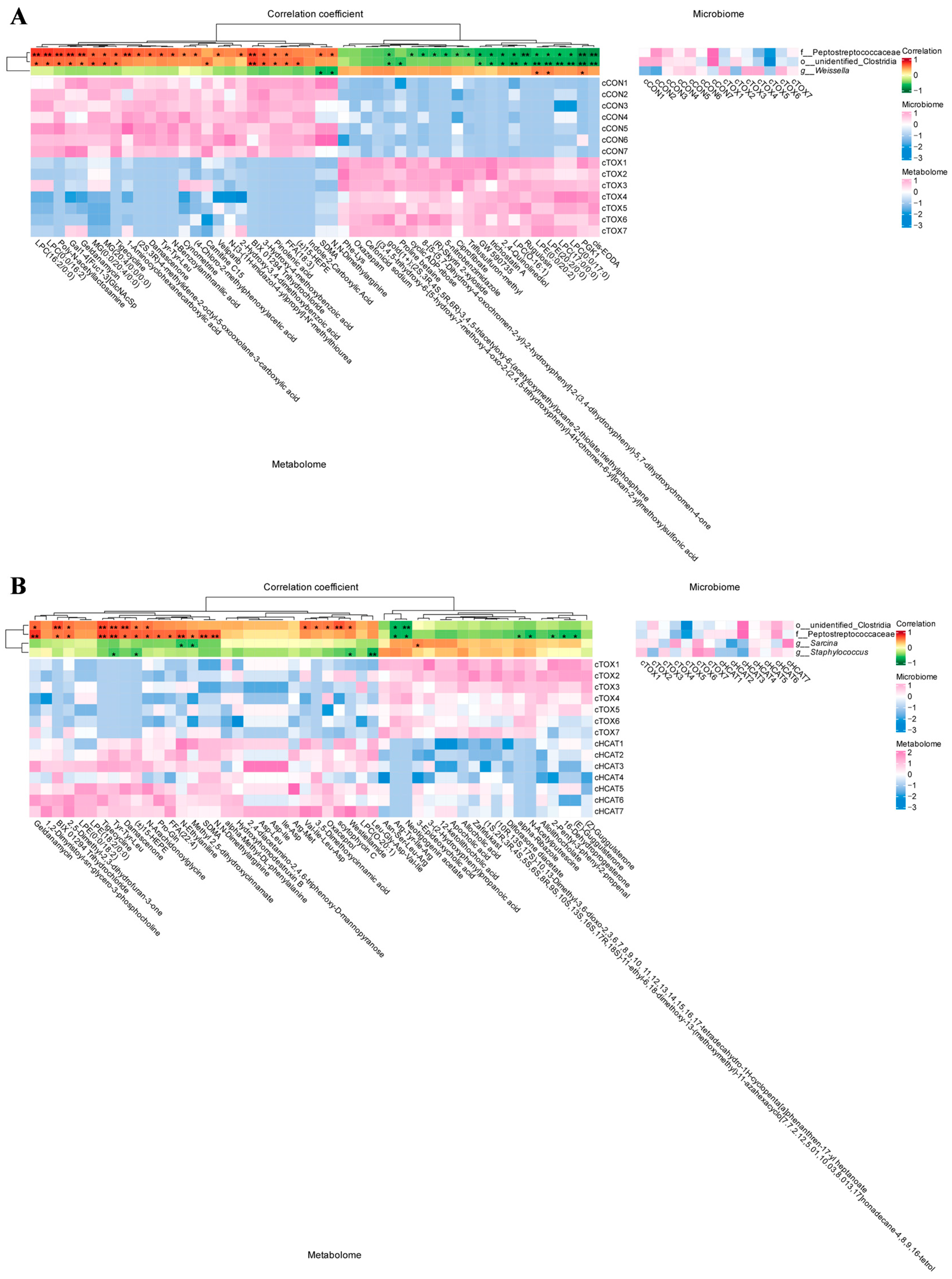
Spearman’s correlation analysis was employed to identify the relationships between ileal microbiota and metabolite profiles of hepatic portal vein blood among groups. As shown in Figure 9A, in the TOX–CON comparison, the family Peptostreptococcaceae had a significant positive correlation with 23 different metabolites such as Tyr-Tyr-Leu (p = 0.018), Damascenone (p = 0.019), Tigecycline (p = 0.013), BIX 01294 Trihydrochloride (p = 0.004), and Geldanamycin (p = 0.004), but showed significant negative correlations (p < 0.05) with 18 different metabolites. Similarly, in the HCAT–TOX comparison, the family Peptostreptococcaceae was significantly positively correlated with Tyr-Tyr-Leu (p = 0.003), (±)15-HEPE (p = 0.012), Damascenone (p = 0.015), Tigecycline (p = 0.003), BIX 01294 Trihydrochloride (p = 0.033), and Geldanamycin (p = 0.001), but displayed significant negative correlations (p < 0.05) with seven different metabolites (Figure 9B). The metabolites listed above are common differential metabolites in the TOX–CON comparison and the HCAT–TOX comparison (Table S4). The genus Staphylococcus, known as a potential pathogen, showed a significant negative correlation with Tyr-Tyr-Leu (p = 0.047) and (±)15-HEPE (p = 0.045).

4. Discussion

Mycotoxins produced by Fusarium graminearum have been shown to cause acute and chronic toxicity in animals [5]. In the present study, the contents of AFB1 and fumonisins were lower than the limit of the Chinese hygienical standard for feeds (GB 13078-2017) [28]. Thus, piglets were mainly poisoned by DON and ZEA in this study. Research on performance and toxicological effects in pigs has primarily concentrated on medium to high levels of DON, specifically ranging from 2 to 10 mg/kg of feed [5]. In contrast, the content of DON and ZEA in the FGM-contaminated diet in this study was 1.87 mg/kg and 1.91 mg/kg, respectively, belonging to the low-dose treatment.
Similarly to a previous study [29], the present study found that FGM caused growth retardation in piglets, as evidenced by the decrease in ADG, accompanied by the increased diarrhea rate and diarrhea index. It was reported that CAT can enhance intestinal antioxidant capacity and change the microbiota composition to ameliorate intestinal mucosal damage induced by lipopolysaccharide [12]. However, limited research has been carried out to investigate the impact of CAT on pigs exposed to FGM. In this study, the CAT supplementation attenuated the damage of FGM on the growth performance of piglets, which showed that 400 U/kg CAT supplementation increased ADG and ADFI, and all doses of CAT supplementation alleviated diarrhea. The organ index, which refers to the relative organ weight, serves as a common indicator for assessing health condition in piglets. According to Xiao et al. [30], dietary 4 mg/kg DON increased the indexes of kidney and spleen of piglets. However, in this study, we found no changes in the indexes of piglets, which may be related to the relatively low dose of FGM in the diet.
The resultant redox imbalance in the organs of the piglet, particularly the liver and intestine, due to FGM exposure, has been well-established [31]. It has been proposed that oxidative stress is associated with increased production of ROS in the intestine and liver [32]. On the contrary, in this study, FGM decreased the level of ROS in ileal tissue of piglets, while CAT supplementation reversed this change. Interestingly, the subsequent analysis indicated that FGM contamination increased the level of ROS in ileal chyme. It was possible that FGM resulted in the intestinal barrier damage along with cell death and rupture, subsequently allowing ROS to move from epithelial cells into the intestinal lumen. The decreased gene expression of intestinal tight junction proteins caused by FGM could lend support to this conjecture. SOD and CAT help prevent redox imbalance by specifically neutralizing superoxide anion and H2O2, respectively [10]. In this study, FGM decreased ROS in ileal tissue, increased ROS in ileal chyme, and impaired antioxidant property, which indicated an oxidative stress in piglets [4,7]. However, FGM-induced changes in ROS level in both ileal tissue and chyme were normalized by CAT supplementation, which also improved the activities of ileal SOD and CAT along with hepatic CAT of piglets exposed to FGM. These findings underscored the efficacy of exogenous CAT in mitigating oxidative stress induced by FGM in piglets.
The Nrf2/Keap1/HO-1 pathway is one of the essential mechanisms for cells to fight against oxidative stress [33]. Upon exposure to excessive free radicals, Nrf2 separates from Keap1 and translocates into the nucleus, ultimately activating the transcription of antioxidant enzymes such as CAT, HO-1, and SOD [34]. This study, like Qiu et al. [35], demonstrated that FGM suppressed the expression of antioxidation-related genes (e.g., CAT, SOD1) in the ileum of piglets. There were no notable changes in the expression of antioxidation-related genes in the liver of FGM-exposed piglets, which did not agree with the study of Ji et al. [36], who found that 3 mg/kg DON reduced the expression of hepatic CAT and SOD1 of piglets. The discrepancy might be due to the difference in the content of FGM in the diet. The CAT supplementation elevated the expression of antioxidation-related genes in the ileum and liver of piglets exposed to FGM, with higher dose of CAT being less efficient. These results demonstrated that a high dose of CAT probably mitigated FGM-induced oxidative stress by directly eliminating radical accumulation, thereby decreasing the consumption of antioxidant enzymes, rather than enhancing the expression of antioxidant enzyme genes via the Nrf2/Keap1/HO-1 pathway.
The intestine represents the first target organ of FGM [37]. As the crucial barrier structure of the intestine, tight junctions comprise various structurally unique proteins, including Occludin, Claudins, and Zonula occluden, which form a paracellular permeability barrier and preserve intestinal integrity [38]. Oxidative damage induced by FGM contamination has been proved to reduce the expression of intestinal tight junction proteins in piglets [35]. As expected, this study showed that supplemental CAT alleviated the decreases in gene expression of Occludin and Claudin-1 in the ileum and colon of piglets exposed to FGM, demonstrating that the improvement of intestinal physical barrier function by CAT addition was probably related to the observed enhancement of intestinal antioxidant capacity.
Intestinal microbiota are widely recognized for their crucial roles in regulating host growth and overall health. An increasing number of studies disclosed that oxidative damage induced by FGM contributes to the disruption of intestinal microbiota in various animal models [35,39]. Previous studies have suggested the improvement of intestinal microbiota by CAT supplementation in both piglets [12] and broilers [16]. In this study, based on the results from the assessment of growth performance, the CON, TOX, and HCAT group were selected for ileal microbiota analysis, which showed that the CAT supplementation did not cause significant changes in intestinal microbial diversity of piglets. However, significant changes were observed in ileal microbial composition, as manifested by the inhibition effect of CAT on the increased abundance of the genus Staphylococcus and the decreased abundance of the genus Lactobacillus induced by FGM. As a part of the typical cutaneous and mucosal microbiota of mammals, the genus Staphylococcus and its affiliated members (such as Staphylococcus aureus and Staphylococcus hycius) frequently serve as primary causative pathogens of infections in animals [40]. In contrast, the genus Lactobacillus, a well-established source of probiotics in the intestine, is involved in regulating intestinal epithelial barrier integrity and oxidative stress as well as improving host immune responses [41]. It was indicated that supplemental CAT improved the intestinal microbial composition in this study. Similarly to the current study, Xu et al. [42] reported the decreased abundance of genus the Lactobacillus in the intestine of pigs exposed to DON.
Alterations in the intestinal microbiota inevitably lead to corresponding changes in the metabolic profiles of the host, which have been extensively documented in a previous study [43]. The intestine and liver are linked through the portal vein and interact via several pathways [44]. Venous blood from the small and large intestine flows into the portal vein, which supplies 75% of the blood of the liver, carrying enterally absorbed nutrients as well as microbial metabolites [45]. In turn, the liver regulates the intestine by releasing bile containing bile acids and antimicrobial molecules directly into the small intestine, thereby completing the cycle by modulating the microbiota [46]. Microbial metabolites like secondary bile acids and ethanol may play a role in the onset of non-alcoholic fatty liver disease [45]. Therefore, we intended to understand the changes in metabolites from the intestine into the liver after FGM and exogenous CAT intervention. The CON, TOX, and HCAT groups were selected for metabolome analysis of hepatic portal vein blood. The results showed that samples from the HCAT group were closer to those from the CON group than those from the TOX group. Moreover, the differential metabolites were primarily enriched in glycerophospholipid metabolism due to the intervention of FGM and exogenous CAT. Among them, seven metabolites were screened from the volcano plot, including Tyr-Tyr-Leu, (±)15-HEPE, Damascenone, etc. Tyr-Tyr-Leu is a tripeptide with antioxidant activity [47]. (±)15-HEPE, which has anti-inflammatory actions [48], exhibits a negative correlation with MDA production [49]. Damascenone has been proved to benefit resistance against oxidative stress [50]. Accordingly, the above metabolites elevated by CAT addition could be deduced to improve the antioxidant capacity and improve the hepatic redox status of piglets.
Then Spearman’s correlation analysis was conducted to identify the relationships between ileal microbiota and metabolite profiles of hepatic portal vein blood among CON, TOX, and HCAT groups. The results showed that the increases in the six differential metabolites (Tyr-Tyr-Leu, (±)15-HEPE, Damascenone, Tigecycline, BIX 01294 Trihydrochloride, and Geldanamycin) were related to the increased abundance of the family Peptostreptococcaceae, whereas the increases in Tyr-Tyr-Leu and (±)15-HEPE levels were related to the decreased abundance of the genus Staphylococcus. The above results indicated that exogenous CAT changed the composition and abundance of intestinal microbiota, which could change the metabolite profiles of hepatic portal vein blood, probably alleviating the redox imbalance in the intestine and liver of piglets. However, further research regarding which metabolites are essential in the above processes deserves to be conducted.

5. Conclusions

Exogenous CAT enhanced the antioxidant capacity in both the ileum and the liver of piglets. Furthermore, supplemental 400 U/kg CAT improved the composition of intestinal microbiota and altered the metabolite profiles in hepatic portal vein blood (mainly through glycerophospholipid metabolism). These could explain the observed mitigating effects of CAT supplementation on FGM-induced impairments of piglets in growth performance and redox balance. The appropriate amount of supplemental CAT was 400 U/kg. Our findings offered a strategy to minimize the negative impacts of dietary FGM contamination on pigs.

Supplementary Materials

The following supporting information can be downloaded at https://www.mdpi.com/article/10.3390/agriculture15171892/s1: Table S1: The contents of primary mycotoxins in feed; Table S2: Assessment of the piglets’ fecal score; Table S3: Sequences for real-time PCR primers; Table S4: Common differential metabolites in the TOX–CON comparison and the HCAT–TOX comparison.

Author Contributions

Conceptualization, S.L. and J.Z.; data curation, Y.J. and C.L.; formal analysis, S.L.; methodology, H.Y. and J.Z.; validation, Q.C. and W.W.; investigation, Y.J. and M.X.; project administration, Q.C.; resources, C.Z. and Z.D.; writing—original draft, S.L. and Y.J.; writing—review and editing, S.L.; funding acquisition, J.Z.; supervision, J.Z. All authors have read and agreed to the published version of the manuscript.

Funding

This research was financially supported by the Biological Breeding-National Science and Technology Major Project (No. 2023ZD04072).

Institutional Review Board Statement

The Animal Care and Use Committee of the South China Agricultural University (China) approved the experimental animal protocols for this study (Ethics Approval Number: 2024F227/15 March 2024).

Data Availability Statement

The data presented in this study are available on request from the corresponding author.

Conflicts of Interest

The authors declare no conflicts of interest.

Abbreviations

The following abbreviations are used in this manuscript:
ADFIAverage daily feed intake
ADGAverage daily gain
AFB1Aflatoxin B1
CATCatalase
CONBasal diet
DONDeoxynivalenol
FBWFinal body weight
FCFold change
FCRFeed conversion ratio
FGMFusarium graminearum mycotoxins
GPX1Glutathione peroxidase 1
HCATFGM-contaminated diet + 400 U/kg CAT
HO-1Heme oxygenase 1
H2O2Hydrogen peroxide
IBWInitial body weight
Keap1Kelch-like ECH-associated protein 1
LCATFGM-contaminated diet + 100 U/kg CAT
LEfSeLDA combined effect size
MCATFGM-contaminated diet + 200 U/kg CAT
NQO1NAD(P)H oxidoreductase 1
Nrf2Nuclear factor erythroid-2-related factor
OPLS-DAOrthogonal partial least squares-discriminant analysis
PCAPrincipal component analysis
ROSReactive oxygen species
SODSuperoxide dismutase
TOXFGM-contaminated diet
VIPVariable importance in projection
ZEAZearalenone

References

  1. Iqbal, S.Z. Mycotoxins in food, recent development in food analysis and future challenges; a review. Curr. Opin. Food Sci. 2021, 42, 237–247. [Google Scholar] [CrossRef]
  2. Rozewicz, M.; Wyzinska, M.; Grabinski, J. The Most Important Fungal Diseases of Cereals-Problems and Possible Solutions. Agronomy 2021, 11, 714. [Google Scholar] [CrossRef]
  3. Tolosa, J.; Rodríguez-Carrasco, Y.; Ruiz, M.J.; Vila-Donat, P. Multi-mycotoxin occurrence in feed, metabolism and carry-over to animal-derived food products: A review. Food Chem. Toxicol. 2021, 158, 112661. [Google Scholar] [CrossRef]
  4. Sun, H.Y.; He, Z.Q.; Xiong, D.W.; Long, M. Mechanisms by which microbial enzymes degrade four mycotoxins and application in animal production: A review. Anim. Nutr. 2023, 15, 256–274. [Google Scholar] [CrossRef] [PubMed]
  5. Escrivá, L.; Font, G.; Manyes, L. In vivo toxicity studies of Fusarium mycotoxins in the last decade: A review. Food Chem. Toxicol. 2015, 78, 185–206. [Google Scholar] [CrossRef]
  6. Sun, Y.; Jiang, J.; Mu, P.Q.; Lin, R.Q.; Wen, J.K.; Deng, Y.Q. Toxicokinetics and metabolism of deoxynivalenol in animals and humans. Arch. Toxicol. 2022, 96, 2639–2654. [Google Scholar] [CrossRef]
  7. Ren, Z.H.; He, H.Y.; Zuo, Z.C.; Xu, Z.W.; Wei, Z.Y.; Deng, J.L. ROS: Trichothecenes’ handy weapon? Food Chem. Toxicol. 2020, 142, 111438. [Google Scholar] [CrossRef] [PubMed]
  8. MatÉs, J.M.; Pérez-Gómez, C.; De Castro, I. Antioxidant enzymes and human diseases. Clin. Biochem. 1999, 32, 595–603. [Google Scholar] [CrossRef] [PubMed]
  9. Veal, E.A.; Day, A.M.; Morgan, B.A. Hydrogen peroxide sensing and signaling. Mol. Cell 2007, 26, 1–14. [Google Scholar] [CrossRef] [PubMed]
  10. Zamocky, M.; Furtmüller, P.G.; Obinger, C. Evolution of catalases from bacteria to humans. Antioxid. Redox Signal. 2008, 10, 1527–1548. [Google Scholar] [CrossRef]
  11. Ali, S.S.; Ahsan, H.; Zia, M.K.; Siddiqui, T.; Khan, F.H. Understanding oxidants and antioxidants: Classical team with new players. J. Food Biochem. 2020, 44, e13145. [Google Scholar] [CrossRef]
  12. Chen, J.L.; Li, F.C.; Yang, W.R.; Jiang, S.Z.; Li, Y. Supplementation with exogenous catalase from Penicillium notatum in the diet ameliorates lipopolysaccharide-induced intestinal oxidative damage through affecting intestinal antioxidant capacity and microbiota in weaned pigs. Microbiol. Spectr. 2021, 9, e00654. [Google Scholar] [CrossRef]
  13. Robert, H.; Payros, D.; Pinton, P.; Théodorou, V.; Mercier-Bonin, M.; Oswald, I.P. Impact of mycotoxins on the intestine: Are mucus and microbiota new targets? Toxicol. Environ. Health Part B 2017, 20, 249–275. [Google Scholar] [CrossRef] [PubMed]
  14. Fan, J.P.; Zhang, Y.H.; Zuo, M.Y.; Ding, S.X.; Li, J.J.; Feng, S.K.; Xiao, Y.P.; Tao, S.Y. Novel mechanism by which extracellular vesicles derived from Lactobacillus murinus alleviates deoxynivalenol-induced intestinal barrier disruption. Environ. Int. 2024, 185, 108525. [Google Scholar] [CrossRef]
  15. Liu, Y.H.; Li, J.Y.; Kang, W.L.; Liu, S.P.; Liu, J.Y.; Shi, M.D.; Wang, Y.B.; Liu, X.J.; Chen, X.X.; Huang, K.H. Aflatoxin B1 induces liver injury by disturbing gut microbiota-bile acid-FXR axis in mice. Food Chem. Toxicol. 2023, 176, 113751. [Google Scholar] [CrossRef] [PubMed]
  16. Wang, W.W.; Zhu, J.Q.; Cao, Q.Y.; Zhang, C.M.; Dong, Z.M.; Feng, D.Y.; Ye, H.; Zuo, J.J. Dietary catalase supplementation alleviates deoxynivalenol-induced oxidative stress and gut microbiota dysbiosis in broiler chickens. Toxins 2022, 14, 830. [Google Scholar] [CrossRef]
  17. GB/T 39235-2020; Nutrient Requirements of Swine. Standards Press of China: Beijing, China, 2020.
  18. GB/T 6432-2018; Determination of Crude Protein in Feeds—Kjeldahl method. Standards Press of China: Beijing, China, 2018.
  19. GB/T 6436-2018; Determination of Calcium in Feeds. Standards Press of China: Beijing, China, 2018.
  20. GB/T 6437-2018; Determination of Phosphorus in Feeds—Spectrophotometry. Standards Press of China: Beijing, China, 2018.
  21. GB/T 6438-2007; Animal Feeding Stuffs—Determination of Crude Ash. Standards Press of China: Beijing, China, 2007.
  22. GB/T 20806-2006; Determination of Neutral Detergent Fiber in Feedstuffs. Standards Press of China: Beijing, China, 2006.
  23. NY/T 1459-2022; Determination of Acid Detergent Fiber in Feed. China Agriculture Press: Beijing, China, 2022.
  24. Li, M.; Sun, M.N.; Hong, X.; Duan, J.S.; Du, D.L. Survey of deoxynivalenol contamination in agricultural products in the Chinese market using an ELISA kit. Toxins 2019, 11, 6. [Google Scholar] [CrossRef]
  25. Marquis, V.; Schulthess, J.; Molist, F.; Santos, R.R. Effect of a Yeast β-Glucan on the performance, intestinal integrity, and liver function of broiler chickens fed a diet naturally contaminated with Fusarium mycotoxins. Toxins 2025, 17, 51. [Google Scholar] [CrossRef]
  26. Huang, S.B.; Cui, Z.J.; Hao, X.Y.; Cheng, C.H.; Chen, J.Z.; Wu, D.Y.; Luo, H.F.; Deng, J.P.; Tan, C.Q. Dietary fibers with low hydration properties exacerbate diarrhea and impair intestinal health and nutrient digestibility in weaned piglets. J. Anim. Sci. Biotechnol. 2022, 13, 142. [Google Scholar] [CrossRef] [PubMed]
  27. Jiang, X.Y.; Ma, Y.; Wang, T.; Zhou, H.N.; Wang, K.S.; Shi, W.G.; Qin, L.; Guan, J.H.; Li, L.S.; Long, B.; et al. Targeting UBE2T potentiates gemcitabine efficacy in pancreatic cancer by regulating pyrimidine metabolism and replication stress. Gastroenterology 2023, 164, 1232–1247. [Google Scholar] [CrossRef]
  28. GB 13078-2017; Hygienical Standard for Feeds. Standards Press of China: Beijing, China, 2017.
  29. Wu, C.M.; Song, J.P.; Liu, X.Y.; Zhang, Y.W.; Zhou, Z.Y.; Thomas, D.G.; Wu, B.; Yan, X.R.; Li, J.; Zhang, R.N.; et al. Effect of iron-manganese oxide on the degradation of deoxynivalenol in feed and enhancement of growth performance and intestinal health in weaned piglets. Ecotoxicol. Environ. Saf. 2024, 286, 117246. [Google Scholar] [CrossRef]
  30. Xiao, H.; Wu, M.M.; Tan, B.E.; Yin, Y.L.; Li, T.J.; Xiao, D.F.; Li, L. Effects of composite antimicrobial peptides in weanling piglets challenged with deoxynivalenol: I. growth performance, immune function, and antioxidation capacity1. J. Anim. Sci. 2013, 91, 4772–4780. [Google Scholar] [CrossRef]
  31. Zheng, W.J.; Ji, X.; Zhang, Q.; Du, W.C.; Wei, Q.W.; Yao, W. Hydrogen-rich water and lactulose protect against growth suppression and oxidative stress in female piglets fed Fusarium toxins contaminated diets. Toxins 2018, 10, 228. [Google Scholar] [CrossRef]
  32. Wang, K.; Tang, Y.; Wu, X.; Liang, H.M.; Chen, D.W.; Yu, B.; He, J.; Mao, X.B.; Huang, Z.Q.; Yan, H.; et al. Eugenol attenuates transmissible gastroenteritis virus-induced oxidative stress and apoptosis via ROS-NRF2-ARE signaling. Antioxidants 2022, 11, 1838. [Google Scholar] [CrossRef] [PubMed]
  33. Magesh, S.; Chen, Y.; Hu, L.Q. Small molecule modulators of Keap1-Nrf2-ARE pathway as potential preventive and therapeutic agents. Med. Res. Rev. 2012, 32, 687–726. [Google Scholar] [CrossRef]
  34. Yamada, M.; Iwata, M.; Warabi, E.; Oishi, H.; Lira, V.A.; Okutsu, M. p62/SQSTM1 and Nrf2 are essential for exercise-mediated enhancement of antioxidant protein expression in oxidative muscle. FASEB J. 2019, 33, 8022–8032. [Google Scholar] [CrossRef]
  35. Qiu, Y.Q.; Yang, J.; Wang, L.; Yang, X.F.; Gao, K.G.; Zhu, C.; Jiang, Z.Y. Dietary resveratrol attenuation of intestinal inflammation and oxidative damage is linked to the alteration of gut microbiota and butyrate in piglets challenged with deoxynivalenol. J. Anim. Sci. Biotechnol. 2021, 12, 71. [Google Scholar] [CrossRef]
  36. Ji, X.; Tang, Z.Q.; Zhang, F.; Zhou, F.; Wu, Y.J.; Wu, D. Dietary taurine supplementation counteracts deoxynivalenol-induced liver injury via alleviating oxidative stress, mitochondrial dysfunction, apoptosis, and inflammation in piglets. Ecotoxicol. Environ. Saf. 2023, 253, 114705. [Google Scholar] [CrossRef] [PubMed]
  37. Tu, Y.; Liu, S.Q.; Cai, P.R.; Shan, T.Z. Global distribution, toxicity to humans and animals, biodegradation, and nutritional mitigation of deoxynivalenol: A review. Compr. Rev. Food Sci. Food Saf. 2023, 22, 3951–3983. [Google Scholar] [CrossRef]
  38. Schoultz, I.; Keita, Å.V. The intestinal barrier and current techniques for the assessment of gut permeability. Cells 2020, 9, 1909. [Google Scholar] [CrossRef]
  39. Zhang, J.; Liu, H.; Shen, Y.; Cheng, D.; Tang, H.; Zhang, Q.; Li, C.; Liu, M.; Yao, W.H.; Ran, R.R.; et al. Macrophage AHR-TLR4 cross-talk drives p-STAT3 (Ser727)-mediated mitochondrial oxidative stress and upregulates IDO/ICAM-1 in the steatohepatitis induced by aflatoxin B1. Sci. Total Environ. 2024, 923, 171377. [Google Scholar] [CrossRef]
  40. Morris, D.O.; Loeffler, A.; Davis, M.F.; Guardabassi, L.; Weese, J.S. Recommendations for approaches to meticillin-resistant staphylococcal infections of small animals: Diagnosis, therapeutic considerations and preventative measures. Vet. Dermatol. 2017, 28, 304. [Google Scholar] [CrossRef]
  41. Rastogi, S.; Singh, A. Gut microbiome and human health: Exploring how the probiotic genus Lactobacillus modulate immune responses. Front. Pharmacol. 2022, 13, 1042189. [Google Scholar] [CrossRef] [PubMed]
  42. Xu, X.X.; Chang, J.; Wang, P.; Liu, C.Q.; Liu, M.J.; Zhou, T.; Yin, Q.Q.; Yan, G.R. Glycyrrhizic acid and compound probiotics supplementation alters the intestinal transcriptome and microbiome of weaned piglets exposed to deoxynivalenol. Toxins 2022, 14, 856. [Google Scholar] [CrossRef] [PubMed]
  43. Geng, S.J.; Cheng, S.S.; Li, Y.; Wen, Z.S.; Ma, X.; Jiang, X.M.; Wang, Y.Z.; Han, X.Y. Faecal microbiota transplantation reduces susceptibility to epithelial injury and modulates tryptophan metabolism of the microbial community in a piglet model. J. Crohns Colitis 2018, 12, 1359–1374. [Google Scholar] [CrossRef] [PubMed]
  44. Tilg, H.; Adolph, T.E.; Trauner, M. Gut-liver axis: Pathophysiological concepts and clinical implications. Cell Metab. 2022, 34, 1700–1718. [Google Scholar] [CrossRef]
  45. Albillos, A.; De Gottardi, A.; Rescigno, M. The gut-liver axis in liver disease: Pathophysiological basis for therapy. J. Hepatol. 2020, 72, 558–577. [Google Scholar] [CrossRef]
  46. Chen, M.J.; Liu, C.; Wan, Y.; Yang, L.; Jiang, S.; Qian, D.W.; Duan, J.A. Enterohepatic circulation of bile acids and their emerging roles on glucolipid metabolism. Steroids 2021, 165, 108757. [Google Scholar] [CrossRef]
  47. Ningrum, A.; Wardani, D.W.; Vanidia, N.; Manikhaeda; Sarifudin, A.; Kumalasari, R.; Ekafitri, R.; Kristanti, D.; Setiaboma, W.; Munawaroh, H.S.H. Evaluation of antioxidant activities from a sustainable source of okara protein hydrolysate using enzymatic reaction. Molecules 2023, 28, 4974. [Google Scholar] [CrossRef]
  48. Rohwer, N.; Chiu, C.Y.; Huang, D.; Smyl, C.; Rothe, M.; Rund, K.M.; Schebb, N.H.; Kühn, H.; Weylandt, K.H. Omega-3 fatty acids protect from colitis via an Alox15-derived eicosanoid. FASEB J. 2021, 35, e21491. [Google Scholar] [CrossRef]
  49. Zhu, Z.Y.; Zhou, W.J.; Yang, Y.; Wang, K.; Li, F.H.; Dang, Y.Q. Quantitative profiling of oxylipin reveals the mechanism of pien-tze-huang on alcoholic liver disease. Evid. Based Complement. Alternat. Med. 2021, 2021, 1–14. [Google Scholar] [CrossRef]
  50. Ma, Q.L.; Xiong, Y.N.; Jie, Z.W.; Li, C.Z.; Wang, C.Y.; Cai, J.W.; Zhang, Y.C.; Li, J.H.; Yao, Y.H.; Chang, M.Z.; et al. Damascenone inhibits osteoclastogenesis by epigenetically modulating Nrf2-mediated ROS scavenge and counteracts OVX-induced osteoporosis. Phytomedicine 2024, 135, 156205. [Google Scholar] [CrossRef] [PubMed]
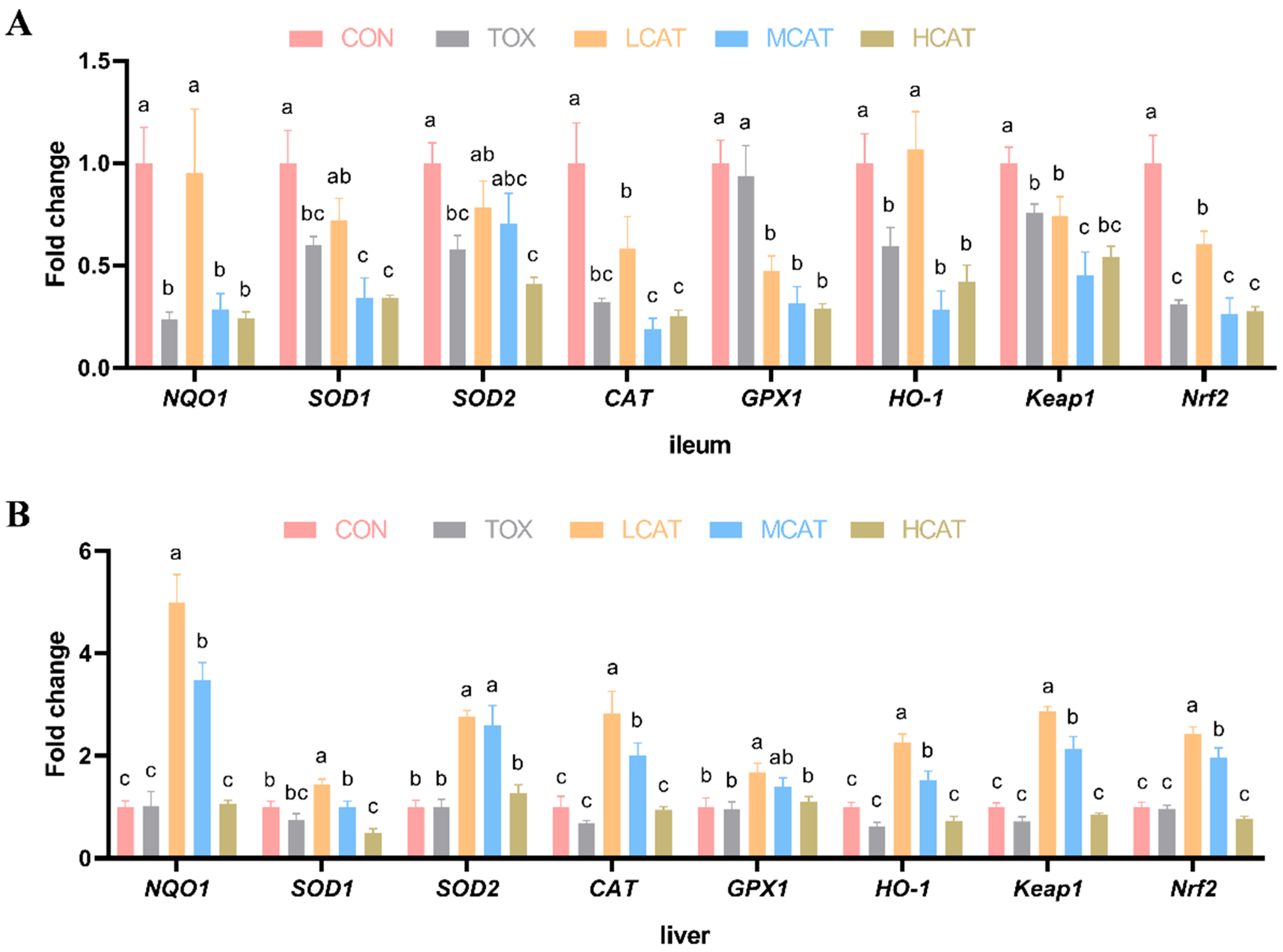
Figure 1. Effects of exogenous CAT on the relative mRNA expression of antioxidation-related genes in the ileum (A) and liver (B) of piglets exposed to FGM. Data are presented as mean ± SEM (n = 8). a–c Values with different superscript letters differ significantly (p < 0.05). NQO1 = NAD(P)H oxidoreductase 1; SOD = superoxide dismutase; CAT = catalase; GPX1 = glutathione peroxidase 1; HO-1 = heme oxygenase 1; Keap1 = Kelch-like ECH-associated protein 1; Nrf2 = nuclear factor erythroid-2-related factor. CON = basal diet; TOX = FGM-contaminated diet; LCAT = FGM-contaminated diet + 100 U/kg CAT; MCAT = FGM-contaminated diet + 200 U/kg CAT; HCAT = FGM-contaminated diet + 400 U/kg CAT.
Agriculture 15 01892 g001
Figure 2. Effects of exogenous CAT on relative mRNA expression of ileal (A) and colonic (B) tight junction proteins of piglets exposed to FGM. Data are presented as mean ± SEM (n = 8). a–c Values with different superscript letters differ significantly (p < 0.05). CON = basal diet; TOX = FGM-contaminated diet; LCAT = FGM-contaminated diet + 100 U/kg CAT; MCAT = FGM-contaminated diet + 200 U/kg CAT; HCAT = FGM-contaminated diet + 400 U/kg CAT.
Agriculture 15 01892 g002
Figure 3. α-diversity and β-diversity of ileal microbiota among groups (n = 7). (A) α-diversity, including the Shannon index, Chao1 index, Simpson index, and ACE index. (B) β-diversity, including PCoA and NMDS analysis. PCoA = principal coordinate analysis; NMDS = non-metric multi-dimensional scaling. CON = basal diet; TOX = FGM-contaminated diet; HCAT = FGM-contaminated diet + 400 U/kg CAT.
Agriculture 15 01892 g003
Figure 4. Ileal microbial composition of piglets at phylum (A) and genus (B) levels (n = 7). CON = basal diet; TOX = FGM-contaminated diet; HCAT = FGM-contaminated diet + 400 U/kg CAT.
Agriculture 15 01892 g004
Figure 5. LEfSe analysis of bacterial richness (p < 0.05, LDA > 2.0) in ileal microbiota of piglets, including the cladogram (A) and LDA score distribution histogram (B) (n = 7). CON = basal diet; TOX = FGM-contaminated diet; HCAT = FGM-contaminated diet + 400 U/kg CAT.
Agriculture 15 01892 g005
Figure 6. Analysis of PCA (A) and OPLS-DA (B) scatter plots of piglets exposed to FGM (n = 7). PCA = principal component analysis; OPLS-DA = orthogonal partial least squares discriminant analysis. CON = basal diet; TOX = FGM-contaminated diet; HCAT = FGM-contaminated diet + 400 U/kg CAT.
Agriculture 15 01892 g006
Figure 7. Analysis of the volcano plot of piglets exposed to FGM (n = 7) by the standard of variable importance in projection (VIP) > 1, p-value of t-test < 0.05, and Log2FC (fold change) > 2. The TOX and CON comparison (A) as well as the HCAT and TOX comparison (B) were shown. CON = basal diet; TOX = FGM-contaminated diet; HCAT = FGM-contaminated diet + 400 U/kg CAT.
Agriculture 15 01892 g007
Figure 8. Kyoto encyclopedia of genes and genomes (KEGG) pathway enrichment analysis of piglets exposed to FGM (n = 7). Comparisons between the TOX and CON group (A) as well as the HCAT and TOX group (B) are shown. CON = basal diet; TOX = FGM-contaminated diet; HCAT = FGM-contaminated diet + 400 U/kg CAT.
Agriculture 15 01892 g008
Figure 9. Heatmap of the correlation between ileal microbiota and differential metabolites of hepatic portal vein blood (n = 7). The TOX and CON comparison (A) as well as the HCAT and TOX comparison (B) are shown. CON = basal diet; TOX = FGM-contaminated diet; HCAT = FGM-contaminated diet + 400 U/kg CAT. * Significant difference (p < 0.05); ** highly significant difference (p < 0.01).
Agriculture 15 01892 g009
Table 1. Composition of the basal diet (air-dry basis).
IngredientsContent (%)Nutrient LevelsContent (%)
Corn57.83Analyzed nutrient level
Soybean meal18.60Crude protein18.69
Soybean oil1.40Organic matter80.94
Extruded soybean meal8.00Neutral detergent fiber7.12
Whey powder10.00Acid detergent fiber3.60
Salt0.32Calcium0.61
Limestone0.50Total phosphorus0.55
Dicalcium phosphate1.23Calculated nutrient level
Choline chloride (50%)0.16Metabolizable energy, MJ/kg13.81
L-lysine (99%)0.58Total lysine1.30
DL-methionine (98%)0.17Total methionine + cystine0.73
L-threonine (98.5%)0.17Total tryptophan0.23
L-tryptophan (98%)0.05Total threonine0.83
Premix (1)1.00
Total100.00
(1) Supplied per kilogram of diet: vitamin B1, 1.25 mg; vitamin B2, 5 mg; vitamin B6, 2.5 mg; vitamin B12, 0.0125 mg; vitamin A, 5000 IU; vitamin D3, 2000 IU; vitamin E, 37.5 mg; vitamin K3, 3 mg; niacin, 25 mg; pantothenic acid, 12.5 mg; folic acid, 2.5 mg; biotin, 0.25 mg; Cu, 35 mg; Fe, 120 mg; Zn, 80 mg; Mn, 65 mg; I, 0.3 mg; Se, 0.3 mg; Co, 0.15 mg.
Table 2. Effects of exogenous CAT on the growth performance of piglets exposed to FGM (n = 8).
ItemsCON (1)TOXLCATMCATHCATSEMp-Value (2)
TreatmentLinearQuadratic
IBW (kg)7.937.897.897.917.910.1131.0000.9430.997
FBW (kg)17.0715.5416.9517.0818.850.3120.0670.0090.035
ADG (g)332.37 ab268.36 b314.22 ab327.05 ab381.96 a9.9450.0390.0040.016
ADFI (g)559.08 ab462.29 b555.74 ab549.03 ab695.91 a14.1460.0020.0010.003
FCR1.691.751.771.681.830.0260.3870.7620.445
Diarrhea rate (%)12.95 c30.61 a24.49 ab16.96 b16.07 b2.533<0.001//
Diarrhea index0.50 c0.99 a0.76 b0.65 b0.58 b0.059<0.001//
SEM = pooled standard error of the mean; IBW = initial body weight; FBW = final body weight; ADG = average daily gain; ADFI = average daily feed intake; FCR = feed conversion ratio. (1) CON = basal diet; TOX = FGM-contaminated diet; LCAT = FGM-contaminated diet + 100 U/kg CAT; MCAT = FGM-contaminated diet + 200 U/kg CAT; HCAT = FGM-contaminated diet + 400 U/kg CAT. (2) Treatment: treatment effect according to Duncan’s test; Linear and Quadratic: linear and quadratic dose trend-effects of exogenous CAT at 0, 100, 200, and 400 U/kg according to polynomial contrasts. a–c Means within a row with different superscripts differ (p < 0.05).
Table 3. Effects of exogenous CAT on the organ indexes of piglets exposed to FGM (n = 8).
ItemsCON (1)TOXLCATMCATHCATSEMp-Value (2)
TreatmentLinearQuadratic
Liver index (g/kg)27.7626.8225.6827.8329.140.3490.1100.0440.066
Thymus index (g/kg)2.592.542.082.972.550.0990.1700.4620.760
Spleen index (g/kg)2.092.071.832.092.140.0380.4600.2270.214
SEM = pooled standard error of the mean. (1) CON = basal diet; TOX = FGM-contaminated diet; LCAT = FGM-contaminated diet + 100 U/kg CAT; MCAT = FGM-contaminated diet + 200 U/kg CAT; HCAT = FGM-contaminated diet + 400 U/kg CAT. (2) Treatment: treatment effect according to Duncan’s test; Linear and Quadratic: linear and quadratic dose-trend effects of exogenous CAT at 0, 100, 200, and 400 U/kg according to polynomial contrasts.
Table 4. Effects of exogenous CAT on antioxidant parameters of piglets exposed to FGM (n = 8).
ItemsCON (1)TOXLCATMCATHCATSEMp-Value (2)
TreatmentLinearQuadratic
ileal tissue
ROS1.00 a0.74 b0.93 a0.90 a0.93 a0.0180.0060.0260.021
H2O2 (mmol/g prot)8.6910.478.359.419.430.3150.3010.6330.349
SOD (U/mg prot)23.2722.3422.4920.5621.880.2720.0570.2080.288
CAT (U/mg prot)7.35 a3.44 c4.21 bc4.90 b3.65 c0.115<0.0010.2800.002
ileal chyme
ROS1.00 b1.95 a0.98 b1.17 b1.17 b0.0860.0180.0680.026
H2O2 (mmol/g prot)1.112.611.932.411.970.1750.0720.4080.661
liver
ROS1.001.060.941.001.000.0400.9400.9070.944
H2O2 (mmol/g prot)2.834.283.933.553.220.2040.2140.0860.237
SOD (U/mg prot)80.61 c88.56 bc98.14 ab108.04 a105.10 a1.600<0.0010.0050.006
CAT (U/mg prot)52.80 c48.20 c42.03 c127.95 b154.94 a2.930<0.001<0.001<0.001
SEM = pooled standard error of the mean; ROS = reactive oxygen species; H2O2 = hydrogen peroxide; SOD = superoxide dismutase; CAT = catalase. (1) CON = basal diet; TOX = FGM-contaminated diet; LCAT = FGM-contaminated diet + 100 U/kg CAT; MCAT = FGM-contaminated diet + 200 U/kg CAT; HCAT = FGM-contaminated diet + 400 U/kg CAT. (2) Treatment: treatment effect according to Duncan’s test; Linear and Quadratic: linear and quadratic dose trend-effects of exogenous CAT at 0, 100, 200, and 400 U/kg according to polynomial contrasts. a–c Means within a row with different superscripts differ (p < 0.05).
Disclaimer/Publisher’s Note: The statements, opinions and data contained in all publications are solely those of the individual author(s) and contributor(s) and not of MDPI and/or the editor(s). MDPI and/or the editor(s) disclaim responsibility for any injury to people or property resulting from any ideas, methods, instructions or products referred to in the content.