Article

Establishment and Application of Loop-Mediated Isothermal Amplification Assays for Pathogens of Rice Bakanae Disease

1
College of Plant Protection, Shenyang Agricultural University, Shenyang 110866, China
2
Donggang City Demonstration and Reproduction Farm, Dandong 118322, China
3
Liaoning Rice Research Institute, Shenyang 110101, China
*
Authors to whom correspondence should be addressed.
Agriculture 2025, 15(12), 1319; https://doi.org/10.3390/agriculture15121319
Submission received: 13 March 2025 / Revised: 24 May 2025 / Accepted: 28 May 2025 / Published: 19 June 2025

Abstract

:
Rice bakanae disease (RBD), a major threat in rice-cropping nations, can reduce rice yield and quality. As it is a seed-borne disease, effective seed detection is crucial. Loop-mediated isothermal amplification (LAMP) can rapidly and specifically amplify DNA at a constant temperature with high sensitivity. This research uses LAMP to develop rapid RBD pathogen detection systems. Primers were designed targeting the NRPS31 gene of Fusarium fujikuroi and conserved TEF1α sequences of Fusarium asiaticum, Fusarium proliferatum, and Fusarium andiyazi. These reactions at 60 °C for 60 min had a detection limit of 100 pg·μL−1, and LAMP proved applicable in field trials.

1. Introduction

Rice (Oryza sativa L.) plays an indispensable role in food security, economic development, and cultural heritage. As a staple food crop, rice not only provides essential nutrition for humans but also offers significant economic value through its by-products. Throughout its growth stages from sowing to maturity, rice is highly susceptible to infections by various pathogens. Once infected, rice may suffer from reduced healthy panicle numbers, underdeveloped grains, and consequently, degraded rice quality [1]. With the continuous growth of the global population and the improvement of economic standards, the global demand for rice and its quality requirements have both increased [2]. Therefore, effectively addressing rice disease infections and enhancing rice yield and quality have become critical issues for future agricultural development.
Rice bakanae disease (RBD) is extensively prevalent in rice-growing regions globally, with notable occurrences in Asia, North America, Africa, South America, and certain parts of Europe [3]. The principal symptoms of RBD include a lightening of the rice stem color, abnormal seedling growth, reverse-growing fibrous roots, and the production of white frost-like mildew layers. In severe instances, it can cause plant death, thereby affecting seed germination, plant growth, and ultimately resulting in yield losses [4]. Nevertheless, the manifestation of symptoms and the magnitude of losses associated with RBD can vary depending on diverse geographical locations, growth temperatures, and rice cultivars [5]. RBD is mainly disseminated through seeds, yet it can also spread via soil and plant residues. Over time, the number of pathogens in the soil and their infestation capacity tend to decline gradually [6]. This disease is a single-cycle one and can be transmitted by environmental factors such as wind and rain [7].
RBD can be induced by one or multiple Fusarium species [8], such as Fusarium fujikuroi, Fusarium proliferatum, Fusarium verticillioides, Fusarium andiyazi, and Fusarium asiaticum [9]. Fusarium fungi generate mycotoxins. Once animals and humans ingest grains contaminated with these mycotoxins, their health will be affected. Toxins like fumonisins and trichothecene toxins can lead to a wide range of diseases, including esophageal cancer, cerebral leukodystrophy, pulmonary edema, and various types of cancer [10]. Gibberellins (GA3), an important phytohormone, is also a secondary metabolite produced by Fusarium spp. [11]. The abnormal seedling growth caused by RBD is mainly induced by GA3 secreted by the fungus, which contributes to the study of the fungus’s virulence mechanism [12].
The detection methods of RBD can be broadly classified into two main categories: morphological identification and molecular biology identification [13]. The classification of bacterial species based on phenotypic characteristics is the most commonly used traditional method in fungal identification. In morphological identification, the pathogens are first isolated using selective media. Subsequently, the genus of the pathogens is identified based on a series of morphological indicators. Finally, the pathogenicity of the pathogens is determined by applying Koch’s postulates. However, this method is time-consuming. Moreover, due to the similar morphological and biological characteristics of various Fusarium spp., it is rather difficult to distinguish them [14]. Common molecular biology detection techniques include the Polymerase Chain Reaction (PCR), the fundamental principle of which relies on the thermal denaturation property of DNA. Through multi-step cyclic processes, DNA polymerase utilizes four deoxyribonucleotides as substrates to synthesize new DNA strands at optimal temperatures [15]. Additionally, the Quantitative Real-time PCR (qPCR) technique is widely employed. Built upon traditional PCR, qPCR integrates fluorescent signal monitoring to track the dynamic progression of PCR reactions, thereby enabling quantitative analysis of target DNA [16].
LAMP is a novel nucleic-acid amplification technique that has witnessed rapid development in recent years [17,18]. Four specific primers, namely two internal primers (F3/B3) and two external primers (FIP/BIP), are designed for six regions of the target gene [19,20]. Among these, the external primers F3/B3 can also serve as conventional PCR primers [21]. The LAMP system utilizes Bst DNA polymerase. It amplifies the target fragment at a constant temperature (60–65 °C), with the 3′ end of the outer-primer segment acting as the replication start-point [22]. Compared with other molecular-biology techniques, the LAMP assay has been widely applied in the detection of bacteria, viruses, parasites, and phytopathogenic fungi. This is attributed to its numerous advantages, such as simplicity, rapidity, high sensitivity, and specificity. The LAMP technique operates under isothermal conditions, which reduces the need for expensive and complex instruments during testing. This reaction exhibits high tolerance to pathogens and other substances, minimizing the risk of false-negative results caused by enzyme inactivation. Typically completed within 1 h, LAMP features simple procedures and low requirements for template DNA quality, making it well-suited for on-site detection. Without the need to invest substantial time and financial resources, this assay can meet the requirements for the rapid on-site detection of pathogenic bacteria. As a result, it enables the timely monitoring of disease occurrence and the effective control of the spread of pathogenic bacteria and disease epidemics [23,24,25].
RBD is caused by several species of GFSC. In thisstudy, Loop-Mediated Isothermal Amplification (LAMP) detection techniques were developed for Fusarium fujikuroi, Fusarium asiaticum, Fusarium proliferatum, and Fusarium andiyazi, respectively. These developments provide more comprehensive coverage of the RBD pathogens. Subsequently, these assays were applied to seed and other field-based detections.

2. Materials and Methods

2.1. Experimental Materials

Source of Isolates: Fusarium fujikuroi, Fusarium asiaticum, Fusarium proliferatum, and Fusarium andiyazi were obtained from diseased rice collected from major rice-producing areas in Liaoning province in 2023. All of the aforementioned strains were identified through morphological, pathogenicity, and molecular biology identifications for ITS, TEF-1α, and TUB sequences (Figure 1).
Rice Seed Varieties: Fifty-five major rice cultivars were collected in 2024 from Liaoning, Henan, and Jilin. The collected seeds were sorted, packaged, and stored for two experiments: detecting rice bakanae disease pathogens naturally present in mature rice seeds and testing seeds artificially inoculated with rice bakanae disease pathogens.

2.2. Fungal DNA Extraction

Fusarium fujikuroi, Fusarium asiaticum, Fusarium proliferatum, Fusarium andiyazi, and other Fusarium strains were cultured on potato dextrose agar medium (PDA) at 28 °C for 4–6 days [26,27]. Several fungal plugs were excised from PDA plates and inoculated into potato dextrose (PD) medium. The cultures were subjected to shaking incubation until hyphal growth was observed. The mycelia were then filtered, dried, and ground into a powder. Genomic DNA was extracted using the Plant Fungi DNA Extraction Kit (Tengen DP320) and the DNA concentrations were subsequently determined. All DNA samples were stored at −20 °C.

2.3. LAMP Primers Design and Screening

The non-ribosomal peptide synthase (NRPS31) gene is unique to the Fusarium fujikuroi, while the TEF-1α gene is conserved across the Fusarium [28,29]. In the research, the NRPS31 gene was selected as the target gene for Fusarium fujikuroi, and the TEF-1α gene for Fusarium asiaticum, Fusarium proliferatum, and Fusarium andiyazi. LAMP primers were designed according to the sequences of the aforementioned target genes (Table 1). Sequence alignment was performed using BioEdit 7.0 software. The target sequences of rice bakanae disease pathogens were subjected to similarity analysis with those of other Fusarium species in the NCBI database to select specific sequence fragments for primer design. Multiple sets of LAMP primers were designed using Primer Explorer v4 software, followed by specificity screening.

2.4. LAMP Reaction and Product Detection

The LAMP reaction volume was 25 μL, comprising 2.5 μL of 10 × ThermoPol Buffer, 2 μL of betaine (10 mM), 1.5 μL of MgSO4 (100 mM), 3.5 μL of dNTPs (10 mM), 4 μL of primers FIP/BIP (20 μM), 1 μL of primers F3/B3 (10 μM), 2 μL of primers LF/LB (10 μM), 2 μL of hydroxynaphthol blue (HNB), 1 μL of Bst DNA polymerase (8 U/μL), 1 μL of DNA template, and ddH2O, resulting in a final volume of 25 μL. Amplification reactions were conducted at a temperature of 60 °C for a period of 60 min. Three replicates were conducted for each sample. At the conclusion of the amplification process, the LAMP products were observed directly by the unaided eye [22,30]. The positive reactions exhibited a sky blue coloration, while the negative reactions displayed a purple hue [31].

2.5. Preparation of Pathogen-Infected Rice Seeds

Fusarium fujikuroi, F. asiaticum, F. proliferatum, and F. andiyazi were separately inoculated into mung bean soup medium and cultured for several days, followed by dark cultivation under blacklight to induce sporulation. After 7 days, rice bakanae pathogens with confirmed sporulation via microscopy were mixed with appropriate sterile water in Petri dishes to prepare spore suspensions.
Six rice seed samples (20 g each) listed in Table 2 were selected, transferred to Petri dishes, and repeatedly washed by stirring with 5% sodium hypochlorite solution and 75% ethanol to remove surface contaminants. After washing, the seeds were rinsed multiple times with sterile water to obtain aseptic seeds, which were then transferred to centrifuge tubes, mixed with spore suspensions, sealed, and shake-cultured for 3 days to generate pathogen-infected seeds.

2.6. DNA Extraction from Rice Seeds

The processing of rice seeds entailed water-washing and grinding procedures. For each rice variety, 20 g of seeds was randomly sampled and placed in a Petri dish. Subsequently, 2 drops of 1% Tween 80 were added, and the seeds were rinsed with sterile water for a duration of 10 min. The resulting washing solution was then filtered into a 2 mL centrifuge tube and centrifuged at an appropriate speed for 2 min. The supernatant was carefully discarded, while the precipitate at the bottom of the centrifuge tube was retained. The retained precipitate was subsequently subjected to a drying process. Following the drying step, the BayBiopure magnetic-bead-basedpolysaccharide–polyphenol plant genomic DNA extraction kit (product code: PSDM-48-K) was employed to extract DNA from both the dried precipitate and the rice seeds.

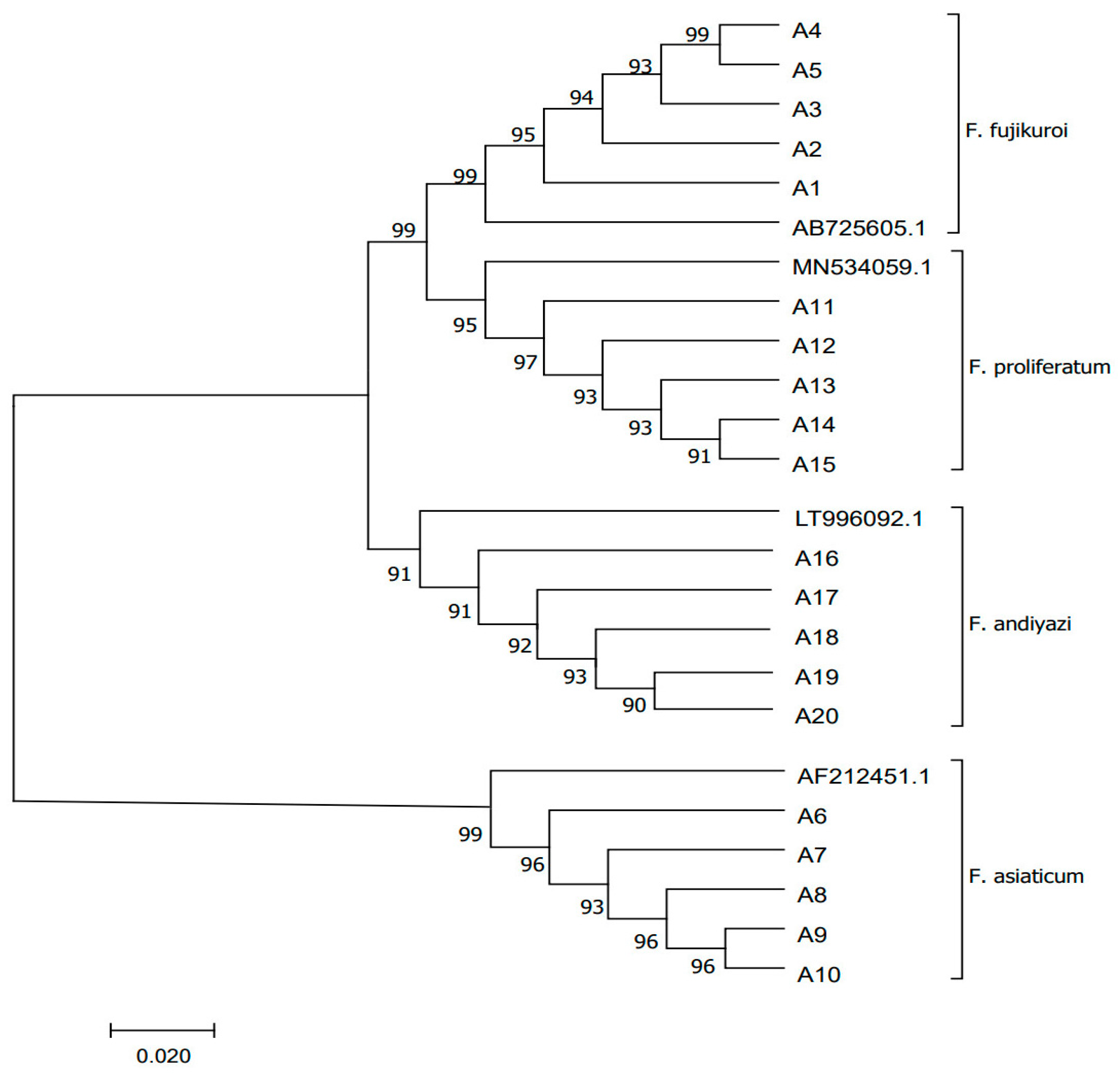
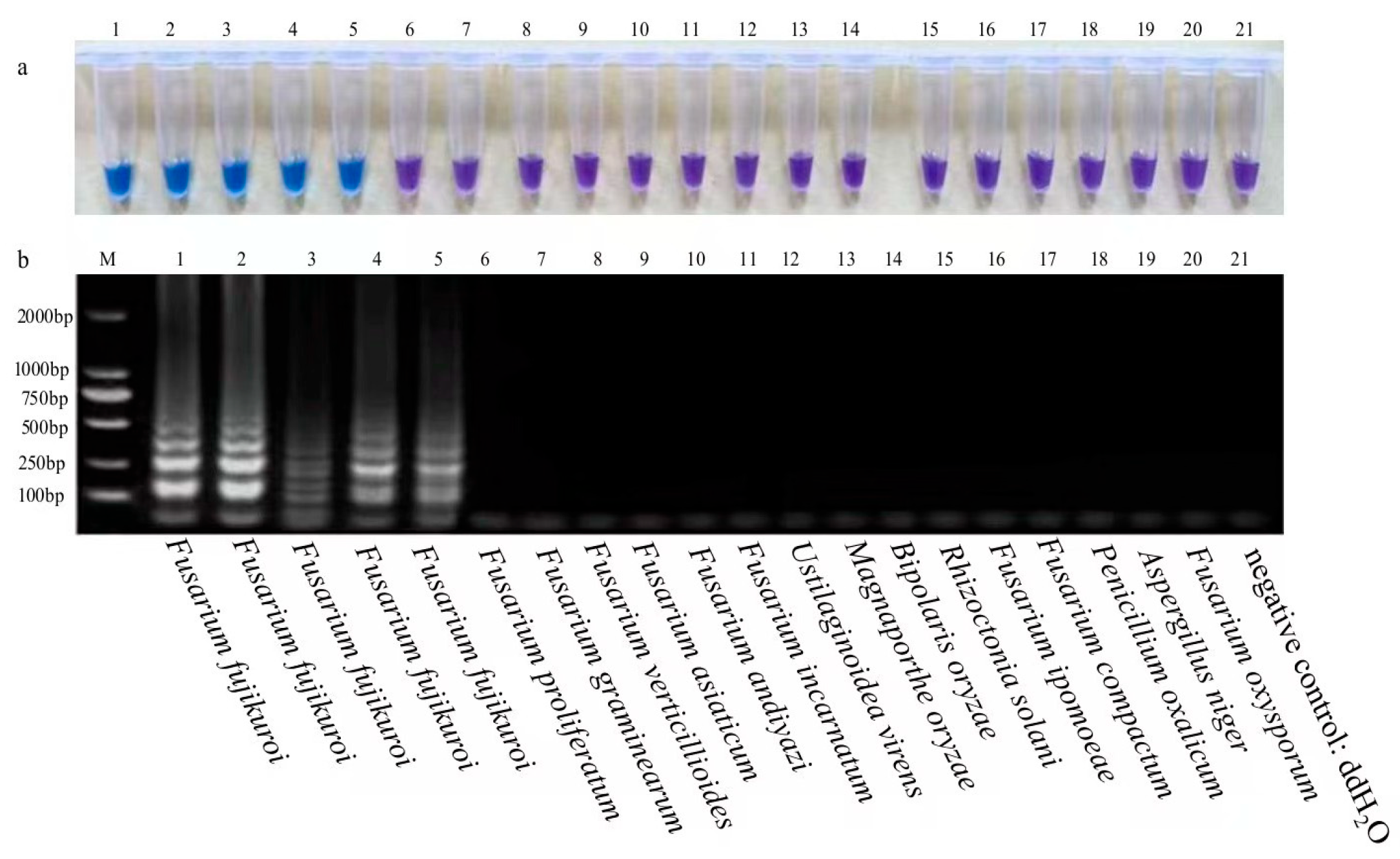
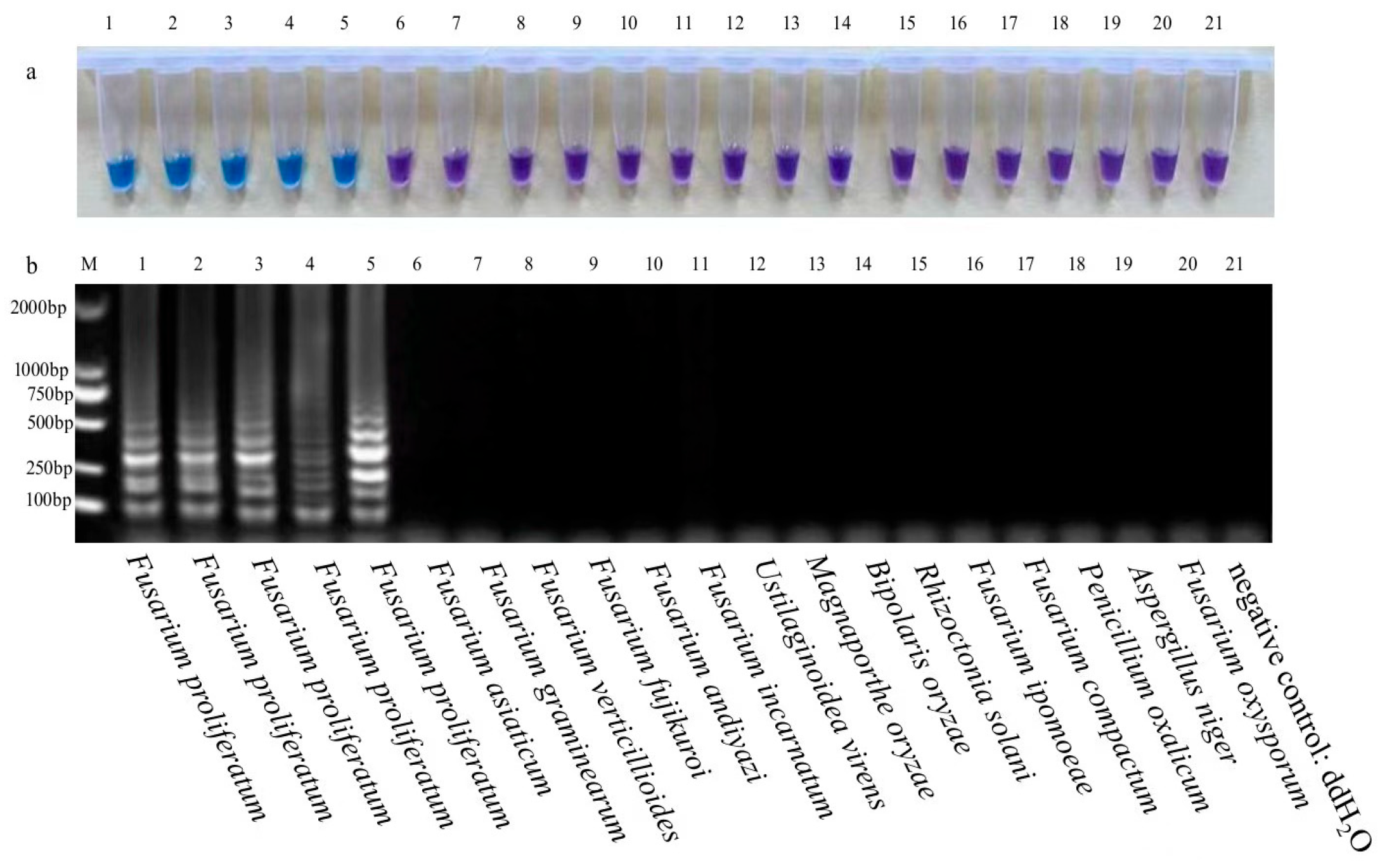
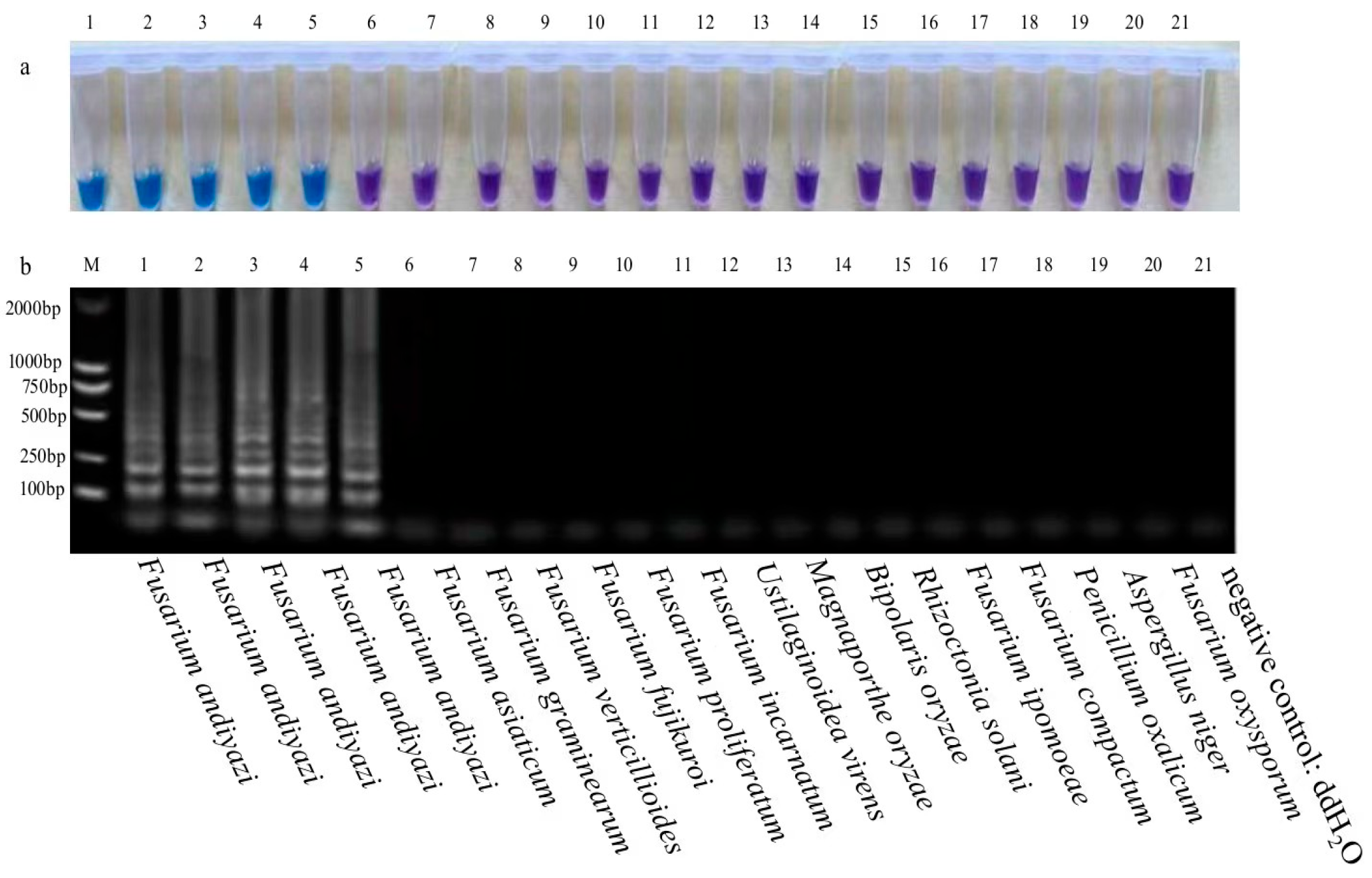
3. Results and Analysis

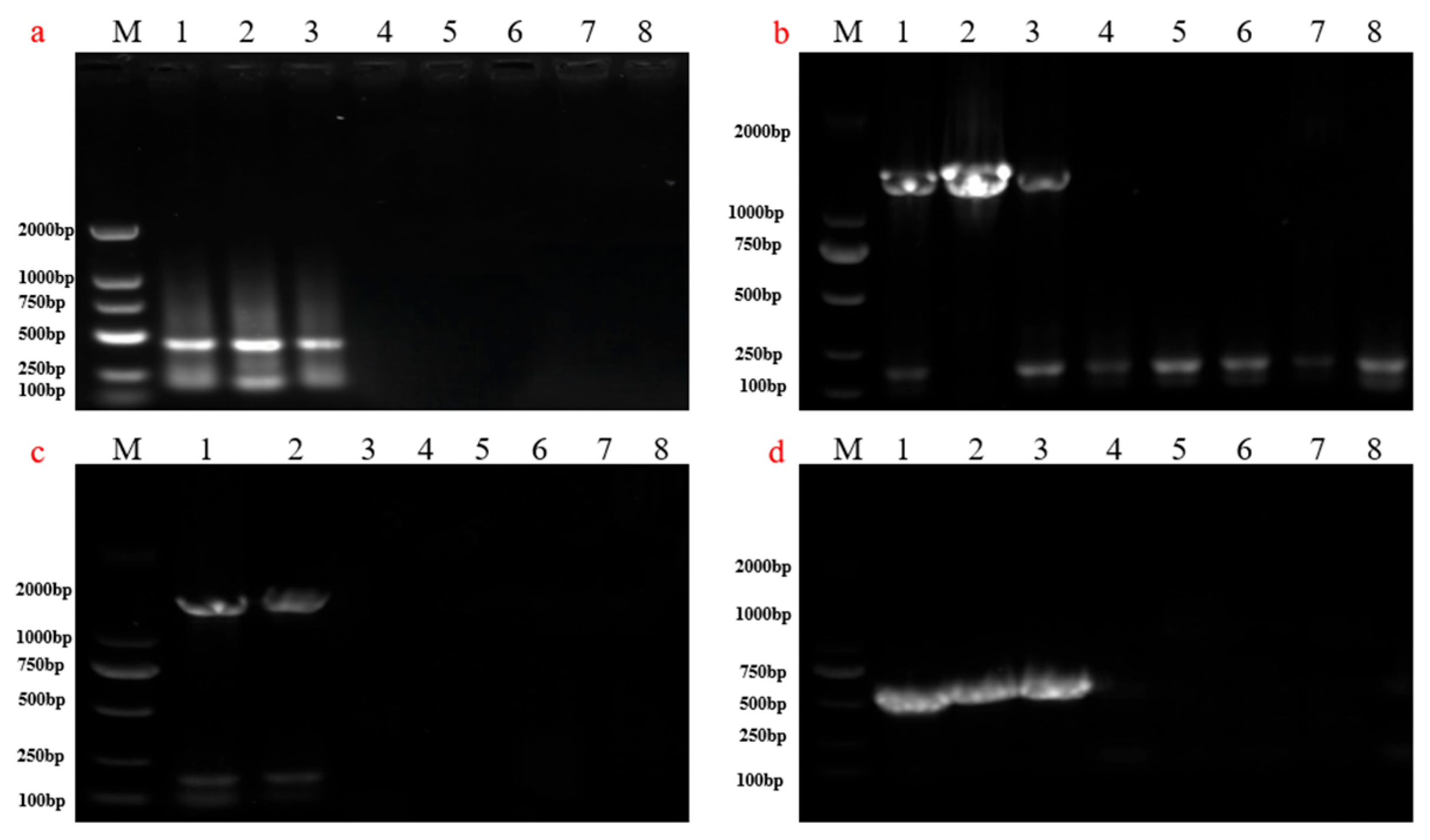
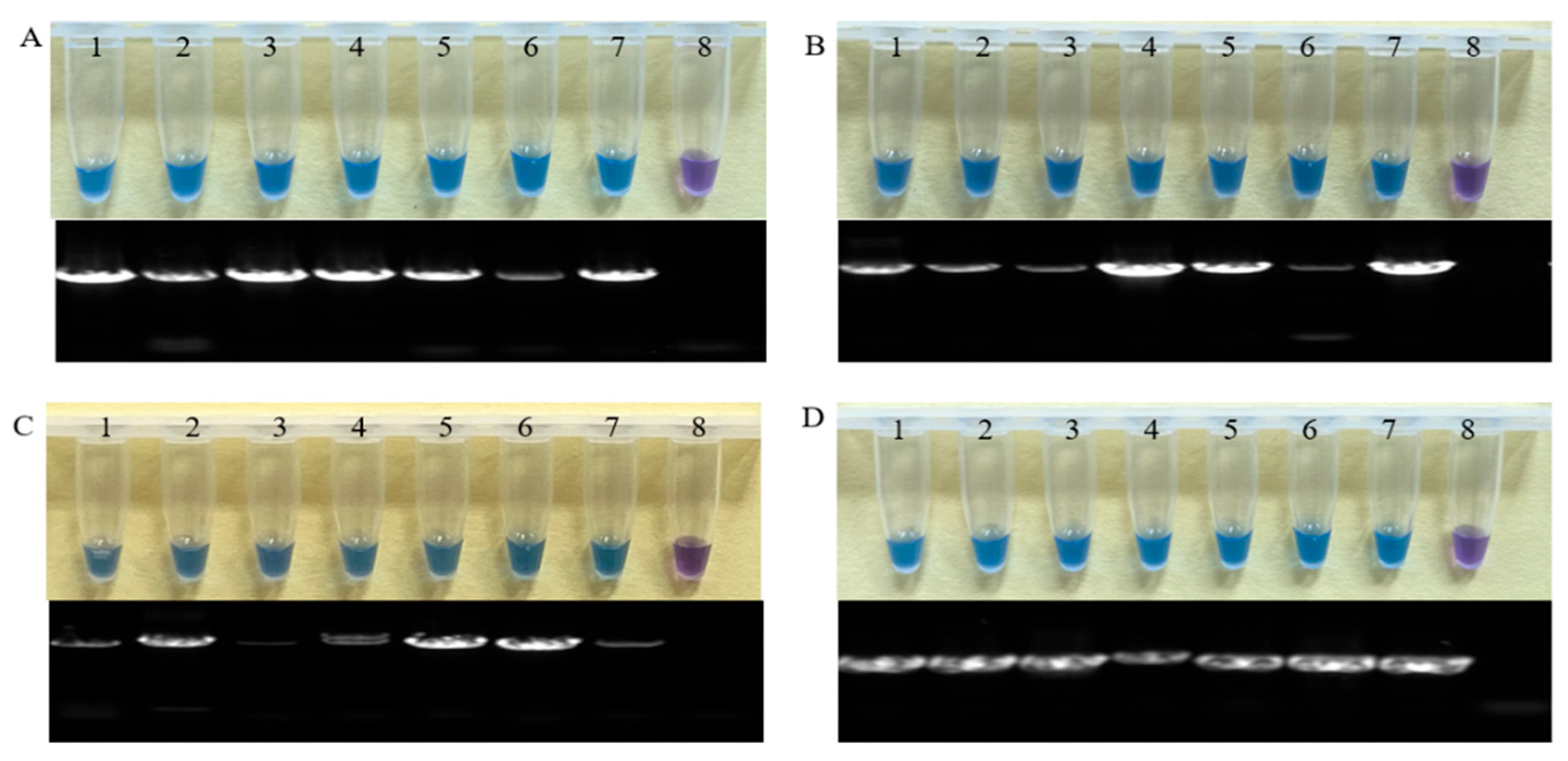
3.1. Specificity of LAMP Assays for RBD

Strains of Fusarium fujikuroi, Fusarium asiaticum, Fusarium proliferatum, Fusarium andiyazi, and other Fusarium species, and strains of rice diseases caused by other fungi, were used to confirm the specificity of the Ff-NRPS31-LAMP, Fa-TEF-LAMP, Fp-TEF-LAMP, and Fan-TEF-LAMP assays. The results were confirmed as positive or negative by the addition of HNB to the reaction system prior to amplification. Upon completion of the reaction, the color of the positive samples underwent a change, becoming sky blue, while the color of the negative samples remained purple. The results demonstrated that Fusarium fujikuroi strains exhibited a positive reaction, whereas other strains of Fusarium and other fungi causing rice diseases displayed a negative reaction (Figure 2). The specificity detections for Fusarium asiaticum, Fusarium proliferatum, and Fusarium andiyazi yielded comparable results, with the amplified reaction of the detected positive strains exhibiting a sky blue coloration, while that of the control strains remained purple (Figure 3, Figure 4 and Figure 5). To verify the specificity of the LAMP reaction, all specificity detections were repeated three times. The results demonstrated that the Ff-NRPS31-LAMP, Fa-TEF-LAMP, Fp-TEF-LAMP, and Fan-TEF-LAMP methods were capable of reliably detecting Fusarium fujikuroi, Fusarium asiaticum, Fusarium proliferatum, and Fusarium andiyazi, respectively, with species specificity.

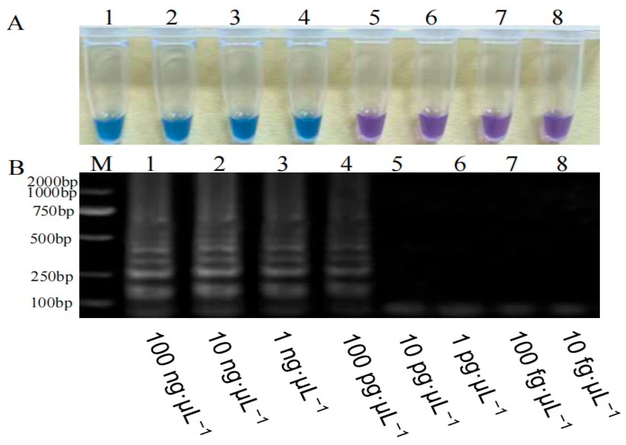
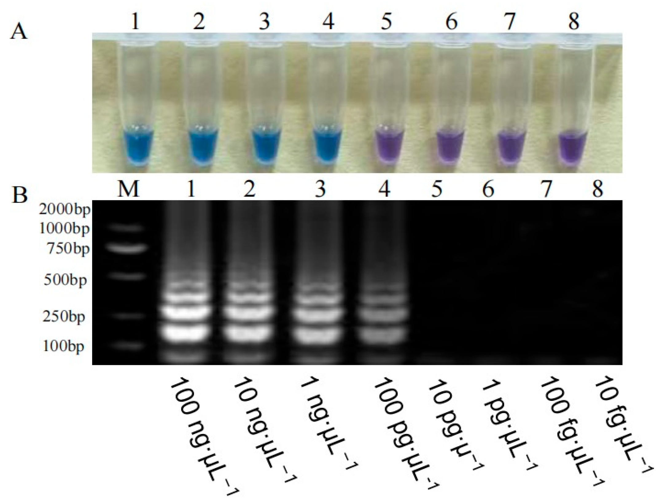
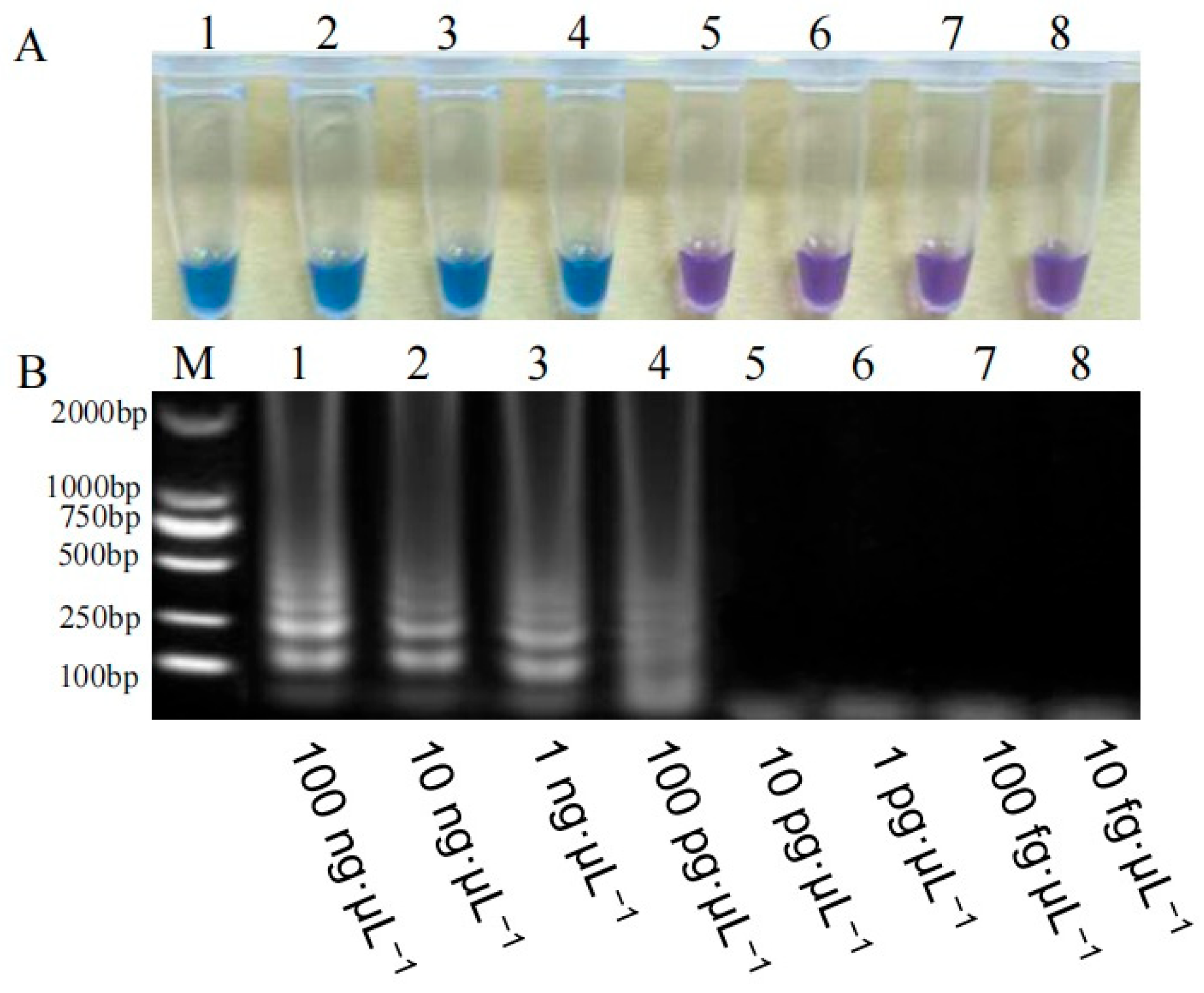
3.2. Sensitivity of the LAMP Assays for RBD

In the LAMP assay sensitivity detection, LAMP was performed based on the HNB visual color reaction using 10-fold serial dilutions of DNA templates (100 ng·μL−1, 10 ng·μL−1, 1 ng·μL−1, 100 pg·μL−1, 10 pg·μL−1, 1 pg·μL−1, 100 fg·μL−1, and 10 fg·μL−1) carried out.
The results are shown in Figure 6, Figure 7, Figure 8 and Figure 9. The DNA concentrations were ranked in descending order. The lowest concentration detected by Ff-NRPS31-LAMP, Fa-TEF-LAMP, Fp-TEF-LAMP, and Fan-TEF-LAMP were 100 pg·μL−1, which are also suitable for high-sensitivity LAMP amplification.

3.2.1. Sensitivity Detection of the Ff-NRPS31-LAMP Assay

To evaluate the sensitivity of the Ff-NRPS31-LAMP assay, the DNA template of Fusarium fujikuroi was subjected to a 10-fold serial dilution. Prior to the experiment, the F. fujikuroi template DNA was adjusted to 100 ng·µL−1 for convenient result observation. As shown in Figure 6, the minimum detection limit of the Ff-NRPS31-LAMP assay was 100 pg·µL−1, demonstrating its high detection sensitivity.
Figure 6. Ff-NRPS31-LAMP assay sensitivity detection. Note: (A): Diagram of color variation; (B): electrophoretogram; M: 2000 bp; 1–8: Fusarium fujikuroi.
Agriculture 15 01319 g006

3.2.2. Sensitivity Detection of the Fa-TEF-LAMP Assay

Before the experiment, the template DNA of Fusarium asiaticum was adjusted to 100 ng·µL−1. As shown in Figure 7, the minimum detection limit of the Fa-TEF-LAMP assay was 100 pg·µL−1. The results of the HNB color change diagram and agarose gel electrophoresis were consistent.
Figure 7. Fa-TEF-LAMP assay sensitivity detection. Note: (A): Diagram of color variation; (B): electrophoretogram; M: 2000 bp; 1–8: Fusarium asiaticum.
Agriculture 15 01319 g007

3.2.3. Sensitivity Detection of the Fp-TEF-LAMP Assay

As shown in Figure 8, when the DNA template concentration was diluted to 100 pg·µL−1, the visual observation results based on the HNB indicator were distinct, allowing for the identification of positive reaction results. The reliability of the results was verified by 2% agarose gel electrophoresis, which showed that although the amplified bands were less obvious at a DNA template concentration of 100 pg·µL−1, bands were still present. Therefore, comprehensive analysis indicated that the minimum DNA concentration detectable by the Fp-TEF-LAMP assay was 100 pg·µL−1.
Figure 8. Fp-TEF-LAMP assay sensitivity detection. Note: (A): Diagram of color variation; (B): electrophoretogram; M: 2000 bp; 1–8: Fusarium proliferatum.
Agriculture 15 01319 g008

3.2.4. Sensitivity Detection of the Fan-TEF-LAMP Assay

As shown in Figure 9, the Fan-TEF-LAMP assay failed to detect the pathogen when the DNA concentration was lower than 100 pg·µL−1. Therefore, the minimum detection limit of the Fan-TEF-LAMP assay was determined to be 100 pg·µL−1.
Figure 9. Fan-TEF-LAMP assay sensitivity detection. Note: (A): Diagram of color variation; (B): electrophoretogram; M: 2000 bp; 1–8: Fusarium andiyazi.
Agriculture 15 01319 g009

3.2.5. Sensitivity Comparison Between PCR and LAMP Assays for Detecting Rice Bakanae Disease Pathogens

The PCR technique is a commonly used method for the molecular identification of rice bakanae pathogens. The fundamental principle of this technique is based on the thermal denaturation property of DNA. Through multiple cyclic steps, DNA polymerase synthesizes new DNA strands using four deoxyribonucleotides as raw materials under appropriate temperatures. In contrast, the LAMP technique can operate at a constant temperature. It utilizes Bst DNA polymerase to drive DNA synthesis, enabling efficient amplification of target DNA. In this study, the sensitivities of the constructed Ff-NRPS31-LAMP, Fa-TEF-LAMP, Fp-TEF-LAMP, and Fan-TEF-LAMP assays were compared with those of the PCR technique for detecting corresponding pathogens. PCR amplification was performed using the outer primers F3/B3 of the Ff-NRPS31-LAMP, Fa-TEF-LAMP, Fp-TEF-LAMP, and Fan-TEF-LAMP systems. The sample DNA was successively diluted tenfold to determine the sensitivity of pathogen detection by the PCR technique, and the results are shown in Figure 10.
Compared with the LAMP sensitivity detection results presented in Figure 6, Figure 7, Figure 8 and Figure 9, it can be concluded that the sensitivities of the four constructed LAMP detection techniques in this study are all higher than those of the PCR technique. These LAMP techniques exhibit stronger sensitivity and are more favorable for detecting pathogens in field environments and under low-concentration DNA conditions.
Figure 10. Results of sensitivity detection by PCR technique. Note: (a) Sensitivity detection of Fusarium fujikuroi. (b) Sensitivity detection of Fusarium asiaticum. (c) Sensitivity detection of Fusarium proliferatum. (d) Sensitivity detection of Fusarium andiyazi. 1–8: 100 ng·μL−1, 10 ng·μL−1, 1 ng·μL−1, 100 pg·μL−1, 10 pg·μL−1, 1 pg·μL−1, 100 fg·μL−1, 10 fg·μL−1.
Agriculture 15 01319 g010

3.3. Detection of Maturity Period of Rice Seeds

The results are shown Table 2. The established LAMP assays for RBD were applied to seed samples of 55 rice varieties collected in the field. Three replicate detections were conducted for each sample. As illustrated in Table 2, the Ff-NRPS31-LAMP assay identified Fusarium fujikuroi in 12 seed samples. The Fusarium asiaticum was detected by the Fa-TEF-LAMP method in five samples, while Fusarium proliferatum was identified by the Fp-TEF-LAMP method in eight samples. Finally, the Fusarium andiyazi was detected by the Fan-TEF-LAMP assay in four samples.
Table 2. Detection of RBD pathogens carried in rice seeds using LAMP assays, respectively.
Sample (Cultivar)SourceFf-NRPS3-LAMPFa-TEF-LAMPFp-TEF-LAMPFan-TEF-LAMP
Xinliangyou9328Henan----
Xinliangyou1319Henan+---
Xinliangyou905Henan++--
XinliangyoufengnuoHenan----
Fliangyou6828Henan--+-
Liangyou7166Henan----
Liangyou2169Henan+---
LiangyouwushanHenan----
LiangyoufengheHenan----
Xingengnuo368Henan----
Xingengnuo216Henan----
Tongyu338Jilin----
Tongyu271Jilin+---
Tonghe885Jilin----
Tonghe875Jilin+---
Zhongkefa5Jilin----
Zhongkefa6Jilin-+--
Jijing338Jilin----
Tongxi968Jilin--+-
Tongxi969Jilin----
Tongxi963Jilin----
Tiejing11Liaoning----
Tiejing1507Liaoning----
Tiejing1603Liaoning----
Tiejing1712Liaoning++--
Tiejing1743Liaoning----
Tiejing1808Liaoning+---
Tiejing1811Liaoning----
Tiejingxiang3haoLiaoning----
Tiejing96Liaoning----
Tiejing1947Liaoning+---
Liaojing1925Liaoning+--+
Liaojingxiang1haoLiaoning----
Liaojing1499Liaoning--+-
Liaojing1402Liaoning----
Liaojing399Liaoning----
Liaojing327Liaoning--+-
Tianlongyou2605Liaoning--+-
Tianlongyou 717Liaoning+---
Beijing705Liaoning----
Beijing1702Liaoning-++-
Beijing1501Liaoning---+
Beijing1604Liaoning+-++
Beijing2Liaoning----
Zhonghua11Liaoning----
RibenqingLiaoning----
MenggudaoLiaoning----
LijiangxintuanheiguLiaoning----
Yanfeng47Liaoning----
Gangyuan8Liaoning--+-
Beijing143Liaoning----
Shennong9816Liaoning++-+
Shennong9903Liaoning----
Beijing1haoLiaoning----
Beijing3haoLiaoning----

3.4. The Detection of Pathogen Seed Carrying

The LAMP assays for RBD were applied to detect artificially prepared pathogen seeds carriers. Six rice varieties were randomly selected for the preparation of carrier seeds, and three replicated detections were performed for each sample. The Ff-NRPS31-LAMP, Fa-TEF-LAMP, Fp-TEF-LAMP, and Fan-TEF-LAMP methods for detecting artificially prepared seeds carrying Fusarium fujikuroi, Fusarium asiaticum, Fusarium proliferatum, and Fusarium andiyazi, respectively, were employed as illustrated in Figure 11. The results demonstrate that the LAMP assays for RBD can be employed for the detection of Fusarium fujikuroi, Fusarium asiaticum, Fusarium proliferatum, and Fusarium andiyazi in rice seeds.

4. Discussion

Rice is one of the three major food crops that human beings depend on for their survival, and the area of rice cultivation in China accounts for one-sixth of the world’s rice cultivation area [32]. RBD is one of many diseases that are widely occurring, harmful, and difficult to control in rice cultivation [33]. In this research, on the basis of LAMP assays, we established four new methods for the detection of RBD pathogens.
Fusarium spp. are morphologically and taxonomically similar to each other and also have similar gene sequences between species [34,35]. In this research, based on the target sequences, NRPS31 and TEF sequences, comparison between Fusarium species and genera were conducted to identify specific fragments, and primers were designed to detect the specificities of Fusarium fujikuroi, Fusarium asiaticum, Fusarium proliferatum, and Fusarium andiyazi. The primers were then subjected to continuous screening to select the most optimal ones. We established the gene sequences species-specific LAMP assays for Fusarium fujikuroi, Fusarium asiaticum, Fusarium proliferatum, and Fusarium andiyazi; these target genes were highly conserved among homologous strains. The specificity of these four LAMP assays was verified using other Fusarium species and rice diseases caused by other fungi as control strains. In the LAMP system sensitivity detection, LAMP was performed based on the HNB visual color reaction using 10-fold serial dilutions of DNA templates (100 ng·μL−1, 10 ng·μL−1, 1 ng·μL−1, 100 pg·μL−1, 10 pg·μL−1, 1 pg·μL−1, 100 fg·μL−1 and 10 fg·μL−1). The amplification was observed. The results showed that the sensitivity of the four LAMP systems can all reach 100 pg·μL−1, and they have good versatility and specificity, which can meet the relevant detection requirements.
Morphological identification, PCR and other molecular biology detection techniques have been widely used in the detection of RBD [36,37]. However, morphological identification is time-consuming, and the method also requires the experimenter to have rich experience and advanced knowledge of fungal taxonomy; PCR detection technology requires expensive equipment and complex operation steps [38]; in contrast, LAMP detection technology has the characteristics of strong primer specificity, high sensitivity, short reaction time, simple operation, visualization of the reaction results, intelligence, high efficiency, and low platform requirements, which can be applied to detect pathogens from field soil, diseased tissues, seeds, and so on [39].
RBD is prevalent across China, where it affects all rice-growing regions [40]. In recent years, pathogens mutation has led to the development of pesticide resistance in RBD, making the control of RBD more challenging [41]. Moreover, the disease is primarily spread through seeds [7], underscoring the significance of developing a rapid and accurate diagnostic method, such as LAMP assays, to detect RBD. This is crucial for the prevention and management of RBD.

5. Conclusions

In this study, we focused on the detection and diagnosis of rice bakanae disease. This disease, caused by diverse and complex fungal pathogens within the Fusarium fujikuroi complex (GFSC), poses a significant threat to rice production and food security. Leveraging the principles and advantages of loop-mediated isothermal amplification (LAMP) technology, we developed and optimized LAMP-based detection systems for GFSC and evaluated their field application, aiming to provide a foundation for the early diagnosis and precision control of rice bakanae disease. The main conclusions are as follows: (1) Compared with traditional PCR detection technology, the LAMP-based detection system for rice bakanae disease pathogens demonstrated superior performance in field applications, particularly in its ability to detect target genes from mixed DNA samples. (2) The four LAMP assays developed for rice bakanae disease pathogens exhibited high specificity, sensitivity, and broad applicability, enabling their use in the early detection of pathogen-infected rice seeds and direct testing of diseased tissues.
In subsequent research, we will focus on translating these findings into field applications for early disease diagnosis and prediction of rice bakanae disease. Additionally, we will investigate quantitative LAMP-based detection methods to further enhance technical support for the prevention, control, and diagnosis of rice bakanae disease infections.

Author Contributions

Conceptualization, X.L., S.W., Y.W. and W.Z.; methodology, X.L., S.W., Y.W. and W.Z.; validation, X.L., Y.Z., J.X., C.L., Y.S. and T.H.; formal analysis, X.L., Y.Z., S.W., Y.W. and W.Z.; investigation, X.L., S.W., J.X., C.L. and Y.S.; resources, X.L., S.W. and Y.W.; data curation, X.L., Y.Z., J.X. and C.L.; writing—original draft preparation, X.L. and Y.Z.; writing—review and editing, X.L., S.W., Y.W. and W.Z.; supervision, X.L., S.W., Y.W. and W.Z.; project administration, X.L., S.W., Y.W. and W.Z.; funding acquisition, S.W., Y.W. and W.Z. All authors have read and agreed to the published version of the manuscript.

Funding

This work was supported by the earmarked fund for China Agriculture Research System (CARS-01); Liao Ning Revitalization Talents Program (XLYC2213046).

Institutional Review Board Statement

Not applicable.

Data Availability Statement

The associated data will be provided by the corresponding authors upon request.

Acknowledgments

The author(s) would like to express gratitude to all the anonymous reviewers for their rigorous comments, which have made this study more readable and helped maintain its relatively high quality.

Conflicts of Interest

The authors declare no conflicts of interest.

References

  1. Kim, D.; Jeong, S.; Kim, B.; Kim, S.J.; Kim, H.; Jeong, S.; Yun, G.Y.; Kim, K.Y.; Park, K. Automated detection of rice bakanae disease via drone imagery. Sensors 2022, 23, 32. [Google Scholar] [CrossRef] [PubMed]
  2. Karthik, C.; Shu, Q. Current insights on rice (Oryza sativa L.) bakanae disease and exploration of its management strategies. J. Zhejiang Univ. Sci. B 2023, 24, 755–778. [Google Scholar] [CrossRef] [PubMed]
  3. Jeon, H.; Kim, J.E.; Yang, J.W.; Son, H.; Min, K. Application of direct PCR for phylogenetic analysis of Fusarium fujikuroi species complex isolated from rice seeds. Front. Plant Sci. 2023, 13, 1093688. [Google Scholar] [CrossRef] [PubMed]
  4. Morcia, C.; Tumino, G.; Gasparo, G.; Ceresoli, C.; Fattorini, C.; Ghizzoni, R.; Carnevali, P.; Terzi, V. Moving from qPCR to chip digital PCR assays for tracking of some Fusarium Species causing Fusarium head blight in cereals. Microorganisms 2020, 8, 1307. [Google Scholar] [CrossRef]
  5. Laha, G.S.; Singh, R.R.; Ladhalakshmi, D.; Sunder, S.; Prasad, M.; Dagar, C.S.; Babu, V.R. Importance and Management of Rice Diseases: A Global Perspective. In Rice Production Worldwide; Springer: Cham, Switzerland, 2017; pp. 303–360. [Google Scholar]
  6. Ito, S.; Kimura, J. Studies on the bakanae disease of the rice plant. Rep. Hokkaido Agric. Exp. Stn. 1931, 27, 1–95. [Google Scholar]
  7. Picco, A.M.; Lorenz, E.; Rodolfz, M.; Biloni, M.; Bruneli, A. A multi-location mycological survey on some Italian rice varieties during the crop season. Atti Delle Giornate Fitopatol. 2002, 2, 487–488. [Google Scholar]
  8. Nguyen, T.T.; Dehne, H.W.; Steiner, U. Histopathological assessment of the infection of maize leaves by Fusarium graminearum, F. proliferatum, and F. verticillioides. Fungal Biol. 2016, 120, 1094–1104. [Google Scholar] [CrossRef]
  9. Gupta, A.K.; Singh, Y.; Jain, A.K.; Singh, D. Prevalence and incidence of bakanae disease of rice in northern India. J. AgriSearch 2014, 1, 233–237. [Google Scholar]
  10. Hsuan, H.M.; Salleh, B.; Zakaria, L. Molecular identification of Fusarium Species in Gibberella fujikuroi species complex from rice, sugarcane and maize from Peninsular Malaysia. Int. J. Mol. Sci. 2011, 12, 6722–6732. [Google Scholar] [CrossRef]
  11. Bashyal, B.M.; Aggarwal, R.; Sharma, S.; Gupta, S.; Rawat, K.; Singh, D.; Singh, A.K.; Krishnan, S.G. Occurrence, identification and pathogenicity of Fusarium species associated with bakanae disease of basmati rice in India. Eur. J. Plant Pathol. 2016, 144, 457–466. [Google Scholar] [CrossRef]
  12. Dissanayake, M.L.; Tanaka, S.; Ito, S. Fumonisin B(1) production by Fusarium proliferatum strains isolated from Allium fistulosum plants and seeds in Japan. Lett. Appl. Microbiol. 2009, 48, 598–604. [Google Scholar] [CrossRef] [PubMed]
  13. Malonek, S.; Bömke, C.; Bornberg-Bauer, E.; Rojas, M.C.; Hedden, P.; Hopkins, P.; Tudzynski, B. Distribution of gibberellin biosynthetic genes and gibberellin production in the Gibberella fujikuroi species complex. Phytochemistry 2005, 66, 1296–1311. [Google Scholar] [CrossRef] [PubMed]
  14. Desjardins, A.E.; Manandhar, H.K.; Planter, R.D.; Manandha, G.G.; Poling, S.M.; Maragos, C.M. Fusarium species from Nepalese rice and production of mycotoxins and gibberellic acid by selected species. Appl. Environ. Microbiol. 2000, 66, 1020–1025. [Google Scholar] [CrossRef] [PubMed]
  15. Ma, L.J.; Geiser, D.M.; Proctor, R.H. Fusarium pathogenomics. Annu. Rev. Microbiol. 2013, 67, 399–416. [Google Scholar] [CrossRef]
  16. Yin, Y.; Liu, X.; Ma, Z. Simultaneous detection of Fusarium asiaticum and Fusarium graminearum in wheat seeds using a real-time PCR method. Soc. Appl. Microbiol. 2009, 48, 680–686. [Google Scholar]
  17. Xu, M.; Ye, W.; Zeng, D.; Wang, Y.; Zheng, X. Rapid diagnosis of wheat head blight caused by Fusarium asiaticum using a loop-mediated isothermal amplification assay. Australas. J. Plant Pathol. 2017, 46, 261–266. [Google Scholar] [CrossRef]
  18. Zhao, L.; Cheng, J.L.; Hao, X.A.; Tian, X.M.; Wu, Y.F. Rapid detection of tobacco viruses by reverse transcription loop-mediated isothermal amplification. Arch. Virol. 2012, 157, 2291–2298. [Google Scholar] [CrossRef]
  19. Ortega, F.S.; Tomlinson, J.; Gilardi, G.; Spadaro, D.; Gullino, M.L.; Garibaldi, A.; Boonham, N. Rapid detection of Fusarium oxysporum f. sp. lactucae on soil, lettuce seeds and plants using loop-mediated isothermal amplification. J. Plant Pathol. 2018, 67, 1462–1473. [Google Scholar] [CrossRef]
  20. Lai, D.W.; Zhang, Y.L.; Huang, Q.X.; Yin, G.H.; Pennerman, K.K.; Liu, Z.X.; Guo, A.P. Reverse transcription loop-mediated isothermal amplification to rapidly detect Rice ragged stunt virus. Saudi J. Biol. Sci. 2018, 25, 1577–1584. [Google Scholar] [CrossRef]
  21. Goto, M.; Honda, E.; Ogura, A.; Nomoto, A.; Hanaki, K. Colorimetric detection of loop-mediated isothermal amplification reaction by using hydroxynaphthol blue. BioTechniques 2009, 46, 167–172. [Google Scholar] [CrossRef]
  22. Notomi, T.; Okayama, H.; Masubuchi, H.; Yonekawa, T.; Watanabe, K.; Amino, N.; Hase, T. Loop-mediated isothermal amplification of DNA. Nucleic Acids Res. 2000, 28, e63. [Google Scholar] [CrossRef] [PubMed]
  23. Sanna, M.; Spadaro, D.; Gullino, M.L.; Mezzalama, M. Optimization of a loop-mediated isothermal amplification assay for on-site detection of Fusarium fujikuroi in rice seed. J. Agron. 2021, 11, 1580. [Google Scholar] [CrossRef]
  24. Quazi, S.A.; Meon, S.; Abidin, B. Characterization of Fusarium proliferatum through species specific primers and its virulence on rice seeds. Int. J. Agric. Biol. 2013, 15, 649–656. [Google Scholar]
  25. Rahjoo, V.; Zad, J.; Javan-Nikkhah, M.; Gohari, M.A. Morphological and molecular identification of Fusarium isolated from maize ears in Iran. J. Plant Pathol. 2008, 90, 463–468. [Google Scholar]
  26. White, T.J.; Bruns, T.D.; Lee, S.B. Amplification and direct sequencing of fungal ribosomal RNA genes for phylogenetics. In PCR Protocols; Academic Press: Cambridge, MA, USA, 1990; pp. 315–322. [Google Scholar]
  27. Zhang, S.Y.; Dai, D.J.; Wang, H.D.; Zhang, C.Q. One-step loop-mediated isothermal amplification (LAMP) for the rapid and sensitive detection of Fusarium fujikuroi in bakanae disease through NRPS31, an important gene in the gibberellic acid bio-synthesis. Sci. Rep. 2019, 9, 3726. [Google Scholar] [CrossRef]
  28. Gerlach, W.; Nirenberg, H. The Genus Fusarium: A pictorial Atlas. Mitteilungen Biol. Bundesanst. Land-Und Forstwirtsch. 1983, 209, 1–406. [Google Scholar] [CrossRef]
  29. Marasas, W.F.O.; Rheeder, J.P.; Lamprecht, S.C.; Zeller, K.A.; Leslie, J.F. Fusarium andiyazi sp. nov., a new species from sorghum. Mycologia 2001, 93, 1203–1210. [Google Scholar] [CrossRef]
  30. Zhang, X.; Harrington, T.C.; Batzer, J.C.; Kubota, R.; Peres, N.A.; Gleason, M.L. Detection of Colletotrichum acutatum sensu lato on strawberry by loop-mediated isothermal amplification. Plant Dis. 2016, 100, 1804–1812. [Google Scholar] [CrossRef]
  31. Denschlag, C.; Vogel, R.F.; Niessen, L. Hyd5 gene-based detection of the major gushing-inducing Fusarium spp. in a loop-mediated isothermal amplification (LAMP) assay. Int. J. Food Microbiol. 2012, 156, 189–196. [Google Scholar] [CrossRef]
  32. Berruezo, L.A.; Mercado Crdenas, G.E.; Harries, E.M.; Stenglein, S.A.; Curti, R.N.; Rodriguero, M.S.; Galvan, M.Z. Characterization of Fusarium species associated with to bacco diseases in Northwestern Argentina. Eur. J. Plant Pathol. 2018, 151, 1065–1079. [Google Scholar] [CrossRef]
  33. Mori, Y.; Nagamine, K.; Tomita, N.; Notomi, T. Detection of loop mediated isothermal amplification reaction by turbidity derived from magnesium pyrophosphate formation. Biochem. Biophys. Res. Commun. 2001, 289, 150–154. [Google Scholar] [CrossRef] [PubMed]
  34. O’Donnell, K.; Cigelnik, E.; Nirenberg, H.I. Molecular systematics and phylogeography of the Gibberella fujikuroi species complex. Mycologia 1998, 90, 465–493. [Google Scholar] [CrossRef]
  35. Liu, Y.L.; Whelen, S.; Hall, B.D. Phylogenetic relationships among ascomycetes: Evidence from an RNA polymerase II subunit. Mol. Biol. Evol. 1999, 16, 1799–1808. [Google Scholar] [CrossRef] [PubMed]
  36. Mule, G.; Susca, A.; Stea, G.; Moretti, A. A species-specific PCR assay based on the calmodulin partial gene for identification of Fusarium verticillioides, F. proliferatum and F. subglutinans. Eur. J. Plant Pathol. 2004, 110, 495–502. [Google Scholar] [CrossRef]
  37. Jurado, M.; Vázquez, C.; Marin, S.; Sanchis, V.; González-Jaén, M.T. PCR-based strategy to detect contamination with mycotoxi-genic Fusarium species in maize. Syst. Appl. Microbiol. 2006, 29, 681–689. [Google Scholar] [CrossRef]
  38. Amoah, B.K.; Macdonald, M.V.; Rezanoor, N.; Nicholson, P. Theuse of the random amplified polymorphic DNA technique to identify matinggroups in the Fusarium section Liseola. J. Plant Pathol. 1996, 45, 115–125. [Google Scholar] [CrossRef]
  39. Almasi, M.A.; Dehabadi, S.M.H.; Moradi, A.; Eftekhari, Z.; Ojaghkandi, M.A.; Aghaei, S. Development and application of loop-mediated isothermal amplification assay for rapid detection of Fusarium oxysporum f. sp. lycopersici. J. Plant Pathol. Microbiol. 2013, 4, 177–183. [Google Scholar] [CrossRef]
  40. Choi, J.H.; Lee, S.; Nah, J.Y.; Kim, H.K.; Paek, J.S.; Lee, S.; Ham, H.; Hong, S.K.; Yun, S.H.; Lee, T. Species composition of and Fumonisin production by the Fusarium fujikuroi species complex isolated from Korean cereals. Int. J. Food Microbiol. 2018, 267, 62–69. [Google Scholar] [CrossRef]
  41. Amoah, B.K.; Rezanoor, H.N.; Nicholson, P.; Macdonald, M.V.P. Variation in the Fusarium section Liseola: Pathogenicity and genetic studies of Fusarium moniliforme sheldon from different hosts in Ghana. J. Plant Pathol. 1995, 44, 563–572. [Google Scholar] [CrossRef]
Figure 1. Joint phylogenetic tree based on TEF, TUB, and ITS sequences. Note: A1–A5: Fusarium fujikuroi; A6–A10: Fusarium asiaticum; A11–A15: Fusarium proliferatum; A16–A20: Fusarium andiyazi.
Agriculture 15 01319 g001
Figure 2. Specificity verification of Ff-NRPS31-LAMP detection system. Note: (a): Diagram of color variation; (b): electrophoretogram; M: 2000 bp.
Agriculture 15 01319 g002
Figure 3. Specificity verification of Fa-TEF-LAMP detection system. Note: (a): Diagram of color variation; (b): electrophoretogram; M: 2000 bp.
Agriculture 15 01319 g003
Figure 4. Specificity verification of Fp-TEF-LAMP detection system. Note: (a): Diagram of color variation; (b): electrophoretogram; M: 2000 bp.
Agriculture 15 01319 g004
Figure 5. Specificity verification of Fan-TEF-LAMP detection system. Note: (a): Diagram of color variation; (b): electrophoretogram; M: 2000 bp.
Agriculture 15 01319 g005
Figure 11. HNB-based LAMP assays for the detection of artificially infested rice seeds carrying RBD pathogens by amplification at 60 °C for 60 min. Note: (A), 1: positive control: Fusarium fujikuroi; 2–7: artificially infested rice seeds carrying Fusarium fujikuroi; 8: negative control: ddH2O; (B), 1: positive control: Fusarium asiaticum; 2–7: artificially infested rice seeds carrying Fusarium asiaticum; 8: negative control: ddH2O; (C), 1: positive control: Fusarium proliferatum; 2–7: artificially infested rice seeds carrying Fusarium proliferatum; 8: negative control: ddH2O; (D), 1: positive control: Fusarium andiyazi; 2–7: artificially infested rice seeds carrying Fusarium andiyazi; 8: negative control: ddH2O.
Agriculture 15 01319 g011
Table 1. Prers used for the development of LAMP assays for specific detection of RBD.
AssayPrimer TypeSequence (5′–3′)Length
Ff-NRPS31-LAMPF3TTTTTTAAGTCCGTATTG18
B3GTGCTCATCGCTTCCTCAAC20
FIPGGGTGGTGGCAGCTC-TTGCACGCGTTGAGTTTAC34
BIPCATTGCACGATAGCTAGC-CTAATCAGATATTTTCTCTA38
LFGCCCCTGATTCTACC15
LBACCTACCTACTCTCAAG17
Fa-TEF-LAMPF3ACCAGTCACTAACCACCTGT20
B3GAGCGTCTGATAGCCATGTT20
FIPTCACGCTCGGCTTTGAGCTTG-GAGCTCGGTAAGGGTTCCT40
BIPATCGCCCTCTGGAAGTTCGAGA-GACAGCAGTGGTGACAACAT42
LFAGAACCCAGGCGTACTTGA19
LBCTCCTCGCTACTATGTCACCGT22
Fp-TEF-LAMPF3CATTTACCCCGCCACTCG18
B3AGGAGTCTCGAACTTCCAGA20
FIPCGGACGGTTAGTGACTGCTTGA-CGTTTGCCCTCTCCACAA40
BIPCGCTGAGCTCGGTAAGGGTTC-GGTGATACCACGCTCACG39
LFGACGATGCGCTCATTGAGG19
LBCTTCAAGTACGCCTGGGTTC20
Fan-TEF-LAMPF3GCATTTACCCCGCCACTC18
B3AGCGAGGAGTCTCGAACTTC20
FIPCTCAGCGGCTTCCTATTGTCGA-CCTCTCCCATTCCACAACC41
BIPAGGGTTCCTTCAAGTACGCCTG-GATGGTGATACCACGCTCAC42
LFCGTGACAATGCGCTCAGTGA20
LBGGTTCTTGACAAGCTCAAGGC21
Disclaimer/Publisher’s Note: The statements, opinions and data contained in all publications are solely those of the individual author(s) and contributor(s) and not of MDPI and/or the editor(s). MDPI and/or the editor(s) disclaim responsibility for any injury to people or property resulting from any ideas, methods, instructions or products referred to in the content.

