Article

Dam Age Differentially Affects Immune Response of Her Calf to Bovine Respiratory Disease Vaccination

by
Cassidy Reddout
1,
Christopher C. L. Chase
2,
Paul Beck
1 and
Janeen L. Salak-Johnson
1,*
1
Department of Animal and Food Sciences, Oklahoma State University, Stillwater, OK 74078, USA
2
Department of Veterinary and Biomedical Sciences, South Dakota State University, Brookings, SD 57007, USA
*
Author to whom correspondence should be addressed.
Agriculture 2024, 14(1), 68; https://doi.org/10.3390/agriculture14010068
Submission received: 21 September 2023 / Revised: 12 December 2023 / Accepted: 27 December 2023 / Published: 29 December 2023
(This article belongs to the Section Farm Animal Production)

Abstract

:
Understanding the biological factors that affect immunological responses may be beneficial in developing more effective bovine respiratory disease vaccination strategies. Dam traits, such as age and vaccination history, are not considered when developing vaccination protocols or assessing vaccination efficacy, even though both factors may influence the transference of passive immunity; therefore, a preliminary study was conducted to determine the effect of dam age on the immune response of their calves to vaccinations. Calves born to dams of various ages were vaccinated with a modified live vaccine at 3 to 4 months old, revaccinated with either the same modified live or an inactivated vaccine at weaning, and boosted with the same vaccine 28 days later. Immune and cortisol measures were taken upon initial vaccination, revaccination at weaning, and then 14 and 28 days post-revaccination and post-booster. Overall, calves from older-aged dams began to exhibit a pattern indicative of a dampened immunoglobulin-G response to revaccination and booster, regardless of vaccination type. In contrast, calves from the youngest and oldest dams who received one modified live and two inactivated vaccines had the highest serum-neutralizing titers for most antigens present in the vaccines. These preliminary data partly imply that it may be plausible that the more doses of modified live vaccines the dam receives, which increase with each pregnancy, may affect the immune response of her progeny to vaccination at weaning and revaccination. This may be due to the effects on maternal passive immunity and is further affected by the type of vaccination her progeny received for some measures. However, more research is necessary to elucidate further the effects biological factors have on the immune response to vaccination, develop better vaccination strategies, and further validate these findings through increased sample sizes.

1. Introduction

Bovine respiratory disease (BRD) continues to be one of the most challenging issues for the cattle industry, despite years of research and development into different vaccination and management strategies. It is the most common feedlot illness, responsible for more than 50% of all deaths and 75% of morbidities in the United States [1]. Vaccination is the most common preventative measure. Theurer et al. [2] reported that adopting a standard vaccination protocol for a cow–calf operation can improve health outcomes at the feedlot. However, the control of BRD is difficult due to the complex nature of the disease and the numerous risk factors involved in the triad complex, including bacterial and viral pathogens, environmental conditions, and host immunity [3,4]. Host immunity is further affected by other biological animal factors, which include age at vaccination [5], vaccination protocol utilized [6], sex [7], and various other factors that may contribute to the continued high BRD prevalence in the feedlot [8]. Minimal studies have assessed the effect of individual animal factors, such as the age of the dam or their vaccination history, on the immune response of their progeny to vaccinations.
The specific immune response to BRD vaccination depends on multiple factors, including vaccine type, such as a modified live virus or an inactivated one [9]. For example, different types of vaccines use different antigens that activate different components of the adaptive immune system. Modified live virus vaccines induce T- and B-cells, resulting in complete cell-mediated and humoral immune responses. Inactivated virus vaccines primarily stimulate B-cells, inducing a strong humoral immune response but a less robust cell-mediated immunity. A booster vaccination is generally administered regardless of vaccine type, but at least two doses of the inactivated vaccine are required to stimulate a more effective immune response [10].
Another factor that influences the immune response to vaccination is age. Signer et al. [11] found that an individual’s immune response to vaccination decreases as age increases. This may be partly due to decreased B-cell production necessary to produce antigenic memory; thus, as animals age, their immune system is less productive in producing serum-neutralizing antibodies, and the antigen affinity is reduced [12]. This, in turn, leads to a longer time to reach peak titers and a faster decline in titers post-vaccination [12]. This age-related reduction in immunity is termed immunosenescence, which impacts the robustness of the adaptive immune system [13]. Thus, it is plausible that as the dam ages (or vaccine doses increase), this may affect her humoral immune response, influencing the vaccine responsiveness of her progeny.
Often, cattle, especially those in breeding herds, receive annual vaccinations, with one before breeding and then again while pregnant. The accumulative effect of annual revaccination may lead to vaccine exhaustion, or the ‘Hoskins Effect’ [14]. It has been shown in humans that repeated vaccination reduces the effectiveness of influenza protection, as evidenced by reduced titer values [15,16] and virus-specific antibody affinity maturation [17]. Some have speculated that a reduced immune response to vaccination may be partly due to the animal’s first exposure to the antigen being the vaccination, followed by subsequent antigenically identical vaccinations. This may result in the antigenic distance hypothesis, in which the virus has mutated from the original vaccine formulation, resulting in new antigen variants and reducing the efficacy of the immune response to the mutation, as it relies on antibodies made for the original form encountered through vaccination [18]. Moreover, it is plausible that a reduced immune response to vaccines in the dam may affect passive immunity, which may have implications for the immune response of the progeny.
Maternal vaccination against BRD is believed to be beneficial for calf protection [19,20]. However, there is some concern that maternal interference may reduce vaccination efficacy in the calf [21]. It may be plausible that maternal interference is reduced in calves born to older dams or that maternal passive immunity is affected, thus affecting the immune response to vaccination in the progeny. A more comprehensive understanding of the effect of the age of the dam related to repeated vaccination is important to implement a more effective vaccination protocol for cow–calf operations. We previously reported the differential immune phenotypes induced by vaccination type in heifer calves [6]; therefore, the objective of this preliminary study was to characterize the effect of dam AGE, and thus her vaccination history, on overall calf response to BRD vaccination and to further characterize if dam AGE affected the calves’ immune response to being vaccinated at weaning and again 28 days later with either modified live or inactivated vaccines.

2. Materials and Methods

All procedures were approved by the Oklahoma State University Institutional Animal Care and Use Committee (Protocol No. AG-15-21 and AG-19-8).

2.1. Animals and Experimental Design

At breeding, Angus dams kept at Oklahoma State University Field and Research Service Unit (Valliant, OK, USA) received a modified live vaccine (MLV; Bovishield® Gold FP5 VL5, Zoetis Animal Health, Parsippany-Troy Hills, NJ, USA) containing infectious bovine rhinotracheitis (IBR), bovine viral diarrhea virus type 1 and 2 (BVD1 and BVD2), parainfluenza 3 (PI3), and bovine respiratory syncytial virus (BRSV), and then a booster four weeks later. This standard vaccination protocol was repeated each breeding period that the dam remained in the breeding herd.
All calves were vaccinated at 3–4 months of age with Titanium®5 (Elanco Animal Health, Greenfield, IN, USA), a modified live vaccine containing IBR, BVD1 and 2, PI3, and BRSV, but blood samples were only taken from a subset of dams and calves (n = 21) at this time. Calves were weaned at 6 to 7 months old and then transported to the Oklahoma State University Willard Sparks Beef Research Center (WSBRC; Stillwater, OK, USA). Dams remained with the breeding herd in Valliant, OK.
Calves were bled before transport. Upon arrival at WSBRC in Stillwater, OK, calves were held overnight in holding pens with ad libitum access to water and hay. The next day, calves were bled, weighed, and allocated to a vaccine treatment group where they were revaccinated with either MLV or an inactivated (INA) (inactivated IBR, BVD1 and 2, PI3, and BRSV) vaccine, and 28 days later, they received a booster with the same treatment vaccine. This resulted in two treatment groups: MLV = 3 MLV (n = 30) or INA = 1 MLV and 2 INA (n = 25) vaccinations. Blood samples were obtained from these calves (n = 55) at 14 and 28 days post-revaccination (PRv) and post-booster (PB) with Titanium®5 (MLV) or Virashield®6 (INA; Elanco Animal Health). Animals were also weighed on blood collection days.
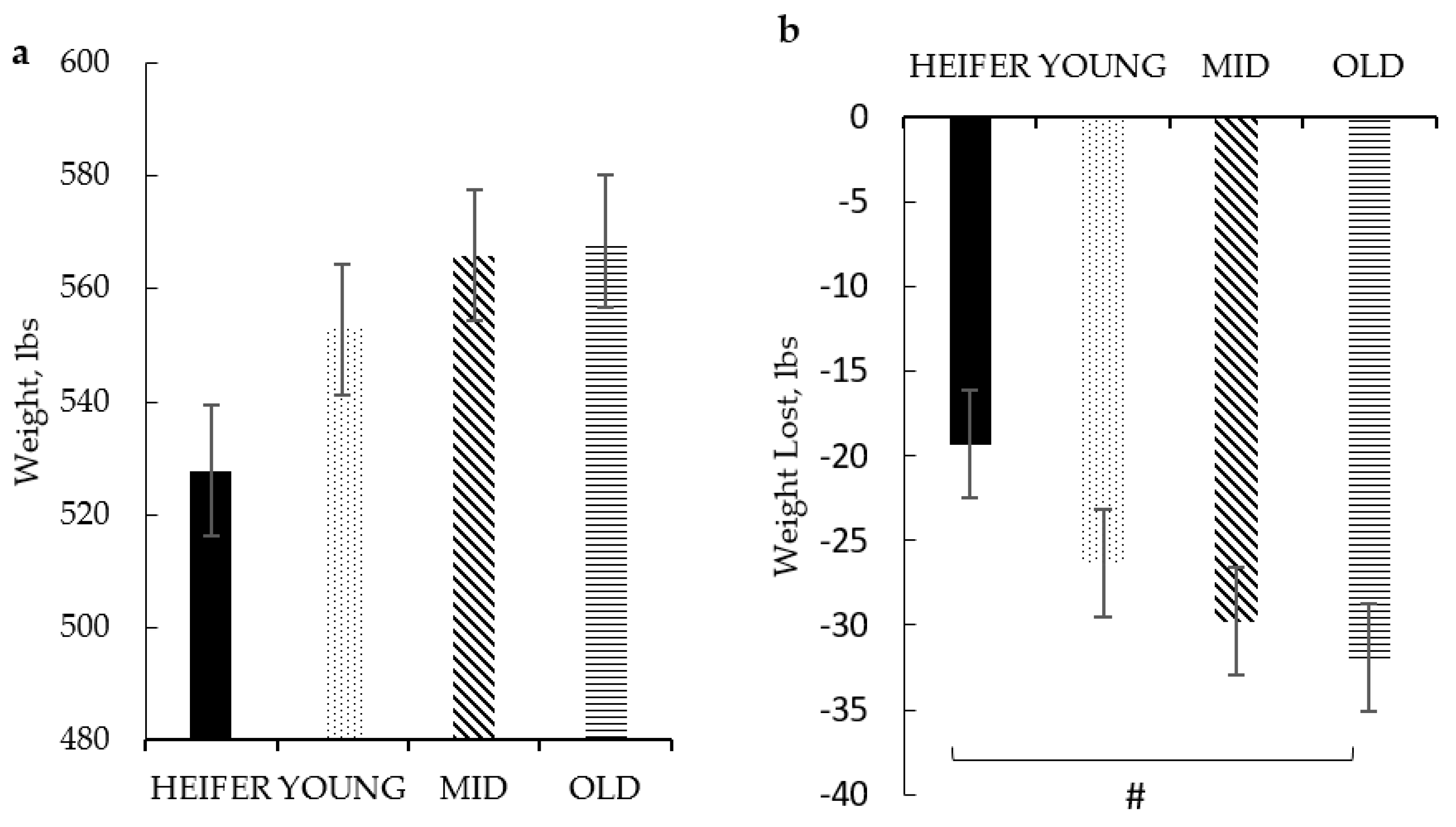
Dam age categories were created based on her age (or parity), indicative of the number of BRD vaccinations she received each pregnancy, which resulted in four age groups: HEIFER (2-year-olds; 2 vaccinations; and n = 9), YOUNG (3- to 4-year-olds; 4–6 vaccinations; and n = 22), MID (5- to 6-year-olds; 8–10 vaccinations; and n = 13), and OLD (≥7-year-olds; ≥12 vaccinations; and n = 11).

2.2. Sample Collection and Blood Processing

Whole blood and serum samples were collected via jugular venipuncture using serum, heparin (plasma), and EDTA (complete cell counts; CBC) vacutainers (BD Vacutainers; Franklin Lakes, NJ, USA). The CBC was determined electronically from whole blood using the Element HT5 Hematology Analyzer (Heska, Loveland, CO, USA). Serum tubes were allowed to clot at room temperature, centrifuged at 3000× g for 20 min, and then aliquoted. Plasma samples were stored at −20 °C and serum at −80 °C until subsequent analysis.

2.3. Cortisol, Cytokines, and Immunoglobulin-G Subsets

Plasma cortisol was measured using a commercially available enzyme-linked immunoassay (ELISA) following the manufacturer’s protocol (Enzo Life Sciences, Farmingdale, NY, USA). Samples were diluted 1:8 in assay buffer and run in duplicate in a 96-well microtiter plate coated with goat anti-mouse IgG. The conjugate (alkaline phosphatase conjugate with cortisol) and antibody (mouse monoclonal antibody to cortisol) were added. Plates were placed on a shaker at 500 rpm for 2 h at room temperature and washed three times, then substrate (P-nitrophenylphosphate) was added, and plates were incubated for 1 h at room temperature. The reaction was stopped with the solution provided. Plates were read using a microplate reader (BioTek Epoch, Winooski, VT, USA) at 405 nm. A standard curve was used to determine the concentration of the unknown samples using the Gen5 Data Analysis Software version 3.0 (Bio Tek). The minimal detectable concentration of the assay was 56.7 pg/mL, and the mean intra- and interassay coefficients of variability were 7.3% and 13.4%, respectively.
Interleukin-4, -6, -10, -17, -21 (Invitrogen Corp., Waltham, MA, USA), and -8 (Biomatik, Wilmington, DE, USA) were measured using commercially available bovine ELISA kits for each cytokine following the manufacturer’s protocols. Briefly, the 96-well microtiter plates were pre-coated for all cytokines except interleukin-6 and -8. Standards and samples were pipetted in duplicate, and plates were incubated at room temperature with gentle shaking. Anti-bovine detection antibodies IL-4 or IL-6 and biotin-conjugated antibodies IL-10, IL-17, or IL-21 were added to the appropriate plate and incubated for 1 h at room temperature. Plates were washed several times, Streptavidin-HRP was added to each well, and plates were incubated with moderate shaking at room temperature for 30 min (IL-6 and IL-4) or 45 min (IL-10, IL-17, and IL-21). Tetramethylbenzidine dihydrochloride (TMB) substrate was added to each well. Plates were incubated for 20 min (IL-6 and IL-4) or 30 min with gentle shaking (IL-10, IL-17, and IL-21); the reaction was terminated with stop solution, and plates were read at 450 nm wavelengths using the BioTek Epoch plate reader. Using the Gen5 Data Analysis Software (BioTek, Winooski, VT, USA), the minimal detectable concentrations of the assays for interleukin-4, -6, -8, -10, -17, and -21 were 15.6 pg/mL, 78.1 pg/mL, 5.9 pg./mL, 0.12 ng/mL, 2 pg./mL, and 0.41 ng/mL, respectively. The mean intra- and interassay coefficients of variability were 10% and 12% for IL-4, -6, -17, and -21; 8% and 10% for IL-8; and 5% and 7.2% for IL-10, respectively.
Immunoglobulin-G (IgG) subset IgG1 and IgG2 concentrations were measured at South Dakota State University (Brookings, SD, USA) using commercially available bovine IgG1 (E11-16) and IgG2 (E11-17) ELISA kits (Bethyl Laboratories Inc., Montgomery, TX, USA), following the manufacturer’s protocol. Briefly, samples were diluted in sample diluting buffer at 1:500,000, and samples and standards were pipetted in duplicate onto 96-well microtiter plates coated with either anti-bovine IgG1 or IgG2 antibody. Plates were incubated at room temperature for 1 h and washed four times. Anti-IgG1 or anti-IgG2 detection antibodies were pipetted into the wells. Plates were incubated at room temperature for 1 h and washed four times. Horseradish peroxidase solution was added to each well, and plates were incubated at room temperature for 30 min and washed. The TMB substrate was added, and plates were incubated in the dark for 30 min at room temperature. The reaction was stopped using the provided solution, and plates were read at 450 nm. The standard curve was plotted using Soft-Max pro software version 7.1.2 (Molecular Devices LLC, San Jose, CA, USA) to estimate concentrations of IgG1 and IgG2. Total IgG was calculated by adding the values of the concentrations of IgG1 and IgG2. The minimal detectable concentration of both assays was 1.0 ng/mL.

2.4. Serum-Neutralizing Antibody Titers

Serum samples were shipped to the Texas Veterinary Medical Diagnostic Laboratory (Canyon, TX, USA) on dry ice to analyze serum-neutralizing (SN) antibody titers for infectious bovine rhinotracheitis (IBR), bovine viral diarrhea virus types 1a, 1b, and 2 (BVDV 1a, 1b, 2), parainfluenza 3 (PI3), and bovine respiratory syncytial virus (BRSV). The reciprocal of the highest dilution of serum that neutralizes the infectivity of the virus was determined as the SN antibody titer.

2.5. Statistical Analysis

Data were analyzed using Pearson’s correlation and mixed procedure in SAS 9.4 (Inst. Inc., Cary, NC, USA) with repeated measures. All traits were tested for departure from a normal distribution through analysis of the residuals, and log transformation was applied to non-normally distributed traits. The model included random effects of dam or calf and fixed effects of dam AGE (HEIFER, YOUNG, MID, or OLD) and vaccination treatment (MLV or INA). Days post-revaccination or booster were used as a repeated measure. The Tukey–Kramer post hoc analysis adjusted the p-value to account for Type I error for multiple comparisons. Significance was set at (p ≤ 0.05), and trends were discussed from (p > 0.05) to (p ≤ 0.10).

3. Results

3.1. Immunophenotype of Dam at 3 to 4 Months Post-Calving and Progeny at 3–4 Months of Age and Weaning

Shown in Table 1 are the effects of dam AGE on her leukocyte populations, cytokines, and immunoglobulin measures at 3 to 4 months post-calving. All measures were similar, regardless of her AGE.
Shown in Table 2 are the effects of dam AGE on measures of her calves at 3 to 4 months old. Specifically, IL-21 was the highest (p = 0.005) among calves from MID-aged dams than calves born to dams of all other AGE groups (Table 2). These calves also tended to have higher IL-17. Interestingly, there was no effect of dam AGE on IgG1, but the calves born to MID-aged dams had the lowest (p = 0.045) IgG2 concentrations, resulting in the highest (p = 0.03) IgG1/IgG2 ratio (Table 2). All other measures were similar among calves regardless of the dam AGE group category at 3 to 4 months old (Table 2). Furthermore, all measures were similar among the calves regardless of dam AGE at weaning (Table 3).
There was no effect of dam AGE on the body weight of calves at 3 to 4 months old or calf weaning weight (Figure 1a); however, calf weight loss during transport increased with dam AGE (p = 0.08; Figure 1b). Calves from HEIFER dams tended to lose less weight than those from OLD dams.

3.2. Calf Measures from Revaccination and Booster Vaccination

3.2.1. Interactive Dam AGE via Vaccination Treatment by Day Effects on IgGs

A three-way interactive effect of dam AGE via vaccine treatment by day was detected for immunoglobulin G 1 (IgG1; p = 0.0009) but not total IgG (p = 0.0293; no relevant comparisons found; Table 4), IgG2, or IgG1/IgG2 ratio (p > 0.10; Table 4). While calves vaccinated with MLV vaccines from OLD dams only had significantly lower IgG1 concentrations than MLV-vaccinated calves from YOUNG dams at 14d-PRv (Table 4), it is interesting to note that despite a lack of significance, MLV-vaccinated calves from OLD dams had the lowest IgG1 concentrations at 14d-PRv and 14d-PB. A similar non-significant pattern was noted for IgG2 and total IgG concentrations for the MLV-vaccinated calves from OLD dams at 14d-PRv and 14d-PB. At these time points, a similar but less robust pattern was noted for these measures in the INA-vaccinated calves of OLD dams. As the lack of significance may be due to the small sample size, these patterns may still be notable for biological relevancy.

3.2.2. Effects of Dam AGE via Vaccine Treatment on Serum-Neutralizing Antibody Titers, Cytokines, and Cortisol

An interactive effect of dam AGE and vaccination treatment was found for IBR (p < 0.0001), BVD1a and b (p < 0.0001 and p = 0.0003, respectively), BVD2 (p < 0.0001), BRSV (p = 0.06), and PI3 (p < 0.0001; Table 5) titers. Within the INA-vaccinated calves, those from HEIFER dams exhibited the highest titer values for all measured antigens except PI3 (Table 5). In contrast, those from OLD dams had higher titer values than those from YOUNG and MID dams for BVD1a and higher IBR and BVD2 titers than those from MID dams. Conversely, within the MLV-only vaccinated calves, those from OLD dams had the lowest overall IBR titer values. In contrast, calves from HEIFER dams had the highest BRSV titers, and calves from YOUNG dams had the highest overall PI3 titer values (Table 5).
A dam AGE via vaccination treatment interaction occurred for IL-10 (p = 0.0002) and IL-21 (p = 0.0018), as shown in Table 6. At 14d-PRv, IL-10, -21, and -17 were higher in calves from OLD-age dams than from HEIFER, YOUNG, and MID dams (Table 6). Cytokine concentrations only significantly differed between calves of different dam AGEs who received only the MLV vaccination. MLV-only vaccinated calves from YOUNG dams exhibited higher overall IL-10 concentrations than those from HEIFER dams (Table 6). While those from OLD dams had higher IL-21 concentrations than their MLV counterparts from MID dams, no significant comparisons within vaccine treatment were found for the remaining cytokines or cortisol concentrations (Table 6).

3.2.3. Effects of Dam AGE by Day on Leukocyte Populations in Calves after Revaccination

A dam AGE x Day interaction occurred for percentages of lymphocytes (p = 0.0013) and N/L ratio (p = 0.04) at 14d-PRv. Specifically, calves from HEIFER dams had a lower percentage of lymphocytes than calves from YOUNG dams at 14d-PB (Table 7). While the percentage of neutrophils and the neutrophil to lymphocyte ratio were not found to be significantly different between age groups over days post-revaccination and booster vaccination, it is interesting to note that calves of HEIFER dams also had higher neutrophil percentages at 14d-PB, resulting in a higher neutrophil to lymphocyte ratio at this time point than their counterparts from other dam AGEs (Table 7).

3.2.4. Overall Effect of Dam AGE on IgG1 to IgG2 Ratio and IL-8

The IgG1 to IgG2 ratio was highest (p = 0.049) in calves from HEIFER dams (1.8), with calves from OLD dams having the lowest (1.09). All other ratios were greater than 1, regardless of dam AGE. (Figure 2). IL-8 concentrations were higher overall (p = 0.05) in calves of YOUNG dams than of those from OLD dams.
Calf body weight was not affected by dam AGE or vaccination treatment post-revaccination.

4. Discussion

Understanding how biological factors (i.e., age) may alter the vaccination response of the progeny and how these factors may interact with vaccination protocol is important to developing efficient vaccination strategies for cow–calf operations to improve calf health upon receiving at the feedlot. More specifically, the age of the dam and her vaccination history in terms of the number of times she received a modified live bovine respiratory disease vaccination may affect passive immunity, which may affect the immune response of her progeny to vaccinations. It should be noted that these data are a subset of a larger study that characterizes the immune phenotype of calves that received either a modified live or inactivated vaccine, resulting in a smaller sample size when dam age is considered. Regardless of the smaller sample size, we found that immunoglobulin concentrations at 14 days post-revaccination and booster were reduced in calves born to older dams (≥7 years of age; ≥12 vaccinations) compared to those born to younger dams (≤6 years of age; ≥8 vaccinations). Also, serum-neutralizing antibody titers were higher in calves vaccinated with one MLV at 3 to 4 months old and then revaccinated with INA vaccines at weaning, and 28 days later, they were higher in calves born to the youngest (2 years of age; 2 vaccinations) and the oldest (≥7 years of age; ≥2 vaccinations) dams than calves born to middle-aged dams (3 to 6 years of age; 4–8 vaccinations) for the majority of tested antigens. These data may begin to indicate that dam AGE may affect passive immunity, and her vaccination history may also differentially affect her calves’ immune response to vaccine type (e.g., modified-live or killed). Old dams seem to have a suppressive effect on the immune response of their progeny, which is indicative of influencing both a cell-mediated (Th-1; IgG2) and humoral (Th-2; IgG1) immune response at the time of vaccination, regardless of vaccination protocol. A robust Th-1 and Th-2 response may need to be initiated for protection and viral clearance of BRD [22,23].
In the current study, calves born to heifers or young dams began to exhibit a pattern of having the highest IgG1 concentrations at weaning and upon revaccination 28 days later. This may begin to indicate, at least in the calves of young-aged dams, higher levels of IgG1 being passed from the dam, as this is the predominant subset in milk [24], or a more robust response. Maternal antibodies may still be detectable in the calf at 6 months old [21]. However, these results are interesting, as it is generally believed that heifers produce lower IgGs in the milk [25,26]. There are multiple speculations about why heifers normally produce reduced IgGs in the colostrum and milk; one theory is that less exposure to antigens due to younger age results in fewer antibodies transferred to the calf [27]. Another speculation is that in heifers, mammary development is not yet complete and may reduce the transfer of IgGs into mammary secretions [28]. Higher IgG production in the calves of these heifers may be due to naivety from reduced maternal antibodies, thus increasing the need of the calf to produce their antibodies when exposed and having fewer specific antibodies to clear pathogens faster and with fewer antibodies produced [29].
Conversely, in calves from older dams, IgG1 and IgG2 levels only peaked at 14 d post-booster vaccination, regardless of vaccination protocol, possibly indicating that calves from older dams may require more exposures to these antigens than those from younger dams in order to mount an efficient immune response, especially since the magnitude of the concentrations at 14 d post-booster was less than their conspecifics from younger dams. However, it should be conceded that there was a lack of statistical significance found in these differences, possibly due to the sample size of the current study. This possible decrease in IgGs may be due to a reduction in passive immunity in older dams, as B cells have been found to decrease as the dam ages [11]. Moreover, the immunoglobulins’ antigen affinity may decrease as individuals age [12], thus indicating a reduction in the quantity or quality of maternal passive antibodies the offspring receives, which may, in the short term, be indicative of reduced adaptive immunity in response to vaccination and a booster partly due to reduced passive immunity. A reduction in antibody quality may also explain why we did not see a similar IgG response to vaccination in calves from old-aged dams as we did from calves from heifer dams. However, both are speculated to receive reduced passive immunity from their dams. However, we did not assess the dam’s adaptive immunity or maternal antibody quantity and quality in the colostrum and milk.
Reduced passive immunity may be partly due to an altered specific immune response to antigens in the BRD vaccine administered to calves born to heifers and old dams. This is more evident in those calves that received one MLV and two INA vaccines since naivety toward these antigens may have led to more robust titer responses in these calves than their counterparts from young or mid-aged dams. Previous research has shown that titer differences due to vaccination type may be due to lower exposure of heifers to antigens via vaccination or natural exposure, thus resulting in a more robust titer response [27]. On the other hand, in humans, it has been reported that as individuals age, their titer response to vaccination results in lower titer levels declining rapidly [12]. It is also plausible that the older dams reached a point of vaccine exhaustion from repetitive vaccinations, similar to what occurs in humans. Individuals subjected to repetitive vaccinations, especially those using antigenically similar vaccine formulations, have a reduced immune response to subsequent vaccinations [14,15,16,17,18]. Moreover, we speculate that this may be occurring in cattle as well. Lee et al. [30] found that dairy cows under 5 years old showed higher titers when vaccinated yearly for BHV, a causative agent for IBR, than cows older than 5 years. For both calves from heifers and old dams, the possible reduction in the dam’s ability to produce specific antibodies may have resulted in less specific BRD antibodies being passed to the offspring. Thus, decreased maternal antibodies may have increased progeny antibody titer values, as maternal antibodies for viruses within the BRD complex may interfere with calf antibody production [13,21,31]. However, these higher titer responses do not necessarily indicate a higher level of protection [32].
Calves of young and mid-aged dams appear to have less variability in IgG and serum-neutralizing antibody titer responses to vaccination. Serum-neutralizing antibody titer responses were fairly similar between calves of young dams who received either vaccination protocol or between calves of mid dams who received either vaccination protocol. While these calves of young and mid-aged dams did not exhibit the highest titer values overall in response to vaccination, excluding PI3 responses, this could indicate they had a more efficient immune response, as some have reported low to medium titer values are protective against immune challenges [32]. Previous research shows that young to mid-age may increase the passive immune factors produced by the dam and the level of these factors detectable in the offspring. Parity 2 to 5 cows, which are 3 to 6 years old, have been found to have increased milk yield [33], which may aid in increased passing of maternal immunity but also increased IgG concentration in the milk and colostrum [24,25,26,27,34]. In other species, such as swine, as parity increases, passive immunity increases in the progeny [35,36], which may indicate an optimal dam age range for maternal passage of humoral immunity. While the current study has characterized the immune responses to BRD vaccination of offspring born to dams of various ages, further research should be conducted to determine if dam age and vaccine history hinder vaccine efficacy. Moreover, as the current study acts as a conduit for preliminary data, more focused studies should be conducted to investigate this possible phenomenon further.

5. Conclusions

Dam AGE may play an influential role in the progeny response to BRD vaccination. It is unclear if this effect is due to physiological age and its consequences on immune function or to a diminished immune response due to a repetitive vaccination protocol. Calves from heifer dams appeared to have a higher magnitude of immunoglobulin and titer responses to vaccination. However, these levels were affected by the vaccination protocol. In contrast, calves from old dams began to show reduced immunoglobulin production regardless of vaccination protocol and more robust titer responses when vaccinated with the inactivated BRD vaccine. Calves from young and mid-age dams exhibited a more consistent response overall, possibly indicating a more efficient vaccination response regardless of vaccination protocol. However, the dam’s age implications for the protective efficacy of BRD vaccination cannot be fully elucidated without an immune challenge. Further research should be conducted to determine the causative reasons behind the effect of dam AGE on progeny vaccination response, as well as to continue to understand the multiple biological animal factors that dictate the immune response to vaccination.

Author Contributions

Conceptualization, J.L.S.-J.; methodology, J.L.S.-J., C.R. and C.C.L.C.; validation, J.L.S.-J., C.R. and C.C.L.C.; formal analysis, J.L.S.-J. and C.R.; investigation, C.R.; resources, J.L.S.-J., C.C.L.C. and P.B.; writing—original draft preparation, C.R.; writing—review and editing, J.L.S.-J. and C.R.; visualization, J.L.S.-J. and C.R.; supervision, J.L.S.-J.; project administration, J.L.S.-J.; funding acquisition, J.L.S.-J. and P.B. All authors have read and agreed to the published version of the manuscript.

Funding

This research was partly funded by Elanco Animal Health (Grant number ELA2000204) and the Temple Grandin Endowed Professorship held by Salak-Johnson and the Oklahoma Agricultural Experiment Station, Stillwater, OK. The mention of trade names or commercial products in this article is solely to provide scientific information. It does not imply recommendation or endorsement by the Oklahoma State Experiment Station.

Institutional Review Board Statement

The study was conducted according to the Guide for the Care and Use of Agricultural Animals in Research and Teaching guidelines and approved by the Oklahoma State University Institutional Animal Care and Use Committee (Protocol No. AG-15-21 approved on 22 May 2020 and Protocol No. AG-19-8 approved on 27 May 2020, Stillwater, OK, USA).

Data Availability Statement

Data supporting the findings from this study are available from the corresponding author upon request.

Acknowledgments

The authors acknowledge Hernandez for the technical assistance and the farm staff at the Willard Sparks Center for caring for these calves.

Conflicts of Interest

The authors declare no conflict of interest. The funders had no role in the design of the study; in the collection, analyses, or interpretation of data; in the writing of the manuscript; or in the decision to publish the results.

References

  1. Loneragan, G.H.; Thomson, D.U.; Montgomery, D.L.; Mason, G.L.; Larson, R.L. Prevalence, Outcome, and Health Consequences Associated with Persistent Infection with Bovine Viral Diarrhea Virus in Feedlot Cattle. J. Am. Vet. Med. Assoc. 2005, 226, 595–601. [Google Scholar] [CrossRef] [PubMed]
  2. Theurer, M.E.; Johnson, M.D.; Fox, T.; McCarty, T.M.; McCollum, R.M.; Jones, T.M.; Alkire, D.O. Bovine Respiratory Disease during the Mid-Portion of the Feeding Period: Observations of Frequency, Timing, and Population from the Field. Appl. Anim. Sci. 2021, 37, 52–58. [Google Scholar] [CrossRef]
  3. Theurer, M.E.; Johnson, M.D.; Fox, T.; McCarty, T.M.; McCollum, R.M.; Jones, T.M.; Alkire, D.O. Bovine Respiratory Disease during the Mid-Portion of the Feeding Period: Observations from Vaccination History, Viral and Bacterial Prevalence, and Rate of Gain in Feedlot Cattle. Appl. Anim. Sci. 2021, 37, 59–67. [Google Scholar] [CrossRef]
  4. Taylor, J.D.; Fulton, R.W.; Lehenbauer, T.W.; Step, D.L.; Confer, A.W. The Epidemiology of Bovine Respiratory Disease: What Is the Evidence for Predisposing Factors? Can. Vet. J. 2010, 51, 1095. [Google Scholar]
  5. Matty, J.M.; Reddout, C.; Adams, J.; Major, M.; Lalman, D.; Biggs, R.; Salak-Johnson, J.L.; Beck, P.A. The Effects of Respiratory Vaccine Type and Timing on Antibody Titers, Immunoglobulins, and Growth Performance in Pre- and Post-Weaned Beef Calves. Vet. Sci. 2023, 10, 37. [Google Scholar] [CrossRef] [PubMed]
  6. Reddout, C.; Hernandez, L.P.; Chase, C.C.L.; Beck, P.; White, F.; Salak-Johnson, J.L. Immune Phenotype Is Differentially Affected by Changing the Type of Bovine Respiratory Disease Vaccine Administered at Revaccination in Beef Heifers. Front. Vet. Sci. 2023, 10, 1161902. [Google Scholar] [CrossRef]
  7. Burdick Sanchez, N.C.; Broadway, P.R.; Carroll, J.A. Sexual Dimorphic Innate Immune Response to a Viral-Bacterial Respiratory Disease Challenge in Beef Calves. Vet. Sci. 2022, 9, 696. [Google Scholar] [CrossRef]
  8. USDA. Feedlot 2011, “Part IV: Health and Health Management on U.S. Feedlots with a Capacity of 1000 or More Head”; USDA–APHIS–VS–CEAH–NAHMS: Fort Collins, CO, USA, 2011. [Google Scholar]
  9. Richeson, J.T.; Falkner, T.R. Bovine Respiratory Disease Vaccination: What Is the Effect of Timing? Bov. Respir. Dis. 2020, 36, 473–485. [Google Scholar] [CrossRef]
  10. Chamorro, M.F.; Palomares, R.A. Bovine Respiratory Disease Vaccination Against Viral Pathogens: Modified-Live Versus Inactivated Antigen Vaccines, Intranasal Versus Parenteral, What Is the Evidence? Vet. Clin. N. Am. Food Anim. Pract. 2020, 36, 461–472. [Google Scholar] [CrossRef]
  11. Signer, R.A.J.; Montecino-Rodriguez, E.; Dorshkind, K. Aging, B: Lymphopoiesis, and Patterns of Leukemogenesis. Exp. Gerontol. 2007, 42, 391–395. [Google Scholar] [CrossRef]
  12. Shah, N.S.; Ershler, W.B. Immune System. In Encyclopedia of Gerontology, 2nd ed.; Birren, J.E., Ed.; Elsevier: New York, NY, USA, 2007; pp. 742–749. ISBN 978-0-12-370870-0. [Google Scholar]
  13. Peters, A.; Delhey, K.; Nakagawa, S.; Aulsebrook, A.; Verhulst, S. Immunosenescence in Wild Animals: Meta-Analysis and Outlook. Ecol. Lett. 2019, 22, 1709–1722. [Google Scholar] [CrossRef] [PubMed]
  14. Hoskins, T.W.; Davies, J.; Smith, A.J.; Miller, C.; Allchin, A. Assessment of Inactivated Influenza-A Vaccine after Three Outbreaks of Influenza A at Christ’s Hospital. Lancet 1979, 313, 33–35. [Google Scholar] [CrossRef] [PubMed]
  15. Kwong, J.C.; Chung, H.; Jung, J.K.; Buchan, S.A.; Campigotto, A.; Campitelli, M.A.; Crowcroft, N.S.; Gubbay, J.B.; Karnauchow, T.; Katz, K.; et al. The Impact of Repeated Vaccination Using 10-Year Vaccination History on Protection against Influenza in Older Adults: A Test-Negative Design Study across the 2010/11 to 2015/16 Influenza Seasons in Ontario, Canada. Eurosurveillance 2020, 25, 1900245. [Google Scholar] [CrossRef] [PubMed]
  16. Sugishita, Y.; Nakayama, T.; Sugawara, T.; Ohkusa, Y. Negative Effect on Immune Response of Repeated Influenza Vaccination and Waning Effectiveness in Interseason for Elderly People. Vaccine 2020, 38, 3759–3765. [Google Scholar] [CrossRef] [PubMed]
  17. Khurana, S.; Hahn, M.; Coyle, E.M.; King, L.R.; Lin, T.-L.; Treanor, J.; Sant, A.; Golding, H. Repeat Vaccination Reduces Antibody Affinity Maturation across Different Influenza Vaccine Platforms in Humans. Nat. Commun. 2019, 10, 3338. [Google Scholar] [CrossRef] [PubMed]
  18. Azim Majumder, M.A.; Razzaque, M.S. Repeated Vaccination and ‘Vaccine Exhaustion’: Relevance to the COVID-19 Crisis. Expert Rev. Vaccines 2022, 21, 1011–1014. [Google Scholar] [CrossRef]
  19. Meyer, G.; Foret-Lucas, C.; Delverdier, M.; Cuquemelle, A.; Secula, A.; Cassard, H. Protection against Bovine Respiratory Syncytial Virus Afforded by Maternal Antibodies from Cows Immunized with an Inactivated Vaccine. Vaccines 2023, 11, 141. [Google Scholar] [CrossRef] [PubMed]
  20. O’Brien, D.; Dever, M. Confirmation of the Passive Transfer of Maternal Antibodies to Calves Following Vaccination of Pregnant Cows with an Inactivated Mannheimia Haemolytica and Bovine Herpes Virus Type 1 Vaccine. Aust. Vet. J. 2023, 101, 187–192. [Google Scholar] [CrossRef]
  21. Chase, C.C.; Hurley, D.J.; Reber, A.J. Neonatal Immune Development in the Calf and Its Impact on Vaccine Response. Vet. Clin. N. Am. Food Anim. Pract. 2008, 24, 87–104. [Google Scholar] [CrossRef]
  22. Oumouna, M.; Mapletoft, J.W.; Karvonen, B.C.; Babiuk, L.A.; Van Drunen Littel-van den Hurk, S. Formulation with CpG Oligodeoxynucleotides Prevents Induction of Pulmonary Immunopathology Following Priming with Formalin-Inactivated or Commercial Killed Bovine Respiratory Syncytial Virus Vaccine. J. Virol. 2005, 79, 2024–2032. [Google Scholar] [CrossRef]
  23. Kalina, W.V.; Woolums, A.R.; Gershwin, L.J. Formalin-Inactivated Bovine RSV Vaccine Influences Antibody Levels in Bronchoalveolar Lavage Fluid and Disease Outcome in Experimentally Infected Calves. Vaccine 2005, 23, 4625–4630. [Google Scholar] [CrossRef]
  24. Kirkpatrick, J.G.; Step, D.L.; Payton, M.E.; Richards, J.B.; McTague, L.F.; Saliki, J.T.; Confer, A.W.; Cook, B.J.; Ingram, S.H.; Wright, J.C. Effect of Age at the Time of Vaccination on Antibody Titers and Feedlot Performance in Beef Calves. J. Am. Vet. Med. Assoc. 2008, 233, 136–142. [Google Scholar] [CrossRef]
  25. Weaver, D.M.; Tyler, J.W.; VanMetre, D.C.; Hostetler, D.E.; Barrington, G.M. Passive Transfer of Colostral Immunoglobulins in Calves. J. Vet. Intern. Med. 2000, 14, 569–577. [Google Scholar] [CrossRef]
  26. Gulliksen, S.M.; Lie, K.I.; Sølverød, L.; Østerås, O. Risk Factors Associated with Colostrum Quality in Norwegian Dairy Cows. J. Dairy Sci. 2008, 91, 704–712. [Google Scholar] [CrossRef]
  27. Conneely, M.; Berry, D.P.; Sayers, R.; Murphy, J.P.; Lorenz, I.; Doherty, M.L.; Kennedy, E. Factors Associated with the Concentration of Immunoglobulin G in the Colostrum of Dairy Cows. Animal 2013, 7, 1824–1832. [Google Scholar] [CrossRef]
  28. Gross, J.J.; Schüpbach-Regula, G.; Bruckmaier, R.M. Rapid Communication: Colostrum Immunoglobulin Concentration in Mammary Quarters Is Repeatable in Consecutive Lactations of Dairy Cows. J. Anim. Sci. 2016, 94, 1755–1760. [Google Scholar] [CrossRef]
  29. Janeway, C.; Travers, P.; Walport, M. Immunological Memory. In Immunobiology: The Immune System in Health and Disease; Garland Science: New York, NY, USA, 2001. [Google Scholar]
  30. Lee, M.; Reed, A.; Estill, C.; Izume, S.; Dong, J.; Jin, L. Evaluation of BHV-1 Antibody Titer in a Cattle Herd against Different BHV-1 Strains. Vet. Microbiol. 2015, 179, 228–232. [Google Scholar] [CrossRef]
  31. Fulton, R.W.; Confer, A.W.; Burge, L.J.; Perino, L.J.; d’Offay, J.M.; Payton, M.E.; Mock, R.E. Antibody Responses by Cattle after Vaccination with Commercial Viral Vaccines Containing Bovine Herpesvirus-1, Bovine Viral Diarrhea Virus, Parainfluenza-3 Virus, and Bovine Respiratory Syncytial Virus Immunogens and Subsequent Revaccination at Day 140. Vaccine 1995, 13, 725–733. [Google Scholar] [CrossRef]
  32. Downey-Slinker, E.D.; Ridpath, J.F.; Sawyer, J.E.; Skow, L.C.; Herring, A.D. Antibody Titers to Vaccination Are Not Predictive of Level of Protection against a BVDV Type 1b Challenge in Bos Indicus-Bos Taurus Steers. Vaccine 2016, 34, 5053–5059. [Google Scholar] [CrossRef]
  33. de Ferreira, M.F.L.; Rennó, L.N.; Rodrigues, I.I.; de Valadares Filho, S.C.; Costa e Silva, L.F.; Detmann, E.; Paulino, M.F. Evaluation of Non-Linear Models to Predict Potential Milk Yield of Beef Cows According to Parity Order Under Grazing. Front. Vet. Sci. 2021, 8, 721792. [Google Scholar] [CrossRef]
  34. Pritchett, L.C.; Gay, C.C.; Besser, T.E.; Hancock, D.D. Management and Production Factors Influencing Immunoglobulin G1 Concentration in Colostrum from Holstein Cows. J. Dairy Sci. 1991, 74, 2336–2341. [Google Scholar] [CrossRef] [PubMed]
  35. Burkey, T.E.; Thomas, E.; Miller, P.; Johnson, R.; Reese, D.; Moreno, R. Does Dam Parity Affect Progeny Health Status? Neb. Swine Rep. 2008. Available online: https://digitalcommons.unl.edu/coopext_swine/36 (accessed on 12 September 2023).
  36. Carney-Hinkle, E.E.; Tran, H.; Bundy, J.W.; Moreno, R.; Miller, P.S.; Burkey, T.E. Effect of Dam Parity on Litter Performance, Transfer of Passive Immunity, and Progeny Microbial Ecology. J. Anim. Sci. 2013, 91, 2885–2893. [Google Scholar] [CrossRef]
Figure 1. Effect of dam AGE on weight at weaning (a) and weight lost during transport (b), n = 55. Data are expressed as means ± standard error of the mean. Means with a # between dam AGE differ at adjusted p < 0.10 using Tukey–Kramer. Dam age p = 0.12 for weight at weaning and p = 0.08 for weight lost during transport.
Agriculture 14 00068 g001
Figure 2. Overall effect of dam AGE on IgG1 to IgG2 ratio after re-vaccination, n = 55. Data are expressed as means ± standard error of the mean. Means with a * between treatments differ at adjusted p < 0.05 using Tukey–Kramer. Dam AGE x Day p = 0.049.
Agriculture 14 00068 g002
Table 1. Effects of dam AGE on leukocyte populations, cytokines, and immunoglobulins at 3 to 4 months post-calving 1,2.
Dam AGE Category
MeasureHEIFERYOUNGMIDOLDSEMp-Value
Neutrophils, %232127262.800.33
Lymphocytes, %465143434.300.47
Neutrophil/Lymphocyte Ratio0.540.430.700.640.110.30
Interleukin-10, ng/mL2.222.072.451.970.430.88
Interleukin-21, ng/mL8.533.976.255.973.100.71
Interleukin-17, pg/mL6.684.648.297.435.100.95
Cortisol, pg/mL805065898866978215800.50
IgG1, ng/mL15.5020.9020.012.23.700.34
IgG2, ng/mL44.4066.657.1061.110.100.36
Total IgG, ng/mL59.9087.4077.1073.311.900.34
Ratio of IgG1 to IgG20.380.350.330.220.060.27
1 Data are expressed as means ± pooled standard error of the mean (SEM), n = 21. 2 Dam AGEs were HEIFER = 2 years old, YOUNG = 3–4 years old, MID = 5–6 years old, and OLD = ≥7 years old.
Table 2. Effect of dam AGE on leukocyte populations, cytokines, and immunoglobulins of her calf at 3–4 months of age 1,2,3,4.
Dam AGE Category
MeasureHEIFERYOUNGMIDOLDSEMp-Value
Neutrophils, %252422323.500.32
Lymphocytes, %595858484.400.36
Neutrophil/Lymphocyte Ratio0.440.480.400.680.110.33
Interleukin-10, pg/mL1.251.543.461.620.800.26
Interleukin-21, pg/mL0.51 a0.42 a2.23 b0.24 a0.350.005
Interleukin-17, pg/mL001.7700.800.11
Cortisol, pg/mL617896406892829921550.64
IgG1, ng/mL14.7017.9010.0013.002.500.20
IgG2, ng/mL2.65 a1.80 ab0.95 b2.50 ab0.400.045
Total IgG, ng/mL29.3017.9012.8032.507.280.24
Ratio of IgG1 to IgG21.04 a1.73 ab3.96 b1.19 a0.650.03
1 Means that differ at adjusted p < 0.05 using Tukey–Kramer are denoted by a different lowercase letters superscript. 2 Interleukins -21 and -17 and IgG2 were log-transformed. 3 Data are expressed as means ± standard error of the mean, n = 21. 4 Dam AGEs were HEIFER = 2 years old, YOUNG = 3–4 years old, MID = 5–6 years old, and OLD = ≥7 years old.
Table 3. Effect of dam AGE on calf measures at weaning 1,2,3.
Dam AGE Category
MeasuresHEIFERYOUNGMIDOLDSEMp-Value
Neutrophils, %211919211.400.59
Lymphocytes, %697172691.500.43
Neutrophil to Lymphocyte Ratio0.310.270.270.310.020.60
Interleukin-10, ng/mL0.570.420.14−0.160.230.15
Interleukin-21, ng/mL0.830.350.270.600.290.51
Interleukin-17, pg/mL0.470.800.670.590.480.96
Interleukin-4, pg/mL4.804.565.123.830.600.44
Interleukin-6, pg/mL5.346.416.76.640.490.18
Interleuking-8, pg/mL1.732.792.402.140.400.34
Cortisol, pg/mL6.216.366.646.150.210.39
IgG1, ng/mL2.952.672.292.260.270.27
IgG2, ng/mL2.402.552.112.160.240.49
Total IgG, ng/mL3.433.322.972.950.240.41
Ratio of IgG1 to IgG21.931.211.811.300.410.36
1 Data are expressed as means ± pooled standard error of the mean (SEM), n = 55. 2 Interleukins, cortisol, IgG1, IgG2, and total IgG were log-transformed. 3 Dam AGEs were HEIFER = 2 years old, YOUNG = 3–4 years old, MID = 5–6 years old, and OLD = ≥7 years old.
Table 4. Dam AGE effects on calves IgG subsets to vaccination treatment across days 1,2,3.
MLVINA
MeasureHEIFERYOUNGMIDOLDHEIFERYOUNGMIDOLDSEMp-Value
IgG1 ng/mL 24.000.0009
14d-PRv131.78 ab141.85 a97.51 ab21.63 b50.9526.1125.5524.81
28d-PRv31.4430.4527.7726.9257.7242.4854.7428.06
14d-PB157.06174.60153.20100.60109.7779.3127.5074.94
28d-PB24.1417.9518.2921.9120.8019.1267.3712.46
IgG2 ng/mL 34.150.400
14d-PRv102.18130.13117.2017.9419.2020.1842.4618.21
28d-PRv24.7518.8525.5020.7049.1817.4931.8020.11
14d-PB165.90138.04150.11101.67101.50100.6038.3170.33
28d-PB58.6953.5972.72101.4674.5235.6599.1161.64
Total IgG ng/mL 53.160.0293
14d-PRv233.96271.98214.7139.5770.1546.2968.0143.02
28d-PRv56.1949.2953.2747.62106.9059.9786.5448.17
14d-PB322.96312.64303.31202.26211.23179.9165.81145.27
28d-PB82.8371.5491.01123.3795.3254.77166.4874.10
IgG1/IgG2 Ratio 0.190.32
14d-PRv1.771.531.641.203.041.310.871.36
28d-PRv1.251.881.641.321.412.881.711.43
14d-PB1.012.251.331.082.540.720.951.11
28d-PB2.051.290.940.810.820.870.600.38
1 Data are expressed as means ± standard error of the mean (SEM), n = 55. 2 Means that differ at adjusted p < 0.05 using Tukey–Kramer within vaccine treatment and within a day are denoted by a different lowercase letters superscript. 3 Dam AGEs were HEIFER = 2 years old, YOUNG = 3–4 years old, MID = 5–6 years old, and OLD = ≥7 years old. Calf vaccination treatments were MLV = 3 doses of MLV, INA = 1 dose of MLV, and 2 doses of INA.
Table 5. Dam AGE effects on calves serum-neutralizing antibody titer to vaccination treatment 1,2,3,4.
MLVINA
TiterHEIFERYOUNGMIDOLDHEIFERYOUNGMIDOLDSEMp-Value
IBR3.00 ab3.21 a2.81 ab1.98 b3.98 a2.68 bc1.40 b3.89 ac0.27<0.0001
BVD1a3.933.794.183.965.69 a4.65 b13.07 b25.96 a0.31<0.0001
BVD1b3.723.593.953.665.41 a4.03 b3.20 b5.00 ab0.320.0003
BVD25.144.924.904.606.31 a5.40 a2.66 b5.69 a0.31<0.0001
BRSV4.00 a2.85 b3.01 ab2.35 b4.70 a3.33 b3.21 b2.94 b0.270.0694
PI33.86 a5.39 b4.36 a4.02 a6.356.666.287.350.32<0.0001
1 Data are expressed as means ± standard error of the mean (SEM), n = 55; all means were log-transformed. 2 Means that differ at adjusted p < 0.05 using Tukey–Kramer within vaccine treatment and within a day are denoted by a different alphabetical superscript. Means that differ at adjusted 0.05 < p ≤ 0.10 using Tukey–Kramer within vaccine treatment and within a day are denoted by a different lowercase letters superscript. 3 IBR, Infectious Bovine Rhinotracheitis; BVD1a, Bovine Viral Diarrhea type 1a; BRSV, Bovine Respiratory Syncytial Virus; and PI3, Parainfluenza 3. 4 Dam AGEs were HEIFER = 2 years old, YOUNG = 3–4 years old, MID = 5–6 years old, and OLD = ≥7 years old. Calf vaccination treatments were MLV = 3 doses of MLV, INA = 1 dose of MLV, and 2 doses of INA.
Table 6. Effect of dam AGE on cytokines 1,2,3.
MLVINA
MeasureHEIFERYOUNGMIDOLDHEIFERYOUNGMIDOLDSEMp-Value
Interleukin-10, ng/mL0.25 a0.61 b0.41 ab0.51 ab0.260.240.270.240.090.0002
Interleukin-21, ng/mL1.04 ab0.76 ab0.54 a1.72 b0.290.810.090.130.280.0018
Interleukin-17, pg/mL0.951.700.931.750.280.770.830.030.440.038
Interleukin-4, pg/mL5.414.383.923.703.913.764.664.880.430.0341
Interleukin-6, pg/mL5.526.786.506.306.516.105.655.870.600.37
Interleukin-8, pg/mL2.162.662.661.871.912.461.901.390.340.50
Cortisol, pg/mL6.916.886.967.086.776.996.616.800.120.11
1 Data are expressed as means ± standard error of the mean (SEM), n = 55; all means were log-transformed. 2 Means that differ at adjusted p < 0.05 within row using Tukey–Kramer are denoted by a different lowercase letters superscript. 3 Dam AGEs were HEIFER = 2 years old, YOUNG = 3–4 years old, MID = 5–6 years old, and OLD = ≥7 years old.
Table 7. Effect of dam AGE on leukocyte populations across days post-revaccination and booster vaccination 1,2,3.
Dam AGE
MeasureHEIFERYOUNGMIDOLDSEMp-Value
Neutrophil, % 2.570.0013
14d-PRv29181619
28d-PRv29252529
14d-PB24212222
28d-PB27262927
Lymphocyte, % 2.650.0013
14d-PRv61 172 1,276 272 1,2
28d-PRv61656560
14d-PB62656664
28d-PB58646264
Neutrophil to Lymphocyte Ratio 0.090.043
14d-PRv0.610.250.230.27
28d-PRv0.550.480.400.57
14d-PB0.430.340.360.33
28d-PB0.500.430.590.43
1 Data are expressed as means ± standard error of the mean (SEM), n = 55. 2 Means that differ at adjusted 0.05 < p < 0.01 using Tukey–Kramer adjustment within a day are denoted by a different numerical superscript. 3 Dam AGEs were HEIFER = 2 years old, YOUNG = 3–4 years old, MID = 5–6 years old, and OLD = ≥7 years old.
Disclaimer/Publisher’s Note: The statements, opinions and data contained in all publications are solely those of the individual author(s) and contributor(s) and not of MDPI and/or the editor(s). MDPI and/or the editor(s) disclaim responsibility for any injury to people or property resulting from any ideas, methods, instructions or products referred to in the content.

