Article

Development of Synbiotic Preparations That Restore the Properties of Cattle Feed Affected by Toxin-Forming Micromycetes

1
Academy of Biology and Biotechnologies, Southern Federal University, 344090 Rostov-on-Don, Russia
2
Don State Technical University, 1 Gagarin Sq., 344003 Rostov-on-Don, Russia
3
FSBSI Agrarian Scientific Center “Donskoy”, Nauchniy Gorodok Str. 3, 347740 Zernograd, Russia
*
Author to whom correspondence should be addressed.
Agriculture 2023, 13(3), 523; https://doi.org/10.3390/agriculture13030523
Submission received: 27 January 2023 / Revised: 17 February 2023 / Accepted: 18 February 2023 / Published: 22 February 2023
(This article belongs to the Section Agricultural Product Quality and Safety)

Abstract

:
A synbiotic preparation based on two strains of B. licheniformis was developed to reduce the harmful effects of mycotoxins in postharvest products used for cattle feed. The genomes of these strains were sequenced to analyze their potential for producing bioactive secondary metabolites. An optimal substrate was selected from the variety of plant substrates, with soybeans being identified as the optimal choice. Based on the results of experiments conducted on experimental animals for a month, the introduction of a synbiotic preparation leveled the negative effect of mycotoxins on weight gain in heifer calves, bringing it to the level of the control groups. There was also a decrease in the level of alkaline phosphatase in serum to normal values, which may indicate the probiotic effects of the preparation, not associated with the elimination of the effect of mycotoxins. The activity of these strains against mycotoxins correlates with the presence of lactonase genes in their genomes. Antimicrobial and bioactive metabolites, such as lichenicidin and lichenisin synthesized by strains CL 33 and CL 56, also seem to contribute to the probiotic effect. The results of the study open up prospects for the creation of new synbiotic preparations to fight contamination with mycotoxins.

1. Introduction

Mycotoxins are toxic secondary metabolites synthesized by mold fungi of the genera Aspergillus, Fusarium, Penicillium, Claviceps and Alternaria. Mycotoxins have a negative impact on aquaculture and agriculture, entering plant feed as a result of the processing of postharvest products obtained from fields infected with phytopathogenic fungi [1]. In dairy farming, the most negative effects are observed from aflatoxins (AF) and zearalenone (ZEA). Of the four main representatives of aflatoxins, namely (AFB1, AFB2, AFG1, AFG2), aflatoxin B1 (AFB1) is the most toxic and is found in the greatest amounts in feed. At the same time, toxin derivativesformed due to a limited degradation of toxins in the rumen can be just as or even more toxic than the original toxins [2].
The negative effect of mycotoxins, in addition to reducing the productive qualities of animals, lies in their transfer in a biotransformed or unchanged form to animal products, which is dangerous to the health of consumers. The adverse health effects of mycotoxins range from acute poisoning to long-term effects, such as im mune deficiency and cancer [3]. Mycotoxin contamination can lead to reduced carcass yield, decreased average daily gain, poor feed conversion efficiency, increased mortality rate and other negative impacts on meat production [4]. Additionally, mycotoxins can contaminate animal products such as milk and meat (especially pork), leading to human exposure through consumption of these foods [3,4,5].
There is an approach in which the effects of mycotoxins in feed are reduced by the addition of probiotics; however, most of the work in this area is associated with lactic acid-producing group of probiotics, while the possibility of using probiotic strains of Bacillus for these purposes has been insufficiently studied in our opinion.
There is reason to believe that this problem can be solved by using probiotic and synbiotic preparations based on Bacillus bacteria. Mold toxins, which have a lactone ring in their composition, can be destroyed by certain enzymes with lactonase activity. These include laccase and AHL lactonase [6,7]. These enzymes are commonly used by bacteria not to fight mycotoxins, but to destroy the signaling molecules of competing species, which they use for intercellular signaling and the development of quorum sensing [8]. Thus, N-acyl homoserine lactone (AHL) is the best known autoinducer of quorum-sensing systems in gram-negative bacteria, and it is known that probiotic Bacilli have a wide range of lactone-degrading enzymes that are potentially useful [9,10,11].
The most promising approach in terms of modulating the health of animals and humans, as well as their intestinal microbiota, seems to be the combined use of probiotics and prebiotics, both as separate preparations and as synbiotics [12]. Synbiotics are not just a mixture of probiotics and prebiotics; components in their composition are synergistic [13].
The aim of this work is to study the effects of Bacillus probiotics on the condition of heifers receiving food contaminated with mycotoxins, and to create a synbiotic preparation based on probiotic Bacillus strains and plant substrates that can reduce the harmful effects of mycotoxins in postharvest products used for cattle feed. The main objectives were genetic characterization of selected strains with lactonase activity and study of their metabolites capable of providing an additional probiotic effect, selection of the optimal substrate for obtaining a synbiotic preparation and studying the influence of the synbiotic preparation on the physiological parameters of cattle.
To achieve the above-described goal, a methodology was developed for obtaining synbiotic preparations associated with the use of various plant substrates (peas, chickpeas, food industry waste). A synbiotic preparation based on two strains of Bacillus licheniformis was tested on cattle. The results showed that the preparation inhibited the negative effects of mycotoxins and led to an increase in weight gain in heifer calves.
The use of such preparations can have a global positive effect on the health of agricultural consumers, reducing the risks of developing socially significant diseases, such as mycotoxicoses, cancer and others. Reducing the burden associated with the presence of mycotoxins in feed can not only have a direct economic effect by increasing animal productivity, but can also facilitate the processing of postharvest products.

2. Materials and Methods

2.1. Probiotic Strains

The strains of microorganisms Bacillus licheniformis CL 33 and CL 56 belonging to Agrokholod LLC were used. The strains from the collection were previously isolated from the feces of healthy cattle and selected by screening for the presence of the aiiA gene, as well as for the presence of quorum quenching (QQ) activity. The technique for analyzing QQ activity is protected by a patent, which is currently in the process of registration.
For identification and genetic analysis of the strains, their whole genome sequencing was carried out.
Genomic DNA of strains CL 33 and CL 56 was isolated from fresh culture biomass grown on solid LB medium with 1.5%, using the QIAamp DNA Mini Kit (Qiagen, Hilden, Germany). Sequencing was performed on Genolab-M equipment. Read quality control was performed using HTQC [14]. Low-quality (Q < 15), short (<100 bp) reads, and adapter sequences were removed using Trimmomatic version 0.40 [15]. Raw filtered reads were assembled into contigs using SPAdes version 3.15.4 software (Center for Algorithmic Biotechnology, Saint-Petersburg, Russia) [16] with a k-mer size of 77. Contigs shorter than 500 bp were removed. The annotation was performed using the Prokka v. 14.5 (Carlton, Australia) [17].
The search for specific genes was performed using Unipro UGENE. Analysis of the presence of genes for the synthesis of bioactive secondary metabolites was performed using Antismash v 6.0.0 (Leiden, The Netherlands) [18].

2.2. Selection of the Optimal Substrate for Cultivation

MPA medium (agarized meat-peptone broth) was used to cultivate primary Bacillus inoculums.
Liquid sterile media based on soybeans, chickpeas, bell-peppers, zucchini and green peas were used as nutrient substrate options. All media were subjected to centrifugation and filtering through filter paper 2–4 times (followed by autoclaving) to achieve the highest possible optical transparency of the medium.
Suspensions of strains of bacilli with a density of 0.5 according to McFarland were obtained in sterile saline. Next, 10 µL of the suspension were taken and added to each nutrient medium in a ratio of 1 to 100.
Then, the resulting preparations were added to the FLUOstar Omega (BMG Labtech) luminometer plate in a volume of 200 μL per well in accordance with the protocol and incubated for 24 h at a temperature of 37 °C, with optical density measured every 30 min. Additionally, as a negative control, pure nutrient media without the addition of bacilli were introduced.
The optical density was measured at a wavelength of 600 nm (OD 600). All experiments were carried out in 3 independent repetitions. The number of CFU/mL was determined based on the optical density of the culture according to the calibration curve.

2.3. Production of Preparation

The experiment used a preparation containing biochar 700 as a prebiotic (provided by the Southern Federal University), which is often used to reduce the load of toxic substances, including mycotoxins. The preparation contained B. licheniformis CL 33 in the amount of 7.4 × 10⁸ CFU/g and B. licheniformis CL 56 in the amount of 8.7 × 10⁸ CFU/g.
The preparation of each strain was obtained by solid-phase cultivation on the surface of soybeans according to the method described by the authors earlier [19]. This method allows obtaining biologically active biofilms and a high titer of the drug.
The finished probiotic preparation was obtained by mixing equal weight amounts of preparations of both selected strains and stored in a refrigerator at a temperature of (4 ± 2°) and a humidity of not more than 60%.

2.4. Animal Experiments

“Cellobacterin” bioadditive (Biotrof, Saint-Petersburg, Russia) was used as a positive control.
The substrate for experiments was prepared using crushed grain of barley, wheat, corn and wheat bran. The sterile substrate was inoculated with 10% of the volume of the bags with fungal mycelium (micromycetes Aspergillus parasiticus, Aspergillus ochraceus, Aspergillus flavus, Fusarium spp.). Then, they were cultivated at 25 °C for 6–8 days and until the substrate was completely overgrown.
The number of mycotoxins in the feed was determined by competitive enzyme immunoassay according to state standard GOST 31653-2012 [20]. Feed samples in the laboratory after grinding were extracted with an aqueous solution of acetonitrile (1:6) for 24 h. In each sample, the amount and presence of 8 mycotoxins were determined: fusariotoxins (T-2 toxin, fumonisin B1, zearalenone, DON), aspergilotoxins (AB1 aflatoxin, sterigmatocystin, ochratoxin A1) and penicillotoxin (citrinin). Further, their concentration was calculated in μg per 1 kg of feed.
For the study, young heifers aged 1–1.5 years were selected. The choice of animal model was based on the fact that heifers are commonly raised for food production, and therefore the safety and quality of their meat and dairy products are of great concern to consumers. Furthermore, heifers are often used in toxicological research as a model organism for other livestock species, making the results of this study potentially relevant to a wide spectrum of animal agriculture.
The total number of animals was 70 heads. All animals used were healthy according to standards for cattle breed (state standard GOST R 50848-96) and free of parasites. Since we needed clinically healthy individuals, all animals were evaluated for a number of indicators. Animals were weighed, and the temperature, heart rate and number of respiratory movements were measured.
Blood samples were taken from each animal from the saphenous vein (blood sampling and blood serum were obtained according to generally accepted methods). The blood formula and a number of the most important biochemical blood parameters were monitored: COE (an indicator of inflammation), bilirubin, ALT, AST, α-amylase, alkaline phosphatase (liver condition), urea (excretory system condition), glucose, hemoglobin and total protein. Animals were used only if the values of all the parameters studied were normal.
The young animals were divided into seven groups of ten heads each and placed in separate pens:
  • Control.
  • Control with mycotoxins, 4500 g of bran.
  • Positive control. “Cellobacterin” 600 g, 3900 g bran.
  • Synbiotic, lower dose. 100 g bacteria preparation, 350 g biochar, 4050 g bran.
  • Synbiotic + mycotoxins, lower dose. 100 g bacteria preparation, 350 g biochar, 4050 g bran.
  • Synbiotic, high dose. 450 g of the preparation, 350 g of biochar, 3700 g of bran.
  • Synbiotic, high dose + mycotoxins 450 g of the preparation, 350 g of biochar, 3700 g of bran.
Each animal in the non-mycotoxin groups received: 4 kg of corn silage per day, 1 kg of concentrate (groats, compound feed), hay from herbs and 2 kg straw. Carbamide was introduced into the diet as a mineral supplement according to standard protocol.
In the groups with the introduction of mycotoxins, the same feeding scheme was used, while the food was affected by toxin-forming micromycetes and contaminated with the mycotoxins T-2-toxin and fumonisin B1.
Then, blood was taken from each animal from the saphenous vein (blood sampling and blood serum were obtained according to generally accepted methods).
Clinical examination of animals, as well as control of physiological parameters in each individual (weight, temperature, pulse, number of respiratory movements) was carried out before the start of the experiment and 4 weeks after the start of the experiment.
Body weight was measured using a digital scale to the nearest 0.1 kg. The temperature was measured using a digital thermometer inserted approximately 10 cm into the rectum, and the values were recorded to the nearest 0.1 °C. Pulse rate was measured by palpation of the femoral artery, and the number of beats per minute was recorded. Respiratory rate was measured by observation of the flank movements, and the number of breaths per minute was recorded.
Several of the most important biochemical blood parameters were also monitored: the number of erythrocytes and platelets (as part of the blood formula), bilirubin, ALT, AST, α-amylase, alkaline phosphatase (liver condition), urea (excretory system condition), and glucose, hemoglobin, total protein. Additionally, the immunological status of animals was assessed by two indicators: leukocyte count and ESR.
Morphological and biochemical blood tests were carried out in the laboratory of the North Caucasian Zonal Research Veterinary Institute according to standard methods.

2.5. Statistical Analysis

Statistical analysis of the data was carried out using R version 3.6.1 (R Foundation for Statistical Computing, Vienna, Austria). Statistically significant differences from control groups were assessed using the two-tailed Student’s t-test, assuming unequal variances. The data were first tested for normal distribution using the Shapiro–Wilk test. For normally distributed data, the t-test was applied. To determine whether there was a significant difference in the means of three or more groups, we used one-way analysis of variance (ANOVA) followed by Tukey’s post-hoc test. All tests were performed at a significance level of 0.05, and results are reported as means ± standard deviation.

3. Results

3.1. Genetic Characteristics of Strains

In both strains, the absence of genetic determinants of resistance to currently used antibiotics was confirmed.
Based on the results of a search using the Antismash v 6.0.0 algorithm, clusters of genes for the synthesis of secondary (non-ribosomally synthesized) metabolites, as well as a number of ribosomally synthesized metabolites (RSPM, ribosomally synthesized and post-translationally modified peptides) were identified, which can mediate probiotics’ activity through modulation of the microbiota composition.
Summarized results for the detected gene clusters of secondary metabolites are presented in Table 1.

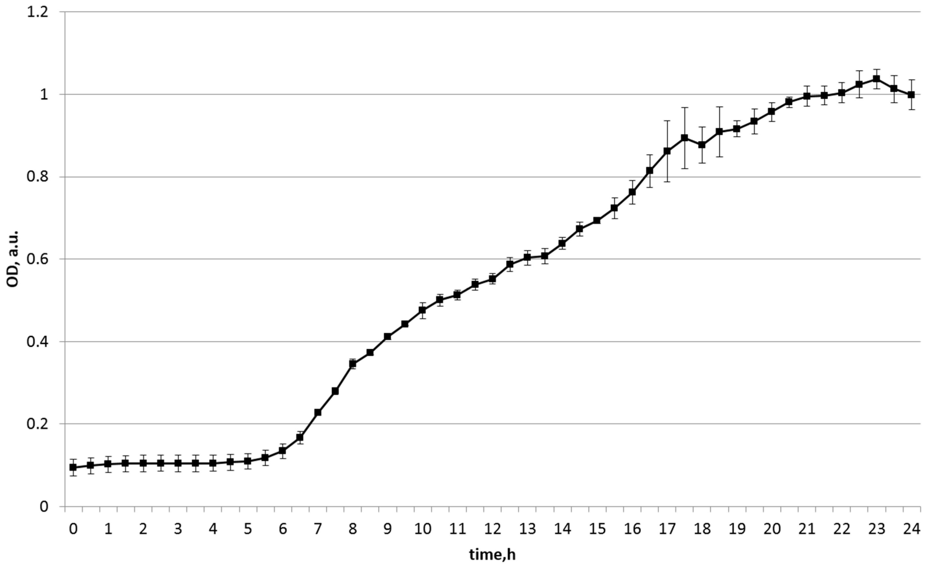
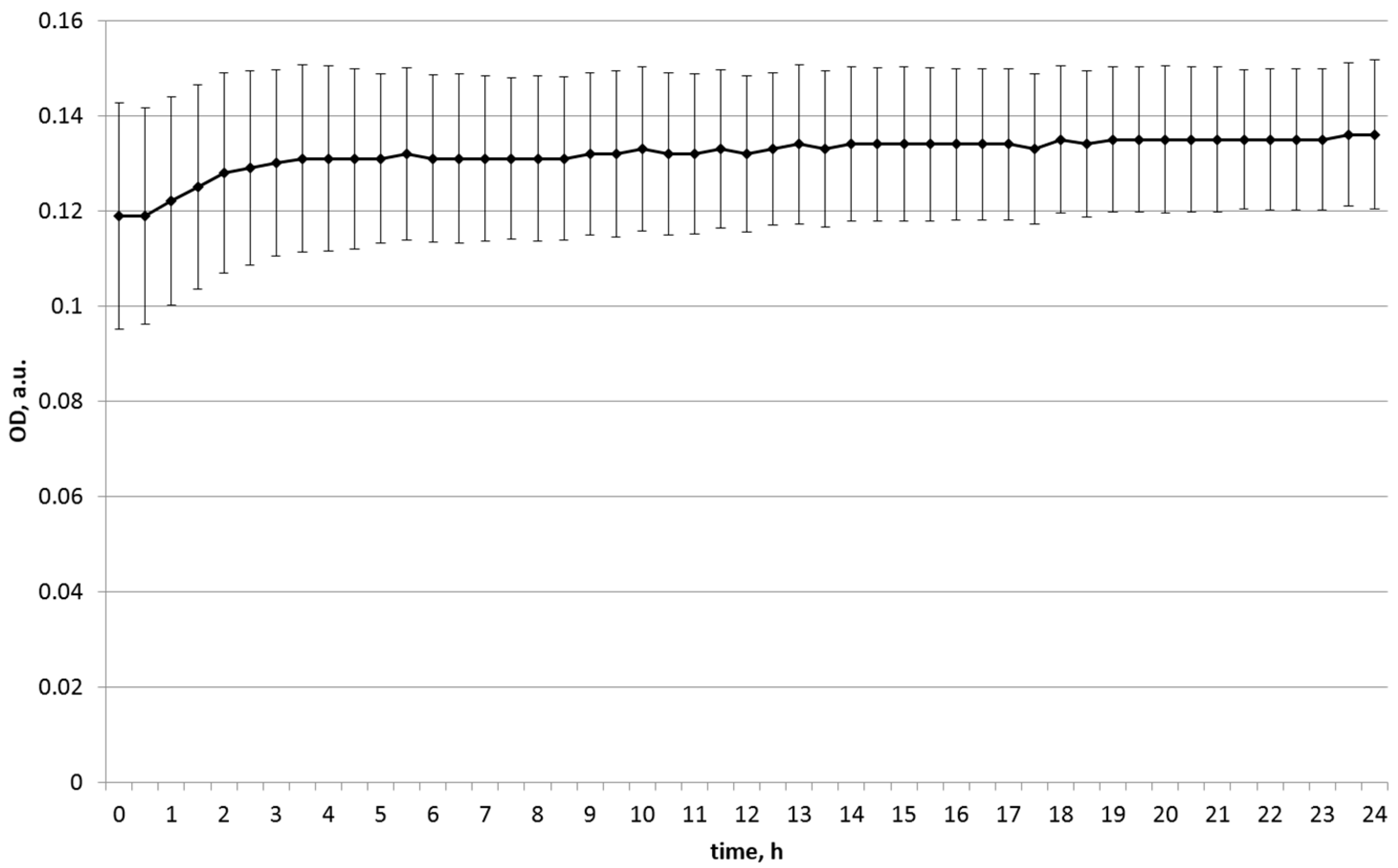
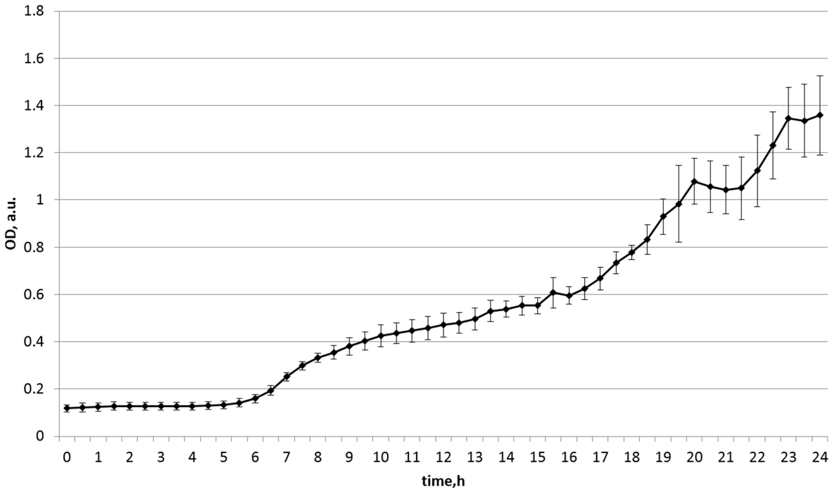
3.2. Selection of the Optimal Substrate

The results of selecting the optimal medium for the B. licheniformis CL 33 strain are shown in Figure 1, Figure 2, Figure 3 and Figure 4 and in Table 2 and Table 3.
As can be seen from the presented data, the most intensive growth of strain CL 33 cells was observed on soy medium. Different initial density was associated with the difference in the initial turbidity of the media due to the substrate; thus, the final density relative to the initial density was estimated, rather than the absolute value of the density. Pea medium was the second-most effective after soy medium, while no significant growth was observed on the other media.
The growth dynamic of B. licheniformis 56 was quite similar.
The most intensive growth of strain CL 56 cells was also observed on soy medium. The chickpea medium appeared to be the second-most effective, while no significant increase was observed on the other media. Thus, we can recommend the soy medium as optimal for growing these strains. On the basis of these results, heat-treated hydrated soybeans were used as a substrate for the production of the preparation.

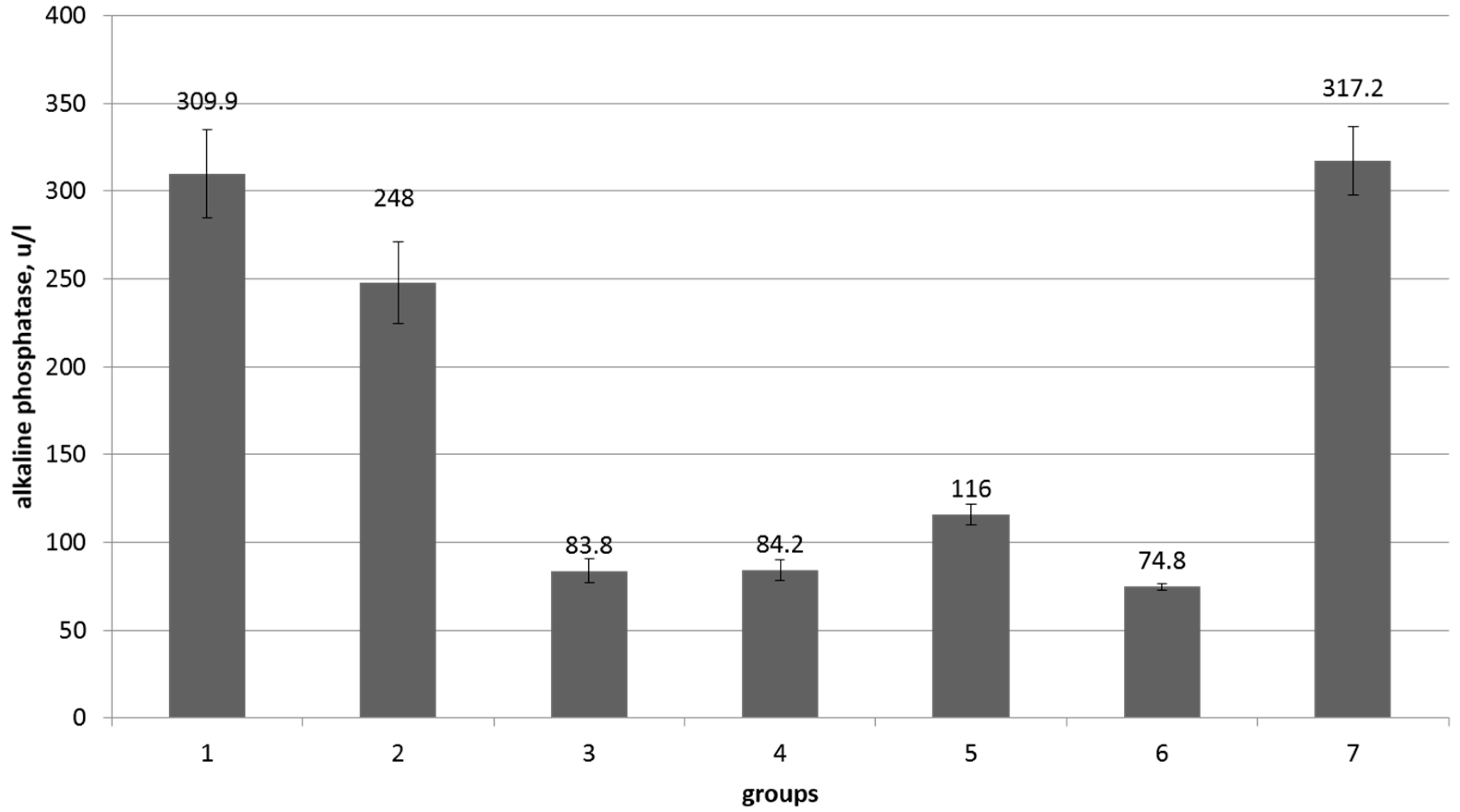
3.3. Influence of the Synbiotic Preparation on the Physiological Parameters of Cattle

The initial morphological and biochemical parameters of the blood of animals in all seven groups were within the reference values. The values of clinical indicators (temperature, pulse, respiration) during the experiment were within the reference values and did not differ statistically when compared between groups. A statistically significant reduction in weight gain was noted in the mycotoxin treated group. The introduction of a synbiotic preparation into the feed leveled this effect (Table 4). At the same time, the average body weight of animals of all experimental and control groups was within the reference values and did not have statistically significant differences with the control group.
At the end of the experiment in all experimental groups, the morphological and biochemical parameters of blood were within acceptable normative values. Blood parameters that did not have significant differences and deviations from the norm are given in Supplementary Tables. The only significant difference was observed in the level of alkaline phosphatase (Figure 5).

4. Discussion

Probiotics are used in cattle breeding to compensate for adverse external influences due to their ability to modulate the immune system and the functioning of the nervous system, reduce the quantities of toxic substances in the blood, participate in the digestion of food at different stages of the digestion process in cows, and also contribute to the normalization of the microbiota [21]. A promising direction is the use of feed with the inclusion of spore-forming probiotic cultures, as well as probiotics based on spore-forming bacteria. In particular, Bacillus strains in the spore stage are resistant to high temperatures and survive the processes of extrusion, granulation and expansion. The dormant spore stage allows these probiotics to have longer shelf life without the danger of losing properties. In commercial preparations, two types of strains are mainly used: Bacillus subtilis and Bacillus licheniformis. They belong to the transient microbiota, which means they should not populate the digestive tract [22].
It appears that the optimal medium for the cultivation of probiotic strains can be prepared based on soybeans. This may be due to the fact that the soy substrate promotes increased production of bioactive lipopeptides in bacilli [23], and thus is a suitable component/substrate for a synbiotic preparation. Soybean meal, soy hydrolysate [24] and soybean flour [25] have also been used as substrates to increase surfactin and other lipopeptides production by Bacillus. In general, oily substrates can be applied in combination with hydrophilic substrates to enhance biosurfactant production [26]. Furthermore, the quantitative and qualitative effect of water immiscible and miscible carbon-rich substrates on the production of biosurfactants has been studied [27], showing that these types of substrates can increase lipopeptide production.
A decrease in weight gain was noted in the group treated with mycotoxins, which is natural, since the action of toxins disrupts the physiological processes of animals. It was shown that the introduction of a synbiotic preparation into the feed reduced this effect. At the same time, the average body weight of animals of all experimental and control groups was within the reference values and did not have statistically significant differences with the control group. Thus, it was shown that the introduction of the synbiotic preparation reduces the harm from the presence of mycotoxins in animal feed.
It is known that in the rumen of cattle, food undergoes microbial fermentation, and only after that does it enter the rest of the stomach and intestines. The rumen is inhabited by a diverse microbiota, which not only digests cellulose and forms the necessary protein, but can also subject the substances contained in the feed to various modifications. Rumen microbial composition influences feed efficiency [28], CH4 emission rate [29], animal health [30] and milk composition [31]. The term “feed efficiency” describes the efficiency with which feed is converted into usable product, and the ratio of feed efficiency is an inherited trait in cattle [32]. Due to the reduced pH, high humidity and temperature, conditions are created in the rumen that are suitable for the development of microorganisms, including those belonging to the genus Bacillus. By adding probiotic bacteria to the feed, we contribute to the enrichment of the rumen microbiota with these strains. Mycotoxins that enter the rumen along with the food, in fact, due to the processes taking place in the rumen, are incubated for quite a long time together with mycotoxin-degrading bacilli, which can significantly reduce the toxicity of the feed.
Bacilli can fight mycotoxins in several ways. It is reported that Bacillus strains isolated from the aquatic environment of the Brazilian Amazon demonstrated activity against 7 mycotoxigenic fungi [33]. Product of BsDyP gene from Bacillus subtilis SCK6 has been shown to enzymatically degrade multiple major mycotoxins [34]. Fengycin produced by Bacillus amyloliquefaciens FZB42 has been shown to inhibit Fusarium graminearum growth and biosynthesis of its associated mycotoxins [35]. Certain Bacillus spp. have also demonstrated decontamination potential on multiple Fusarium mycotoxins [36], while other species, such as B. cereus 342-2, have been investigated for their biocontrol properties against toxigenic mycobiota and associated toxins found in date palm fruits [37]. Additionally, B. subtilis BIOUFLA2 has been evaluated for its ability to reduce fungal colonization and subsequent contamination with rot and/or mycotoxin levels in kernels used for animal feed production [38].
The strains used in the preparation were characterized as having quorum-quenching activity. It is known that some microorganisms have a system of protection against the negative effects of pathogens, based on the destruction of signals from the quorum-sensing system; in particular, homoserine lactones. This enzyme can hydrolyze the lactone ring and the amide bond of AHL into inactive molecules and thereby block quorum-sensing systems and reduce symptoms caused by pathogens [8].
Enzymes capable of inactivating lactones are also able to destroy mycotoxins that include the lactone ring, therefore, selection for this trait makes it possible to find strains that can not only disrupt the sense of quorum in potential pathogens, but also protect the host from mycotoxins from food.
Among bacteria of the genus Bacillus, genes encoding AHL lactonase (for example, the aiiA gene) are widespread, which makes these microorganisms a promising object for study. Microorganisms belonging to the genus Bacillus also contain the laccase gene, which has quorum-inhibiting activity, cotA [39].
It can be assumed that the inactivation of mycotoxins was not the only effect of these metabolites, but was also accompanied by a violation of intercellular signaling, which reduces the activity of potential pathogens.
The effect of reducing the content of alkaline phosphatase in the serum of animals deserves a separate discussion. Although when mycotoxins were added to the feed, it decreased slightly compared to the control, it should be noted that its level in the control group was initially high (309.9 units/L), while the norm for cattle is 55–80 units/L [40], and the use of probiotic and synbiotic preparations led to a decrease to exactly this value (except for the group that received a large dose of synbiotic and mycotoxins). This phenomenon was likely due to the systemic effects of probiotics. The increase in control enzyme levels was likely due to sub-optimal diet and should be addressed in further studies. Interestingly, the alkaline phosphatase-lowering effect of probiotics has been observed in human studies [41] and is indicative of improved liver health.
Comparing the effects obtained by us with similar studies, it can be seen that the action of our drug fits into the well-known paradigm of the action of probiotics. It was shown that probiotic bacteria are able to inactivate, adsorb or degrade such substances as AFB1—aflatoxin B1, AFs—aflatoxins, AFB2—aflatoxin B2, AFG1—aflatoxin G1, AFG2—aflatoxin G2 and AFM1—aflatoxin M1 [42]. However, according to this review, most studies in this area have been conducted on strains of lactobacilli, while other bacteria and yeasts are found in a few isolated studies, including as part of complex preparations. Much of this research has been carried out in rat models and not in livestock.
There are in vitro studies on Bacillus strains, including a study on B. licheniformis showing activity against AFB1 [43], a study characterizing a B. licheniformis strain with ZEA-removal ability and a B. amyloliquefaciens strain with the same activity [44] and a study on Bacillus subtilis and Bacillus natto [45]. Authors [46] studied the combination of B. subtilis, Lactobacillus casei and Candida utilis to eliminate AFB1 and ZEA from animal feeding.
As for in vivo studies, there are data on ducks [47] and chickens [48]. No in vivo studies have been performed on heifers in the manner of this research. It is interesting to note, however, a parallel with [49], where the authors noted that bull calves prefer food enriched with the probiotic B. amyloliquefaciens H57 over conventional food. The authors attribute this to the ability of the probiotic to prevent the formation of harmful substances including the toxic metabolites of fungi, in the feed that change its organoleptic qualities.
It is worth noting that CL 33 and CL 56 strains contain metabolites that can provide other probiotic properties of Bacillus strains. Thus, lichenicidin is a two-peptide lantibiotic produced by Bacillus licheniformis [50]. It is composed of the two peptides Blia and Blib, which are linked together by lanthionine bridges [51]. It has antimicrobial activity against Gram-positive bacteria and can inhibit the growth of clinically relevant strains, such as methicillin-resistant Staphylococcus aureus (MRSA) [52]. The bactericidal activity of lantibiotics is based on specific binding to the peptidoglycan precursor lipid II, which predetermines its antimicrobial activity [53]. Lichenicidin has been shown to increase the net surface charge of S. aureus cells as measured by zeta potential without achieving electrical neutralization. In addition, lichenicidin causes disturbances in the cell surface of pathogen cells, which leads to leakage of internal contents, as observed using atomic force microscopy [54].
Lichenisin is known as a surface active biosurfactant [55]. Lichenisins are anionic cyclic lipoheptapeptide biosurfactants produced by Bacillus licheniformis on a carbohydrate-free medium with extreme glucose. They have the ability to reduce the surface tension of water from 72 to 27 mN/m [56].
Fengycin(s) is a type of lipopeptide produced by Bacillus species, which has been extensively explored for its biological control properties. It was shown to be able to cause alterations in the bacterial surface topography [57], promote the formation of pores and cause leakage from pathogen cells [58]. Among other properties, fengycins can reduce the mitochondrial membrane potential, induce production of reactive oxygen species (ROS) and downregulate antioxidant enzyme production in fungi cells. Fengycin BS155 can cause chromatin condensation in fungal hyphal cells, which leads to significant changes in protein expression [59]. Fengycins have antimicrobial activity against a wide range of fungi and bacteria [59,60,61].
Bacillibactin is a catechol-type siderophore. It has been reported to have antibacterial, antifungal and cytotoxic activity [62], as well as broad-spectrum biocontrol potential [63]. Some forms show cytotoxicities against human cancer cell lines HepG2 and MCF7 [64]. Additionally, it may be involved in the production of other antimicrobial compounds, such as fengycin, surfactin, bacillaene and bacillomycin [61].
These compounds can modulate the composition of the microbiota, leading to systemic beneficial effects in animals. There are also suggestions that some of the antimicrobial peptides have a dual effect and can also be used by microorganisms as signaling molecules at naturally achievable subinhibitory concentrations [65].

5. Conclusions

The optimal substrate was selected for obtaining a synbiotic preparation based on two strains of B. licheniformis. According to the results of experiments carried out on heifers, the introduction of substrates affected by toxin-forming micromycetes into feed led to a decrease in weight gain, but the introduction of a synbiotic drug, especially at a high dose, leveled this negative effect, bringing it to the level of the control groups. There was also a decrease in the level of alkaline phosphatase in serum to normal values, which may characterize probiotic effects of the drug not associated with the elimination of the effect of mycotoxins.
In the genomes of the studied bacteria, genes for the synthesis of compounds with antimicrobial and other biologically significant activity were found. Their presence may provide the effects of the drug through an indirect effect, that is, through the modulation of the composition of the rumen microbiota.
The results of the study open up prospects for the creation of a new generation of synbiotic preparations for veterinary medicine, which will not only have a general systemic positive effect on the animal organism, but also reduce the negative effects of feed contamination with mycotoxins.

Supplementary Materials

The following supporting information can be downloaded at: https://www.mdpi.com/article/10.3390/agriculture13030523/s1. Table S1: Initial morphological and biochemical blood parameters of experimental animals; Table S2: Morphological and biochemical parameters of the blood of experimental animals at the end of the experiment.

Author Contributions

Conceptualization, V.C. and A.G.; methodology, M.M. and A.B.; software, Y.D. and D.R.; experiments, M.M., E.P. and A.G.; genomic analysis, E.P. and V.P.; manuscript draft, Y.D., A.B. and A.O.; manuscript editing, E.P. and A.B. All authors have read and agreed to the published version of the manuscript.

Funding

This research was funded by Agrokholod LLC, and Ministry of Industry and Trade of the Russian Federation through Agreement No. 020-11-2020-2040. EVP was also funded by the Ministry of Science and Higher Education of the Russian Federation, agreement no. 075-15-2022-1122. M.M. and A.B. were funded by the Strategic Academic Leadership Program of the Southern Federal University (“Priority 2030”).

Institutional Review Board Statement

The animal study protocol was approved by the Commission on Bioethics of the Southern Federal University Protocol No. 3-14 dated 17 February 2014 and Protocol No. 2 dated 18 April 2022.

Data Availability Statement

The data presented in this study are available on request from the corresponding author.

Acknowledgments

Animals and the place for their maintenance were provided by Agrokholod LLC. Morphological and biochemical blood tests were carried out in the laboratory of the North Caucasian Zonal Research Veterinary Institute.

Conflicts of Interest

The authors declare no conflict of interest.

References

  1. Anater, A.; Manyes, L.; Meca, G.; Ferrer, E.; Luciano, F.B.; Pimpão, C.T.; Font, G. Mycotoxins and their consequences in aquaculture: A review. Aquaculture 2016, 451, 1–10. [Google Scholar] [CrossRef]
  2. Connolly, L.; Ropstad, E.; Verhaegen, S. In vitro bioassays for the study of endocrine-disrupting food additives and contaminants. TrAC Trends Anal. Chem. 2011, 30, 227–238. [Google Scholar] [CrossRef] [Green Version]
  3. Kępińska-Pacelik, J.; Biel, W. Alimentary Risk of Mycotoxins for Humans and Animals. Toxins 2021, 13, 822. [Google Scholar] [CrossRef]
  4. Cortinovis, C.; Pizzo, F.; Spicer, L.J.; Caloni, F. Fusarium mycotoxins: Effects on reproductive function in domestic animals-A review. Theriogenology 2013, 80, 557–564. [Google Scholar] [CrossRef]
  5. Mihalache, O.A.; Dellafiora, L.; Dall’Asta, C. Assessing the Mycotoxin-related Health Impact of Shifting from Meat-based Diets to Soy-based Meat Analogues in a Model Scenario Based on Italian Consumption Data. Expo. Health 2022, 1–15. [Google Scholar] [CrossRef]
  6. Enguita, F.J.; Martins, L.O.; Henriques, A.O.; Carrondo, M.A. Crystal structure of a bacterial endospore coat component. A laccase with enhanced thermostability properties. J. Biol. Chem. 2003, 278, 19416–19425. [Google Scholar] [CrossRef] [Green Version]
  7. Liu, D.; Momb, J.; Thomas, P.W.; Moulin, A.; Petsko, G.A.; Fast, W.; Ringe, D. Mechanism of the Quorum-Quenching Lactonase (AiiA) from Bacillus thuringiensis. Product-Bound Structures. Biochemistry 2008, 47, 7706–7714. [Google Scholar] [CrossRef] [Green Version]
  8. Rasch, M.; Buch, C.; Austin, B.; Slierendrecht, W.J.; Ekmann, K.S.; Larsen, J.L.; Johansen, C.; Riedel, K.; Eberl, L.; Givskov, M.; et al. An inhibitor of bacterial quorum sensing reduces mortalities caused by Vibriosis in Rainbow Trout (Oncorhynchus mykiss, Walbaum). Syst. Appl. Microbiol. 2004, 27, 350–359. [Google Scholar] [CrossRef]
  9. Pan, J.; Huang, T.; Yao, F.; Huang, Z.; Powell, C.A.; Qiu, S.; Guan, X. Expression and characterization of aiiA gene from Bacillus subtilis BS-1. Microbiol. Res. 2008, 163, 711–716. [Google Scholar] [CrossRef]
  10. Ming, L.-J.; Epperson, J.D. Metal binding and structure-activity relationship of the metalloantibiotic peptide bacitracin. J. Inorg. Biochem. 2002, 91, 46–58. [Google Scholar] [CrossRef]
  11. Tazehabadi, M.H.; Algburi, A.; Popov, I.V.; Ermakov, A.M.; Chistyakov, V.A.; Prazdnova, E.V.; Weeks, R.; Chikindas, M.L. Probiotic Bacilli inhibit Salmonella biofilm formation without killing planktonic cells. Front. Microbiol. 2021, 12, 242. [Google Scholar] [CrossRef]
  12. Frei, R.; Akdis, M.; O’Mahony, L. Prebiotics, probiotics, synbiotics, and the immune system: Experimental data and clinical evidence. Curr. Opin. Gastroenterol. 2015, 31, 153–158. [Google Scholar] [CrossRef] [Green Version]
  13. Swanson, K.S.; Gibson, G.R.; Hutkins, R.; Reimer, R.A.; Reid, G.; Verbeke, K.; Scott, K.P.; Holscher, H.D.; Azad, M.B.; Delzenne, N.M.; et al. The International Scientific Association for Probiotics and Prebiotics (ISAPP) consensus statement on the definition and scope of synbiotics. Nat. Rev. Gastroenterol. Hepatol. 2020, 17, 687–701. [Google Scholar] [CrossRef]
  14. Yang, X.; Liu, D.; Liu, F.; Wu, J.; Zou, J.; Xiao, X.; Zhao, F.; Zhu, B. HTQC: A fast quality control toolkit for Illumina sequencing data. BMC Bioinform. 2013, 14, 33. [Google Scholar] [CrossRef] [Green Version]
  15. Bolger, A.M.; Lohse, M.; Usadel, B. Trimmomatic: A flexible trimmer for Illumina sequence data. Bioinformatics 2014, 30, 2114–2120. [Google Scholar] [CrossRef] [Green Version]
  16. Bankevich, A.; Nurk, S.; Antipov, D.; Gurevich, A.; Dvorkin, M.; Kulikov, A.S.; Lesin, V.; Nikolenko, S.; Pham, S.; Prjibelski, A.; et al. SPAdes: A New Genome Assembly Algorithm and Its Applications to Single-Cell Sequencing. J. Comput. Biol. 2012, 19, 455–477. [Google Scholar] [CrossRef] [Green Version]
  17. Seemann, T. Prokka: Rapid prokaryotic genome annotation. Bioinformatics 2014, 30, 2068–2069. [Google Scholar] [CrossRef] [Green Version]
  18. Blin, K.; Shaw, S.; Kloosterman, A.M.; Charlop-Powers, Z.; Van Wezel, G.P.; Medema, M.H.; Weber, T. antiSMASH 6.0: Improving cluster detection and comparison capabilities. Nucleic Acids Res. 2021, 49, W29–W35. [Google Scholar] [CrossRef]
  19. Fedorenko, G.M.; Fedorenko, A.G.; Chistyakov, V.A.; Prazdnova, E.V.; Usatov, A.V.; Chikindas, M.L.; Mazanko, M.S.; Weeks, R. Method of preparation, visualization and ultrastructural analysis of a formulation of probiotic Bacillus subtilis KATMIRA1933 produced by solid-phase fermentation. MethodsX 2019, 6, 2515–2520. [Google Scholar] [CrossRef]
  20. GOST 31653-2012; Feed. Method of Enzyme Immunoassay of Mycotoxins. Standardinform: Moscow, Russia, 2012.
  21. Wochner, K.F.; Becker-Algeri, T.A.; Colla, E.; Badiale-Furlong, E.; Drunkler, D.A. The action of probiotic microorganisms on chemical contaminants in milk. Crit. Rev. Microbiol. 2018, 44, 112–123. [Google Scholar] [CrossRef]
  22. Jeżewska-Frąckowiak, J.; Seroczyńska, K.; Banaszczyk, J.; Jedrzejczak, G.; Żylicz-Stachula, A.; Skowron, P.M. The promises and risks of probiotic Bacillus species. Acta Biochim. Pol. 2018, 65, 509–519. [Google Scholar] [CrossRef] [Green Version]
  23. Wang, Q.; Chen, S.; Zhang, J.; Sun, M.; Liu, Z.; Yu, Z. Co-producing lipopeptides and poly-γ-glutamic acid by solid-state fermentation of Bacillus subtilis using soybean and sweet potato residues and its biocontrol and fertilizer synergistic effects. Bioresour. Technol. 2008, 99, 3318–3323. [Google Scholar] [CrossRef]
  24. Rangarajan, V.; Clarke, K.G. Process development and intensification for enhanced production of Bacillus lipopeptides. Biotechnol. Genet. Eng. Rev. 2015, 31, 46–68. [Google Scholar] [CrossRef]
  25. Umar, A.; Zafar, A.; Wali, H.; Siddique, M.P.; Qazi, M.A.; Naeem, A.H.; Malik, Z.A.; Ahmed, S. Low-cost production and application of lipopeptide for bioremediation and plant growth by Bacillus subtilis SNW3. AMB Express 2021, 11, 165. [Google Scholar] [CrossRef]
  26. Subsanguan, T.; Khondee, N.; Rongsayamanont, W.; Luepromchai, E. Formulation of a glycolipid: Lipopeptide mixture as biosurfactant-based dispersant and development of a low-cost glycolipid production process. Sci. Rep. 2022, 12, 16353. [Google Scholar] [CrossRef]
  27. Ndlovu, T.; Rautenbach, M.; Khan, S.; Khan, W. Variants of lipopeptides and glycolipids produced by Bacillus amyloliquefaciens and Pseudomonas aeruginosa cultured in different carbon substrates. AMB Express 2017, 7, 109. [Google Scholar] [CrossRef] [Green Version]
  28. Li, F.; Guan, L.L. Metatranscriptomic profiling reveals linkages between the active rumen microbiome and feed efficiency in beef cattle. Appl. Environ. Microbiol. 2017, 83, e00061-17. [Google Scholar] [CrossRef] [Green Version]
  29. Kittelmann, S.; Pinares-Patiño, C.S.; Seedorf, H.; Kirk, M.R.; Ganesh, S.; McEwan, J.C.; Janssen, P.H. Two different bacterial community types are linked with the low-methane emission trait in sheep. PLoS ONE 2014, 9, e103171. [Google Scholar] [CrossRef]
  30. Silberberg, M.; Chaucheyras-Durand, F.; Commun, L.; Mialon, M.M.; Monteils, V.; Mosoni, P.; Morgavi, D.P.; Martin, C. Repeated acidosis challenges and live yeast supplementation shape rumen microbiota and fermentations and modulate inflammatory status in sheep. Animal 2013, 7, 1910–1920. [Google Scholar] [CrossRef] [Green Version]
  31. Jami, E.; White, B.A.; Mizrahi, I. Potential role of the bovine rumen microbiome in modulating milk composition and feed efficiency. PLoS ONE 2014, 9, e85423. [Google Scholar] [CrossRef]
  32. Berry, D.P.; Crowley, J.J. Cell biology symposium: Genetics of feed efficiency in dairy and beef cattle. J. Anim. Sci. 2013, 91, 1594–1613. [Google Scholar] [CrossRef] [Green Version]
  33. Veras, F.F.; Correa AP, F.; Welke, J.E.; Brandelli, A. Inhibition of mycotoxin-producing fungi by Bacillus strains isolated from fish intestines. Int. J. Food Microbiol. 2016, 238, 23–32. [Google Scholar] [CrossRef]
  34. Qin, X.; Su, X.; Tu, T.; Zhang, J.; Wang, X.; Wang, Y.; Wang, Y.; Bai, Y.; Yao, B.; Luo, H.; et al. Enzymatic degradation of multiple major mycotoxins by dye-decolorizing peroxidase from Bacillus subtilis. Toxins 2021, 13, 429. [Google Scholar] [CrossRef]
  35. Hanif, A.; Zhang, F.; Li, P.; Li, C.; Xu, Y.; Zubair, M.; Zhang, M.; Jia, D.; Zhao, X.; Liang, J.; et al. Fengycin produced by Bacillus amyloliquefaciens FZB42 inhibits Fusarium graminearum growth and mycotoxins biosynthesis. Toxins 2019, 11, 295. [Google Scholar] [CrossRef] [Green Version]
  36. Hassan, Z.U.; Al Thani, R.; Alsafran, M.; Migheli, Q.; Jaoua, S. Selection of Bacillus spp. with decontamination potential on multiple Fusarium mycotoxins. Food Control 2021, 127, 108119. [Google Scholar] [CrossRef]
  37. Abdallah, Y.; Ul Hassan, Z.; Al-Thani, R.; Al-Shamary, N.; Al-Yafei, T.; Alnaimi, H.; Jaoua, S. Prevalence of toxigenic mycobiota and mycotoxins in date palm fruits and investigation on Bacillus cereus 342-2 as biocontrol agent. Biocontrol Sci. Technol. 2022, 32, 1372–1388. [Google Scholar] [CrossRef]
  38. Guimarães, R.A.; Pherez-Perrony, P.E.; Müller, H.; Berg, G.; Medeiros FH, V.; Cernava, T. Microbiome-guided evaluation of Bacillus subtilis BIOUFLA2 application to reduce mycotoxins in maize kernels. Biol. Control 2020, 150, 104370. [Google Scholar] [CrossRef]
  39. McKenney, P.T.; Driks, A.; Eichenberger, P. The Bacillus subtilis endospore: Assembly and functions of the multilayered coat. Nat. Rev. Microbiol. 2012, 11, 33–44. [Google Scholar] [CrossRef]
  40. Ghazali, M.F.; Koh-Tan, H.H.; McLaughlin, M.; Montague, P.; Jonsson, N.N.; Eckersall, P.D. Alkaline phosphatase in nasal secretion of cattle: Biochemical and molecular characterisation. BMC Vet. Res. 2014, 10, 204. [Google Scholar] [CrossRef] [Green Version]
  41. Rodrigo, T.; Dulani, S.; Nimali, S.S.; De Silva, A.P.; Fernando, J.; De Silva, H.J.; Wickramasinghe, V.P. Effects of probiotics combined with dietary and lifestyle modification on clinical, biochemical, and radiological parameters in obese children with nonalcoholic fatty liver disease/nonalcoholic steatohepatitis: A randomized clinical trial. Clin. Exp. Pediatr. 2022, 65, 304. [Google Scholar] [CrossRef]
  42. Baralić, K.; Živančević, K.; Bozic, D.; Đukić-Ćosić, D. Probiotic cultures as a potential protective strategy against the toxicity of environmentally relevant chemicals: State-of-the-art knowledge. Food Chem. Toxicol. 2022, 172, 113582. [Google Scholar] [CrossRef]
  43. Hsu, T.C.; Yi, P.J.; Lee, T.Y.; Liu, J.R. Probiotic characteristics and zearalenone removal ability of a Bacillus licheniformis strain. PLoS ONE 2018, 13, e0194866. [Google Scholar] [CrossRef]
  44. Ju, J.; Tinyiro, S.E.; Yao, W.; Yu, H.; Guo, Y.; Qian, H.; Xie, Y. The ability of Bacillus subtilis and Bacillus natto to degrade zearalenone and its application in food. J. Food Process Preserv. 2019, 43, e14122. [Google Scholar] [CrossRef]
  45. Lee, A.; Cheng, K.C.; Liu, J.R. Isolation and characterization of a Bacillus amyloliquefaciens strain with zearalenone removal ability and its probiotic potential. PLoS ONE 2017, 12, e0182220. [Google Scholar] [CrossRef] [Green Version]
  46. Huang, W.; Chang, J.; Wang, P.; Liu, C.; Yin, Q.; Zhu, Q.; Lu, F.; Gao, T. Effect of the combined compound probiotics with mycotoxin–degradation enzyme on detoxifying aflatoxin B1 and zearalenone. J. Toxicol. Sci. 2018, 43, 377–385. [Google Scholar] [CrossRef] [Green Version]
  47. Zhang, L.; Ma, Q.; Ma, S.; Zhang, J.; Jia, R.; Ji, C.; Zhao, L. Ameliorating effects of Bacillus subtilis ANSB060 on growth performance, antioxidant functions, and aflatoxin residues in ducks fed diets contaminated with aflatoxins. Toxins 2016, 9, 1. [Google Scholar] [CrossRef] [Green Version]
  48. Zhang, N.-Y.; Qi, M.; Zhao, L.; Zhu, M.-K.; Guo, J.; Liu, J. Curcumin prevents aflatoxin B1 hepatoxicity by inhibition of cytochrome P450 isozymes in chick liver. Toxins 2016, 8, 372. [Google Scholar] [CrossRef] [Green Version]
  49. Ngo, T.T.; Bang, N.N.; Dart, P.; Callaghan, M.; Klieve, A.; Hayes, B.; McNeill, D. Feed preference response of weaner bull calves to Bacillus amyloliquefaciens H57 probiotic and associated volatile organic compounds in high concentrate feed pellets. Animals 2020, 11, 51. [Google Scholar] [CrossRef]
  50. Begley, M.; Cotter, P.D.; Hill, C.; Ross, R.P. Identification of a novel two-peptide lantibiotic, lichenicidin, following rational genome mining for LanM proteins. Appl. Environ. Microbiol. 2009, 75, 5451–5460. [Google Scholar] [CrossRef] [Green Version]
  51. Caetano, T.; Krawczyk, J.M.; Mösker, E.; Süssmuth, R.D.; Mendo, S. Heterologous expression, biosynthesis, and mutagenesis of type II lantibiotics from Bacillus licheniformis in Escherichia coli. Chem. Biol. 2011, 18, 90–100. [Google Scholar] [CrossRef] [Green Version]
  52. Barbosa, J.C.; Silva, Í.C.; Caetano, T.; Mösker, E.; Seidel, M.; Lourenço, J.; Süssmuth, R.D.; Santos, N.C.; Gonçalves, S.; Mendo, S. Assessing the potential of the two-peptide lantibiotic lichenicidin as a new generation antimicrobial. World J. Microbiol. Biotechnol. 2022, 38, 18. [Google Scholar] [CrossRef]
  53. Panina, I.S.; Balandin, S.V.; Tsarev, A.V.; Chugunov, A.O.; Tagaev, A.A.; Finkina, E.I.; Antoshina, D.V.; Sheremeteva, E.V.; Paramonov, A.S.; Rickmeyer, J.; et al. Specific Binding of the α-Component of the Lantibiotic Lichenicidin to the Peptidoglycan Precursor Lipid II Predetermines Its Antimicrobial Activity. Int. J. Mol. Sci. 2023, 24, 1332. [Google Scholar] [CrossRef]
  54. Barbosa, J.C.; Gonçalves, S.; Makowski, M.; Silva, Í.C.; Caetano, T.; Schneider, T.; Mendo, S. Insights into the mode of action of the two-peptide lantibiotic lichenicidin. Colloids Surf. B Biointerfaces 2022, 211, 112308. [Google Scholar] [CrossRef]
  55. Coronel-León, J.; de Grau, G.; Grau-Campistany, A.; Farfan, M.; Rabanal, F.; Manresa, A.; Marqués, A.M. Biosurfactant production by AL 1.1, a Bacillus licheniformis strain isolated from Antarctica: Production, chemical characterization and properties. Ann. Microbiol. 2015, 65, 2065–2078. [Google Scholar] [CrossRef]
  56. Anuradha, S.N. Structural and molecular characteristics of lichenysin and its relationship with surface activity. Biosurfactants 2010, 672, 304–315. [Google Scholar] [CrossRef]
  57. Medeot, D.B.; Fernandez, M.; Morales, G.M.; Jofré, E. Fengycins from Bacillus amyloliquefaciens MEP218 exhibit antibacterial activity by producing alterations on the cell surface of the pathogens Xanthomonas axonopodis pv. vesicatoria and Pseudomonas aeruginosa PA01. Front. Microbiol. 2020, 10, 3107. [Google Scholar] [CrossRef] [PubMed] [Green Version]
  58. Patel, H.; Tscheka, C.; Edwards, K.; Karlsson, G.; Heerklotz, H. All-or-none membrane permeabilization by fengycin-type lipopeptides from Bacillus subtilis QST713. Biochim. Et Biophys. Acta (BBA)-Biomembr. 2011, 1808, 2000–2008. [Google Scholar] [CrossRef] [Green Version]
  59. Zhang, L.; Sun, C. Fengycins, cyclic lipopeptides from marine Bacillus subtilis strains, kill the plant-pathogenic fungus Magnaporthe grisea by inducing reactive oxygen species production and chromatin condensation. Appl. Environ. Microbiol. 2018, 84, e00445-18. [Google Scholar] [CrossRef] [Green Version]
  60. González-Jaramillo, L.M.; Aranda, F.J.; Teruel, J.A.; Villegas-Escobar, V.; Ortiz, A. Antimycotic activity of fengycin C biosurfactant and its interaction with phosphatidylcholine model membranes. Colloids Surf. B Biointerfaces 2017, 156, 114–122. [Google Scholar] [CrossRef]
  61. Su, Z.; Chen, X.; Liu, X.; Guo, Q.; Li, S.; Lu, X.; Zhang, X.; Wang, P.; Dong, L.; Zhao, W.; et al. Genome mining and UHPLC–QTOF–MS/MS to identify the potential antimicrobial compounds and determine the specificity of biosynthetic gene clusters in Bacillus subtilis NCD-2. BMC Genom. 2020, 21, 767. [Google Scholar] [CrossRef]
  62. Hertlein, G.; Müller, S.; Garcia-Gonzalez, E.; Poppinga, L.; Süssmuth, R.D.; Genersch, E. Production of the catechol type siderophore bacillibactin by the honey bee pathogen Paenibacillus larvae. PLoS ONE 2014, 9, e108272. [Google Scholar] [CrossRef] [PubMed] [Green Version]
  63. Chen, L.; Heng, J.; Qin, S.; Bian, K. A comprehensive understanding of the biocontrol potential of Bacillus velezensis LM2303 against Fusarium head blight. PLoS ONE 2018, 13, e0198560. [Google Scholar] [CrossRef] [Green Version]
  64. Zhou, M.; Liu, F.; Yang, X.; Jin, J.; Dong, X.; Zeng, K.W.; Liu, D.; Zhang, Y.; Ma, M.; Yang, D. Bacillibactin and bacillomycin analogues with cytotoxicities against human cancer cell lines from marine Bacillus sp. PKU-MA00093 and PKU-MA00092. Mar. Drugs 2018, 16, 22. [Google Scholar] [CrossRef] [Green Version]
  65. Vasilchenko, A.S.; Rogozhin, E.A. Subinhibitory Effects of Antimicrobial Peptides. Front. Microbiol. 2019, 10, 1160. [Google Scholar] [CrossRef] [Green Version]
Figure 1. Growth curve of B. licheniformis CL 33 on soybean medium.
Agriculture 13 00523 g001
Figure 2. Growth curve of B. licheniformis CL 33 on green pea medium.
Agriculture 13 00523 g002
Figure 3. Growth curve of B. licheniformis CL 56 on soybean medium.
Agriculture 13 00523 g003
Figure 4. Growth curve of B. licheniformis CL 56 on green pea medium.
Agriculture 13 00523 g004
Figure 5. Alkaline phosphatase levels in serum. Groups: 1—control, 2—control with mycotoxins, 3—positive control, 4—synbiotic, lower dose, 5—synbiotic, lower dose + mycotoxins, 6—synbiotic, high dose, 7—synbiotic, high dose + mycotoxins.
Agriculture 13 00523 g005
Table 1. Genes for the synthesis of secondary metabolites in the genomes of the studied strains.
StrainMetabolitePercentage of Similarity with a Known Analogue, %
B. licheniformis CL 33Lichenicidin100%
Lichenisin100%
Fengycin-like peptide53%
Bacillibactin-like peptide53%
Butyrosine-like metabolite7%
B. licheniformis CL 56Lichenicidin100%
Lichenisin100%
Bacillibactin-like peptide52%
Fengycin-like peptide53%
Table 2. Optical density (OD) of B. licheniformis CL 33 cultures on different media.
Substrate SourceStart ODMaximum ODFinal OD (24 h)
Soy0.0991.0370.999
Chickpea0.1450.9020.531
Bell pepper0.7380.8560.856
Zucchini0.1580.2540.254
Green pea0.1190.1360.136
Table 3. Optical density (OD) of B. licheniformis CL 56 cultures on different media.
Substrate SourceStart ODMaximum ODFinal OD (24 h)
soy0.1181.3581.358
chickpea0.1160.7330.567
bell pepper0.7320.8460.846
zucchini0.1250.3860.386
green pea0.1130.1440.144
Table 4. Body weight and indicators of the clinical status of experimental animals after the end of the experiment.
Indicators, UnitsReference ValuesControlControl + Myco-ToxinsCello-BacterinSynbiotic Low DoseSynbiotic Low Dose + MycotoxinsSynbiotic High DoseSynbiotic High Dose + Mycotoxins
Weight gain, kg-12.5 ± 1.29.6 ± 2.212.5 ± 1.413.2 ± 1.3 **11.9 ± 1.013.3 ± 1.3 *12.4 ± 1.2 *
Weight, kg280–330316.7 ± 2.5317.6 ± 3.8316.1 ± 2.0306.4 ± 26.4309.7 ± 3.6312.9 ± 3.2319.6 ± 2.8
Temperature, °C37.5–39.038.3 ± 0.0838.14 ± 0.0938.2 ± 0.0638.1 ± 0.1238.0 ± 0.0938.2 ± 0.0738.1 ± 0.04
Pulse beats/min.50–8078.8 ± 2.663.8 ± 2.175.7 ± 3.6670.1 ± 2.166 ± 1.673.7 ± 2.976.6 ± 2.8
Breathing in/min15–3021.5 ± 0.621.9 ± 1.220.6 ± 0.521.9 ± 1.122 ± 0.821.1 ± 1.323 ± 1.07
Differences from the option with mycotoxins: * p < 0.05 ** p < 0.01.
Disclaimer/Publisher’s Note: The statements, opinions and data contained in all publications are solely those of the individual author(s) and contributor(s) and not of MDPI and/or the editor(s). MDPI and/or the editor(s) disclaim responsibility for any injury to people or property resulting from any ideas, methods, instructions or products referred to in the content.

