Communication

First Report on Purpureocillium lilacinum Infection of Indoor-Cultivated Morel Primordia

1
Applied Mycology and Microbiology Department, MIGAL-Galilee Research Institute, Kiryat Shmona 11016, Israel
2
Department of Food Sciences, Tel Hai College, Upper Galilee 12210, Israel
Agriculture 2022, 12(5), 695; https://doi.org/10.3390/agriculture12050695
Submission received: 17 April 2022 / Revised: 13 May 2022 / Accepted: 13 May 2022 / Published: 14 May 2022
(This article belongs to the Special Issue Interactions between Microorganisms in Plant Diseases)

Abstract

:
The cultivation of morel mushrooms (Morchella spp.) outdoors or in controlled indoor systems is a relatively new practice, and infections are beginning to be observed. Infection of indoor-cultivated Morchella rufobrunnea initials (primordia) occurred at our research facilities in Israel. The mushroom initials turned brown, were covered with a dense white mycelium of a foreign fungus and were disintegrated soon after. The isolation of a fungal contaminant from the infected mushroom revealed small colonies with a pinkish spore zone on potato dextrose agar medium. Molecular identification using partial large subunit 28S ribosomal DNA and rRNA internal transcribed spacer sequences identified the fungus as Purpureocillium lilacinum. Inoculation of Morchella colony on agar plat with the isolated fungus caused browning and inhibition of mycelial growth. Inoculation of a healthy primordium with P. lilacinum spores resulted in its browning and deterioration. This is the first report of an infection of indoor-cultivated mushroom and the first showing P. lilacinum as a pathogen of morels.

1. Introduction

Morchella species mushrooms (morels) (Ascomycota, Pezizales) are known for their delicate taste and aroma and for their wide range of antioxidant and health-related biological activities [1,2]. These edible mushrooms are mostly harvested from their natural habitat. For over a century, efforts have been made to cultivate them. However, due to the complex life cycle of this genus and a limited understanding of the factors affecting the fruiting process, the first report on indoor morel cultivation was only published in 1982 by Ower [3], followed by Ower et al. [4] in 1986. Several companies (e.g., Gourmet Mushrooms Inc.) then successfully grew it in the United States. In 2010, a successful indoor cultivation of Morchella rufobrunnea in a soilless system was achieved at our research facilities in Israel [5]. At the same time, reports of outdoor cultivation systems began to emerge, mainly in China, as summarized by Liu et al. [6] and Sambyal and Singh [7], showing controlled fruitification of Morchella importuna, Morchella sextelata, Morchella eximia and Morchella conica.
With the increasing cultivation of morels in artificial systems, infections began to appear. Commercial indoor cultivation of morels in the United States was completely abandoned due to bacterial contamination and reduced output [8], although recently, reports on indoor cultivation are reemerging [9]. In China, home to the world’s largest outdoor cultivation of Morchella (over 1200 ha in 2015–2016), there have also been reports of infections caused by fungi, including Fusarium incarnatum–equiseti species complex [10], Diploöspora longispora [11], Paecilomyces penicillatus causing white mold disease and infecting cultivated M. importuna [12,13], as well as Cladobotryum protrusum [14], among other pathogens [15]. There are no sufficient studies regarding morel diseases in indoor cultivation systems to date.
In 2016, infected young fruiting bodies of morels were observed for the first time in the growing bed of our indoor cultivation facility. The growing bed surface was covered with white mycelium, which also infected the young fruiting bodies (ascocarps of up to 2 cm in height), resulting in ageing and disintegration after severe browning. The aim of this work was to isolate and identify the contaminating fungus and to study its possible pathogenicity to the morel primordia.

2. Materials and Methods

2.1. Isolation of the Contaminating Fungus

The suspected pathogenic contaminating fungus was isolated from infected young fruiting bodies of M. rufobrunnea developing in a growth chamber at 17–20 °C and 90% air humidity. The contaminated ascomata were cut to reveal surface-underlying tissue and smeared aseptically on rose-bengal agar medium (Oxoid, Basingstoke, UK) to obtain separate, defined colonies and to limit the spread of the isolated filamentous fungal colonies [16]. The medium was supplemented with 200 mg/L chloramphenicol to prevent bacterial growth. Plates were incubated at 22 °C, in darkness. After 3 days, individual colonies from germinated spores were observed. Spores from the colonies were re-inoculated on rose-bengal-chloramphenicol medium to obtain colonies originating from single spores. The mycelium from these colonies was transferred to fresh potato dextrose agar (PDA, Difco) medium and regrown.

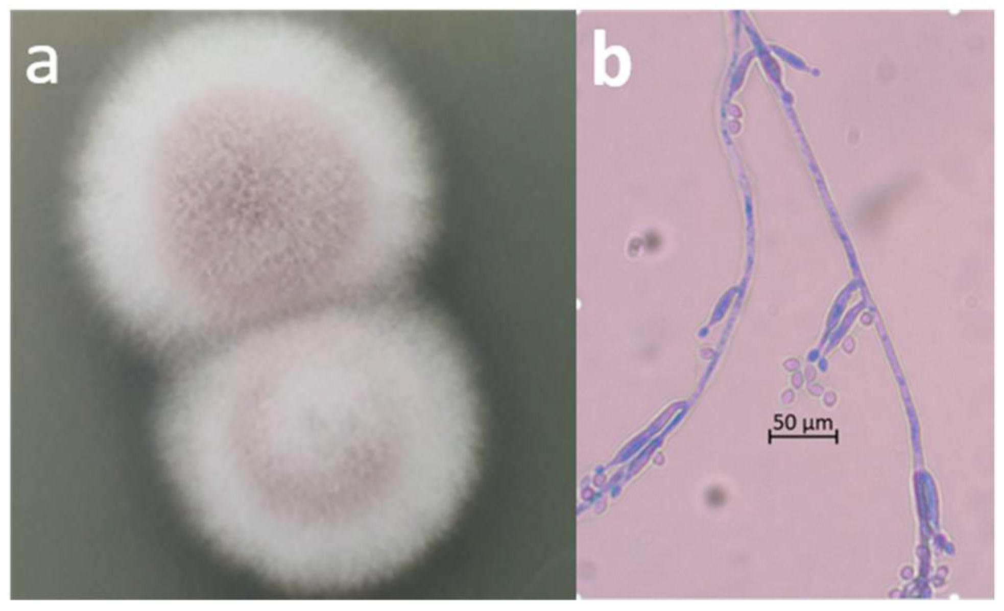
2.2. Microscopic Observation and Molecular Identification of the Contaminating Fungus

The morphological characterization of the isolated fungus was based on microscopic observation of 7-day-old colonies grown on PDA medium. A glass coverslip was overlaid on the surface of the colony and transferred to a microscope slide, where the fungus was stained with lactophenol cotton blue for microscopic observations.
For molecular identification of the fungus, fresh spores bearing mycelia were used. The surface part of the grown colony was removed from the agar medium, washed with sterile DW and centrifuged. Genomic DNA was extracted from the fungal pellet using the ZymoBIOMICSTM DNA Miniprep Kit (Zymo Research, Irvine, CA, USA) according to the manufacturer’s instructions. DNA isolation was performed for three single colonies growing on PDA. The primer pairs LROR + LR6 and ITS1 + ITS4 were used to amplify the large subunit (LSU) ribosomal DNA region and the rRNA internal transcribed spacer (ITS) region, respectively, for phylogenetic analyses [17]. PCR amplification was carried out using a Flexigene thermocycler (Techne, UK) under the conditions described by Raja et al. [17] using 10 µL HY-Taq ReadyMix *2x (Hylabs, Rehovot, Israel), 0.5 µL of each primer, 2 µL DNA (50 ng/µL) and 7 µL nuclease-free water. PCR reactions involved 35 cycles at 94 °C for 1 min, 54° for 1 min and 72° for 1 min. Amplification products were sequenced by HyLabs (Israel). The sequences of the partial 28S LSU gene and the rRNA ITS region of the isolated contaminant, designated 1925-1, were submitted to GenBank (accession numbers OK178301 and OL911053, respectively). The partial 28S LSU gene and rRNA ITS region sequences were assembled and edited by SeqMan program.

2.3. Pathogenicity Test (Koch’s Postulates)

The pathogenicity of the isolated P. lilacinum on Morchella was assayed by inoculation of either Morchella mycelial culture grown on PDA medium or the primordial stage of ascocarps produced in the growing system with P. lilacinum spores. In the first system, after 3 days of M. rufobrunnea mycelial growth at 22 °C on PDA medium, 2–5 µL of P. lilacinum spore suspension was applied on the colony surface. Changes in the colony morphology of M. rufobrunnea with continued incubation as above were monitored for 72 h. A control culture of healthy M. rufobrunnea without the isolated fungus was also grown. In the second assay, an aqueous suspension of 105 spores/mL from 7-day-old pre-cultured P. lilacinum was spray inoculated on freshly produced 3-day-old M. rufobrunnea primordia (3–8 mm long) in an indoor cultivation system. Phenotypic changes in the infected primordia were inspected. Three days after inoculation, the infected primordia were removed from the growing system and placed on PDA in a Petri dish to re-isolate the fungus and enhance the pathogenicity test. In addition, the contaminating fungus was re-isolated directly from infected tissue of the primordia as described above in Section 2.1.

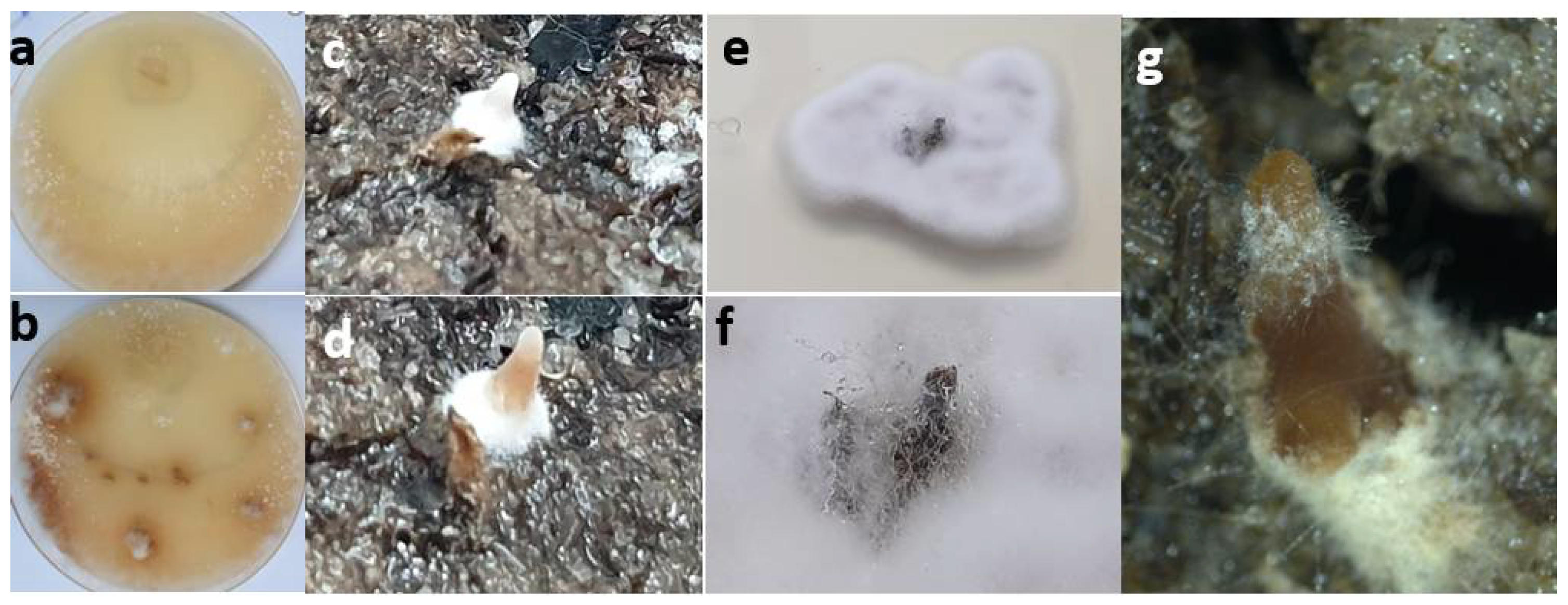
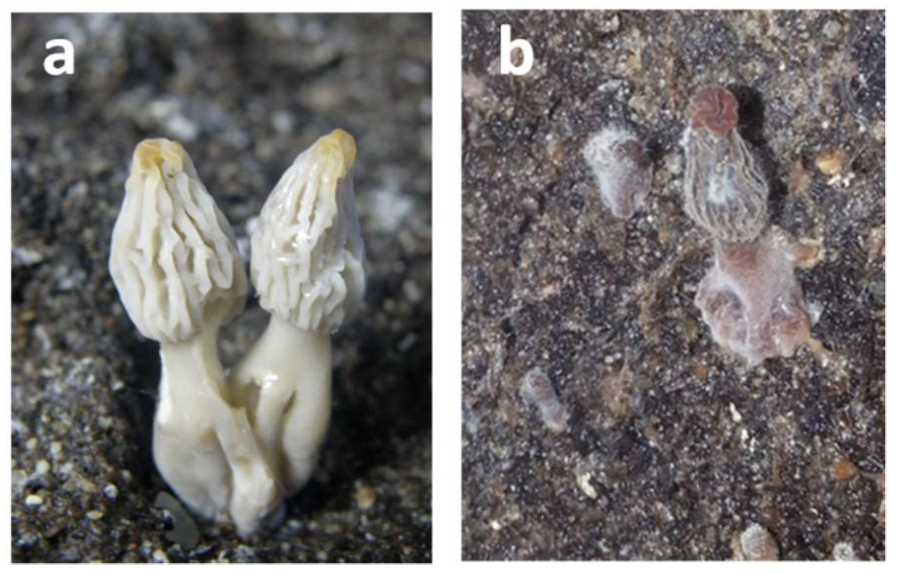
3. Results and Discussion

The infection of M. rufobrunnea was first observed in the growing room, with Morchella ascocarps already infected in their early stages of development (Figure 1). The surface of the growth substrate, as well as the initials and young ascocarps, were covered with white mycelium. The ascocarps ceased to increase in size, turned dark brown and disintegrated.
The isolation of the contaminating fungus on rose-bengal medium and further transfer to PDA medium resulted in defined colonies with white mycelium at the edge and pinkish conidia at the center (Figure 2a). The morphological observation of the fungal colony showed typical conidiophores and phialides with chains of ellipsoid conidia (Figure 2b), in accordance with P. lilacinum characterization [18].
Molecular analysis yielded a 1000 bp partial sequence of the 28S ribosomal DNA region and a 591 bp sequence of the rRNA ITS region. Both sequences were aligned with several sequences from the GenBank database. For both regions, the highest similarity occurred with several P. lilacinum sequences. The ITS region of P. lilacinum 1925-1 showed 100% identity with the ITS region of range of P. lilacinum strains isolated from different substrates that were considered for species characterization by Luangsa-Ard et al. [18] (Table 1).
After identifying the isolated fungus, we further confirmed its pathogenic activity against M. rufobrunnea using an in vitro agar plate system. When spores of the isolated fungus were applied on the surface of an already established 3-day-old M. rufobrunnea colony, the mycelium turned brown at the site of application within 1 day (Figure 3a). The browning process expanded with time, indicating toxic activity of the isolated fungus, which produced a white and dense mycelium on top of the M. rufobrunnea mycelium (Figure 3b, showing the culture 72 h after adding the isolated fungal spores). M. rufobrunnea mycelium, which was far (>1.5 cm) from the P. lilacinum inoculation point, continued its normal development, producing sclerotia (Figure 3b).
We then confirmed the pathogenicity of the isolated P. lilacinum against the developing M. rufobrunnea primordia in the mushroom growing system. The life cycle of Morel mushroom has been described by Volk and Leonard in 1990 [19], as well as in a work from our lab [20]. The primordial stage was chosen over the mature mushroom stage for the Koch’s test because infection of the morels in the cultivation system was originally observed at this stage of ascocarp development. An aqueous spore suspension of P. lilacinum was sprayed on the soil surface and primordia when they were still white and only 3–8 mm long (Figure 3c). Two days later, the primordial head turned brown and ceased to develop (Figure 3d). A few days later, white hyphae were observed to cover the young brown primordia (Figure 3g). Bbrown primordia were transferred to PDA medium, resulting in growth of a typical P. lilacinum colony on the infected primordium (Figure 3e), which eventually disintegrated under the isolated fungal hyphae (Figure 3f).
The results indicated that the isolated P. lilacinum 1925-1 spores could reproduce the disease symptoms, that is, browning of the mycelium and the developing ascocarp and further degradation of the Morchella tissue (Figure 3g), similar to those observed on the originally infected mushrooms from which P. lilacinum was isolated. The browning may reflect oxidative enzyme activity under stress conditions in the infected Morchella, similar to other cases of mushroom infection [21,22]. However, it should be noted that, in contrast to the current study, ascocarp browning was not prominent when M. importuna was infected with the pathogenic fungus Diploöspora longispora in an artificial inoculation assay [12].
The identified contaminating fungus P. lilacinum belongs to the phylum Ascomycota, subphylum Pezizomycotina, order Hypocreales, family Ophiocordycipitaceae [18]. This family contains many parasitic fungi. Originally designated Paecilomyces lilacinus (Thom) Samson, this fungus was later given the name P. lilacinum [18,23]. However, in several reports, it is described using its former designation of P. lilacinus, a biological control agent against root-knot nematodes and cotton aphids [24,25]. In a wide survey conducted by Luangsa-Ard et al. [18], P. lilacinus species were found to share sequence similarity with P. lilacinum.
Many recent reports have shown antifungal activity of P. lilacinum against a range of fungi, such as Phytophthora infestans [26], gray mold [27], Verticillium dahliae [28], Sclerotinia sclerotiorum [29] and green mold (Penicillium digitatum) [30], making it a candidate biocontrol agent against phytopathogenic fungi. Ali [31] reported that the filtrate of P. lilacinum inhibits Stromatinia cepivora mycelial growth and sclerotium formation. Moreover, treating soil with the filtrate caused a loss of S. cepivora sclerotial activity and significantly decreased disease incidence and severity. Whole-genome sequencing has additionally elucidated some of its mycoparasitic activities [32].
This is the first report of the isolation and identification of the fungus P. lilacinum on cultivated morel and, specifically, in indoor cultivation. A phenotypically similar disease of M. importuna cultivated outdoors was reported by He et al. [12], where the contaminating fungus was identified as Paecilomyces penicillatus. As in earlier publications, where the species name P. lilacinus was replaced by P. lilacinum [20,21], it is suggested that the P. penicillatus identified by He et al. [12] and the P. lilacinum reported in the present study may be related.
The results presented here show that P. lilacinum, a mycoparasitic fungus, attacks the vegetative mycelium, as well as young morel mushrooms. While P. lilacinum is naturally disseminated in the environment, it is also spread artificially for crop-pest management [33]. This is an important point when considering outdoor cultivation of morels, as it damages mushrooms, similar to the case of another mycoparasitic fungus, Trichoderma, from the same order (Hypocreales), which caused infections in the mushroom industry [34] while being used as a beneficial biocontrol agent for crops [35].
The same characteristics that define P. lilacinum as a good candidate for biocontrol of pathogenic fungi make it harmful to cultivated morel mushrooms. Further studies should be performed to prevent or control such contaminants. As with other indoor-cultivated mushrooms, treatments of all facilities, including growth substrates and the air, are required.

Funding

This research received no external funding.

Institutional Review Board Statement

Not applicable.

Informed Consent Statement

Not applicable.

Acknowledgments

The author thanks Limor Zabari for technical assistance and the MIGAL research authority for support of the research.

Conflicts of Interest

The author declares no conflict of interest.

References

  1. Tietel, Z.; Masaphy, S. True morels (Morchella)—nutritional and phytochemical composition, health benefits and flavor: A review. Crit. Rev. Food Sci. Nutr. 2018, 58, 1888–1901. [Google Scholar] [CrossRef] [PubMed]
  2. Sun, X.M.; Zhang, W.M.; Wu, S.L.; Shi, J.; Zhang, G.L. Preparing of new health food Morchella capsule. Edible Fungi China 2001, 20, 38–40. [Google Scholar]
  3. Ower, R. Notes on the development of the morel ascocarp: Morchella esculenta. Mycologia 1982, 74, 142–144. [Google Scholar] [CrossRef]
  4. Ower, R.; Mills, G.L.; Malachowski, J.A. Cultivation of Morchella. U.S. Patent 4,594,809, 17 June 1986. [Google Scholar]
  5. Masaphy, S. Biotechnology of morel mushrooms: Successful fruiting body formation and development in a soilless system. Biotechnol. Lett. 2010, 32, 1523–1527. [Google Scholar] [CrossRef]
  6. Liu, Q.; Ma, H.; Zhang, Y.; Dong, C. Artificial cultivation of true morels: Current state, issues and perspectives. Crit. Rev. Biotechnol. 2018, 38, 259–271. [Google Scholar] [CrossRef] [PubMed]
  7. Sambyal, K.; Singh, R.V. A comprehensive review on Morchella importuna: Cultivation aspects, phytochemistry, and other significant applications. Folia Microbiol. 2021, 66, 147–157. [Google Scholar] [CrossRef] [PubMed]
  8. Tan, F.H. History, current status and prospects of morel cultivation. Edible Med. Mushrooms 2016, 24, 140–144. (In Chinese) [Google Scholar]
  9. Longley, R.; Benucci, G.M.N.; Mills, G.; Bonito, G. Fungal and bacterial community dynamics in substrates during the cultivation of morels (Morchella rufobrunnea) indoors. FEMS Microbiol. Lett. 2019, 366, fnz215. [Google Scholar] [CrossRef]
  10. Guo, M.P.; Chen, K.; Wang, G.Z.; Bian, Y.B. First report of stipe rot disease on Morchella importuna caused by Fusarium incarnatum–F. equiseti species complex in China. Plant Dis. 2016, 100, 2530. [Google Scholar] [CrossRef]
  11. He, P.; Li, C.; Cai, Y.; Zhang, Y.; Bian, Y.; Liu, W. First report of pileus rot disease on cultivated Morchella importuna caused by Diploöspora longispora in China. J. Gen. Plant Pathol. 2018, 84, 65–69. [Google Scholar] [CrossRef]
  12. He, X.L.; Peng, W.H.; Miao, R.Y.; Tang, J.; Chen, Y.; Liu, L.X.; Wang, D.; Gan, B.C. White mold on cultivated morels caused by Paecilomyces penicillatus. FEMS Microbiol. Lett. 2017, 364, fnx037. [Google Scholar] [CrossRef] [PubMed] [Green Version]
  13. Wang, X.; Peng, J.; Sun, L.; Bonito, G.; Guo, Y.; Li, Y.; Fu, Y. Genome sequencing of Paecilomyces penicillatus provides insights into its phylogenetic placement and mycoparasitism mechanisms on morel mushrooms. Pathogens 2020, 9, 834. [Google Scholar] [CrossRef] [PubMed]
  14. Lan, Y.F.; Cong, Q.Q.; Wang, Q.W.; Tang, L.N.; Li, X.M.; Yu, Q.W.; Cui, X.; An, X.R.; Yu, C.X.; Kong, F.H.; et al. First report of Cladobotryum protrusum causing cobweb disease on cultivated Morchella importuna. Plant Dis. 2020, 104, 977. [Google Scholar] [CrossRef]
  15. Liu, W.; Cai, Y.; He, P.; Ma, X.; Bian, Y. Occurrence and control of pests and diseases in field cultivation of Morchella mushrooms. Acta Edulis Fungi 2019, 26, 128–134. [Google Scholar]
  16. Albaum, S.; Masaphy, S. Comparison of rose bengal-chloramphenicol and modified aureomycin-rose bengal-glucose-peptone agar as media for the enumeration of molds and yeasts in water by membrane filtration techniques. J. Microbiol. Methods 2019, 76, 310–312. [Google Scholar] [CrossRef] [PubMed]
  17. Raja, H.A.; Miller, A.N.; Pearce, C.J.; Oberlies, N.H. Fungal identification using molecular tools: A primer for the natural products research community. J. Nat. Prod. 2017, 80, 756–770. [Google Scholar] [CrossRef] [PubMed]
  18. Tamura, K.; Dudley, J.; Nei, M.; Kumar, S. MEGA4: Molecular Evolutionary Genetics Analysis (MEGA) software version 4.0. Mol. Biol. Evol. 2007, 24, 1596–1599. [Google Scholar] [CrossRef]
  19. Saitou, N.; Nei, M. The neighbor-joining method: A new method for reconstructing phylogenetic trees. Mol. Biol. Evol. 1987, 4, 406–425. [Google Scholar]
  20. Luangsa-Ard, J.; Houbraken, J.; van Doorn, T.; Hong, S.B.; Borman, A.M.; Hywel-Jones, N.L.; Samson, R.A. Purpureocillium, a new genus for the medically important Paecilomyces lilacinus. FEMS Microbiol. Lett. 2011, 321, 141–149. [Google Scholar] [CrossRef]
  21. Volk, T.J.; Leonard, T.J. Cytology of the life-cycle of Morchella. Mycol Res. 1990, 94, 399–406. [Google Scholar] [CrossRef]
  22. Masaphy, S. External ultrastructure of fruit body initiation in Morchella. Myco Res. 2005, 109, 508–512. [Google Scholar] [CrossRef] [PubMed]
  23. Kim, S. Antioxidant compounds for the inhibition of enzymatic browning by polyphenol oxidases in the fruiting body extract of the edible mushroom Hericium erinaceus. Foods 2020, 9, 951. [Google Scholar] [CrossRef] [PubMed]
  24. Soler-Rivas, C.; Jolivet, S.; Arpin, N.; Olivier, J.M.; Wichers, H.J. Biochemical and physiological aspects of brown blotch disease of Agaricus bisporus. FEMS Microbiol. Rev. 1999, 23, 591–614. [Google Scholar] [CrossRef] [PubMed]
  25. Samson, R.A. Paecilomyces and some allied hyphomycetes. Stud. Mycol. 1974, 6, 1–119. [Google Scholar]
  26. Lopez, D.C.; Zhu-Salzman, K.; Ek-Ramos, M.J.; Sword, G.A. The entomopathogenic fungal endophytes Purpureocillium lilacinum (formerly Paecilomyces lilacinus) and Beauveria bassiana negatively affect cotton aphid reproduction under both greenhouse and field conditions. PLoS ONE 2014, 9, e103891. [Google Scholar] [CrossRef] [Green Version]
  27. Singh, S.; Pandey, R.K.; Goswami, B.K. Bio-control activity of Purpureocillium lilacinum strains in managing root-knot disease of tomato caused by Meloidogyne incognita. Biocontr. Sci. Technol. 2013, 23, 1469–1489. [Google Scholar] [CrossRef]
  28. Wang, G.; Liu, Z.; Lin, R.; Li, E.; Mao, Z.; Ling, J.; Yang, Y.; Yin, W.-B.; Xie, B. Biosynthesis of antibiotic leucinostatins in bio-control fungus Purpureocillium lilacinum and their inhibition on Phytophthora revealed by genome mining. PLoS Pathog. 2016, 12, e1005685. [Google Scholar] [CrossRef] [Green Version]
  29. Yuan, H.; Xu, P.; Xiao, H.; Zhang, Z.; Zhao, J.; Wang, G.; Xiao, X.; Xiao, Y. Efficacy of Purpureocillium lilacinus on gray mold of post-harvest tomato fruits. Chin. J. Biol. Control 2017, 33, 525–530. [Google Scholar]
  30. Lan, X.; Zhang, J.; Zong, Z.; Ma, Q.; Wang, Y. Evaluation of the biocontrol potential of Purpureocillium lilacinum QLP12 against Verticillium dahliae in eggplant. BioMed Res. Int. 2017, 2017, 4101357. [Google Scholar] [CrossRef] [Green Version]
  31. Elsherbiny, E.A.; Taher, M.A.; Elsebai, M.F. Activity of Purpureocillium lilacinum filtrates on biochemical characteristics of Sclerotinia sclerotiorum and induction of defense responses in common bean. Eur. J. Plant Pathol. 2019, 155, 39–52. [Google Scholar] [CrossRef]
  32. Elsherbiny, E.A.; Taher, M.A.; Abd El-Aziz, M.H.; Mohamed, S.Y. Action mechanisms and biocontrol of Purpureocillium lilacinum against green mould caused by Penicillium digitatum in orange fruit. J. Appl. Microbiol. 2021, 131, 1378–1390. [Google Scholar] [CrossRef] [PubMed]
  33. Ali, M.A.S. Role of Purpureocillium lilacinum cultural filtrate in controlling onion white rot. J. Plant Protect. Pathol. 2020, 11, 175–184. [Google Scholar] [CrossRef]
  34. Prasad, P.; Varshney, D.; Adholeya, A. Whole genome annotation and comparative genomic analyses of bio-control fungus Purpureocillium lilacinum. BMC Genom. 2015, 16, 1004. [Google Scholar] [CrossRef] [PubMed] [Green Version]
  35. Kumar, K.K.; Arthurs, S. Recent advances in the biological control of citrus nematodes: A review. Biol. Control 2021, 157, 104593. [Google Scholar] [CrossRef]
Figure 1. Healthy (a) and infected (b) young fruiting bodies of M. rufobrunnea.
Agriculture 12 00695 g001
Figure 2. Colony of isolated fungus P. lilacinum 1925-1 on PDA (a) and conidiophores bearing ellipsoid conidia at 1000× magnification (b).
Agriculture 12 00695 g002
Figure 3. Pathogenicity assays of P. lilacinum 1925-1 against M. rufobrunnea. (a,b) M. rufobrunnea mycelial culture after inoculation of P. lilacinum spores on day 3 of growth (a) and 72 h later (b); (c,d) Young primordia in the growing system before (white, 3–8 mm long) (c) and 48 h after (d) inoculation with P. lilacinum spores; (e) Infected primordia with established P. lilacinum colony after transfer from the growing system to PDA medium; (f) Infected primordia covered by P. lilacinum hyphae on PDA; (g) Infected primordia on cultivation soil.
Agriculture 12 00695 g003
Table 1. Sequence homology of the rRNA ITS regions of isolate 1925-1 with previously identified P. lilacinum strains considered by Luangsa-Ard et al. [18].
NCBI
Accession No.
Homology (%)From
HQ842812100Nematoda
HQ842815100Other
HQ842816100Human
HQ842817100Entomogenous
HQ842819100Nematoda
HQ842820100Miscellaneous
HQ842821100Other
HQ842824100Human
HQ842825100Entomogenous
HQ842828100Human
HQ842838100Environmental
HQ84284197.51Environmental
Publisher’s Note: MDPI stays neutral with regard to jurisdictional claims in published maps and institutional affiliations.

