Article

Antiviral Activity of TiO2 NPs against Tobacco Mosaic Virus in Chili Pepper (Capsicum annuum L.)

by
Noemi L. Acuña-Fuentes
1,
Marcela Vargas-Hernandez
2,*,
Samantha de Jesus Rivero-Montejo
1,
Luisa K. Rivas-Ramirez
1,
Israel Macias-Bobadilla
3,
Viviana Palos-Barba
4,
Eric M. Rivera-Muñoz
4,
Ramon G. Guevara-Gonzalez
1 and
Irineo Torres-Pacheco
1,*
1
Laboratory of Biosystems Engineering, Faculty of Engineering, Campus Amazcala, Autonomous University of Queretaro, Carretera a Chichimequillas, km 1 S/N, El Marques 76265, Queretaro, Mexico
2
Faculty of Engineering, Campus Amealco, Autonomous University of Queretaro, Carretera Amealco Temazcaltzingo, km 1, Centro, Amealco de Bonfil 76850, Queretaro, Mexico
3
Faculty of Engineering, Campus Conca, Autonomous University of Queretaro, Valle Agrícola S/N, Arroyo Seco 76410, Queretaro, Mexico
4
Department of Nanotechnology, Center of Applied Physics and Advanced Technology, National Autonomous University of Mexico, A.P. 1-1010, Querétaro 76010, Queretaro, Mexico
*
Authors to whom correspondence should be addressed.
Agriculture 2022, 12(12), 2101; https://doi.org/10.3390/agriculture12122101
Submission received: 3 November 2022 / Revised: 26 November 2022 / Accepted: 30 November 2022 / Published: 8 December 2022
(This article belongs to the Section Crop Protection, Diseases, Pests and Weeds)

Abstract

:
Tobacco mosaic virus is the etiological agent of one of the most critical diseases limiting chili pepper production. Various practices have been used to manage the disease, e.g., the use of resistant varieties and interference with the vector through chemical control. However, these practices are not helpful once the virus has been established in the plant. There is still no effective method for the sustainable management of the disease; therefore, exploring new options is required. Currently, some studies have reported the activity of TiO2 NPs against viruses in plants, although not against TMV in chili pepper. The present work aims to determine a possible direct action of TiO2 NPs against TMV and if there is a relationship between the amount of virus and symptoms. The application of TiO2 NPs at 150 μg/mL in infected pepper plants reduced symptoms and viral load and improved the morphological characteristics compared to the control. Incubation of 150 µg/mL TiO2 NPs with the virus for 6 and 8 h before infection decreased viral concentration significantly after infection compared to the control. In this work, it is reported, for the first time, that the use of TiO2 NPs is a novel practice for the control of TMV in chili pepper.

1. Introduction

Nanotechnology has received significant attention in developing particles with dimensions less than <100 nm [1]. Their physical and chemical properties depend on the high area/volume relationship. The smaller they are, the greater the surface area, presenting more significant activity compared to their bulk materials [2]. In agriculture, at specific doses, they have been used in a promising way in disease management and the improvement of plant health. Therefore, the application of nanotechnology can contribute to guaranteeing global food security [3,4]. Studies have highlighted the potential applications of metal oxide NPs as elicitors or biostimulants, improving the nutraceutical and nutritional quality of plants [5]. On the other hand, they also have been shown to be a promising practice for integrated strategy disease management due to their antimicrobial, antifungal, and antiviral activity against different types of these histological agents. Tobacco mosaic virus (TMV) belongs to the Tobamovirus genus [6,7]. TMV disease is one of the most critical limitations in the production of chili crops. TMV are viruses with a size of 300 x 15 nm, which can encode for four proteins: the 126 and 183 kDa replication-associated proteins, the structural capsid or coat protein, and movement protein [8]. This virus in plants causes mosaic, mottling, necrosis, chlorosis, and leaf distortion in plants such as Capsicum annuum, Solanum lycopersicum, and Nicotiana tabacum virus [9]. Currently, various chemical compounds (ex. ribavirin and oseltamivir) [10,11] and biological compounds (ex. seco-pregnane steroids, ningnanmycin, tagitinin C (Ses-2), and 1β-methoxydiversifolin-3-0-methyl ether (Ses-5)) limit the infection and proliferation of the TMV [12]. However, most of these strategies are established in plants cultivated under in vitro conditions since they are not very effective once the virus has been established in plants cultivated in the open field.
Considering the above, it is appropriate to develop new practices focused on reinforcing the integrated management of TMV diseases. The scientific references for incorporating NPs into TMV management practices have reported evidence that these compounds possess antiviral activity against TMV. For example, chitosan Schiff–base nano-silver (S-cos-Ag) has been reported to prevent and treat the TMV virus and reduce the number of lesions in TMV-infected tobacco leaves, showing a degree of control of up to 78% [7]. On the other hand, pretreatment of ZnO NPs and SiO2 NPs exhibited substantial aggregation of TMV particles and lysis in vitro, and inoculated tobacco plants showed significantly lower virus colonization [6]. TiO2 NPs are very popular since they are produced on a large scale due to their properties, including low toxicity and a wide range of applications in the industry. Although there are no reports of the antiviral activity of this type of nanoparticles against TMV, they have been found to have antiviral activity against the virus as the turnip mosaic virus (TuMV) in Nicotiana bethamiana plants [13] and broad bean stain virus in Vicia faba L. [14]. The antiviral activity of TiO2 NPs against TMV in chili pepper is currently unknown. Considering that ZnO NPs and SiO2 NPs may be feasible practices for managing the disease caused by TMV in a solanaceous such as tobacco, the purpose of this work is to expand the options for the integrated management of this pathogen in another solanaceous, namely, chili. It is also desirable to determine if there is a direct action of TiO2 NPs on the virus and if there is a relationship between the amount of virus and symptoms.

2. Materials and Methods

2.1. Plant Establishment

The plant material used in the experiment was a variety of Jalapeño Don Pancho pepper obtained from Mexico’s National Institute for Forestry, Agriculture, and Livestock Research (INIFAP). The plants were germinated and transplanted when they had 6 to 8 true leaves in 9 L bags with tezontle substrate, humus, and soil where chili is usually grown, in a 2:2:5 ratio. Plant nutrition consisted of a 90% Stainer solution. The experiment was carried out in a greenhouse where the average temperature was 21.3 °C and with an RH% of 59%.

2.2. Experimental Design

The experimental design arrangement was in randomized blocks with seven plants per experimental unit, five treatments, and seven replicates. The concentrations of TiO2 NPs evaluated were 50, 100, 150, and 200 µg/mL. The application of the TiO2 NPs was carried out at 72, 144, and 216 h after the plants had been inoculated. The negative control was water, and the positive control was the plants infected with the TMV virus.

2.3. Synthesis and Characterization of TiO2 Nanoparticles

Titanium dioxide nanoparticles were synthesized using the sol–gel method, in which 5 mL of titanium isopropoxide (97%) and 100 mL of 2-propanol (99.5%) were placed in a beaker with slight agitation. Subsequently, 30 mL of deionized water was added to the solution, and the mixture was kept in reaction with slow stirring for 24 h. After this, the solid was recovered by filtration and washed several times with deionized water. The solid was dry at room temperature and was finally subjected to muffle drying at 110 °C for 18 h and calcined at 500 °C for 4 h [15]. The characterization of the nanoparticles was carried out with a HITACHI model SU8230 High-Resolution Scanning Electron Microscope at the Center for Applied Physics and Advanced Technology (CFATA) of the UNAM-Juriquilla to determine the morphology of the material obtained and the size of the nanoparticles. In addition, the crystalline structure of the material from 20 to 80° 2θ was analyzed through X-ray diffraction using Cu Kα radiation at a wavelength of 1.54 Ǻ with a Bruker model Advance D8 instrument at the facilities of the Autonomous University of Queretaro, Campus Aeropuerto. Additionally, the diffraction pattern was refined to determine the average crystal size and the weight concentration of the phases found.

2.4. TMV Inoculation in Capsicum annuum L. Plants

The TMV virus was provided by the Autonomous University of Mexico (UNAM) Campus Iztacala. A 1:10 solution of infected material and extraction buffer (GEB) from the Agdia DAS-ELISA kit was prepared. When the plants presented six true leaves, the virus was inoculated mechanically with a swab impregnated with the viral solution and Carburundum 100.

2.5. Measurement of Morphological Variables and Evaluation of Symptoms

The following morphological variables were measured: plant height, stem diameter, number of leaves, flowers, flower buds, and fruits. For the evaluation of the symptoms, the severity scale reported by Torres-Pacheco [16] was used. This scale was adapted to TMV symptoms and includes values between 1 and 5 (Table 1).

2.6. Immunoassay DAS-ELISA

Leaves were collected 20 days after virus inoculation and frozen with liquid nitrogen. The leaves were taken at the base of the plant and the apical part to determine the virus’s movement. Viral load was performed with the Agdia DAS-ELISA PathoScreen Kit according to the manufacturer’s instructions [17]. Color measurement was performed spectrophotometrically using a Thermo Scientific MULTISKAN GO spectrophotometer model SN 1510-02385C at 405 nm.

2.7. Reactivity Determination

The reactivity test was carried out to determine the direct effect of the TiO2 NPs on the virus. To carry out this test, the following treatments were applied: (1) water control (without NPs, without viruses); (2) control NPs (TiO2 NPs 150 μg/mL); (3) virus control (only viruses without TiO2 NPs); (4) treatment (TiO2 NPs 150 μg/mL with the virus). The viral extract was mixed with the solution with TiO2 NPs 150 µg/mL TMV extract and allowed to incubate for 6 h; then, the pepper plants were inoculated. The next stage involved incubating the mixture of TiO2 NPs at 150 µg/mL and TMV extract for 12 h and inoculating the pepper plants. The treatments were then inoculated in plants with 6–8 leaves. Viral quantification was performed at 48, 96, 144, and 192 h inoculation with the Agdia DAS-ELISA Reagent Set Kit [17]. Five viral load measurements were taken every 48 h to see how the viral load evolved.

2.8. Hydrogen Peroxide Determination

The content of hydrogen peroxide in the leaves of the infected plants treated with the NPs was determined using the technique of Junglee et al. [18]. The method was based on the colorimetric determination of the hydrogen peroxide content in plants using potassium iodide. A total of 100 mg of sample was homogenized with 0.25 mL of 10 mM K/Na-phosphate buffer (pH 5.8), 0.25 mL of 0.1% trichloroacetic acid, and 0.5 mL of 1 M KI. The supernatant obtained was centrifuged at 4 °C, for 15 min, at 16,000× g and kept for 20 min in the dark. The samples were measured at 350 nm. Reported units were nanomoles of peroxide per milligram of protein.

2.9. Enzymatic Activity

For the extracts, 300 mg of the samples were weighed, and 2 mL of 0.05 M potassium phosphate extraction buffer, pH 7.8, at 4 °C was added. It was made up to 250 mL with distilled water to be later centrifuged at 12,000 rpm for 15 min at 4 °C. The supernatant was removed and stored at 4 °C for later use. The amount of protein was determined according to the methodology reported by Bradford [19], using bovine serum albumin as a standard for protein concentration (Sigma-Aldrich, St. Louis, MO, USA).

2.9.1. Phenylalanine Ammonia-Lyase (PAL) Activity

PAL activity was performed according to the methodology reported by Toscano et al. [20] with modifications. Briefly, 1.15 mL of borate buffer (pH 8.8 containing 10 mM L-phenylalanine) was placed, 100 µL of enzyme extract was added, and the mixture was calibrated to 2 mL. The mixture was incubated for 60 min in a water bath at 40 °C. After 60 min, 0.5 mL of 1N HCl was added (to stop the reaction), and the mixture was allowed to stand for 10 min. The calibration curve consisted of a cinnamic acid solution of 2–12 µg cinnamic acid/ml. Absorbance at 290 nm was measured on a MULTISKAN GO, Thermo Scientific spectrophotometer, SN 1510-02385C. The blank was made with 1.5 mL of the reaction buffer solution of borate of 0.1 M and L-phenylalanine of 10 mM, pH 8.8. PAL activity was expressed as µg cinnamic acid/mL produced under the specific conditions and expressed as U/mg protein content.

2.9.2. Catalase (CAT) Activity Assay

Spectrophotometric determination of CAT activity was performed according to the method reported by Afiyanti and Chen [21] with modifications. Briefly, for the assay, 2 mL of reaction buffer (50 mM potassium phosphate buffer, pH 8.0), 0.2 mL of H2O2, and 0.1 mL of enzyme extract were placed directly in the quartz cell. The mixture was stirred, and the change in absorbance at λ 240 nm was measured instantly for 6 min (one reading every minute) in a MULTISKAN GO Thermo Scientific spectrophotometer, SN 1510-02385C. The change in absorbance at 240 nm was measured for 1 min and used to determine the rate of decomposition of H2O2 by CAT; 1 U CAT decomposes 1 mmol H2O2/min at pH 8.0 and 25 °C. Data are expressed as U/mg protein.

2.9.3. Superoxide Dismutase (SOD) Activity Assay

SOD activity was performed by nitro blue tetrazolium (NBT) photochemical reduction inhibition according to the method reported by Hayat et al. [22]. Briefly, 1.5 mL of reaction buffer (50 mM potassium phosphate buffer, pH 7.8), 0.3 mL of 0.1 mM EDTA-Na2, 0.3 mL of 0.13 M methionine, 0.3 mL of 0.75 mM NBT, 0.3 mL of 0.03 mM riboflavin, 0.05 mL of enzyme extract, and 0.25 mL of distilled water was mixed by inverting clear glass tubes. The mixture was exposed to sunlight for 30 min, waiting for the development of a blue color. The blank was made with the previous solutions replacing the enzyme extract with distilled water. Absorbance at λ560 nm was measured on a MULTISKAN GO Thermo Scientific spectrophotometer. A total of 1 U SOD inhibited the reduction of NBT by 50% at pH 7.8 and 25 °C. Data are expressed as U/mg protein.

2.9.4. Peroxidase Activity (POD)

The spectrophotometric determination of POD activity was performed with the Amplex® Red Hydrogen Peroxide/Peroxidase Assay Kit from Invitrogen. The methodology suggested by the brand was followed. A total of 1.1 μL of the enzymatic extract and 48.9 μL of the reaction buffer were added directly to the microplate, 50 μL of the 100 μM Amplex® Red–2 mM H2O2 solution was added, and the mixture was incubated in the dark for 30 min. The absorbance was measured at 560 nm. The negative control consisted of 50 µL of 1X reaction buffer, and the positive control consisted of 50 µL of 2 mU/mL HRP.

2.10. Antioxidant Activity

The determination of antioxidant activity in each treatment was evaluated through 1,1-diphenyl-2-picrylhydrazyl (DPPH•−) and 2,2-azinobis-(3-ethylbenzothiazoline-6-sulfonic acid) (ABTS•+). The methanolic extracts were prepared by adding 9 mg of sample to 15 mL of methanol, and the mixture was kept under stirring for 24 h.

2.10.1. DPPH• Radical Scavenging Activity (DRSA)

DRSA was determined according to the method of Chen et al. [23] with modifications. A total of 0.5 mL of the methanolic extract and 0.5 mL of the 0.1 mM DPPH• radical were mixed (3.94 mg were weighed and made up to 100 mL with methanol). The mixture was shaken vigorously and allowed to incubate in the dark for 30 min at room temperature. Finally, the absorbance was taken at a wavelength of 515 nm; the reaction blank was methanol, and the control was distilled water. %DRSA was calculated by the following equation:
%DRSA = [1 − (Asample − Ablank)/Acontrol)] × 100

2.10.2. ABTS •+ Radical Scavenging Activity (ARSA)

The determination of ARSA was carried out according to the method of Re et al. [24] with modifications. The ABTS•+ radical was generated by mixing the 7 mmol/L ABTS stock solution with 2.45 mmol/L potassium persulfate under darkness (ABTS•+ radical generation) 12 h prior to analysis. The ABTS•+ solution was then diluted in phosphate-buffered saline (PBS) at 0.15 M, pH 7.4, until a reading of 0.7 Abs was obtained at a wavelength of 734 nm. Subsequently, 3 mL of this final solution was mixed with 150 µL of the methanolic extract in a glass cell; finally, the change in absorbance at a wavelength of 734 nm was measured every minute for 6 min. The reaction blank used was PBS, and the control was distilled water. %ARSA was calculated by the following equation:
%ARSA = [1 − (Asample − Ablank)/Acontrol)] × 100

2.11. Statistical Analysis

Data were subjected to analysis of variance (ANOVA) and Tukey test (α = 0.05) using the Statgraphics Centurion XVI statistical software (StatPoint Technologies, Bedford, MA, USA).

3. Results

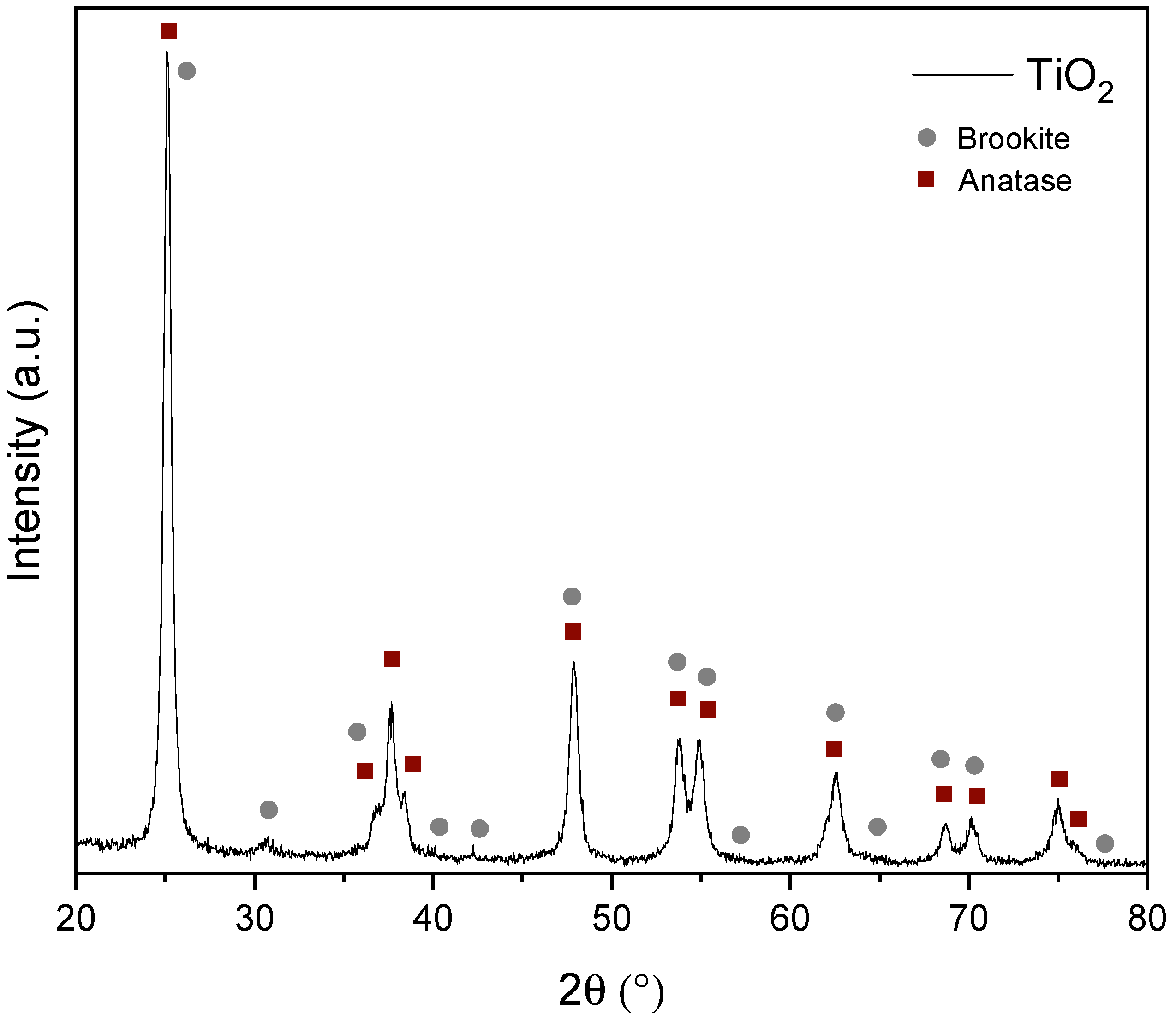
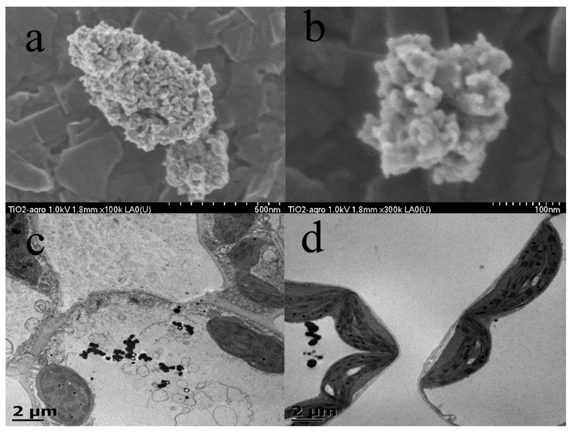
3.1. Characterization of TiO2 Nanoparticles

The characterization of the morphology of the synthesized TiO2 nanoparticles was carried out by scanning electron microscopy (SEM). The formed nanoparticles appeared hemispherical with an approximate size of 20 nm, according to Figure 1. The nanoparticles aggregated in irregular clusters of approximately 0.6 microns, forming interstitial voids (Figure 1a,b). The obtained images made it possible to identify a uniform size of the nanoparticles formed, which can benefit the spraying process of the material and enhance its interaction with plant cells. On the other hand, the X-ray diffraction study was used to determine the crystalline phase corresponding to the prepared TiO2 nanoparticles. A combination of polymorphs of the anatase phase was identified, with a minor brookite phase, as observed in the diffraction pattern obtained in Figure 2. In addition, a quantitative analysis was performed using the Rietveld method with the GSAS-II software, and it was found that the weight percentage of the polymorphs corresponded to 91% for anatase and 9% for brookite, with average crystal sizes of 11.6 nm and 13.5 nm, respectively. Figure 1c,d shows the accumulation and distribution of TiO2 nanoparticles as black particles within plant cells, including chloroplasts.

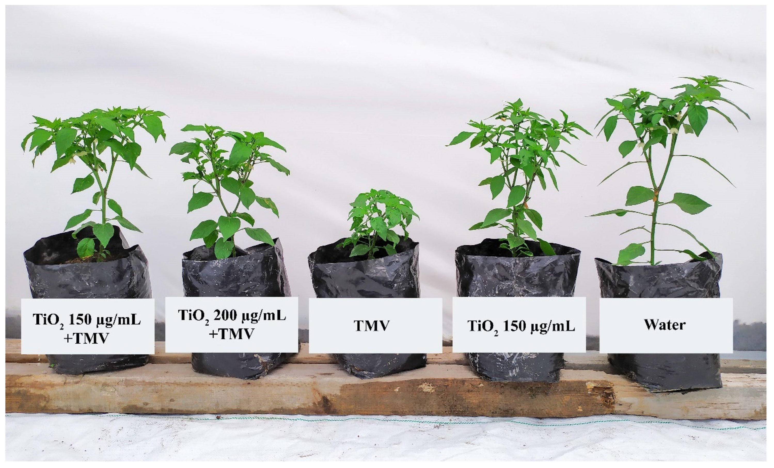
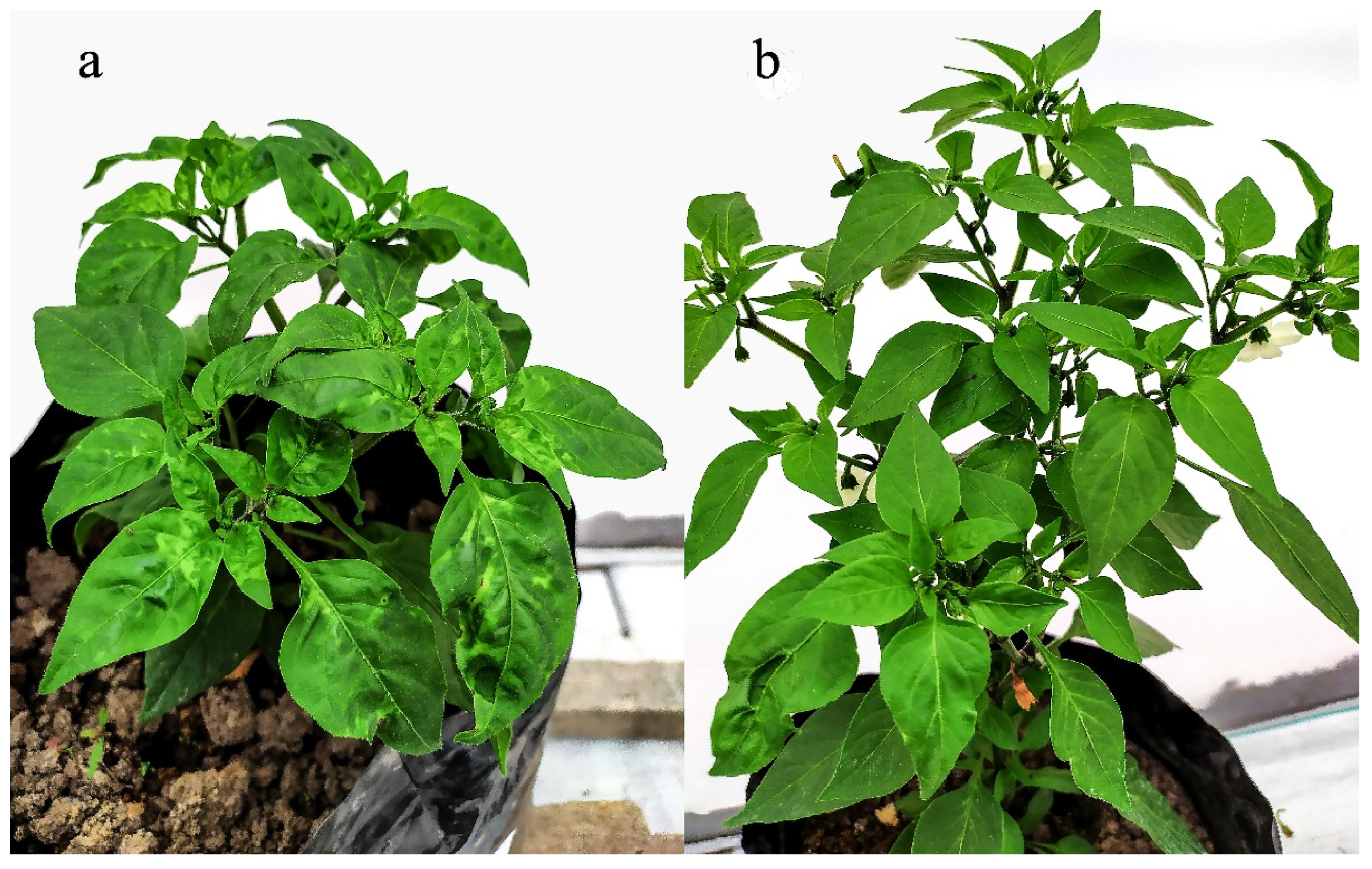
3.2. Evaluation of the Morphological Characteristics and Symptomatology Scale of the Plants Infected and Treated with TiO2 NPs

Concentrations of TiO2 NPs from 50 to 250 μg/mL were evaluated to determine the impact of TiO2 NPs on some morphological characteristics of pepper plants infected with TMV, and height, diameter, and symptom scale were measured (Table 2). The application of TiO2 NPs seemed to increase the height in infected plants by 122% compared to plants infected with TMV without treatment. Likewise, compared to the control (water), there was no significant difference. Once again, the application of TiO2 increased the diameter in infected plants, with the treatment of TiO2 150 μg/mL becoming significantly equal to the control. Plants infected with TMV showed symptoms from day 9 after inoculation. After 20 days, the disease’s symptomatology level was measured according to the health scale proposed by Torres-Pacheco [16] (Table 1). The treatments that decreased the symptoms caused by the virus were the highest concentrations of TiO2 NPs, 150 μg/mL and 250 μg/mL, since they did not present severe symptoms such as the mosaic pattern, deformed leaves, and curling. Although the best treatment was 150 μg/mL, the application of the NPs cannot completely suppress the symptoms at the same level shown by the control plant sprayed with water (Figure 1). In general, for this study, the results show that the treatment of 150 μg/mL improved the antiviral activity, reducing the appearance of the symptoms caused by TMV in pepper plants. In addition, as shown in Figure 3, this antiviral effect of TiO2 in TMV-infected plants was maintained in the most advanced stages of vegetative development. Figure 4 shows the severe symptoms of the virus in plants infected with TMV and plants with TMV treated with TiO2 NPs 150 μg/mL.

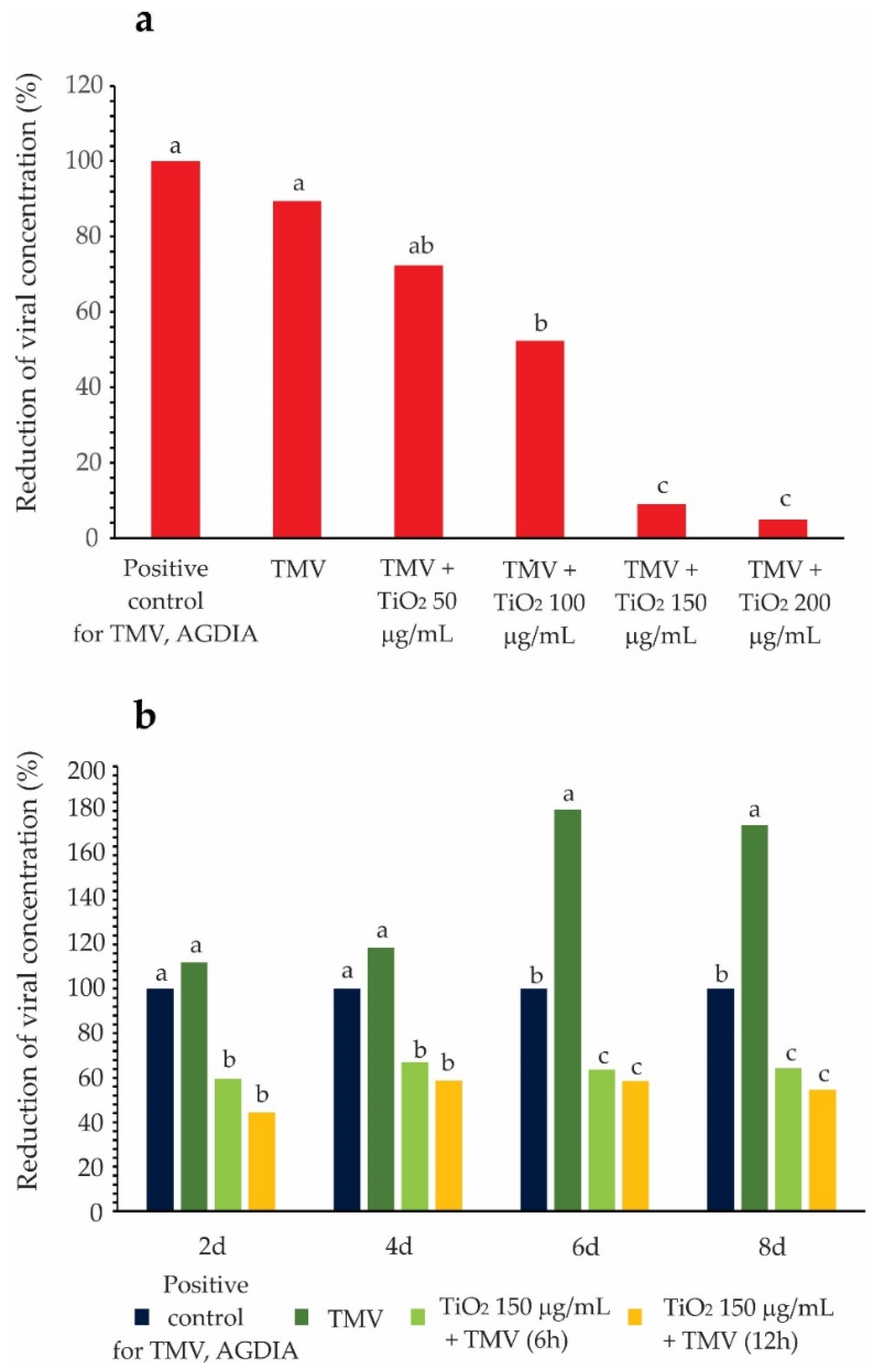
3.3. TMV Detection and Reactivity Test

According to Figure 5a, TiO2 NPs showed a decrease in the virus concentration in the leaves as the nanoparticle concentration increased. The concentrations that showed the least amount of the virus were 150 and 200 μg/mL, reducing it by approximately 90%. From the results obtained above, the concentration of TiO2 at 150 μg/mL was chosen to carry out the reactivity test to observe the direct antiviral activity with the virus. For this, the nanoparticles were incubated with the TMV virus for 6 and 12 h before infecting the plant. Once the virus was incubated with the NPs, it was observed that viral replication and proliferation were significantly inhibited at 2, 4, 6, and 8 days later in the plant compared to plants infected without treatment.
The invasion of pathogens causes defense responses in plants associated with the generation of ion fluxes; the induction of kinase cascades and nitric oxide (NO); and the accumulation of ROS, and among the most crucial molecules, H2O2. The accumulation of H2O2 was detected to understand the role of ROS in resistance against TMV. As shown in Figure 6, TMV induced hydrogen peroxide production while the application of TiO2 NPs in infected and uninfected plants showed a lower concentration, indicating that the nanoparticle can reduce reactive oxygen species (ROS) accumulation in leaves caused by TMV, thus reducing oxidative damage in the plant.

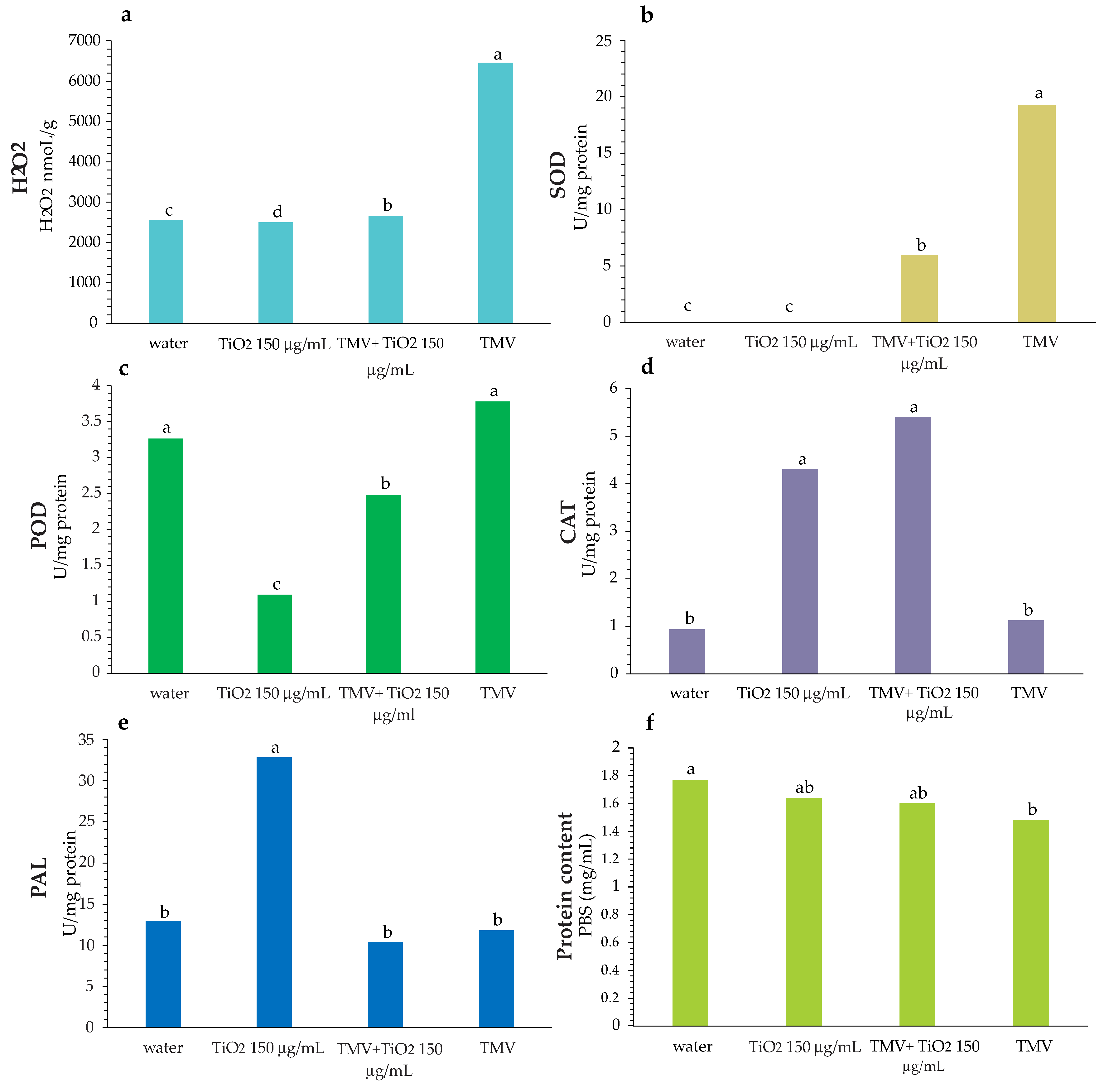
3.4. Enzymatic Activity

CAT, SOD, and POD are related enzymes in cellular ROS homeostasis. Antioxidant activity was evaluated in leaves treated with TiO2 NPs in healthy and TMV-infected plants. As shown in Figure 6, TMV infection induced SOD synthesis in plants with and without NP treatment, being higher in plants that did not have the nanoparticles. For those plants not infected, both the control and those treated with TiO2 NPs did not show the synthesis of this enzyme. As far as POD is concerned, nanoparticle treatments significantly decreased the effect of the enzyme compared to control and infected plants.
In contrast, the activity of the CAT enzyme increased in the plants sprayed with metal oxide nanoparticles, both in the infected and non-infected plants. The foregoing shows that TiO2 NPs enhanced disease tobacco resistance by setting their defensive enzyme activity differently. On the other hand, the total protein content was decreased in plants infected with TMV. NPs also can increase PAL activity, an enzyme responsible for promoting phenylalanine metabolism substances. PAL is related to plant resistance to pathogens, including viruses.

3.5. Antioxidant Activity

Finally, even though PAL is strongly related to antioxidant activity since it induces the synthesis of bioactives such as phenols and flavonoids, no differences in antioxidant activity were observed by the DPPH• and ABTS•+ methods of the different treatments, even though TiO2 150 μg/mL induced the expression of this enzyme (data not shown).

4. Discussion

Viral infections in plants induce a hypersensitive response associated with the accumulation of ROS and changes in different oxidoreductase enzymes [25]. In this work, TMV infection in chili plants not treated with TiO2 NPs accumulated H2O2, which is the molecule responsible for initiating the defense process and central cellular signaling molecules [26]. Moreover, the H2O2 generation was followed by increases in the activities of antioxidant enzymes such as SOD (Figure 6). However, it is notable that the defense response of the pepper plants did not appear or was insufficient to contend against the effects of the virus; that is, it did not reduce the symptoms in plants (Figure 3). In other words, applying TiO2 NPs was needed to observe a significant decrease in the syndrome. These results agree with those reported by Bhatia et al. [27], wherein the authors indicated that in tobacco plants infected with TMV, the superoxide dismutase and peroxidase increased while the activity of catalase decreased. Other reports indicated that CAT does not increase in the presence of TMV and may even decrease its activity [28], which correlates with our results. This is attributed to the fact that the production of salicylic acid (SA) as a defense response inhibits CAT, leading to the accumulation of H2O2, and this then switches on the expression of defense genes and activation of systemic acquired resistance (SAR) [29]. Moreover, other workers reported that TiO2 NP 150 μg/mL has the ability to induce the CAT enzyme in plants with or without virus infection; this enzyme plays an essential role in the rapid removal of ROS [26]. Therefore, in this work, this enzyme was probably responsible for maintaining H2O2 levels at a level similar to the control treated with water.
In general, for this research, the result suggests that the treatment of 150 μg/mL improved the antiviral activity, reducing the appearance of the symptoms caused by TMV in pepper plants. In addition, as shown in Figure 3, this antiviral effect of TiO2 in TMV-infected plants was maintained in the most advanced stages of vegetative development. This work also suggests that the TiO2 NPs inhibited viral symptoms, which may have been due to a direct action on viral proliferation and was reflected in a decrease in the concentration of viral load (Figure 5a) and correlated with the low level of severe symptoms in plants infected with TMV, such as yellow spots, as well as rolling and protuberance characteristics of the presence of the virus (Figure 3).
According to the reactivity test (Figure 5b), the best treatment was the incubation of TMV with TiO2 150 μg/mL for 8 h, since it showed a lower concentration of the virus. However, foliar application of the NPs post-infection at 72, 144, and 216 h had a greater decrease in the amount of virus than the treatments in the reactivity test. This may have been because, as observed in Figure 1c,d, the metal nanoparticles can accumulate and distribute themselves in the cells of the chili pepper leaves, which can facilitate their interaction with viruses.
The reactivity test also shows that TMV-infected plants increased virus concentration over time (2–8 days). However, plants infected with viruses in which the viruses were incubated with 150 μg/mL TiO2 nanoparticles for 6 and 8 h showed no increase. However, in pepper plants infected with TMV, the induction of H2O2, SOD, and POD failed to suppress the virus infection; the application of TiO2 in infected plants induced the activity of these in comparison with the control (Figure 6). This suggests that the inhibitory effect of the symptoms was mainly due to the direct action of the TiO2 nanoparticles on the virus and, to a lesser extent, to the induction of defense responses in the plant. That is, in the reactivity test, the incubation of the viruses with TiO2 nanoparticles repressed the multiplication of the viruses but it remained at low levels over time, probably due to the cooperation that occurred due to the change in the antioxidant response caused by the variability of H2O2, SOD, and CAT induced by NPs.
Although there were results on the possible action effect of TiO2 NPs on the TMV virus, more research is required to determine if the compound block viral replication, protein synthesis, or the assembly process. It may be that the effect of TiO2 nanoparticles has a more general character of action on viruses, which implies an interesting challenge to determine the generic element of the mechanism of action. For example, it has been reported that in the influenza virus (H3N2), TiO2 NPs destroy the virion envelope and cause aggregation of viral particles [30]. In plant studies, only data have been reported suggesting that the effect of TiO2 NPs against turnip mosaic virus infection in tobacco (Nicotiana benthamiana) is very effective in limiting viral infection and inhibiting viral replication in plant cells [13]. On the other hand, TiO2 NPs also reduce the disease severity of the broad bean stain virus (BBSV) in Vicia faba L. plants [14]. Regarding the antiviral action of other metallic nanoparticles on the TMV virus, studies have only been reported on Nicotiana benthamiana with the use of Schiff base nanosilver NPs [7] and zinc oxide NPs [6], which reduced the invasion and damage by TMV. The above information suggests that the foliar application of TiO2 NPs may be incorporated as a practice on TMV-integrated disease management because it has a protective effect once the virus has established itself.
In addition, our dates also suggest a possible biostimulant activity that can influence in a certain way to inhibit the symptoms and viral load and increase the defense of chili pepper against TMV. The treatment with TiO2 showed behavior in the morphological variables similar to that of the uninfected plants. The above because infected and non-infected plants, to which the treatment with nanoparticles was applied at a concentration of 150 μg/mL, did not show damage, and the magnitudes of their morphological variables such as height and diameter increased compared to the control (Table 2). This last piece of information is consistent with that provided in some reports that state that foliar application of TiO2 nanoparticles improves some growth characters in Hordeum vulgare L., Coriandrum sativum L., Vitis vinifera L., Triticum aestivum L., Helianthus annuus L., Dracocephalum moldavica, and Lycopersicum sculentum L. [31,32,33]. Some reports indicate that the above may be due to the photocatalytic capacity of TiO2 NPs that favors photosynthesis and plant growth under specific wavelengths [33].
The results generated in this study suggest that a TiO2 concentration of 150 μg/mL has a protective effect. Therefore, a new approach is opened to managing concentration and/or exposure variables that allows for the obtaining of favorable results without negatively affecting the plant and even promoting the biostimulant or elicitor effect. On the other hand, on the basis of the results of reactivity where the interaction of the nanoparticle and the virus decreases its infectivity, the spraying of nanoparticles in the pre-infection period can also be considered since contact with the virus and the nanoparticle would be more direct and faster.

5. Conclusions

In this study, the results suggest that the foliar application of TiO2 NPs at 150 μg/mL is a promising new practice to control the TMV virus in the chili pepper Capsicum annuum L. In addition, the study showed a biostimulant effect in infected and non-infected pepper plants from a concentration of 150 μg/mL. The foregoing is a reference for future research in controlling another type of geminivirus in other crops of economic importance against TMV.

Author Contributions

I.T.-P. is the author who established the idea of the research project. I.T.-P. and M.V.-H. designed the research and sought funding. N.L.A.-F., S.d.J.R.-M. and L.K.R.-R. carried out the research work in the greenhouse and the laboratory. V.P.-B. and E.M.R.-M. were in charge of providing and characterizing the nanoparticles. I.M.-B., E.M.R.-M. and R.G.G.-G. were in charge of reviewing the work. All authors have read and agreed to the published version of the manuscript.

Funding

Torres-Pacheco acknowledges Ciencia Básica 2018(SEPCONACYT A1-S-33677).

Institutional Review Board Statement

Not applicable.

Data Availability Statement

The original contributions presented in this study are included in the article; further inquiries can be directed to the corresponding authors.

Acknowledgments

Acuña-Fuentes, N.L.; Rivero-Montejo, S.d.J.; and Rivas-Ramírez, L.K. acknowledge CONACYT for the scholarships provided.

Conflicts of Interest

All the authors declare that they have no conflict of interest.

References

  1. Bayda, S.; Adeel, M.; Tuccinardi, T.; Cordani, M.; Rizzolio, F. The History of Nanoscience and Nanotechnology: From Chemical-Physical Applications to Nanomedicine. Molecules 2019, 25, 112. [Google Scholar] [CrossRef] [PubMed] [Green Version]
  2. Vargas-Hernandez, M.; Macias-Bobadilla, I.; Guevara-Gonzalez, R.G.; Rico-Garcia, E.; Ocampo-Velazquez, R.V.; Avila-Juarez, L.; Torres-Pacheco, I. Nanoparticles as Potential Antivirals in Agriculture. Agriculture 2020, 10, 444. [Google Scholar] [CrossRef]
  3. Rodrigues, I.C.P.; Campo, K.N.; Arns, C.W.; Gabriel, L.P.; Webster, T.J.; Lopes, É.S.N. From Bulk to Nanoparticles: An Overview of Antiviral Materials, Its Mechanisms, and Applications. Part. Part. Syst. Charact. 2021, 38, 2100044. [Google Scholar] [CrossRef]
  4. Vrandečić, K.; Ćosić, J.; Ilić, J.; Ravnjak, B.; Selmani, A.; Galić, E.; Pem, B.; Barbir, R.; Vinković Vrček, I.; Vinkovićet, T. Antifungal activities of silver and selenium nanoparticles stabilized with different surface coating agents. Pest Manag. Sci. 2020, 76, 2021–2029. [Google Scholar] [CrossRef]
  5. Rivero-Montejo, S.D.; Vargas-Hernandez, M.; Torres-Pacheco, I. Nanoparticles as Novel Elicitors to Improve Bioactive Compounds in Plants. Agriculture 2021, 11, 134. [Google Scholar] [CrossRef]
  6. Cai, L.; Liu, C.; Fan, G.; Liu, C.; Sun, X. Preventing viral disease by ZnONPs through directly deactivating TMV and activating the plant immunity in Nicotiana benthamiana. Environ. Sci. Nano. 2019, 6, 3653–3669. [Google Scholar] [CrossRef]
  7. Wang, Y.; Sun, C.; Xu, C.; Wang, Z.; Zhao, M.; Wang, C.; Liu, L.; Chen, F. Preliminary experiments on nano-silver against tobacco mosaic virus and its mechanism. Tob. Sci. Technol. 2016, 49, 22–30. [Google Scholar]
  8. Liu, C.; Nelson, R. The cell biology of Tobacco mosaic virus replication and movement. Front. Plant Sci. 2013, 4, 12. [Google Scholar] [CrossRef] [Green Version]
  9. Hu, Q.; Niu, Y.; Zhang, K.; Liu, Y.; Zhou, X. Virus-derived transgenes expressing hairpin RNA give immunity to Tobacco mosaic virus and Cucumber mosaic virus. Virol. J. 2011, 8, 41. [Google Scholar] [CrossRef] [Green Version]
  10. Lerch, B. On the inhibition of plant virus multiplication by ribavirin. Antivir. Res. 1987, 7, 257–270. [Google Scholar] [CrossRef]
  11. Luvisi, A.; Panattoni, A.; Triolo, E. Eradication trials of tobacco mosaic virus using chemical drugs. Acta Virol. 2012, 56, 159–162. [Google Scholar] [CrossRef] [PubMed]
  12. Zhao, L.; Dong, J.; Hu, Z.; Li, S.; Su, X.; Zhang, J.; Yin, Y.; Xu, T.; Zhang, Z.; Chen, H. Anti-TMV activity and functional mechanisms of two sesquiterpenoids isolated from Tithonia diversifolia. Pestic. Biochem. Physiol. 2017, 140, 24–29. [Google Scholar] [CrossRef] [PubMed]
  13. Hao, Y.; Yuan, W.; Ma, C.; White, J.; Zhang, Z.; Adeel, M.; Zhou, T.; Rui, Y.; Xing, B. Engineered nanomaterials suppress Turnip mosaic virus infection in tobacco (Nicotiana benthamiana). Environ. Sci. Nano. 2018, 5, 1685–1693. [Google Scholar] [CrossRef]
  14. Elsharkawy, M.; Derbalah, A. Antiviral activity of titanium dioxide nanostructures as a control strategy for broad bean strain virus in faba bean Pest Manag. Sci. 2018, 75, 828–834. [Google Scholar]
  15. Peza-Ledesma, C.L.; Escamilla-Perea, L.; Nava, R.; Pawelec, B.; Fierro, J.L.G. Supported gold catalysts in SBA-15 modified with TiO2 for oxidation of carbon monoxide. Appl. Catal. A Gen. 2010, 375, 37–48. [Google Scholar] [CrossRef]
  16. Torres-Pacheco, I.; Garzon-Tiznado, J.A.; Brown, J.; Becerra-Flora, A.; Rivera-Bustamante, R. Detection and distribution of geminiviruses in Mexico and the southern United States. Phytopathology 1996, 86, 1186–1192. [Google Scholar] [CrossRef]
  17. Hernández-Santiago, R.; Vargas-Hernández, M.; Zamora-Macorra, E.J. Evaluation of TMV resistance inducers in tomato. Rev. Mex. Cienc. Agric. 2020, 11, 377–390. [Google Scholar]
  18. Junglee, S.; Urban, L.; Huguette, S.; Lopez, F. Optimized Assay for Hydrogen Peroxide Determination in Plant Tissue Using Potassium Iodide. Am. J. Anal. Chem. 2014, 5, 730–736. [Google Scholar] [CrossRef] [Green Version]
  19. Bradford, M.M. A rapid and sensitive method for the quantitation of microgram quantities of protein utilizing the principle of protein-dye binding. Anal. Biochem. 1976, 72, 248–254. [Google Scholar] [CrossRef]
  20. Toscano, S.; Ferrante, A.; Leonardi, C.; Romano, D. PAL activities in asparagus spears during storage after ammonium sulfate treatments. Postharvest Biol. Technol. 2018, 140, 34–41. [Google Scholar] [CrossRef]
  21. Afiyanti, M.; Chen, H.J. Catalase activity is modulated by calcium and calmodulin in detached mature leaves of sweet potato. J. Plant Physiol. 2014, 171, 35–47. [Google Scholar] [CrossRef] [PubMed]
  22. Hayat, S.; Ahmad, H.; Ali, M.; Ren, K.; Cheng, Z. Aqueous garlic extract stimulates growth and antioxidant enzymes activity of tomato (Solanum lycopersicum). Sci. Hortic. 2018, 240, 139–146. [Google Scholar] [CrossRef]
  23. Chen, N.; Yang, H.; Sun, Y.; Niu, J.; Liu, S. Purification and identification of antioxidant peptides from walnut (Juglans regia L.) protein hydrolysates. Peptides 2012, 38, 344–349. [Google Scholar] [CrossRef] [PubMed]
  24. Re, R.; Pellegrini, N.; Proteggente, A.; Pannala, A.; Yang, M.; Rice-Evans, C. Antioxidant activity applying an improved ABTS radical cation decolorization assay. Free Radic. Biol. Med. 1999, 26, 1231–1237. [Google Scholar] [CrossRef]
  25. Akbar, S.; Wei, Y.; Yuan, Y.; Khan, M.T.; Qin, L.; Powell, A.; Chen, B.; Zhang, M. Gene expression profiling of reactive oxygen species (ROS) and antioxidant defense system following Sugarcane mosaic virus (SCMV) infection. BMC Plant Biol. 2020, 20, 532. [Google Scholar] [CrossRef]
  26. Smirnoff, N.; Arnaud, D. Hydrogen peroxide metabolism and functions in plants. New Phytol. 2019, 221, 1197–1214. [Google Scholar] [CrossRef] [Green Version]
  27. Bhatia, S.; Kapoor, H.C.; Lodha, M.L. Modification of Antioxidant Status of Host Cell in Response to Bougainvillea Antiviral Proteins. J. Plant Biochem. Biotechnol. 2004, 13, 113–118. [Google Scholar] [CrossRef]
  28. Yu, F.; Huaxia, Y.; Lu, W.; Wu, C.A.; Cao, X.; Guo, X. GhWRKY15, a member of the WRKY transcription factor family identified from cotton (Gossypium hirsutum L.), is involved in disease resistance and plant development. BMC Plant Biol. 2012, 12, 144. [Google Scholar] [CrossRef] [Green Version]
  29. Yi, S.Y.; Yu, S.H.; Choi, D. Involvement of hydrogen peroxide in repression of catalase in TMV-infected resistant tobacco. Mol. Cells 2003, 15, 364–369. [Google Scholar]
  30. Mazurkova, N.A.; Spitsyna, Y.; Shikina, N.; Ismagilov, Z.; Zagrebelny, S.; Ryabchikova, E. Interaction of titanium dioxide nanoparticles with influenza virus. Nanotechnol. Russ. 2010, 5, 417–420. [Google Scholar] [CrossRef]
  31. Qi, M.; Liu, Y.; Li, T. Nano-TiO2 improve the photosynthesis of tomato leaves under mild heat stress. Biol. Trace Elem. Res. 2013, 156, 323–328. [Google Scholar] [CrossRef] [PubMed]
  32. Gohari, G.; Mohammadi, A.; Akbari, A.; Panahirad, S.; Dadpour, M.R.; Fotopoulos, V.; Kimura, S. Titanium dioxide nanoparticles (TiO2 NPs) promote growth and ameliorate salinity stress effects on essential oil profile and biochemical attributes of Dracocephalum moldavica. Sci. Rep. 2020, 10, 912. [Google Scholar] [CrossRef] [PubMed] [Green Version]
  33. Šebesta, M.; Kolenčík, M.; Sunil, B.R.; Illa, R.; Mosnáček, J.; Ingle, A.P.; Urík, M. Field Application of ZnO and TiO2 Nanoparticles on Agricultural Plants. Agronomy 2021, 11, 2281. [Google Scholar] [CrossRef]
Figure 1. TiO2 NPs at a scale of 500 nm (a) and 100 nm (b); absorption and distribution of TiO2 NPs in leaves of C. annuum (c,d).
Agriculture 12 02101 g001
Figure 2. Diffractogram of the TiO2 nanoparticles.
Agriculture 12 02101 g002
Figure 3. Visual comparison of the plants infected and treated with TiO2 NPs, TMV-infected plants, plants treated with 150 μg/mL TiO2 NPs, and water.
Agriculture 12 02101 g003
Figure 4. (a) TMV-infected plants. (b) TMV-infected plants treated with TiO2 NPs 150 μg/mL.
Agriculture 12 02101 g004
Figure 5. (a) TMV concentration in plants treated with TiO2 NPs. (b) TMV concentration in reactivity test. The letters indicate the significant difference between treatments (α = 0.05).
Agriculture 12 02101 g005
Figure 6. (a) Peroxide concentration; (b) SOD; (c) POD; (d) CAT; (e) PAL; (f) protein in leaves of chili pepper infected with TMV and treated with TiO2 NPs. The letters indicate the significant difference between treatments (α = 0.05).
Agriculture 12 02101 g006
Table 1. Symptoms scale in infected plants proposed by Torres-Pacheco [16].
Symptom Description LevelCategoryPercentage in Leaf (%)
A few apical wrinkles
Small yellow spots were approximately 1 mm in diameter, only visible with light exposure
10–10
Light yellow spots on apical leaves were clearly visible210–20
These spots appeared to form networks primarily at the base of the apical leaves320–30
Evident network and little protuberances430–40
The protuberance rolled up the leaves and necrosis540–50
Table 2. Morphological characteristics and symptom level in plants infected with TMV and treated with TiO2 NPs.
TreatmentHigh (mm)Diameter (mm)Disease Symptoms
Water21.0 b4.3 a0.88 d
TiO2 150 μg/mL20.2 b3.7 b1.00 d
TMV13.7 c3.02 c3.65 a
TMV + TiO2 50 μg/mL22.2 b4.1 ab3.50 ab
TMV + TiO2 100 μg/mL22.6 b4.0 ab3.58 a
TMV + TiO2 150 μg/mL30.4 a4.5 a2.10 c
TMV + TiO2 200 μg/mL21.4 b3.7 b2.1 b
The letters indicate the significant difference between treatments (α = 0.05).
Publisher’s Note: MDPI stays neutral with regard to jurisdictional claims in published maps and institutional affiliations.

