Review

Beyond Digestion: Can Animals Shape the Landscape According to Their Species–Specific Salivary Secretions?

by
Maria Grazia Cappai
1,*,
Sabine Aboling
2 and
Cecilia Dall’Aglio
3
1
Department of Veterinary Medicine, Università degli Studi di Sassari, Via Vienna 2, 07100 Sassari, Italy
2
Institute of Animal Nutrition, University of Veterinary Medicine Hannover, Foundation, Bischofsholer Damm 15, 30163 Hannover, Germany
3
Department of Veterinary Medicine, University of Perugia, Via San Costanzo 4, 06126 Perugia, Italy
*
Author to whom correspondence should be addressed.
Agriculture 2021, 11(9), 817; https://doi.org/10.3390/agriculture11090817
Submission received: 19 July 2021 / Revised: 24 August 2021 / Accepted: 25 August 2021 / Published: 28 August 2021
(This article belongs to the Section Farm Animal Production)

Abstract

:
Several functions are acknowledged for saliva secretion in different animal species following prehension and mastication of feed. Most of such are linked to the specific role of lubrication and softening of the bolus to allow taste perception and easy swallowing. Moreover, enzymatic components are produced in the saliva, some of which are destined to contribute to the digestion of different nutrients (to various extents according to animal species) and to exert antimicrobial activity (lysozyme). In addition, the buffering power and the virtuous recycle of water, electrolytes, and other metabolites are of particular importance for proper digestion and for nutrition–related aspects. Moreover, salivation appears to be involved in a number of other functions. Recent studies on salivary production and roles point to salivary glands as target organs of neuroendocrine regulation in response to many external stimuli coming from the outer world, for which feed still represents the chief external stimulus. Various animal species establish an adaptive strategy when coming into contact with different feeding stuffs and/or dietary substances by modifying both the composition and amount of saliva produced. In the light of recent updates, this review provides a focus on the functional roles of saliva secretions, showing the broad involvement of salivary response in several mechanisms beyond the digestive function and influencing feed selection.

1. Introduction

Saliva is the product of salivary glands, which pour into the mouth, exhibiting different volumes and compositions of their secretion, following specific internal and external stimuli. Although animals show diverse extents of continuous saliva production, when such production responds to the digestive needs from baseline levels to ad hoc situations of feed intake, saliva serves several other purposes, some of which are very interesting from an evolutionary and adaptive point of view. Indeed, the peculiar animal response of salivation is driven by a series of factors, capable of promoting qualitative and quantitative changes of secretions. External stimuli (exteroception), alongside conditioned reflexes and proprioceptive stimuli (from internal stimuli), can lead to different salivary secretion responses by an animal, either varying in amount or composition of secretions or both. In the following sections, the founding roles of salivation are updated in view of the most recent studies and implications, chiefly oriented to natural feed selection and plant–animal interactions.

2. Anatomical and Physiological Principles of Major Salivary Glands and Salivation

Major salivary glands are exocrine and extraenteral glands of the digestive apparatus, with different morphometries and topographies across animal species. Major salivary glands are symmetrically morphometric glands (left and right), namely, the parotid glands (glandula parotis), the mandibular glands (glandula mandibularis), and the sublingual glands (glandula sublingualis) (Figure 1) [1].
Together with the major salivary glands, minor salivary glands are disseminated beneath the oral mucosa of the mouth of all animal species, responsible for spontaneous and continuous low–rate secretion to keep the mouth constantly moist [1]. Beyond the different distributions, salivary glands also produce saliva with different compositions (Table 1).
When food enters the mouth, it is softened and rendered wet by the saliva, for which the autonomic nervous system regulates salivary secretions [2]. In fact, although prehension and mastication of food are voluntary acts, salivation is an unconscious process (autonomic innervation). This autonomic response can take place even before the food is perceived in the mouth, elicited by sensory stimuli other than taste, such as the sight or the smell of food, or because of association with different stimuli under the so-called conditioned reflexes. It is worthy to mention that the basic knowledge of conditioned reflex related to salivation dates back in time (1910) [3]. The principles of the psychic regulation of salivary secretions indicate how the different presentations of the same feed bring about different types of salivary secretions. In dogs, for instance, it was observed that the pure sight of meat was responsible for eliciting a proportionally higher stimulation of the mandibular and sublingual glands (rich in mucins) in spite of the secretion from the parotid glands (serous secretions), which, instead, turned out to be markedly activated when dry meat or meat flour was presented.
The dilution effect of saliva has different physiological meanings when the feed enters the mouth. Molecules from the feed can be dissolved in the aqueous medium to allow the perception of taste, thanks to the taste buds distributed in the mucosal layer of the tongue.
However, such perception is not immediate, as the mucous film covering the taste pores (in which taste cells protrude with a modification of the apical membrane into microvilli) plays a pivotal role in the surface–to–surface interaction with the more fluid saliva on top [4]. For this reason, changes in the composition of saliva are supposed to affect taste perception. Together with major and minor salivary glands, other glands serve the perception of taste. This is the case for von Ebner’s glands, which pour small amounts of serous saliva into the cleft of papillae circumvallatae and foliatae to refresh taste cells and start new taste perception (conscious perception of taste brought to the cerebral cortex). Moreover, saliva is responsible for maintaining the number of taste buds, as taste cells display a turnover of about 10 days [4]. In support of this, it has been well established that in sialectomized rats, the degeneration of taste buds could be observed [5]. In addition, salivation can affect the taste acuity of taste perception itself [6]. That way, saliva secretions are of absolute importance to allow animals to feel the taste of the feed, select feedstuffs, and categorize flavours.
Taste buds are innervated by the chorda tympani, and afferent and efferent nerves are shown in the scheme (Figure 2).
If the taste sensation is pleasant, then gastric secretions take place, showing positive feedback also on the smooth muscle contractile activity of the stomach (peristalsis). In case of an unpleasant sensation, the salivation is even more copious in an attempt to protect the oral mucosa and the whole animal, to a broader extent. In that case, antiperistaltic movements are stimulated, and the saliva extends the protection to the oesophageal mucosa, which can be potentially insulted by the low pH of the bolus coming from the stomach and reaching the mouth. However, under normal conditions, the progression of ingesta follows normal swallowing in order to reach the stomach, where food, saliva, and microbes can be found. Such modulation differently engages the salivary glands according to the type and level of secretion.

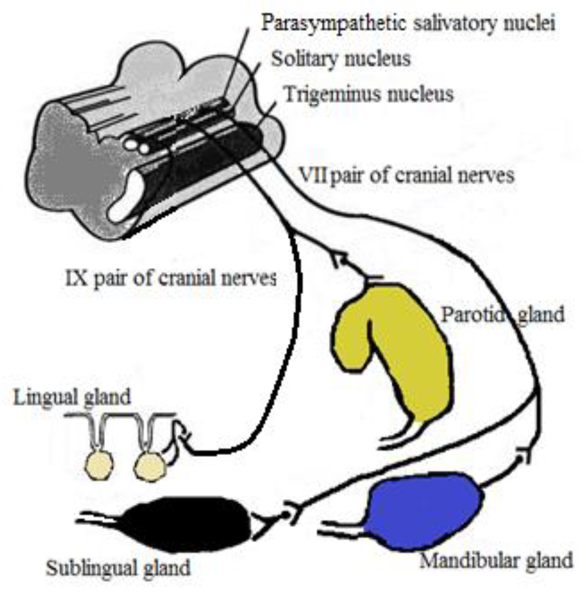
3. Autonomic Control of Salivary Secretion

Salivation is chiefly controlled by the autonomic nervous system. The activity of the salivary glands is under the control of the sympathetic (preganglia of thoracic segments) and parasympathetic autonomic systems [7]. The parotid gland is by far the most studied among the major salivary glands of lab animals and humans. However, recently, the role of the mandibular glands is becoming clearer as they are involved in the salivary response to different extents related to different requests of adapting secretions. Salivary gland activity is regulated by the wide presence of muscarinic receptors, which, under the release of acetylcholine by the presynaptic fibres of the parasympathetic mesencephalic nervous system, modulate the intensity and composition of saliva production. Additionally, the sympathetic nervous system can modulate the secretion of saliva with adrenergic receptors in terms of composition, leading to a saliva rich in amylase content. In general, the presence of muscarinic receptors is characterized by five subtypes of receptors, M1–M5. In the parotid gland, M1 and M3 receptors are both described, the latter more largely represented in the acinar cells [8].
In the parotid gland, parasympathetic and sympathetic nervous systems play a positive effect in stimulating salivation, whereas in the mandibular gland, the parasympathetic fibres induce an increased production of saliva. Although the volume of the latter is reduced, the gland can still produce more mucins under the sympathetic effect. Therefore, different receptors can act under the control of the nervous central system through the autonomic fibres. As a matter of fact, parotid secretions are directed towards the production of serous saliva, rich in digestive enzymes (the parotid is innervated by the superior salivatory nucleus, whereas the mandibular gland is innervated by the inferior salivatory nucleus of the parasympathetic nervous system in the mesencephalus when salivation is called forth). The mandibular gland is a mixed gland with both serous and mucin production, similarly more involved in softening the bolus. In humans, the mandibular glands also express receptors for histamine (H1 and H2) [9]. This means that saliva production and composition are mediated by the autonomic nervous system centrally.

4. Response to External Stimuli

Bitter taste or astringent substances are capable of calling forth a rapid and copious salivary secretion as a first attempt to dilute the presence of potentially harmful molecules introduced in the mouth, thus alleviating this kind of sensation. Perception of adverse substances with potentially harmful effects on the animal depends on smell and taste sensory nerve endings. The sorting of edible feeding sources is based on the development of competence with regard to olfactory and gustative peculiarities of feeds. Thus, the association between feed and sensory learning is likely considered the first step in the identification of edible feeds, which is useful to avoid future negative experiences [10,11]. Species–specific adaptation to certain feeding sources is successful, and the animal elects such feeds in the diet (plants or parts of plants, in particular). Modification of saliva composition can also occur—as usual in wild animals—if the animal has to face different types of feeds. The possibility of varying saliva production across the different animal species can be arguably associated with the mechanism ruling the feeding ecology. Herbivores, omnivores, and carnivores can reasonably display different saliva compositions, which, however, can change between individuals very rapidly, as well as in humans, in which the same individual can respond and rapidly adapt to the diet [12]. Taste receptors and peripheral sensitivity (via V, VII, and IX cranial nerve pairs) [2] are responsible for conditioning different amounts and compositions of salivary secretions. As a consequence, the chemical composition of the diet can modulate salivary glands’ activity and secretion.
The term xenobiotic is derived from the Greek word xenos, meaning foreign (external to the body), to be distinguished from endogenous compounds. If feed selection and avoidance are based on free choice, the presence of some xenobiotic substances, in case of potentially harmful effects, would lead the animal to adapt the salivary secretion and composition (if evolved in this direction) or avoid that feed.
It was found that pigs are able to counteract the detrimental effects of dietary tannins by stimulating parotid gland (PG) secretion and inducing an increase in proline–rich proteins (PRPs). Such proteins belong to a specific family of salivary proteins, identified to have high binding affinity to dietary tannins. The protein–precipitating activity (PPA) exerted by tannins is sensibly contrasted by the production of salivary tannin–binding proteins (TBPs, histatins, and proline–rich proteins) by PG in different species and to different extents and successes [13,14]. As reported in the literature, the common characteristics of proteins and polypeptides with high affinity for tannins are their open loose structure, high proline content, and small size [15,16]. The capability of some animal species to counteract high dietary tannins has been seen to be linked to PRPs’ ancestral function in the maintenance of oral homeostasis at a basal level of secretion, increased by the high presence of tannins in the diet [17,18,19]. Due to selective pressure, the evolutionary adaptation of animal species induced by a mild to marked ability to cope with high tannins in the diet can explain why some species are able to counteract the PPA of tannins, while others are not. Such evolutionary adaptation appears to be better met in animals that might get advantage from a pulsed production of salivary TBP, increased from basal levels, depending on the seasonal variation of tannins ingested with feed.
However, sometimes, contact with biologically active compounds in the diet with potential harmful effects does not act much more specifically against xenobiotics such as hydrolysable tannins. Thus, to counteract the negative effects, ptyalism may not be efficacious and may simply serve as a diluting strategy. Such ptyalism is characterized by a hyperacute continuous production of watery saliva, as observed in horses after ingestion of ergot alkaloids, such as Claviceps purpurea, in infested pasture [20] or in sheep after ingestion of thapsigargin contained in the roots of Drias plants [21]. In all cases, when watery, serous saliva is needed and sialorrhea is started, a bilateral parotidomegaly can be observed [20,21].

5. Regulatory Neuroendocrine Systems of Salivary Composition Elicited by the Diet

To make the link active between the perception of stimuli and the response involving the salivary glands, essentially, chemical communication must occur. In fact, molecules of different natures are endogenously synthesized to orchestrate the salivary response to stimuli. Several systems have been discovered in neuroendocrine modulation in response to stimuli (present in the diet) composed of active molecules synthesized on purpose, with the expression of receptors that render their activity effective at the cellular level, along with the direct effect of sympathetic and parasympathetic regulation.
In this section, a list of systems is briefly reported.
Orexinic system: The orexin family consists of two peptides, orexin–A (OXA) and orexin–B (OXB), derived from the proteolysis of a common 130–amino–acid precursor, prepro–orexin. They share 40% homology, and the sequence of OXA is unvaried among rats, humans, mice, pigs, and cows [22]. Orexin’s biological action is mediated by two G–protein–coupled receptors, orexin type 1 receptor (OX1R) and orexin type 2 receptor (OX2R). OX1R selectively binds to OXA, while OX2R binds to both OXA and OXB [22]. The receptors are 64% homologous and highly preserved among species [23]. Initially, their presence has been highlighted in the neurons of the lateral hypothalamic area in rats [22]. Consequently, this area is known to be involved in appetite control [24]; this justification has been given due to their presence in this part of the hypothalamus. It has been demonstrated that when these substances are injected into the lateral ventricle, they cause an increase in food consumption in subjects fed regularly [25] and that, in any case, there is an inverse relationship between nutritional status and production of orexins. Then, many studies conducted on humans and laboratory animals have revealed the ubiquitous distribution of these molecules, which are localized in the neuroendocrine cells and nerves of the digestive tract [26,27,28] and in other organs and systems that are not strictly related to the functionality of the digestive system. This has led to the hypothesis that they can play a peripheral action in the control of the digestive system′s functionality and in intervening in the functional control of all the organs where they have been observed. Regarding the exocrine glands and, in particular, the salivary glands, their presence was investigated and evidenced in the major salivary glands of pigs [29], where both orexins and receptors were identified only in the excretory striated ducts in the mandibular glands, while the acinar structures were not immunoreactive. Characteristically, also the parasympathetic neurons and axons showed a positive immunoreaction.
Leptin and receptors: Leptin (LEP) is a 16.4 kDa protein encoded by the obese gene (ob gene) and synthesized mainly by the adipocytes [30] and, to a lesser extent, by several tissues and organs, such as the placenta [31,32] and stomach [33]. It plays an important role in the control of food intake and metabolism, acting as a link between the adipose tissue and the central nervous system. Leptin’s action is mediated by the activation of a receptor (ObR), of which there are six isoforms arising from mRNA splice variants and having identical extracellular domains but different lengths of intracellular domains. The so-called “long form” represents the functional structure able to transduce the biological signal conveyed by the hormone. Leptin regulates fat mass through the modulation of the introduction of food and energy consumption; this is its predominant role, as suggested by the appearance of obesity when the gene for leptin or its receptor is missing (ob/ob mice or db/db mice, respectively). Numerous immunohistochemical studies in humans and laboratory animals have shown ubiquitous distribution that goes also beyond the known adipose tissue and nervous system and that involves the various sections of the digestive tract, the male and female reproductive system, and numerous exocrine glands. Regarding the latter and in particular the salivary glands, the presence of the leptin receptor was investigated and evidenced in the major salivary glands of horses [34], while the whole system of “leptin and receptor” was studied and observed in the minor salivary glands of donkeys [35] and in the mandibular glands of growing pigs [36]. In the latter, in particular, the effects of different physical forms of one diet on the expression of these molecules were investigated and analysed. All these results, taken together, indicate that salivary glands are likely both producer and target organs, suggesting a functional role of leptin in these organs.
Cannabinoid receptors: Endogenous cannabinoids or endocannabinoids (anandamide, 2–arachidonoylglycerol, etc.) are molecules synthesized in the brain from arachidonic acid. They interact with some specific receptors, CB1 and CB2 receptors, which belong to the superfamily of the G–protein–linked receptors. The CB1 receptor is mainly expressed in the nerve cells of the central nervous system, primarily in the area for the control of feeding, but also in different kinds of cells of some peripheral organs and tissues, including the skeletal muscles, the liver, and parts of the digestive and urinary tracts. CB2 has a much more limited localization than the previous one and seems to be limited to the immunocompetent system, although its presence has also been highlighted in striated muscle and brain: the biological action consequent to their stimulation has yet to be clarified. It has been hypothesized that endocannabinoids may perform a check on the general and specific metabolisms to each apparatus in which their receptors have been highlighted and, in particular, exert hyperphagic action at central the level [37,38]. Between the peripheral organs, the presence and distribution of CB1 have been evidenced in the major salivary glands of dogs using immunohistochemical techniques. The positive immunoreaction is localized in the cytoplasm of the cells of the striated ducts near or on the apical membrane.
The presence and distribution of such regulatory systems in the salivary glands of different animal species manifest the complexity of neuroendocrine regulation of salivary secretions. As a common driver, all the systems cited before are differently expressed according to the composition of the diet or simply according to the physical form of the diet [36]. In particular, endocannabinoid receptors have been seen to be expressed in the mandibular glands (mucins in saliva) according to the increased demand of chewing by the feed (gross physical form). Additionally, leptin receptors have been seen to be increasingly expressed in the mandibular glands if more intense chewing activity is required to swallow the bolus. In this latter case, leptin is also known to possess mitogenic activity and is predominantly expressed in striated ducts of the mandibular glands of pigs, for physicochemical properties of salivary viscosity. In this regard, recent updates point to aquaporin expression in the mandibular glands for modulating the fluidity of saliva according to the physical form of the diet [39].
The modulation of saliva secretions, therefore, appears to be a powerful means of interaction between the animal and the feed, which is particularly evident in animals under free–choice conditions in the natural environment. In this case, selection and intake of certain feed sources, as available in nature and varying according to season, can produce different feeding habits by the animal, driven by different extents of adaptation to feeds and coping ability.

6. Dynamics of Interaction between Natural Feeding Sources and Animals

Extensive livestock farming can rely on available feeding sources present in the natural landscape so that animals can freely consume a heterogeneous diet and different molecules along with it. Such feeding regimes based on a variety of natural feedstuffs may be responsible for different effects, other than those of strict nutritional value, capable of eliciting the neuroendocrine modulation of salivation [40,41]. In some cases, such effects are beneficial to the animal (antioxidants, for instance), but in some others, they can be detrimental, or worse, lethal, depending on the animal species [10]. That way, under natural conditions, browsers and grazers have developed competences on the selection of edible stuffs, which can safely cover energy and nutrient requirements.
However, the presence of xenobiotics may represent a challenge, and in particular instances, salivation contributes to buffering adverse or undesirable effects from a variety of vegetal compounds.
Animal–plant interaction is based on the continuous and reciprocal survival on both sides. In fact, while the animal tries to get advantage of the energy and nutrients present in the different organs of the plant (leaf, seed, fruits, stems, roots), the plant develops different strategies to avoid such “predation”. The antiherbivory traits of the plant can either be preingestive (antipastoral traits, such as spikes or volatile compounds) or postingestive (such as bitter taste or astringency, causing different grades of aversion after intake). On such basis, aversion to certain feedstuffs can have an impact on the selection of available feeding sources, and thus animals can actively shape the natural environment from accurate selection of feeds [42].
The dynamic interplay of plants and animals can lead to the shaping of the natural landscape, and this phenomenon can be considered to evolve into survival strategies from both sides. In fact, ecosystems can differ in both composition and distribution of plant and animal species, in either scrublands or woodlands, at different latitudes and across seasons. When considering browsing and grazing herbivores, in fact, the different selections of feeds are also ruled by the time spent on feed intake per day, for which salivary secretions can explain the phenomenon of the pulsed (seasonal) adaptation. In general, the more selective the herbivore, the more frequent the feed intake during 24 h. This feeding behaviour is also ruled by the different volumes of the prestomachs of ruminants, for which grazers show higher volumes due to a less energy–dense feed selection, requiring continuous salivary production to keep the metabolic activity of fibre digestion by ruminal microflora. When browsers or intermediate grazers are considered, the selection of some particular feeding stuffs may induce the differentiation of the types of saliva produced in a more efficient way. The physical form of the diet changes from lengthy fibrous feeds as the main component of the diet of grazers to energy–dense organs of plants selected for relatively lower content of digestible fibrous components [43]. In the latter, mostly consumed by browsers or intermediate feeders from scrublands and woodlands (prairies and pastures are preferred by grazers), several other compounds can be found as defensive mechanisms of plants, including tannins, alkaloids, and volatile molecules [21].
The salivary response to xenobiotics present in the diet varies according to the salivary glands of different animal species, as reported in Table 2.

7. Conclusions

Salivation plays a pivotal role in bridging animal feeding habits with natural feeding sources available in the environment. In these extremely various scenarios, the complexity of interactions between plant molecules and salivary secretions is also witnessed by the fine regulation of synthesis of biochemical compounds from both animals and plants. To such an extent, the coevolution of plants and animals can give rise to huge differences among animal species, which, in turn, can explain how feed selection could shape the natural landscape. Species–specific differences point out that domestic animals can select natural feedstuffs in different ways, according to their ability to modulate the composition of their saliva. As detailed in this review, adaptation can rely on the neuroendocrine modulation of salivary secretions, upon which the different coping abilities of animal species can be explained, mirrored by the different feeding behaviours. In view of the literature explored, it appears that domestic animals can show successful salivary adaptation, markedly expressed in pigs, followed by goats, rabbits, and to a progressively lesser extent, horses, sheep, and cattle. In view of a comparative diet composition, browsers and intermediate feeders may be more efficient in shaping the landscape than grazers. As a result, the opportunity offered by the pulsed variation of salivary secretions elicited by feed diversity and seasonal availability can contribute, among other anatomo–physiological peculiarities, to determining the feeding habits of animals. The natural environment displays different sources of chemical stimuli coming from feeding sources, useful for sensory learning. Animal response to xenobiotics in the natural feed has evolved to allow for the safe selection or avoidance of feeding sources, potentially shaping the natural landscape.

Author Contributions

All authors conceived, interpreted, wrote, reviewed and edited the manuscript. All authors have read and agreed to the published version of the manuscript.

Funding

This research received no external funding.

Data Availability Statement

No new data were created or analyzed in this study. Data sharing is not applicable to this article.

Conflicts of Interest

The authors declare no conflict of interest.

References

  1. Barone, R. Anatomie compareé des mammiféres domestique, 4th ed.; Edagricole: Bologna, Italy, 1996. [Google Scholar]
  2. Dukes, H.H. The Physiology of Domestic Animals; Comstock Publishing Associates: Ithaca, NY, USA, 1955. [Google Scholar]
  3. Pavlov, I.P. The Work of the Digestive Glands; Griffin C & Co. Ltd.: London, UK, 1910. [Google Scholar]
  4. Matsuo, R. Central connection for salivary innervation and efferent impulse formation. In Neural Mechanism of Salivary Gland Secretion; Frontiers oral biol; Garrett, J.R., Ekström, J., Anderson, L.C., Eds.; Karger: Basel, Switzerland, 1999. [Google Scholar]
  5. Cano, J.; Rodrìguez-Enchandia, E.L. Degenerating taste buds in sialectomized rats. Cells Tissues Organs 1980, 106, 487–492. [Google Scholar] [CrossRef] [PubMed]
  6. Catalanotto, F.A.; Sweeney, E.A. The effects of surgical desalivation of the rat upon taste acuity. Arch. Oral Biol. 1972, 17, 1455–1465. [Google Scholar] [CrossRef]
  7. Proctor, G.B.; Carpenter, G.H. Regulation of salivary gland function by autonomic nerves. Auton. Neurosci. 2007, 133, 3–18. [Google Scholar] [CrossRef] [PubMed]
  8. Proctor, G.B. Muscarinic receptors and salivary secretion. J. Appl. Physiol. 2006, 100, 1103–1104. [Google Scholar] [CrossRef]
  9. Kim, J.H.; Park, S.H.; Moon, Y.W.; Hwang, S.; Kim, D.; Jo, S.H.; Oh, S.B.; Kim, J.S.; Jahng, J.W.; Lee, J.H.; et al. Histamine H1 receptor induces cytosolic calcium increase and aquaporine translocation in human salivary gland cells. J. Pharmacol. Experim. Ther. 2009, 330, 403–412. [Google Scholar] [CrossRef]
  10. Cappai, M.G.; Aboling, S. Toxic or harmful components of aromatic plants in animal nutrition. In Feed Additives: Aromatic Plants and Herbs in Animal Nutrition and Health; Florou-Paneri, P., Christaki, E., Giannenas, I., Eds.; Elsevier: Amsterdam, The Netherlands, 2019; pp. 147–158. [Google Scholar]
  11. Cappai, M.G.; Pinna, W. Feed intake and selection behavior of the Asinara albino donkey: The fascination of adaptation. Tierarztl. Umsch. 2020, 75, 84–87. [Google Scholar]
  12. Ekström, J.; Khosravani, N.; Castagnola, M.; Messana, I. Saliva and the control of its secrection. In Dysphagia. Medical Radiology; Ekberg, O., Ed.; Springer: Cham, Switzerland, 2017; pp. 21–57. [Google Scholar]
  13. Ann, D.K.; Lin, H.H. Macaque salivary proline-rich protein: Structure, evolution, and expression. Crit. Rev. Oral Biol. Med. 1993, 4, 545–551. [Google Scholar] [CrossRef] [Green Version]
  14. Takahashi, T.; Shimada, T. Selective consumption of acorns by the Japanese wood mouse according to tannin content: A behavioral countermeasure against plant secondary metabolites. Ecol. Res. 2008, 23, 1033–1038. [Google Scholar] [CrossRef]
  15. Bennick, A. Salivary proline-rich proteins. Mol. Cell. Biochem. 1982, 45, 83–99. [Google Scholar] [CrossRef] [PubMed]
  16. Mehansho, H.; Butler, L.; Carlson, D.M. Dietary tannins and salivary proline rich proteins: Interactions, Induction, and Defense Mechanism. Annu. Rev. Nutr. 1987, 7, 423–440. [Google Scholar] [CrossRef] [PubMed]
  17. Bacon, J.R.; Rhoades, M.J. Development of a competition assay for the evaluation of the human parotid salivary proteins to dietary complex phenolics and tannins using a peroxidas-labelled tannin. J. Agric. Food Chem. 1998, 46, 5083–5088. [Google Scholar] [CrossRef]
  18. Clauss, M.; Gehrke, J.; Hatt, J.M.; Dierenfeld, E.S.; Flach, E.J.; Hermes, R.; Castell, J.; Streike, J.; Fickel, J. Tannin-binding salivary proteins in three captive rhinoceros species. Comp. Biochem. Physiol. Part A 2004, 140, 67–72. [Google Scholar] [CrossRef] [PubMed]
  19. Cappai, M.G.; Wolf, P.; Dimauro, C.; Pinna, W.; Kamphues, J. The bilateral parotidomegaly (hypertrophy) induced by acorn consumption in pigs is dependent on individual’s age but not on intake duration. Livest. Sci. 2014, 167, 263–268. [Google Scholar] [CrossRef]
  20. Aboling, S.; Drotleff, A.M.; Cappai, M.G.; Kamphues, J. Contamination with ergot bodies (Claviceps purpurea sensu lato) of two horse pastures in Northern Germany. Mycotoxin Res. 2016, 32, 207–219. [Google Scholar] [CrossRef] [PubMed]
  21. Cappai, M.G.; Wolf, P.; Pinna, W.; Kamphues, J. Pigs use endogenous proline to cope with acorn (Quercus pubescens Willd.) combined diets high in hydrolysable tannins. Livest. Sci. 2013, 155, 316–322. [Google Scholar] [CrossRef]
  22. Sakurai, T.; Amemiya, A.; Ishii, M.; Matsuzaki, I.; Chemlli, R.M.; Tanaka, H.; Williams, S.C.; Richardson, J.A.; Kozlowski, G.P.; Wilson, S.; et al. Orexins and orexin receptors: A family of hypothalamic neuropeptides and G protein-coupled receptors that regulate feeding behavior. Cell 1998, 92, 573–585. [Google Scholar] [CrossRef] [Green Version]
  23. Smart, D.; Jerman, J.C. The physiology and pharmacology of the orexins. Pharmacol. Ther. 2002, 94, 51–61. [Google Scholar] [CrossRef]
  24. Bernardis, L.L.; Bellinger, L.L. The lateral hypothalamic area revisited: Ingestive behavior. Neurosci. Biobehav. Rev. 1996, 20, 189–287. [Google Scholar] [CrossRef]
  25. Kukkonen, J.P.; Holmqvist, T.; Ammoun, S.; Åkerman, K.E.O. Functions of the orexinergic/hypocretinergic system. Am. J. Physiol.-Cell Physiol. 2002, 283, C1567–C1591. [Google Scholar] [CrossRef]
  26. Ehrström, M.; Gustafsson, T.; Finn, A.; Kirchgessner, A.; Grybäc, P.; Jacobsson, H.; Hellstrom, P.M.; Naslund, E. Inhibitory effect of exogenous orexin A on gastric emptying, plasma leptin and the distribution of orexin and orexin receptors in the gut and pancreas in man. J. Clin. Endocrinol. Metabolism. 2005, 90, 2370–2377. [Google Scholar] [CrossRef] [Green Version]
  27. Nakabayashi, M.; Suzuki, T.; Takahashi, K.; Totsune, K.; Muramatsu, Y.; Kaneko, C.; Date, F.; Takeyama, J.; Darnel, A.D.; Moriya, T.; et al. Orexin-A expression in human peripheral tissues. Mol. Cell. Endocrinol. 2003, 205, 43–50. [Google Scholar] [CrossRef]
  28. Näslund, E.; Ehrström, M.; Ma, J.; Hellström, P.M.; Kirchgessner, A.L. Localization and effects of orexin on fasting motility in the rat duodenum. Am. J. Physiol.-Gastrointest. Liver Physiol. 2002, 282, 470–479. [Google Scholar] [CrossRef] [Green Version]
  29. Dall’Aglio, C.; Zannoni, A.; Mercati, F.; Forni, M.; Bacci, M.L.; Boiti, C. Differential gene expression and immune localization of the orexin system in the major salivary glands of pigs. Regul. Pept. 2011, 172, 51–57. [Google Scholar] [CrossRef]
  30. Zhang, Y.; Proenca, R.; Maffei, M.; Barone, M.; Leopold, L.; Friedman, J.M. Positional cloning of the mouse obese gene and its human homologue. Nature 1994, 372, 425–432. [Google Scholar] [CrossRef] [PubMed]
  31. Akerman, F.; Lei, Z.M.; Rao, C.V. Human umbilical cord and fetal membranes co-express leptin and its receptor genes. Gynecol. Endocrinol. 2002, 16, 299–306. [Google Scholar] [CrossRef] [PubMed]
  32. Señarís, R.; Garcia-Caballero, T.; Casabiell, X.; Gallego, R.; Castro, R.; Considine, R.V.; Dieguez, C.; Casanueva, F.F. Synthesis of leptin in human placenta. Endocrinology 1997, 138, 4501–4504. [Google Scholar] [CrossRef]
  33. Bado, A.; Levasseur, S.; Attoub, S.; Kermorgant, S.; Laigneau, J.P.; Bortoluzzi, M.N.; Moizo, L.; Lehy, T.; Guerre-Millo, M.; Le Marchand-Brustel, Y.; et al. The stomach is a source of leptin. Nature 1998, 394, 790–793. [Google Scholar] [CrossRef]
  34. Dall’Aglio, C.; Maranesi, M.; Pascucci, L.; Mercati, F.; Ceccarelli, P. Immunohistochemical distribution of leptin receptor in the major salivary glands of horses. Res. Vet. Sci. 2012, 93, 1116–1118. [Google Scholar] [CrossRef]
  35. Dall’Aglio, C.; Bazucchi, C.; Mercati, F.; Ceccarelli, P. Presence and distribution of leptin and its receptor in the minor salivary glands of the donkey. Acta Histochem. 2015, 117, 305–308. [Google Scholar] [CrossRef] [PubMed]
  36. Cappai, M.G.; Dall’Aglio, C.; Sander, S.J.; Ratert, C.; Dimauro, C.; Pinna, W.; Kamphues, J. Different physical forms of one diet fed to growing pigs induce morphological changes in mandibular glands and local leptin (Ob) production and receptor (ObR) expression. J. Anim. Physiol. Anim. Nutr. 2016, 100, 1067–1072. [Google Scholar] [CrossRef] [PubMed]
  37. Chen, Y.; Buck, J. Cannabinoids protect cells from oxidative cell death: A receptor-independent mechanism. J. Pharmacol. Exp. Ther. 2000, 293, 807–812. [Google Scholar] [PubMed]
  38. Mercati, F.; Dall’Aglio, C.; Pascucci, L.; Boiti, C.; Ceccarelli, P. Identification of cannabinoid type 1 receptor in dog hair follicles. Acta Histochem. 2012, 114, 68–71. [Google Scholar] [CrossRef]
  39. Dall’Aglio, C.; Mercati, F.; De Felice, E.; Tardella, F.M.; Kamphue, J.; Cappai, M.G.; Scocco, P. Influence of different feed physical forms on mandibular gland in growing pigs. Animals 2020, 10, 910. [Google Scholar] [CrossRef]
  40. Jacobson, M.D.; Wusteman, M.; Downes, P. Muscarinic receptors and hydrolysis of inositol phospholipids in rat cerebral cortex and parotid gland. J. Neurochem. 1985, 44, 465–472. [Google Scholar] [CrossRef]
  41. Weiss, S.J.; Putney, J.W., Jr. The relationship of phosphatidylinositol turnover to receptors and calcium channels in rat parotid acinar cells. Biochem. J. 1981, 194, 463–468. [Google Scholar] [CrossRef] [PubMed] [Green Version]
  42. Provenza, F.D.; Lynch, J.J.; Burritt, E.A.; Scott, C.B. How goats learn to distinguish between novel foods that differ in postingestive consequences. J. Chem. Ecol. 1994, 20, 609–624. [Google Scholar] [CrossRef] [PubMed]
  43. Van Soest, J.P. Nutritional Ecology of the Ruminant, 2nd ed.; Cornell University Press: New York, NY, USA, 1994. [Google Scholar]
  44. Lamy, E.; da Costa, G.; e Silva, F.C.; Potes, J.; Coelho, A.V.; Baptista, E.S. Comparison of electroforetic protein profiles of sheep and goat parotid saliva. J. Chem. Ecol. 2008, 34, 388–397. [Google Scholar] [CrossRef]
  45. da Costa, G.; Lamy, E.; e Silva, F.C.; Andersen, J.; Baptista, E.S.; Coelho, A.V. Salivary amylase induction by tannin-enriched diets as a possible countermeasure against tannins. J. Chem. Ecol. 2008, 34, 376–387. [Google Scholar] [CrossRef] [Green Version]
  46. Patterson, J.; Brightling, P.; Titchen, D.A. β-Adrenergic effects on composition of parotid salivary secretion of sheep on feeding. Q. J. Exp. Physiol. 1982, 67, 57–67. [Google Scholar] [CrossRef] [Green Version]
  47. Narjisse, H.; Elhonsali, M.A.; Olsen, J.D. Effects of oak (Quercus ilex) tannins on digestion and nitrogen balance in sheep and goats. Small Rumin. Res. 1995, 18, 201–206. [Google Scholar] [CrossRef]
  48. Salem, A.Z.M.; Lòpez, S.; Ranilla, M.J.; Gonzàlez, J.S. Short- to medium-term effects of consumption of quebracho tannins on saliva production and composition in sheep and goats. J. Anim. Sci. 2013, 91, 1341–1349. [Google Scholar] [CrossRef] [Green Version]
  49. Fickel, J.; Göritz, F.; Joest, B.A.; Hildebrandt, T.; Hoffman, R.R.; Breves, G. Analysis of parotid and mixed saliva in roe deer. J. Comp. Physiol. B 1998, 168, 257–264. [Google Scholar] [CrossRef] [PubMed]
  50. Austin, P.J.; Suchar, L.A.; Robbins, C.T.; Hagerman, A.E. Tannin-binding proteins in saliva of deers and their absence in saliva of sheep and cattle. J. Chem. Ecol. 1989, 15, 1335–1347. [Google Scholar] [CrossRef] [PubMed]
  51. Lamy, E.; Ridrigues, L.; Guerreiro, O.; Soldado, D.; Francisco, A.; Lima, M.; e Silva, F.C.; Jerònimo, E. Changes in salivary protein composition of lambs supplemented with aerial parts and condensed tannins: Extract from Cystus ladanifer L.—A preliminary study. Agrofor. Syst. 2020, 94, 1501–1509. [Google Scholar] [CrossRef]
  52. Wolf, P.; Cappai, M.G. Response of fattening rabbits with acorns (Quercus pubescens Will.) combined in the diet: First acquaintances on growth performance, carcass traits, and perirenal fatty acid profile. Animals 2020, 10, 1394. [Google Scholar] [CrossRef] [PubMed]
Figure 1. Anatomical topography of major salivary glands in some domestic animal species (clockwise: horse, cattle, goat, rabbit, and pig). Salivary glands are schematically reported with different colours in the region of the head, following the comparative extension of the glands: parotid glands (PG, in yellow), mandibular glands (MG, in blue), and sublingual glands (SL, in black). Secretions of the different pairs of major salivary glands differ in composition according to animal species and the species–specific capability to cope with a variety of feeds, nutrients, and secondary metabolites in the natural diet.
Agriculture 11 00817 g001
Figure 2. Simple scheme of parasympathetic innervations of the major salivary glands and von Ebner’s glands of taste buds. Interestingly, the induction of salivary secretion is carried out thanks to parasympathetic ganglia. Purely serous glands (parotid and von Ebner’s glands) are innervated by the IX pairs of cranial nerves. Mixed salivary glands (mandibular and sublingual glands) are innervated by the VII pair of cranial nerves, as shown in the figure. In the mesencephalic nuclei (solitary, parasympathetic, and trigeminus), information is centrally modulated for the response, with autonomic (unconscious, noncortical) response.
Agriculture 11 00817 g002
Table 1. Major salivary glands of domestic animals. Type of gland and secretion.
Salivary GlandType of GlandType of Secretion
Parotid glandExocrineSerous, pure
Mandibular glandExocrineMixed, mucous and serous, prevalently serous (demilune of Giannuzzi)
Sublingual glandExocrineMixed, mucous and serous, prevalently mucous
Table 2. Animal species and salivary response to biologically active substances (xenobiotics) potentially present in the natural diet.
Animal SpeciesSalivary ResponseBiologically Active SubstanceFeed Sources
PigParotid glandsHydrolyzable tannins [19,20,21]Oak acorns
HorseParotid glandsErgot alkaloids [20]Contaminated Poaceae by Claviceps purpurea
GoatParotid glandsTannins [44,45,46,47]Different seeds and foliage
DeerParotid glands and Mandibular glandsTannins [48,49,50]Different seeds and foliage; quebracho
SheepParotid glandsCondensed tannins [51]Cistus ladanifer L.
RabbitParotid glandsHydrolyzable tannins [52]Oak acorns
SquirrelParotid glandsTannins [14]Oak acorns
Hamster, mouseParotid glandsTannins [16]Different seeds and nuts; isoproterenol
Publisher’s Note: MDPI stays neutral with regard to jurisdictional claims in published maps and institutional affiliations.

