Next Article in Journal
Association of Hypertensive Intracerebral Hemorrhage with Left Ventricular Hypertrophy on Transthoracic Echocardiography
Next Article in Special Issue
Association of the Hemoglobin to Serum Creatinine Ratio with In-Hospital Adverse Outcomes after Percutaneous Coronary Intervention among Non-Dialysis Patients: Insights from a Japanese Nationwide Registry (J-PCI Registry)
Previous Article in Journal
A Comprehensive Machine-Learning Model Applied to Magnetic Resonance Imaging (MRI) to Predict Alzheimer’s Disease (AD) in Older Subjects
Previous Article in Special Issue
Insulin Resistance Predicts Severity of Coronary Atherosclerotic Disease in Non-Diabetic Patients
 
 
jcm-logo
Article Menu

Article Menu

Article

Sequential Strategy Including FFRCT Plus Stress-CTP Impacts on Management of Patients with Stable Chest Pain: The Stress-CTP RIPCORD Study

by
Andrea Baggiano
1,†,
Laura Fusini
1,†,
Alberico Del Torto
1,2,
Patrizia Vivona
1,2,
Marco Guglielmo
1,
Giuseppe Muscogiuri
1,
Margherita Soldi
1,
Chiara Martini
3,
Enrico Fraschini
1,
Mark G. Rabbat
4,5,
Francesca Baessato
6,
Gloria Cicala
7,
Maria L. Danza
8,
Annachiara Cavaliere
9,
Antonella Loffreno
10,
Vitanio Palmisano
11,
Francesca Ricci
12,
Giulia Rizzon
9,
Elisabetta Tonet
13,
Giacomo M. Viani
1,2,
Saima Mushtaq
1,
Edoardo Conte
1,
Andrea D. Annoni
1,
Alberto Formenti
1,
Maria E. Mancini
1,
Franco Fabbiocchi
1,
Piero Montorsi
1,2,
Daniela Trabattoni
1,
Alexia Rossi
14,15,
Fabio Fazzari
15,
Nicola Gaibazzi
16,
Daniele Andreini
1,2,
Emilio M. Assanelli
1,
Antonio L. Bartorelli
1,17,
Mauro Pepi
1,
Andrea I. Guaricci
18 and
Gianluca Pontone
1,*
add Show full author list remove Hide full author list
1
Centro Cardiologico Monzino IRCCS, 20138 Milan, Italy
2
Department of Clinical Sciences and Community Health, Cardiovascular Section, University of Milan, 20122 Milan, Italy
3
Department of Radiology, Azienda Ospedaliero-Universitaria, 43126 Parma, Italy
4
Loyola University Medical Center, Center for Heart & Vascular Medicine, Maywood, IL 60153, USA
5
Edward Hines Jr. VA Hospital, Hines, IL 60141, USA
6
Divisione di Cardiologia, Dipartimento di Medicina, Università degli Studi, 37134 Verona, Italy
7
Dipartimento di Medicina e Chirurgia, Azienda Ospedaliero-Universitaria di Parma, Università degli Studi, 43126 Parma, Italy
8
Division of Cardiology, University of Rome Tor Vergata, 00133 Rome, Italy
9
Dipartimento di Medicina, Istituto di Radiologia, Università degli Studi, 35128 Padova, Italy
10
U.O.C. Cardiologia 1, Ospedale di Circolo e Fondazione Macchi, Università degli Studi, 21100 Varese, Italy
11
Diagnostica per Immagini, Università degli Studi di Cagliari, Monserrato, 09042 Cagliari, Italy
12
Department of Biomedicine and Prevention Division of Diagnostic Imaging University of Rome Tor Vergata, 00133 Rome, Italy
13
Cardiovascular Institute, Azienda Ospedaliero-Universitaria di Ferrara, Cona, 44124 Ferrara, Italy
14
Department of Biomedical Sciences, Humanitas University, Pieve Emanuele, 20090 Milan, Italy
15
Department of Diagnostic Imaging, Humanitas Research Hospital, Rozzano, 20089 Milan, Italy
16
Unità Operativa di Cardiologia, Azienda Ospedaliero-Universitaria di Parma, Università degli Studi, 43126 Parma, Italy
17
Department of Biomedical and Clinical Sciences “Luigi Sacco”, University of Milan, 20157 Milan, Italy
18
Institute of Cardiovascular Disease, Department of Emergency and Organ Transplantation, University Hospital Policlinico of Bari, 70124 Bari, Italy
*
Author to whom correspondence should be addressed.
Equal contribution as first author.
J. Clin. Med. 2020, 9(7), 2147; https://doi.org/10.3390/jcm9072147
Submission received: 17 May 2020 / Revised: 3 July 2020 / Accepted: 3 July 2020 / Published: 8 July 2020
(This article belongs to the Special Issue Recent Advances in Coronary Artery Disease)

Abstract

:
Stress computed tomography perfusion (Stress-CTP) and computed tomography-derived fractional flow reserve (FFRCT) are functional techniques that can be added to coronary computed tomography angiography (cCTA) to improve the management of patients with suspected coronary artery disease (CAD). This retrospective analysis from the PERFECTION study aims to assess the impact of their availability on the management of patients with suspected CAD scheduled for invasive coronary angiography (ICA) and invasive FFR. The management plan was defined as optimal medical therapy (OMT) or revascularization and was recorded for the following strategies: cCTA alone, cCTA+FFRCT, cCTA+Stress-CTP and cCTA+FFRCT+Stress-CTP. In 291 prospectively enrolled patients, cCTA+FFRCT, cCTA+Stress-CTP and cCTA+FFRCT+Stress-CTP showed a similar rate of reclassification of cCTA findings when FFRCT and Stress-CTP were added to cCTA. cCTA, cCTA+FFRCT, cCTA+Stress-CTP and cCTA+FFRCT+Stress-CTP showed a rate of agreement versus the final therapeutic decision of 63%, 71%, 89%, 84% (cCTA+Stress-CTP and cCTA+FFRCT+Stress-CTP vs cCTA and cCTA+FFRCT: p < 0.01), respectively, and a rate of agreement in terms of the vessels to be revascularized of 57%, 64%, 74%, 71% (cCTA+Stress-CTP and cCTA+FFRCT+Stress-CTP vs cCTA and cCTA+FFRCT: p < 0.01), respectively, with an effective radiation dose (ED) of 2.9 ± 1.3 mSv, 2.9 ± 1.3 mSv, 5.9 ± 2.7 mSv, and 3.1 ± 2.1 mSv. The addition of FFRCT and Stress-CTP improved therapeutic decision-making compared to cCTA alone, and a sequential strategy with cCTA+FFRCT+Stress-CTP represents the best compromise in terms of clinical impact and radiation exposure.

1. Introduction

Coronary computed tomography angiography (cCTA) was introduced as an anatomic imaging method to rule out the presence of coronary artery disease (CAD) [1] and also for improving prognostic assessment beyond baseline risk factor evaluation and functional stress test findings [2,3,4]. However, several factors limit its specificity and positive predictive value [5,6]. In this regard, there is an emerging body of literature on the added value of stress myocardial perfusion utilizing computed tomography (Stress-CTP) [7,8,9,10,11,12] and computed tomography-derived fractional flow reserve (FFRCT) on top of cCTA [9,13,14]. Moreover, several studies have compared the diagnostic accuracy of these novel CT functional techniques and they have shown favorable and comparable results, with increased diagnostic performance in detecting flow-limiting stenoses when compared to cCTA alone [15,16]. However, no comparison between the influence of FFRCT and Stress-CTP on the management of patients has been performed [17,18]. Therefore, the aim of the study is to evaluate the allocation of patients to optimal medical treatment (OMT) or revascularization using cCTA, cCTA+FFRCT and cCTA+Stress-CTP, and the rate of agreement in terms of the vessels to be revascularized in relation to invasive coronary angiography (ICA) and FFR-defined significance.

2. Experimental Methods

The retrospective analysis of this study was conducted on subjects prospectively enrolled in the PERFECTION Study, a longitudinal prospective study with consecutive cohorts designed to compare the feasibility and accuracy of integrated cCTA+FFRCT versus cCTA+Stress-CTP for the diagnosis of functionally significant CAD [19]. This study complied with the Declaration of Helsinki. Moreover, the institutional ethical study committee approved the protocol (R250/15-CCM 262), and all patients meeting the selection criteria were asked to sign an informed consent. A structured interview was performed to collect a clinical history and cardiac risk factors.
Two consecutive cohorts of patients with a pre-test likelihood of CAD > 50% according to the updated Diamond-Forrester risk model score were evaluated. The first cohort of 147 patients were evaluated with the Static Stress-CTP, while the second cohort of 144 patients were tested with the Dynamic Stress-CTP. The first cohort of patients was screened with consecutive symptomatic patients with suspected CAD referred for non-emergent, clinically indicated ICA between October 2015 and May 2017, while the second cohort was screened following the same criteria between June 2017 and April 2019.
The study flow diagram and the study protocol are shown in Appendix A (Figure A1 and Figure A2, respectively).
Appendix B details the methods used to perform and interpret the results of the studied techniques, including Rest cCTA, FFRCT, Static Stress-CTP, Dynamic Stress-CTP, ICA and invasive FFR. The method used to calculate radiation exposure is also described.
Clinical management according to cCTA results only (Pathway A). As a first step, the clinical management of patients according to cCTA results was assessed according to the criteria as shown in Figure 1: if all coronary stenoses were less than 50%, OMT was chosen; if at least one coronary stenosis was estimated in the range of 50% to 70% of vessel lumen reduction, more information was required; if at least one coronary stenosis resulted in more than 70% of vessel lumen reduction, invasive assessment was indicated.
Clinical management according to addition of FFRCT analysis to cCTA (Pathway B). Clinical management when both cCTA and FFRCT were available was investigated according to the following criteria (Figure 1): (1) in the case of stenoses ≤ 50%, OMT was prescribed with FFRCT values ≥ 0.7 for all vessels, while ICA was indicated if FFRCT was < 0.7 for at least one vessel; (2) in the case of cCTA with at least one stenosis > 50% of lumen reduction, if FFRCT values were > 0.8 for all vessels, OMT was prescribed, while if FFRCT was in the range of 0.7 to 0.8 for at least one vessel, more information was required; finally, if FFRCT was less than 0.7 for at least one vessel, ICA was indicated.
Clinical management according to addition of Stress-CTP results to cCTA (Pathway C). Clinical management when both cCTA and Stress-CTP (where Static or Dynamic protocol was used) were available was investigated according to the following criteria (Figure 1): (1) in the case of coronary stenoses ≤ 50% of lumen reduction, OMT was prescribed regardless of the Stress-CTP results; (2) in the case of cCTA with at least one stenosis > 50% of lumen reduction, if perfusion defects were present, ICA was indicated, while in the absence of perfusion defects, OMT was prescribed.
Clinical management according to addition of Stress-CTP on top of cCTA and FFRCT analysis (Pathway D). Clinical management when Stress-CTP was available on top of cCTA and FFRCT analysis was investigated according to the following criteria (Figure 1). In the setting of cCTA ≤ 50%, we indicated: (a) OMT in the case of FFRCT ≥ 0.8 or FFRCT < 0.8 but with a negative matched Stress-CTP; and (b) ICA in the case of FFRCT < 0.8 with a positive matched Stress-CTP. In the setting of cCTA > 50%, we indicated: (a) OMT in the case of FFRCT ≥ 0.8 or FFRCT between 0.7–0.8 but with a negative matched Stress-CTP; (b) ICA in the case of FFRCT between 0.7–0.8 with a positive matched Stress-CTP or in the case of FFRCT < 0.7, regardless of Stress-CTP findings.
Endpoints. The primary endpoint of the rate of agreement with the final management decision (defined as the ratio between the number of patients in which the therapeutic decision matched the final decision to the total number of patients) was measured for each strategy.
The following endpoints were also measured as part of a secondary analysis:
(a)
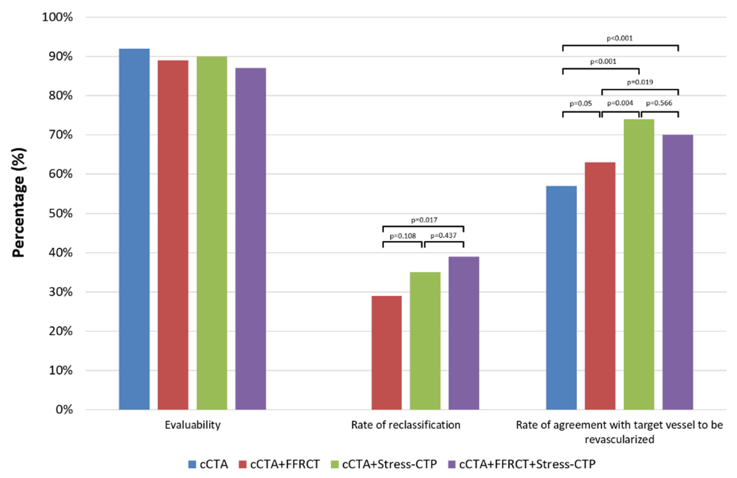
Overall evaluability, defined as the ratio between the number of patients in which the strategy information was applied to the total number of patients
(b)
Rate of reclassification, defined as the ratio between the number of patients who underwent a different management as compared to the initial decision to the total number of patients
(c)
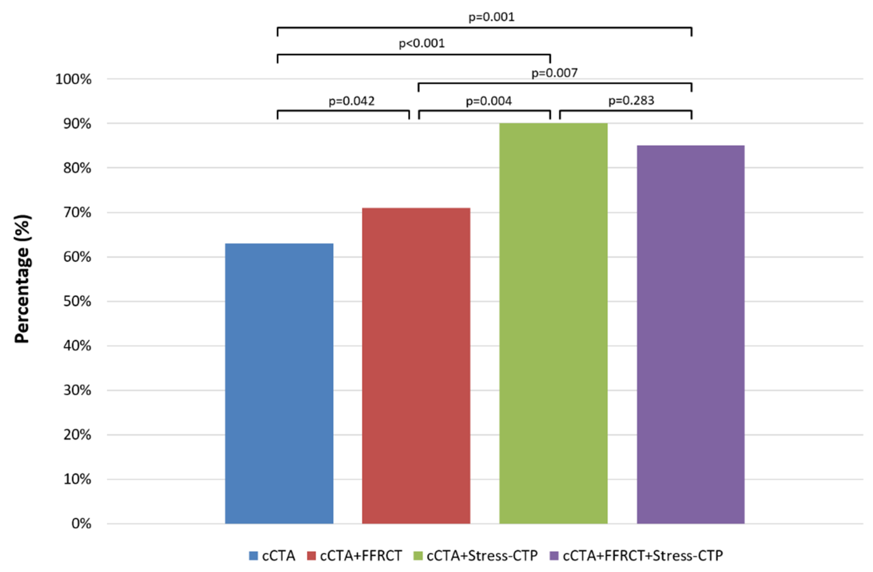
Rate of agreement on the vessels to be revascularized, defined as the ratio between the number of patients in which the vessel to be revascularized matched the final decision to the total number of patients
(d)
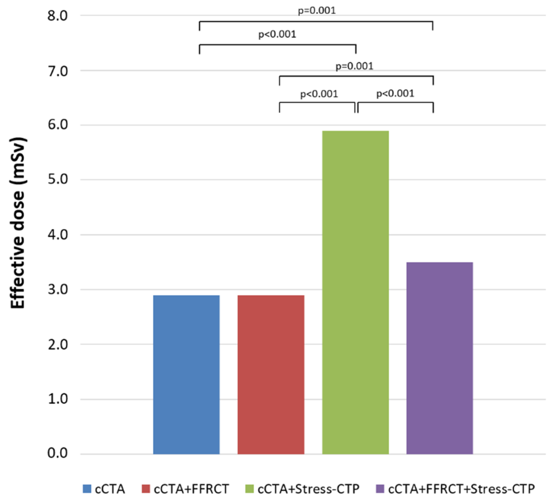
Cumulative effective dose (ED), defined as the cumulative radiation exposure of the strategy.
Statistical analysis. Continuous variables were expressed as mean ± standard deviation (SD) and discrete variables as absolute numbers and percentages. Comparisons between each endpoint of all strategies were assessed with a χ2 test. A p-value < 0.05 was considered significant. Statistical analysis was performed with SPSS, version 25 software (SPSS Inc., Chicago, IL, USA) and R version 3.5.1.

3. Results

Study population.Table 1 shows the patients’ baseline characteristics. The study population consisted of 291 patients (Mean age: 65 ± 9 years; Male: 76%) with a prevalence of obstructive and functionally significant CAD at invasive evaluation of 67% and 49%, respectively.
Management according to cCTA only. Rest cCTA was successfully performed in all patients with a mean ED of 2.9 ± 1.3 mSv (Table 1). The overall evaluability was 92% (267 out of 291 patients). The clinical management according to cCTA alone is illustrated in Figure 2. Seventy-four (25%) patients were treated with OMT, 129 (44%) were sent to ICA, while 88 (30%) patients needed more information for a clinical decision. Once patients were sent to assessment with ICA and invasive FFR, the number of patients treated with OMT increased to 148 (51%), while the number of patients that were treated with revascularization increased to 143 (49%).
The rate of agreement with the final decision was 63% (184 out of 291 patients) and the rate of agreement of vessels to be revascularized was 57% (165 out of 291 patients).
Management according to addition of FFRCT to cCTA. FFRCT analysis was obtained in 280 out of 291 patients (96%) with an overall evaluability of the cCTA+FFRCT strategy of 89% (258 out of 291 patients). The clinical management according to FFRCT analysis on top of cCTA is shown in Figure 3. The addition of FFRCT to cCTA increased the number of patients referred to OMT, from 74 (26%) to 99 (35%), and also increased direct referral to ICA, from 124 (45%) to 134 (48%). The “grey-zone”, in which more information was necessary for a clinical decision, was reduced from 82 (29%) to 47 (17%) patients. Once patients underwent invasive assessment, the number of patients treated with OMT increased to 146 (52%), while the number of patients treated with revascularization remained unchanged at 134 (48%).
The rate of reclassification of patients with FFRCT was 28% (82 out of 291 patients), the rate of agreement with the final treatment decision was 71% (207 out of 291 patients) and the rate of agreement on vessels to be revascularized was 63% (183 out of 291 patients).
Management according to addition of Stress-CTP to cCTA. Stress-CTP was performed in 287 out of 291 patients (99%) with an overall evaluability of the cCTA+Stress-CTP strategy of 90% (263 out of 291 patients). The mean ED of cCTA+Stress-CTP strategy was 5.9 ± 2.7 mSv.
The clinical management according to Stress-CTP analysis on top of cCTA is illustrated in Figure 4. The addition of Stress-CTP led to 87 (30%) patients who needed more information to decide on clinical management, with 50 patients treated with OMT, and 37 patients sent to ICA. After all patients were assessed with the invasive reference standard, the number of patients treated with OMT or revascularization changed minimally, with a mild increase in patients managed with OMT, and a proportional reduction in patients treated with revascularization.
The rate of reclassification of patients with CTP was 34% (100 out of 291 patients), the rate of agreement with the final treatment decision was 89% (261 out of 291 patients) and the rate of agreement on vessels to be revascularized was 74% (216 out of 291 patients).
Management according to sequential addition of FFRCT and Stress-CTP to cCTA. The sequential strategy was available in 254 patients, with an overall evaluability of 87%. The mean ED of the cCTA+FFRCT+Stress-CTP strategy was 3.5 ± 2.1 mSv.
The clinical management according to the sequential addition of FFRCT and Stress-CTP to cCTA is displayed in Figure 5. The addition of functional tools led to a progressive increase in the number of patients treated with OMT from 72 (26%) with cCTA only to 97 (35%) when FFRCT was added, and 121 (44%) with the addition of Stress-CTP. The number of patients in the “grey-zone” in the cCTA analysis was nearly halved from 81 to 46 patients by the addition of FFRCT, and this remaining population was further divided into two approximately equal parts with the addition of Stress-CTP. At the end of this sequential approach, 121 patients (44%) were managed with OMT while 155 (56%) were referred to ICA. After invasive assessment, the number of patients treated with OMT increased to 143 (52%), while patients treated with revascularization were reduced to 133 (48%). The rate of reclassification of patients was 37% (109 out of 291 patients), the rate of agreement with the final treatment decision was 84% (246 out of 291 patients) and the rate of agreement on vessels to be revascularized was 70% (205 out of 291 patients).
Comparison between cCTA, cCTA+FFRCT, cCTA+Stress-CTP and the sequential strategy.Figure 6 and Figure 7 show the comparison between the primary and secondary endpoints of different strategies, respectively. The rate of agreement with the final decision was significantly higher for cCTA+Stress-CTP and for cCTA+FFRCT+Stress-CTP versus other approaches (p < 0.01), with no difference between them. No differences were found in terms of evaluability between the different strategies. Both the cCTA+Stress-CTP and cCTA+FFRCT+Stress-CTP strategies showed a higher rate of patient reclassification and a higher rate of target vessels to be revascularized as compared to the cCTA+FFRCT strategy.
The overall radiation exposure according to each diagnostic strategy is reported in Figure 8. The cCTA+Stress-CTP strategy was associated with the highest ED with a progressive reduction in the ED for cCTA+FFRCT+Stress-CTP and cCTA+FFRCT (p < 0.001).
A representative case example is illustrated in Figure 9.

4. Discussion

The main findings of this study are (a) the addition of both FFRCT and Stress-CTP on top of cCTA led to the reclassification of approximately one third of patients with intermediate to high risk for CAD; (b) compared to the cCTA+FFRCT strategy, cCTA+Stress-CTP showed a better performance in terms of final therapeutic decision-making and target vessels to be revascularized, however, it is also associated with higher radiation exposure; and (c) a sequential strategy including cCTA+FFRCT+Stress-CTP where FFRCT is the gatekeeper for the decision to perform a Stress-CTP, seems to provide the best balance between performance and radiation exposure.
The first step in assessing CAD is the identification of coronary plaque, as the presence of coronary atherosclerosis is a crucial element in determining patients’ prognosis [4,20,21]. Unfortunately, anatomic assessment alone is often not enough to fully determine patient management, especially when patients with high a pre-test likelihood of CAD are evaluated. This is due to the increased coronary calcium burden and disease extension, which are factors that impair the ability of coronary cCTA to correctly rule out significant CAD [5].
Recently, there has been extensive data published regarding the role of FFRCT in this clinical context. This tool is not only accurate in assessing for ischemia [22], but it has been shown to be very helpful in guiding patient management [13,23] in real-word experiences, such as in the ADVANCE Registry [9,24] and in Aarhus, Denmark [25]. Our study of 291 patients confirmed the increased discrimination of functionally relevant stenoses assessed by FFRCT, even in a population with a high burden of significant disease. Even though we used a wide “grey-zone”, between 0.7 and 0.8 in our study, this tool was able to reclassify two-thirds of the patients that required further assessment in order to receive a specific treatment. Moreover, compared to invasive assessment, FFRCT is able to increase the overall diagnostic performance and correct pattern of treatment at both the patient and vessel-specific level.
There is less evidence to support the clinical role of Stress-CTP, and this paucity of data is mainly related to the relatively recent introduction of new generation scanners. Wong et al. used a 320-slice scanner to demonstrate that the combined approach of cCTA+Static Stress-CTP is superior to cCTA alone in assessing the presence of functionally relevant stenosis, using invasive FFR as a reference standard [26]. Using the same reference standard, Coenen et al. used dual energy scanners to show an increase in the diagnostic accuracy of a combined cCTA and Dynamic Stress-CTP approach compared to anatomical assessment alone (AUC 0.78 vs. 0.70) [16]. Finally, Pontone et al. used a last generation whole-heart scanner and showed that an integrated approach (cCTA+Static or Dynamic CTP) had better diagnostic accuracy in the assessment of patients with intermediate-to-high pre-test likelihood of CAD evaluated, again, using invasive FFR as the reference standard [15,27]. Importantly, it was possible to achieve acceptable radiation exposure (<6 mSv for cCTA+Static Stress-CTP and <9 mSv for cCTA+Dynamic Stress-CTP) as well as high diagnostic accuracy (AUC of 0.92 for cCTA+Static Stress-CTP and 0.88 for cCTA+Dynamic Stress-CTP in a per-vessel analysis).
Therefore, the question regarding which of these functional non-invasive techniques should be used, and in which patients is of great interest. Recently, we demonstrated that both Static [15] and Dynamic Stress-CTP [27] showed similar diagnostic performance as compared to invasive evaluation. However, a comparison between Stress-CTP and FFRCT in terms of impact on clinical decision-making is not available. In a sub-study of the RIPCORD trial, Curzen et al. demonstrated that the availability of FFRCT has a substantial effect on the ability to identify significant CAD, and therefore, on the management of patients with stable chest pain compared to cCTA alone [17]. This finding mimics the results found for invasive ICA and FFR in the RIPCORD study.
Our study not only confirmed the additional value of FFRCT and Stress-CTP to cCTA alone, but also demonstrated that a sequential strategy including cCTA alone, followed by FFRCT as a gatekeeper of Stress-CTP represents the best compromise in terms of clinical decision-making and radiation exposure. Due to the recent clinical application of this functional strategy, large-scale multi-center randomized trials addressing the cost-effectiveness and prognostic implications of CT myocardial perfusion are not yet available. To fill this gap in evidence, the CTP-PRO study, an international, multi-center, prospective, open-label, randomized controlled study evaluating the cost-effectiveness and the prognosis of a cCTA+Stress-CTP strategy versus usual care in intermediate-high risk patients with suspected or known CAD has been designed [28] and the results are eagerly awaited.
This study has limitations. Mainly, we used an existing dataset of patients in order to test our hypothesis, to deliver a retrospective analysis of a prospectively collected population, and therefore, the final number of subjects was estimated according to a predefined statistical power for other endpoints.

5. Conclusions

In conclusion, this retrospective analysis of the PERFECTION study demonstrates that the addition of functional assessment with FFRCT and Stress-CTP provides incremental therapeutic decision-making value compared to cCTA alone. A sequential strategy with cCTA+FFRCT+Stress-CTP is associated with the best compromise in terms of clinical impact and radiation exposure.

Author Contributions

Conceptualization, Methodology and Writing Original Data, Data analysis, Supervision: A.B., L.F., G.P.; Data Curation, Data collecting and Data analysis: A.D.T., P.V., M.G., G.M., M.S., F.B., G.C., M.L.D., A.C., A.L., V.P., F.R., G.R., E.T., G.M.V., S.M., E.C., A.D.A., A.F., M.E.M.; Methodology: C.M., E.F., F.F. (Franco Fabbiocchi), P.M., D.T., A.R., F.F. (Fabio Fazzari), N.G., D.A., E.M.A., A.L.B., A.I.G. Writing: M.G.R.; Supervision: M.P.; All authors have read and agreed to the published version of the manuscript.

Funding

The study received a grant from General Electric (GE Healthcare, Milwaukee, WI, USA). FFRCT analysis was provided by HeartFlow (Heartflow Inc., Redwood City, CA, USA).

Acknowledgments

We thank the following people for their contribution to this work: Giuseppe Calligaris, Stefano De Martini, Cristina Ferrari, Stefano Galli, Sebastiano Gili, Luca Grancini, Paolo Olivares, Paolo Ravagnani, Giovanni Teruzzi. Centro Cardiologico Monzino IRCCS, Milan, Italy.

Conflicts of Interest

Pontone G. declares an institutional research grant and/or honorarium as speaker from General Electric, Bracco, Medtronic, Bayer, Heartflow. Andreini D. declares an institutional research grant and/or honorarium as speaker from General Electric, Bracco, Heartflow. Other authors have no conflict of interest to disclose.

Appendix A

Figure A1. Study flow diagram. Consecutive symptomatic patients referred for suspected CAD and scheduled for clinically indicated ICA were screened. After the application of inclusion and exclusion criteria, 291 patients were enrolled, 147 in the first arm, in which Static Stress-CTP was performed, and 144 in the second arm, in which Dynamic Stress-CTP was performed. CAD: coronary artery disease; ICA: invasive coronary angiography; Stress-CTP: stress computed tomography perfusion.
Jcm 09 02147 g0a1
Figure A2. Study protocol. Patients were asked to refrain from smoking and caffeine for 24 h and to maintain fasting for 6 h before the scan. All patients were treated with sublingual nitrates to ensure vasodilatation. In patients with a resting HR > 65 beats/min, metoprolol was intravenously administered, with a titration dose up to 15 mg to achieve a target HR ≤ 65 beats/min. However, all patients were studied even if the target HR was not reached. cCTA and FFRCT were evaluated with the acquisition of the first dataset; then, after a period of fifteen minutes, perfusion evaluation was performed during adenosine administration with the acquisition of the second dataset (Static or Dynamic protocols were used according to the specific arm of the study). HR: heart rate; cCTA: coronary computed tomography angiography; FFRCT: computed tomography-derived fractional flow reserve.
Jcm 09 02147 g0a2

Appendix B

Rest cCTA performance and interpretation. Rest cCTA was performed with a Revolution CT scanner (GE Healthcare, Milwaukee, Wisconsin). The following parameters were used: slice configuration 256 × 0.625 mm with scintillator detector, gantry rotation time 280 ms, tube voltage 120 KVp and 100 KVp in patients with body mass index >30 kg/m2 and <30 kg/m2, respectively, and an effective tube current of 500 mA. One beat axial scan was used in all patients with variable padding ranging from 70% to 80% and 40% to 80% of the cardiac cycle in patients with HR <65 beats/min and >65 beats/min, respectively. All patients received a 70-mL bolus of iodixanol 320 (Visipaque 320 mg/mL, GE Healthcare, Oslo, Norway) at an infusion rate of 6.2 mL/s followed by 50 mL of saline solution at the same rate of infusion. Datasets for each cCTA examination were transferred to an image-processing workstation (Advantage Workstation 4.7; GE Healthcare, Milwaukee, Wisconsin) and independently analyzed according to the Society of Cardiovascular Computed Tomography (SCCT) guidelines for reporting by two cardiac imagers (M.G. and G.M.) who were blinded to the clinical history of the patients. Coronary arteries were segmented as suggested by the American Heart Association. Impaired image quality was classified according to the Likert score. All non-evaluable coronary artery segments were censored as positive. Obstructive CAD was defined as the presence of stenosis >50%. A third cardiac imager (A.I.G.) with more than 5 years’ experience in cCTA adjudicated the scores in cases of disagreement.
FFRCT performance. All cCTA datasets were sent to HeartFlow (Redwood City, California) for FFRCT analysis. In brief, for each patient a quantitative 3-dimensional anatomic model combining the aortic root and epicardial coronary arteries was generated from the cCTA dataset. Subsequently, coronary blood flow and pressure were computed under specific conditions simulating maximal hyperemia.
Static Stress-CTP performance and interpretation. Static Stress-CTP scans was performed after vasodilation induced by intravenous administration of adenosine (0.14 mg/kg/min over 4 min). At the end of the third minute of adenosine infusion, a single data sample was acquired during the first-pass enhancement of cCTA by using the same protocol described for rest-cCTA. All datasets of Static Stress-CTP were transferred to a dedicated image-processing workstation (Advantage Workstation Version 4.7, GE Healthcare), and two cardiac imagers (A.B. and G.P.) blinded to the clinical history, rest cCTA, and invasive evaluation findings, independently evaluated the reconstructed images. The myocardial wall was evaluated on short-axis (apical, mid, and basal slices) and long-axis views (2-, 3- and 4-chamber projections) with 4-mm-thick average multiplanar reformatted images in diastolic phases (70% and 80% of cardiac cycle). A narrow window width and level (350 W and 150 L) was used for perfusion defect evaluation. Each myocardial segment was correlated to the specific coronary territory according to a modified AHA classification as described by Cerci et al. Perfusion defects were defined as subendocardial hypo-enhancements encompassing ≥ 25% of transmural myocardial thickness within a specific coronary territory.
Dynamic Stress-CTP performance and interpretation. Similar to Static Stress-CTP, Dynamic Stress-CTP scans were performed after vasodilatation induced with an intravenous adenosine injection (0.14 mg/kg/min over 4 min). At the end of the third minute of adenosine injection, 0.7 mL/kg of iodixanol 320 (Visipaque 320 mg/mL; GE Healthcare, Oslo, Norway) at an infusion rate of 5 mL/s followed by 0.5 mL/kg of saline solution at an infusion rate of 5 mL/s was injected and, at the same time, the Stress-CTP acquisition was performed in free breathing. Three consecutive series of datasets were acquired that covered the entire heart volume. Multiple short-axis views of the left ventricle from the base to the apex corrected for breathing-related displacement were generated and endocardial and epicardial borders were selected. Accordingly, the change of attenuation in the myocardium over time was computed. For quantification of myocardial blood flow (MBF), the arterial input function (AIF) was sampled in the ascending aorta and the myocardial time/attenuation curves were coupled with the AIF using a hybrid deconvolution model. Finally, MBF was computed by dividing the convoluted maximal slope of the myocardial time-attenuation curve by the maximum AIF. MBF maps were then reconstructed as a stack of color-coded images with a slice thickness of 3.0 mm. Measurements of MBF were obtained from regions of interest of at least 0.5 cm2 positioned in a representative area of each myocardial segment according to a standard 16-segment model. Blinded adjudication was performed to verify co-registration of CTP-defined perfusion defects with culprit vessels as defined using cCTA by two cardiac imagers (A.B. and G.P.) with more than 5 years of experience. Perfusion defects were defined as the presence of MBF values less than 101 mL/100 gr/min within a specific coronary territory.
ICA and invasive FFR performance and interpretation. Certified interventional cardiologists performed ICA in all patients within 60 days after the cCTA examination. Coronary angiograms were analyzed at the clinical site by an interventional cardiologist who was blinded to cCTA and Stress-CTP findings. The severity of coronary stenosis was quantified in two orthogonal planes by identifying the minimum diameter and reference diameter for all stenosis, and the percentage of stenosis was derived. Functionally significant CAD was defined as the presence of coronary artery stenoses ≥ 80%, a totally occluded vessel, or stenoses with invasive FFR < 0.8.
Radiation exposure. The effective radiation dose (ED) was calculated by multiplying the dose-length product and a conversion coefficient for the chest (K = 0.014 mSv/mGy × cm). For ICA, ED was calculated by multiplying the dose area product by a conversion factor (K = 0.21 mSv/mGy × cm2) for lateral and posterior-anterior radiation exposure in the chest.

References

  1. Pontone, G.; Andreini, D.; Quaglia, C.; Ballerini, G.; Nobili, E.; Pepi, M. Accuracy of multidetector spiral computed tomography in detecting significant coronary stenosis in patient populations with differing pre-test probabilities of disease. Clin. Radiol. 2007, 62, 978–985. [Google Scholar] [CrossRef] [PubMed]
  2. Min, J.K.; Dunning, A.; Lin, F.Y.; Achenbach, S.; Al-Mallah, M.; Budoff, M.J.; Cademartiri, F.; Callister, T.Q.; Chang, H.J.; Cheng, V.; et al. Age- and sex-related differences in all-cause mortality risk based on coronary computed tomography angiography findings results from the International Multicenter CONFIRM (Coronary CT Angiography Evaluation for Clinical Outcomes: An International Multicenter Registry) of 23,854 patients without known coronary artery disease. J. Am. Coll. Cardiol. 2011, 58, 849–860. [Google Scholar] [CrossRef] [Green Version]
  3. Pontone, G.; Andreini, D.; Bartorelli, A.L.; Bertella, E.; Cortinovis, S.; Mushtaq, S.; Foti, C.; Annoni, A.; Formenti, A.; Baggiano, A.; et al. A long-term prognostic value of CT angiography and exercise ECG in patients with suspected CAD. JACC Cardiovasc. Imaging 2013, 6, 641–650. [Google Scholar] [CrossRef] [Green Version]
  4. Investigators, S.-H.; Newby, D.E.; Adamson, P.D.; Berry, C.; Boon, N.A.; Dweck, M.R.; Flather, M.; Forbes, J.; Hunter, A.; Lewis, S.; et al. Coronary CT Angiography and 5-Year Risk of Myocardial Infarction. N. Engl. J. Med. 2018, 379, 924–933. [Google Scholar] [CrossRef]
  5. Pontone, G.; Bertella, E.; Mushtaq, S.; Loguercio, M.; Cortinovis, S.; Baggiano, A.; Conte, E.; Annoni, A.; Formenti, A.; Beltrama, V.; et al. Coronary artery disease: Diagnostic accuracy of CT coronary angiography--a comparison of high and standard spatial resolution scanning. Radiology 2014, 271, 688–694. [Google Scholar] [CrossRef]
  6. Dewey, M.; Vavere, A.L.; Arbab-Zadeh, A.; Miller, J.M.; Sara, L.; Cox, C.; Gottlieb, I.; Yoshioka, K.; Paul, N.; Hoe, J.; et al. Patient characteristics as predictors of image quality and diagnostic accuracy of MDCT compared with conventional coronary angiography for detecting coronary artery stenoses: CORE-64 Multicenter International Trial. AJR Am. J. Roentgenol. 2010, 194, 93–102. [Google Scholar] [CrossRef] [PubMed] [Green Version]
  7. Rossi, A.; Merkus, D.; Klotz, E.; Mollet, N.; de Feyter, P.J.; Krestin, G.P. Stress myocardial perfusion: Imaging with multidetector CT. Radiology 2014, 270, 25–46. [Google Scholar] [CrossRef] [PubMed] [Green Version]
  8. Pontone, G.; Andreini, D.; Guaricci, A.I.; Baggiano, A.; Fazzari, F.; Guglielmo, M.; Muscogiuri, G.; Berzovini, C.M.; Pasquini, A.; Mushtaq, S.; et al. Incremental Diagnostic Value of Stress Computed Tomography Myocardial Perfusion With Whole-Heart Coverage CT Scanner in Intermediate- to High-Risk Symptomatic Patients Suspected of Coronary Artery Disease. JACC Cardiovasc. Imaging 2019, 12, 338–349. [Google Scholar] [CrossRef]
  9. Fairbairn, T.A.; Nieman, K.; Akasaka, T.; Norgaard, B.L.; Berman, D.S.; Raff, G.; Hurwitz-Koweek, L.M.; Pontone, G.; Kawasaki, T.; Sand, N.P.; et al. Real-world clinical utility and impact on clinical decision-making of coronary computed tomography angiography-derived fractional flow reserve: Lessons from the ADVANCE Registry. Eur. Heart J. 2018, 39, 3701–3711. [Google Scholar] [CrossRef] [Green Version]
  10. Rossi, A.; Wragg, A.; Klotz, E.; Pirro, F.; Moon, J.C.; Nieman, K.; Pugliese, F. Dynamic Computed Tomography Myocardial Perfusion Imaging: Comparison of Clinical Analysis Methods for the Detection of Vessel-Specific Ischemia. Circ. Cardiovasc. Imaging 2017, 10, e005505. [Google Scholar] [CrossRef] [PubMed] [Green Version]
  11. Pontone, G.; Andreini, D.; Guaricci, A.I.; Guglielmo, M.; Baggiano, A.; Muscogiuri, G.; Fusini, L.; Soldi, M.; Fazzari, F.; Berzovini, C.; et al. Quantitative vs. qualitative evaluation of static stress computed tomography perfusion to detect haemodynamically significant coronary artery disease. Eur. Heart J. Cardiovasc. Imaging 2018, 19, 1244–1252. [Google Scholar] [CrossRef] [PubMed]
  12. Pontone, G.; Baggiano, A.; Andreini, D.; Guaricci, A.I.; Guglielmo, M.; Muscogiuri, G.; Fusini, L.; Soldi, M.; Del Torto, A.; Mushtaq, S.; et al. Diagnostic accuracy of simultaneous evaluation of coronary arteries and myocardial perfusion with single stress cardiac computed tomography acquisition compared to invasive coronary angiography plus invasive fractional flow reserve. Int. J. Cardiol. 2018, 273, 263–268. [Google Scholar] [CrossRef] [PubMed]
  13. Douglas, P.S.; Pontone, G.; Hlatky, M.A.; Patel, M.R.; Norgaard, B.L.; Byrne, R.A.; Curzen, N.; Purcell, I.; Gutberlet, M.; Rioufol, G.; et al. Clinical outcomes of fractional flow reserve by computed tomographic angiography-guided diagnostic strategies vs. usual care in patients with suspected coronary artery disease: The prospective longitudinal trial of FFR(CT): Outcome and resource impacts study. Eur. Heart J. 2015, 36, 3359–3367. [Google Scholar] [CrossRef] [Green Version]
  14. Collet, C.; Onuma, Y.; Andreini, D.; Sonck, J.; Pompilio, G.; Mushtaq, S.; La Meir, M.; Miyazaki, Y.; de Mey, J.; Gaemperli, O.; et al. Coronary computed tomography angiography for heart team decision-making in multivessel coronary artery disease. Eur. Heart J. 2018, 39, 3689–3698. [Google Scholar] [CrossRef]
  15. Pontone, G.; Baggiano, A.; Andreini, D.; Guaricci, A.I.; Guglielmo, M.; Muscogiuri, G.; Fusini, L.; Fazzari, F.; Mushtaq, S.; Conte, E.; et al. Stress Computed Tomography Perfusion Versus Fractional Flow Reserve CT Derived in Suspected Coronary Artery Disease: The PERFECTION Study. JACC Cardiovasc. Imaging 2019, 12, 1487–1497. [Google Scholar] [CrossRef]
  16. Coenen, A.; Rossi, A.; Lubbers, M.M.; Kurata, A.; Kono, A.K.; Chelu, R.G.; Segreto, S.; Dijkshoorn, M.L.; Wragg, A.; van Geuns, R.M.; et al. Integrating CT Myocardial Perfusion and CT-FFR in the Work-Up of Coronary Artery Disease. JACC Cardiovasc. Imaging 2017, 10, 760–770. [Google Scholar] [CrossRef]
  17. Curzen, N.; Rana, O.; Nicholas, Z.; Golledge, P.; Zaman, A.; Oldroyd, K.; Hanratty, C.; Banning, A.; Wheatcroft, S.; Hobson, A.; et al. Does routine pressure wire assessment influence management strategy at coronary angiography for diagnosis of chest pain?: The RIPCORD study. Circ. Cardiovasc. Interv. 2014, 7, 248–255. [Google Scholar] [CrossRef] [Green Version]
  18. Curzen, N.P.; Nolan, J.; Zaman, A.G.; Norgaard, B.L.; Rajani, R. Does the Routine Availability of CT-Derived FFR Influence Management of Patients With Stable Chest Pain Compared to CT Angiography Alone?: The FFRCT RIPCORD Study. JACC Cardiovasc. Imaging 2016, 9, 1188–1194. [Google Scholar] [CrossRef]
  19. Pontone, G.; Andreini, D.; Guaricci, A.I.; Guglielmo, M.; Mushtaq, S.; Baggiano, A.; Beltrama, V.; Trabattoni, D.; Ferrari, C.; Calligaris, G.; et al. Rationale and design of the PERFECTION (comparison between stress cardiac computed tomography PERfusion versus Fractional flow rEserve measured by Computed Tomography angiography In the evaluation of suspected cOroNary artery disease) prospective study. J. Cardiovasc. Comput. Tomogr. 2016, 10, 330–334. [Google Scholar] [CrossRef]
  20. The SCOT-HEART investigators. CT coronary angiography in patients with suspected angina due to coronary heart disease (SCOT-HEART): An open-label, parallel-group, multicentre trial. Lancet 2015, 385, 2383–2391. [Google Scholar] [CrossRef] [Green Version]
  21. Nielsen, L.H.; Botker, H.E.; Sorensen, H.T.; Schmidt, M.; Pedersen, L.; Sand, N.P.; Jensen, J.M.; Steffensen, F.H.; Tilsted, H.H.; Bottcher, M.; et al. Prognostic assessment of stable coronary artery disease as determined by coronary computed tomography angiography: A Danish multicentre cohort study. Eur. Heart J. 2017, 38, 413–421. [Google Scholar] [CrossRef] [PubMed] [Green Version]
  22. Norgaard, B.L.; Leipsic, J.; Gaur, S.; Seneviratne, S.; Ko, B.S.; Ito, H.; Jensen, J.M.; Mauri, L.; De Bruyne, B.; Bezerra, H.; et al. Diagnostic performance of noninvasive fractional flow reserve derived from coronary computed tomography angiography in suspected coronary artery disease: The NXT trial (Analysis of Coronary Blood Flow Using CT Angiography: Next Steps). J. Am. Coll. Cardiol. 2014, 63, 1145–1155. [Google Scholar] [CrossRef] [PubMed] [Green Version]
  23. Douglas, P.S.; De Bruyne, B.; Pontone, G.; Patel, M.R.; Norgaard, B.L.; Byrne, R.A.; Curzen, N.; Purcell, I.; Gutberlet, M.; Rioufol, G.; et al. 1-Year Outcomes of FFRCT-Guided Care in Patients With Suspected Coronary Disease: The PLATFORM Study. J. Am. Coll. Cardiol. 2016, 68, 435–445. [Google Scholar] [CrossRef]
  24. Patel, M.R.; Norgaard, B.L.; Fairbairn, T.A.; Nieman, K.; Akasaka, T.; Berman, D.S.; Raff, G.L.; Hurwitz Koweek, L.M.; Pontone, G.; Kawasaki, T.; et al. 1-Year Impact on Medical Practice and Clinical Outcomes of FFRCT: The ADVANCE Registry. JACC Cardiovasc. Imaging 2020, 13, 97–105. [Google Scholar] [CrossRef]
  25. Norgaard, B.L.; Hjort, J.; Gaur, S.; Hansson, N.; Botker, H.E.; Leipsic, J.; Mathiassen, O.N.; Grove, E.L.; Pedersen, K.; Christiansen, E.H.; et al. Clinical Use of Coronary CTA-Derived FFR for Decision-Making in Stable CAD. JACC Cardiovasc. Imaging 2017, 10, 541–550. [Google Scholar] [CrossRef] [PubMed] [Green Version]
  26. Wong, D.T.; Ko, B.S.; Cameron, J.D.; Leong, D.P.; Leung, M.C.; Malaiapan, Y.; Nerlekar, N.; Crossett, M.; Troupis, J.; Meredith, I.T.; et al. Comparison of diagnostic accuracy of combined assessment using adenosine stress computed tomography perfusion + computed tomography angiography with transluminal attenuation gradient + computed tomography angiography against invasive fractional flow reserve. J. Am. Coll. Cardiol. 2014, 63, 1904–1912. [Google Scholar] [CrossRef]
  27. Pontone, G.; Baggiano, A.; Andreini, D.; Guaricci, A.I.; Guglielmo, M.; Muscogiuri, G.; Fusini, L.; Soldi, M.; Del Torto, A.; Mushtaq, S.; et al. Dynamic Stress Computed Tomography Perfusion With a Whole-Heart Coverage Scanner in Addition to Coronary Computed Tomography Angiography and Fractional Flow Reserve Computed Tomography Derived. JACC Cardiovasc. Imaging 2019, 10, 2460–2471. [Google Scholar] [CrossRef] [PubMed]
  28. Pontone, G.; De Cecco, C.; Baggiano, A.; Guaricci, A.I.; Guglielmo, M.; Leiner, T.; Lima, J.; Maurovich-Horvat, P.; Muscogiuri, G.; Nance, J.W.; et al. Design of CTP-PRO study (impact of stress Cardiac computed Tomography myocardial Perfusion on downstream resources and PROgnosis in patients with suspected or known coronary artery disease: A multicenter international study). Int. J. Cardiol. 2019, 292, 253–257. [Google Scholar] [CrossRef]
Figure 1. Clinical management algorithm. The clinical management algorithms to OMT, revascularization, or further information for cCTA alone (A), for the combined approach of cCTA+FFRCT (B), for the combined approach of cCTA+Stress-CTP (C) and for the sequential approach of cCTA+FFRCT+Stress-CTP (D) are illustrated. OMT: optimal medical treatment; cCTA: coronary computed tomography angiography; OMT: optimal medical treatment; ICA: invasive coronary angiography; FFRCT: computed tomography-derived fractional flow reserve; Stress-CTP: stress computed tomography perfusion.
Jcm 09 02147 g001
Figure 2. Management according to cCTA alone. Nearly one third of the population needed further assessment to be allocated to OMT or revascularization. cCTA: coronary computed tomography angiography; OMT: optimal medical treatment; ICA: invasive coronary angiography; iFFR: invasive fractional flow reserve.
Jcm 09 02147 g002
Figure 3. Management according to the addition of FFRCT to cCTA. If FFRCT is added to cCTA, there is a substantial equal redistribution of patients previously without a clear management indication to OMT or to revascularization. cCTA: coronary computed tomography angiography; OMT: optimal medical treatment; ICA: invasive coronary angiography; FFRCT: computed tomography-derived fractional flow reserve; iFFR: invasive fractional flow reserve.
Jcm 09 02147 g003
Figure 4. Management according to the addition of Stress-CTP to cCTA. If Stress-CTP is added to cCTA, more than two-thirds of patients who were previously without a clear management indication were allocated to OMT. cCTA: coronary computed tomography angiography; OMT: optimal medical treatment; ICA: invasive coronary angiography; Stress-CTP: stress computed tomography perfusion; iFFR: invasive fractional flow reserve.
Jcm 09 02147 g004
Figure 5. Management according to the sequential addition of FFRCT and Stress-CTP to cCTA. If a sequential approach is used, patients without a clear management indication after FFRCT analysis can be allocated to OMT or revascularization after Stress-CTP assessment. cCTA: coronary computed tomography angiography; OMT: optimal medical treatment; ICA: invasive coronary angiography; FFRCT: computed tomography-derived fractional flow reserve; Stress-CTP: stress computed tomography perfusion; iFFR: invasive fractional flow reserve.
Jcm 09 02147 g005
Figure 6. Primary endpoint of the study between different diagnostic strategies. Both the cCTA+Stress-CTP and cCTA+FFRCT+Stress-CTP strategies showed a higher rate of agreement with the final decision as compared to cCTA alone and the cCTA+FFRCT strategy. cCTA: coronary computed tomography angiography; Stress-CTP: stress computed tomography perfusion; FFRCT: computed tomography-derived fractional flow reserve.
Jcm 09 02147 g006
Figure 7. Secondary endpoints of the study between different diagnostic strategies. No differences were found in terms of evaluability between the different strategies. Both the cCTA+Stress-CTP and cCTA+FFRCT+Stress-CTP strategies showed a higher rate of patient reclassification and rate of agreement for the target vessel to be revascularized as compared to cCTA alone and the cCTA+FFRCT strategy. cCTA: coronary computed tomography angiography; Stress-CTP: stress computed tomography perfusion; FFRCT: computed tomography-derived fractional flow reserve.
Jcm 09 02147 g007
Figure 8. Radiation exposure. In terms of radiation exposure, the cCTA+Stress-CTP strategy was associated with the highest ED with a progressive reduction in the ED for cCTA+FFRCT+Stress-CTP and cCTA+FFRCT. cCTA: coronary computed tomography angiography; Stress-CTP: stress computed tomography perfusion, ED: effective dose; FFRCT: computed tomography-derived fractional flow reserve.
Jcm 09 02147 g008
Figure 9. Clinical Case. A 54 y/o male with hypertension, dyslipidaemia and recent onset of atypical chest pain. Panels (AC): cCTA showing significant stenosis in the proximal LAD (A), moderate stenosis in the proximal LCx (B), and moderate stenosis in the mid RCA (C). Panels (D,E): FFRCT showing grey-zone values in the apical LAD, negative values in the RCA and LCx. Panels (FH): Static Stress-CTP with short axis views (F,G) and 2-chamber long axis view (H) showing absence of perfusion defects. Panels (IK): ICA showing moderate stenoses in the proximal LAD (I) and LCx (J), and mild stenosis in the mid RCA (K), all with negative FFR values. cCTA: coronary computed tomography angiography; LAD: left anterior descending; LCx: left circumflex; RCA: right coronary artery; FFRCT: computed tomography-derived fractional flow reserve; Stress-CTP: stress computed tomography perfusion; ICA: invasive coronary angiography; FFR: fractional flow reserve.
Jcm 09 02147 g009
Table 1. Study population characteristics.
Baseline Characteristics
 Number, n291
 Age, years (mean ± SD)65 ± 9
 Male, n (%)222 (76)
 BMI, kg/m2 (mean ± SD)26.7 ± 4.2
Risk Factors
 Hypertension, n (%)214 (74)
 Smoker, n (%)93 (32)
 Hyperlipidaemia, n (%)189 (65)
 Diabetes, n (%)55 (19)
 Family History, n (%)179 (62)
Symptoms
 Typical angina, n (%)218 (75)
 Atypical angina, n (%)73 (25)
Pre-test likelihood of CAD, % (mean ± SD)65 ± 15
Reasons for invasive coronary angiography
 Symptoms, n (%)147 (50)
 Positive exercise-EKG, n (%)79 (27)
 Positive stress echocardiography, n (%)11 (4)
 Positive single photon emission computed tomography, n (%)49 (17)
 Positive stress cardiac magnetic resonance, n (%)5 (2)
cCTA scan protocol, REST
 HR before scanning, bpm (mean ± SD)69 ± 11
 β-blocker, n (%)161 (55)
 β-blocker dosage, mg (mean ± SD)10 ± 5
 HR during scanning, bpm (mean ± SD)62 ± 9
 Dose length product, mGy-cm (mean ± SD)204 ± 92
 Effective dose, mSv (mean ± SD)2.9 ± 1.3
cCTA scan protocol, STRESS
 HR during scanning, bpm (mean ± SD)81 ± 15
 Effective dose for Static CTP, mSv (mean ± SD)2.6 ± 1.0
 Effective dose for Dynamic CTP, mSv (mean ± SD)5.4 ± 0.6
Prevalence of obstructive CAD (≥50%) at ICA
 No disease, n (%)97 (33)
 One-vessel disease, n (%)98 (34)
 Two-vessels disease, n (%)50 (17)
 Three-vessels disease, n (%)46 (16)
Prevalence of functionally significant CAD *143 (49)
SD: standard deviation; BMI: body mass index; CAD: coronary artery disease; EKG: electrocardiogram; HR: heart rate; ICA: invasive coronary angiography: cCTA: coronary computed tomography angiography; CTP: computed tomography perfusion. *: >80% diameter reduction stenosis or fractional flow reserve (FFR) < 0.8 in intermediate stenosis (30–80% diameter reduction).

