Review

Impact of Air Pollution and Weather on Dry Eye

1
Miller School of Medicine, University of Miami, Miami, FL 33136, USA
2
Bascom Palmer Eye Institute, University of Miami, Miami, FL 33136, USA
3
Epidemiology and Public Health, University of Miami, Miami, FL 33136, USA
4
Ophthalmology, Miami Veterans Affairs (VA) Medical Center, Miami, FL 33136, USA
*
Author to whom correspondence should be addressed.
J. Clin. Med. 2020, 9(11), 3740; https://doi.org/10.3390/jcm9113740
Submission received: 17 August 2020 / Revised: 16 November 2020 / Accepted: 19 November 2020 / Published: 20 November 2020
(This article belongs to the Special Issue Climate, Environment, and Disease)

Abstract

:
Air pollution has broad effects on human health involving many organ systems. The ocular surface is an excellent model with which to study the effects of air pollution on human health as it is in constant contact with the environment, and it is directly accessible, facilitating disease monitoring. Effects of air pollutants on the ocular surface typically manifest as dry eye (DE) symptoms and signs. In this review, we break down air pollution into particulate matter (organic and inorganic) and gaseous compounds and summarize the literature regarding effects of various exposures on DE. Additionally, we examine the effects of weather (relative humidity, temperature) on DE symptoms and signs. To do so, we conducted a PubMed search using key terms to summarize the existing literature on the effects of air pollution and weather on DE. While we tried to focus on the effect of specific exposures on specific aspects of DE, environmental conditions are often studied concomitantly, and thus, there are unavoidable interactions between our variables of interest. Overall, we found that air pollution and weather conditions have differential adverse effects on DE symptoms and signs. We discuss these findings and potential mitigation strategies, such as air purifiers, air humidifiers, and plants, that may be instituted as treatments at an individual level to address environmental contributors to DE.

1. Introduction

Air pollution is becoming an increasing global concern, especially outdoor pollution in low-income countries and indoor air pollution in high income countries [1]. Most studies have focused on the link between air pollution and cardiopulmonary diseases, such as asthma, chronic obstructive pulmonary disease (COPD), lung cancer, heart disease, and stroke [2,3]. Emerging literature further suggests that air pollution affects other systems as well, such as the reproductive system, neurological system, and the eye. For example, studies have linked air pollution to male and female infertility [4,5], psychological burden [6], and neurodegeneration [7].
Air pollution includes a complex mixture of particulate matter (PM) and harmful gases. Organ specific PM effects are dictated by the size, morphology, and composition of particles. The two well-established PM sizes PM2.5 (≤2.5 µm) and PM10 (≤10 µm) have different target organs. For example, as PM2.5 passes through the pulmonary system, some of it is deposited in the lower airways and the rest reaches alveoli and therefore the circulatory system. Thus, it affects the cardiopulmonary systems. PM10, on the other hand, does not reach the alveoli, and its effects are restricted to the pulmonary system. If particles are in ultrafine ranges (PM0.1 (≤0.1 µm)), they can further penetrate endothelial layers. PM is further categorized by composition into organic and inorganic matter. Organic PM includes airborne bioaerosols, which are particles from microbes, plants, and animals that are suspended in the air. All other airborne particles fall under the category of inorganic PM, which can include dust (silica), calcium and heavy metal particles such as lead. Gaseous air pollutants include reactive gases, legislated by the US Environmental Protection Agency (EPA), as carbon monoxide (CO), ozone (O3), nitrogen dioxide (NO2), sulfur dioxide (SO2), and volatile organic compounds (VOCs). The pathologic mechanisms of air pollutants that underlie disease likely vary by air pollution type, but shared mechanisms include induction of oxidative stress and inflammation with subsequent nerve, vascular, and DNA damage [8,9].
Until recently, most studies have focused on the effects of outdoor air pollution on human health [10]. However, indoor air pollution is an important contributor to human health, especially given that individuals spend an estimated 80–90% of their day in indoors [11]. Moreover, indoor and outdoor environments are not mutually exclusive as bioaerosols, gases, and particulate matter exchange between indoor and outdoor environment through windows, doors, and other openings, with meteorological conditions present in both settings (Figure 1). However, sources of indoor and outdoor air pollutants are quite different and rate of air exchange between indoor and outdoor environment impacts the levels and types of air pollutants found indoors. Understanding the unique consequences of a particular air pollutant on the body is important given that indoor air is potentially modifiable through simple interventions.
The ocular surface is an excellent model with which to study the effect of various air pollutants on human health as it is in constantly in direct contact with the environment and airborne matter and it can be non-invasively accessible, facilitating disease monitoring. The ocular surface includes the corneal and conjunctival epithelial layer, nerves, and tear lake [12] and serves as a barrier to chemicals, microbes, water, and other substances to protect the eye. A healthy eye should maintain a moist surface in various conditions thereby allowing individuals to remain asymptomatic throughout various physical and biological aggressions. However, air pollutants and weather conditions can compromise tear film and ocular surface health and affect the eye’s ability to lubricate and protect itself. In addition, adverse environmental conditions can activate corneal nerves, leading the dry eye symptoms that include sensations of dryness, aching, tenderness, and burning, to name a few [13]. The ocular surface is easily examined with a slit lamp and disease metrics quantified such as tear break up time (TBUT, lower scores more abnormal), corneal epithelial cell disruption (e.g., staining, higher scores more abnormal), and tear production via Schirmer strips (lower scores more abnormal). While with other organs, biospecimen collection can be challenging, in the eye tear collection is easy and non-invasive. For example, impression cytology can be performed to examine the superficial layers of the ocular epithelium.
The goal of this review is to assess the state of knowledge on the effects of air pollution (PM and gases) and weather on symptoms and signs of dry eye (DE) and to examine whether severity and types of adverse health effects differ by levels and types of air pollutants. This information is needed so that source specific interventions can be developed to mitigate and prevent the adverse health effects of air pollution on the ocular surface, particularly in the indoor air environment as its quality is not regulated by the EPA.

2. Methods

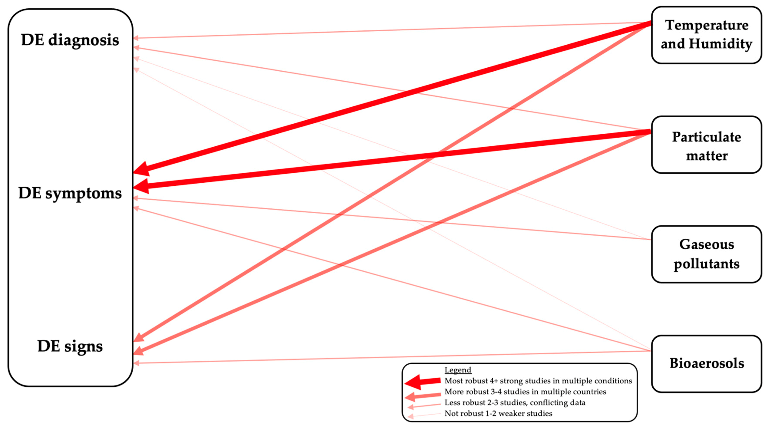
A PubMed search was conducted using the key terms “dry eye” AND either “temperature”, “humidity”, “controlled chamber”, “pollution”, “mold”, “ozone”, “nitrogen dioxide”, “sick building syndrome” and “eye” AND “volatile organic compounds”. All published primary scientific articles were considered, and review articles were excluded. All searches were limited to the English language. Eligible articles were reviewed and summarized. We organized our review by exposure type (particulate matter, gaseous pollutants, and weather) although we are cognizant of overlaps in these categories. We further organized each section by exposure location (outdoor vs. indoor) and DE sub-types (diagnosis, DE symptoms, and DE signs). We chose these groupings as clinically, DE symptoms and signs are often discordant [14,15], suggesting that different contributors may underlie different DE manifestations. In addition, mitigation approaches differ between indoor and outdoor exposures. We summarize our interpretations on the strength of relationships between different exposures and DE parameters in Figure 2.

3. Dry Eye as a Disease Model

Dry eye (DE) is a prevalent, chronic disease that is often encountered by both eye care and primary care providers [16]. DE is defined as a multifactorial disease of the ocular surface classified by a loss of homeostasis of the tear film that may be accompanied by ocular symptoms. Tear film instability and hyperosmolarity, ocular surface inflammation and damage, and neurosensory abnormalities are hypothesized to play etiological roles [17]. DE has been referred to as a “vicious cycle” as an initial insult (e.g., allergy, environmental factors, surgery, Meibomian and lacrimal gland dysfunction) propagates inflammation, an unstable tear film, and apoptosis that perpetuates symptoms and signs of DE even if the initial insult is gone [18]. Symptoms of DE include pain characterized as dryness, discomfort, irritation, burning, itchiness, stinging, foreign body sensation, or light sensitivity and visual complaints such as unstable or poor quality vision [19]. In humans, DE is primarily first identified through symptoms. However, objective signs of DE include decreased tear film stability, decreased aqueous tear production, elevated or unstable tear osmolarity, and inflammation on the ocular surface. DE symptoms are usually assessed through standardized questionnaires such as the Ocular Surface Disease index (OSDI) [20] and DE signs through objective tests such as measurement of TBUT to evaluate tear stability, lissamine green or fluorescein staining to evaluate epithelial disruption, and Schirmer test to measure tear production [21]. Oftentimes, DE symptoms and signs do not correlate with each other, and this complicates diagnosis and management [14,15].

4. Effect of Particulate Matter (PM) on the Ocular Surface

4.1. Outdoor PM

4.1.1. The Effect of Outdoor PM on a Dry Eye Diagnosis

Epidemiological studies have examined the impact of outdoor air pollution on a DE diagnosis. In one study using data from the National Veterans Administration (VA) database (n = 3.41 million patients), DE cases were defined as individuals with an International Classification of Disease (ICD)-9 code for DE (375.15) and medication use (e.g., artificial tears) or procedure (e.g., punctal plugs) for DE [22]. Aerosol optical depth (AOD) was used as an indicator for concentration of aerosols and served as a surrogate measure of air pollution. Higher levels of AOD (measured at each treatment facility) elevated the risk for a DE diagnosis (1.126, 95% CI = 1.125–1.127, p < 0.001). Using the same sampling strategy, the diagnosis of DE was found to have a seasonal pattern with peaks in winter and spring, with the highest prevalence in April (20.9 ± 0.14%), coinciding with elevated pollen indices from www.pollen.com [23]. In a Taiwanese study of 25,818 subjects with DE identified by ICD-9-CM codes from a 1,000,000 person database of insured patients, the first occurrence of DE was also the highest in spring and early summer (26.2% and 25.4%), again aligning with the allergy season [24].
Air pollution has also been linked to a DE diagnosis in Asia. In a Chinese retrospective case-crossover study, 5062 individuals were identified as having an outpatient visit for DE by ICD-10 codes [25]. Daily concentrations of PM2.5 and PM10 were collected by eight urban background air quality monitors covering the city districts of Hangzhou. Using a single pollutant model with subjects serving as their own controls (on the same weekday within the same month), same day exposure of PM2.5 (1.02, 95% CI = 1.01–1.03, p < 0.01) and PM10 (1.01, 95% CI = 1.003–1.02, p < 0.01) increased the risk of an outpatient DE visit. Additionally, PM2.5 and PM10 had a lasting effect on DE diagnosis up to 2 and 3 days, respectively, after exposure using a lag model, with no significant change after 3–7 days. In another Chinese cross-sectional study, 23,922 subjects were recruited from ophthalmic clinics in 32 cities [26]. DE was defined by the presence of ≥1 symptoms such as dryness, foreign body sensation, burning, eye fatigue, discomfort, or vision fluctuation, and one abnormal sign such as a TBUT ≤5 s, presence of fluorescein staining, or Schirmer test ≤ 5 mm/5 min. Pollution levels were considered “extreme” if the daily average PM2.5 level for a specific city was greater than 81.21 μg/m3. Extreme pollution levels for >143 days in a year increased the risk of a DE diagnosis by 2.01 (95% CI = 1.79–2.25; p < 0.0001).
In summary, several US and Asian studies have found a link between a DE diagnosis and air pollution [22,25]. However, one big limitation in using ICD codes to diagnose DE is the inability to examine which facet of DE is related to air pollution.

4.1.2. The Effect of Outdoor PM on Dry Eye Symptoms

DE symptoms have been linked to air pollution. In an Indian study, 500 healthy health care workers answered questions regarding the presence of eye redness, irritation, tearing, blurred vision, photophobia, eye strain, burning, and dryness [27]. Of note, levels of suspended PM in New Delhi are five times (~210–360 µg/m3) higher than the annual average level set by the WHO (60 µg/m3). Individuals were divided based on mode of traveling to the hospital: (a) 250 individuals who commuted to the hospital via open vehicle (e.g., scooter, bicycle, or motorcycle) and (b) 250 individuals who lived on the medical campus and walked to work. Individuals who commuted had higher frequencies of redness (42% vs. 20%), irritation (50% vs. 26%), tearing (38% vs. 18%), burning (28% vs. 0%), and dryness (8% vs. 0%) compared to those who walked to work, p < 0.05 for all. Interestingly, visual quality was similar in both groups. The underlying assumption of this study is that individuals who commute are exposed to higher levels of air pollution than those that do not commute. However, a limitation of the study is that commuters are likely exposed to a variety of environmental insults (PM, gaseous pollutants, weather conditions), and thus, the effect of PM on DE cannot be evaluated in isolation. In contrast to the New Delhi findings, in a prospective study from Thailand, 3025 participants answered 19-yes/no questions regarding various eye symptoms daily for 4 months [28]. All subjects lived within a 10-km radius of an air quality monitoring station where ambient PM10 levels were continuously measured. In this study, PM10 was positively associated with blurred vision (OR = 1.009; 95% CI = 1.004–1.014) but not with irritation or redness.
Furthermore, not all studies have reported a link between air pollution and DE symptoms. In a cross-sectional South Korean study, 16,824 subjects from The Korea National Health and Nutrition Examination Survey (KNHANES) answered the questions: “Do your eyes frequently feel dry or irritated?” and “Have you ever been diagnosed by an ophthalmologist as having dry eye syndrome?” [29]. Of the participants, 21.7% answered yes to having symptoms and 11.8% to having a diagnosis. Air pollution data were pulled from 283 atmospheric monitoring stations located throughout South Korea over a 3-year period using each participant’s residential division as a proxy for their location. Levels of PM10 were not associated with DE symptoms (R = −0.032, 95% CI, −0.313 to 0.254, p = 0.83) or a diagnosis (R = 0.1080, 95% CI, −0.3805 to 0.1818, p = 0.47). Similarly, in a Brazilian study, 71 healthy male taxi drivers or traffic controllers carried a passive sampling device to assess PM2.5 concentration [30]. Individuals were then seen in the eye clinic on 4 occasions where they underwent an eye examination, filled out the OSDI questionnaire, and levels of PM2.5 were measured. At visit 1, mean OSDI score was 9.18 ± 6.81, which is relatively low as the OSDI ranges from 0–100. There was no significant change observed in OSDI scores throughout the study although the subjects were exposed to high levels of PM2.5 (40 mg/m3).
Other studies have focused on particular PM types. In a Swedish study, 89 children aged 7–18 with known pollen allergy (positive skin prick test or specific IgE) graded their eye symptoms from 0 “no symptoms” to 3 “severe symptoms” each day for 42 days [31]. Pollen levels were monitored through a centrally located rooftop volumetric spore trap both in and out of pollen season. DE symptoms scores increased linearly with pollen grain exposure until about 150 grains/cm3 where the curve seems to flatten out. Overall, ocular symptoms were significantly dependent on pollen count throughout the study period.
In summary, some [27,28], but not all [29,30], studies have found a associations between DE symptoms and outdoor PM. Furthermore, there are inconsistencies between studies regarding the types of symptoms that relate to air pollution (pain vs. visual disturbance).

4.1.3. The Effect of Outdoor PM on Dry Eye Signs

Several studies have reported on relationship between outdoor PM and DE signs, but again, with variability in findings across studies. In the New Delhi study [27], individuals who commuted had more rapid TBUT and less tear production than individuals who walked to work (TBUT: 12.97 ± 6.12 vs. 19.23 ± 5.70 s, p < 0.001; Schirmer’s test 13.42 ± 6.67 mm vs. 15.95 ± 6.14 mm, p < 0.001); however, values in both groups were in the normal range, and thus, the clinical significance of these differences is not clear.
Similar data have been found in South American studies. In an Argentinian study of individuals living in an industrial (n = 34) vs. urban (n = 44) zone, annual PM10 levels obtained from sampling sites significantly varied between groups (52.2 ± 16.7 μg/m3 vs. 31.9 ± 13.9 μg/m3, p < 0.05) [32]. DE signs were defined as TBUT < 10 s, corneal staining >grade 3, lid parallel conjunctival folds >grade 3, or Schirmer ≤ 10 mm/5 min. A higher percentage of individuals had ≥3 signs in the industrial vs. urban zone (61.7% n = 21 vs. 25%, n = 11, p < 0.001). In the Brazilian study of 71 healthy male taxi drivers or traffic controllers [30], generalized estimating equation analysis revealed that each 10 μg/m3 increase in PM2.5 resulted in a 10.9 mOsm/kg (p < 0.05) decrease in tear osmolarity with a dose–response pattern. However, the clinical significance of this finding is unclear as DE has been associated with increased or unstable (but not lower) osmolarity [21]. In the Brazilian study, other DE signs, such as staining and Schirmer, were not related to PM concentration.
Overall, the association between outdoor PM and DE signs is less strong than for symptoms (Figure 2), with fewer studies available, inconsistent findings across studies, and fewer number of participants in individual studies. Furthermore, the clinical significance of the findings (e.g., lower osmolarity) is not clear [27,30]. Overall, TBUT has been more consistently linked to PM exposure, as compared to corneal staining or tear production.

4.2. Indoor PM

4.2.1. The Effect of Indoor PM on Dry Eye Symptoms

Short term exposure to an adverse indoor environment can negatively impact DE symptoms. In a prospective study of 88 subjects exiting an older building with a higher concentration of airborne PM (24,436 particles ≥ 0.5 µm/feet3) and a higher number of microbial colonies (1066 colonies/m3) and 102 subjects exiting a newer building with a lower concentration of airborne PM (12,313 particles ≥ 0.5 µm/feet3) and lower number of microbial colonies (400 colonies/m3), DE symptoms were assessed by two yes/no questions, “Are you currently experiencing any dry eye symptoms, such as dryness, irritation, or burning sensation?” and “Have your dry eye symptoms changed since the morning?” [33]. After adjusting for confounding variables, subjects leaving the older building were more likely to report DE symptoms (OR 3.89; 95% CI = 1.21–12.5; p < 0.05) and worsening DE symptoms (OR 13.30; 95% CI = 2.80–63.31.60; p < 0.01) compared to subjects leaving the newer building.
Similar findings have been seen with regards to long term exposures. In a US study of 97 individuals, subjects filled out the OSDI questionnaire and underwent home environment evaluation to assess PM level with a handheld particle counter [34]. In multivariate analyses when adjusting for demographics, comorbidities, medications and other interaction variables, a 1 unit increase in PM2.5 was associated with a 1.59 increased in OSDI score (95% CI, 0.58–2.59; p = 0.002). Similar findings were observed in Brazil. In a cross-sectional study of 936 workers in an older, non-sealed office building with natural ventilation and 1736 workers in a newer, sealed office building with HVAC systems, the prevalence eye symptoms were studied in relation to concentration of PM and other building conditions [35]. In each building, indoor air quality (IAQ) was measured on 6 or more floors at 2 different sampling points for 8 h. Subjects completed an 11-question symptom survey that included a section on dry, itchy or watery eyes. Subjects ranked the frequency of their symptoms from 1 being “every day” to 5 being “less than every 2–3 weeks” and answered if their symptoms were better on days away from the office. PM levels were high (80–800 μg/m3) or very-high (>800 μg/m3) on all 6 floors of the older building while 3 of 7 floors in the newer building had low PM (<80 μg/m3). Eye dryness (33.3% vs. 27.1%, p = 0.01) was more frequent in the older building compared to the newer building. In addition, symptoms of itching were more likely to improve upon leaving the older vs. newer building (64.0% vs. 72.9%, p < 0.05).
Similarly, organic PM like bioaerosols have been associated with DE symptoms in schools and homes. In a Malaysian study of 462 middle school students, vacuumed dust levels from 32 classrooms were analyzed and studied with responses to a self-administered questionnaire [36]. Subjects rated the frequency of eye irritation in the last 3 months as occurring every day, 1–4 times a week, 1–3 times a month, or never. The level of vacuumed fine dust on the filters was associated with weekly ocular irritation (OR = 4.19; 95% CI = 2.31–7.62; p < 0.001). Concerning home exposures, 3485 randomly selected healthy adults in China answered questions regarding the presence of mold/damp stains, moldy odor, and water damage in the home and rated eye itchiness and burning as weekly, sometimes, or never [37]. Individuals who lived in homes with multiple signs of dampness and mold were at an increased risk for ocular itching, irritation, or burning (OR 3.20; 95% CI 1.67–6.15; p < 0.01).
To conclude, the link between indoor PM exposure and painful DE symptoms is strong (Figure 2) with multiple studies across different countries and building situations reporting a link between office and home PM exposure and dryness and irritation.

4.2.2. The Effect of Indoor PM on Dry Eye Signs

Less information is available on the link between an adverse indoor environment and DE signs. In the study of 97 South Florida veterans [34], a one unit increase in PM2.5 was also associated with a 0.39 reduction in Schirmer score (95% CI, −0.75 to −0.03; p = 0.04), a 0.07 increase in meibomian gland dropout (95% CI, 0.01–0.13; p = 0.02), and a 0.06 increase in inflammation measured via Inflammadry strips (95% CI, 0.02–0.11; p = 0.009). In a follow up study by the same group, indoor air was sampled in the home of 157 veterans with a biostage impactor and incubated on nutrient and soy media [38]. Using a multivariate linear regression, elevated microbial counts were associated with meibomian gland drop out when adjusting for age, gender, log (total suspended particles), smoke exposure, allergy status, and number of observations ((OR 67.42, 95% CI = 1.18 to 3864.59, p < 0.05). Corneal epithelial disruption was significantly associated with higher numbers of colonies in the model adjusting for age and gender only (OR 28.07, 95% CI = 1.78 to 443.79, p < 0.05).
Similar findings were seen outside the U.S. In a study of children in Malaysia, TBUT was evaluated in relation to fungal DNA in dust collected by vacuuming 32 classrooms in 8 different middle schools [39]. Fungal DNA was associated with a 5.1 s decrease in TBUT (95% CI = −9.1 to −1.2, p < 0.05) per 105 target copies per gram dust, linking exposure to fungus with tear instability.
Environmental chamber studies support these findings. An environmental chamber is an enclosure that can be used to test the effects of specified environmental conditions (e.g., temperature, humidity, PM exposure) on biological items (in our case, facets of DE). In a chamber study in Denmark, 10 individuals were exposed to clean (~41 μg/m3 dust) and polluted air (~394 μg/m3 dust) in a randomized order for 3 h while in a controlled chamber. TBUT was significantly impacted by dust exposure with 7 of 10 subjects having a decrease in TBUT after exposure to dust, p < 0.05 [40]. To conclude, while fewer studies are available, they are consistent in highlighting a link between tear instability and dust. Interesting, TBUT is also the DE sign most affected by outdoor PM, as summarized above.

4.3. Potential Pathophysiology

There is biologic plausibility linking air pollution to DE. First, as summarized above, some studies have found an inverse correlation between air pollution and TBUT [27,41]. In a similar manner, mouse studies have supported this clinical observation [42]. In a study of male mice, mice were exposed to either 5.0 mg/mL PM2.5 or saline. The PM2.5 group showed significantly decreased TBUT at day 14 compared to the saline group (~2.5 ± 0.75 s vs. 6.5 ± 1.6 s, p < 0.05). In the same study, the PM2.5 group were also found to have higher levels of inflammatory markers compared to the saline group (tumor necrosis factor-α (TNF-α) ~0.38 ± 0.17 vs. 0.32 ± 0.10, p < 0.05; NF-κB ~2.75 ± 0.6 vs. ~0.33 ± 0.1, p < 0.05). In fact, inflammation has been found to be a key component of DE in humans [43] and animal models [44].
Similar to the human studies [36,37,39,40], organic bioaerosols have also been reported to negatively impact ocular surface health. In an in-vitro study, human corneal epithelial (HCE) cells were exposed to various concentrations of mycotoxins (AFB1 and gliotoxin) vs. a solvent control, and in vitro assays were done to asses cellular viability and inflammatory responses [45]. Exposure to AFB1 (16 µg/mL) increased expression of several inflammatory markers (380-fold increase in IL-8, 160-fold increase in C-X-C motif chemokine ligand 1, 21-fold increase in TNF-α) compared to the control. Exposure to gliotoxin (125 ng/mL) insignificantly increased interleukin-8 (IL-8) and TNF-α expression but significantly increased toll like receptor 4 (TLR-4) expression 1.9-fold (p = 0.0041) and poly ADP-ribose polymerase (PARP) expression 1.4-fold (p < 0.0001). These data support the clinical findings that mycotoxins can trigger inflammatory responses.
While inorganic and organic PM influence inflammation on the ocular surface, inorganic PM like dust is also associated with oxidative stress. In a study evaluating the effect of house dust on HCE, dust was collected from 16 homes in Nanjing, China, from the air conditioning units [46]. Primary HCE cells were exposed to either a fresh medium containing between 5 to 320 μg/100 μL of dust or a control medium. Oxidative stress induced DNA damage was assessed by measuring accumulation of 8-OHdG. At the lower level of 5 μg/100 μL, 8-OHdG levels were similar to controls (214 ± 6.50 pg/mL). However, 8-OHdG levels greatly increased to 400 ± 38.8 pg/mL after exposure to 80 μg/100 μL of dust. In fact, a concentration-dependent relationship was revealed between house dust level and ROS production.
Together, these in-vitro and animal studies suggest that inflammatory responses, which play a large role in DE, are triggered by inorganic and organic PM. Additionally, oxidative stress is triggered by non-organic PM.

5. Effect of Gaseous Pollutants on the Ocular Surface

5.1. Outdoor Gaseous Pollutants

5.1.1. The Effect of Outdoor Gaseous Pollutants on a DE Diagnosis

Gaseous pollutants include reactive gases such as NO2, SO2, and O3 and VOCs such as formaldehyde, toluene, acetone, ethanol, and more that are present in both in the outdoor and indoor environment. These pollutants have various sources including car and fuel emissions, wood preservatives, aerosol sprays, disinfectants, combustion, cigarette smoke, and many other sources that may impact human health. Outdoor exposure to reactive gases has been linked to a DE diagnosis but with discrepancies across studies in relation to the specific gases involved, making the data less robust (Figure 2). In a case-crossover study in Taiwan [24], multivariable analyses found significant associations between NO2 (OR 1.07; 95% CI = 1.04–1.10; p < 0.001) and CO (OR 1.12; 95% CI = 1.03–1.21; p < 0.01) and a DE diagnosis. Specifically, every 10-ppb increase in NO2 was associated with a 6.8–7.5% increase in a DE diagnosis and every 1-ppm increase in CO was associated with a 10.5–11.6% increase in a DE diagnosis. In another study in Hangzhou, China [25], NO2 (1.03; 95% CI = 1.01–1.05; p < 0.05), SO2 (1.07; 95% CI = 1.02–1.11; p < 0.01) and CO (1.0; 95% CI = 1.0–1.0; p < 0.01) were associated with DE outpatient visits in a multi-pollutants model. O3 did not significantly associate with DE. Other studies, however, have reported associations between O3 and a DE diagnosis. In a South Korean population-based cross-sectional study [29], using multivariable models, a 0.003-ppm increase in O3 level was associated with both a diagnosis and symptoms of DE (OR 1.31; 95% CI = 1.12–1.53; p = 0.002). A 0.003-ppm increase in the NO2 level was associated with a diagnosis of DE but not symptoms (OR 1.12; 95% CI = 1.02–1.23; p = 0 .02), and SO2 was not associated with either DE diagnosis or symptoms (OR 1.10; 95% CI = 0.84–1.43; p = 0.50). There is a paucity of literature relating VOCs to DE diagnosis.

5.1.2. The Effect of Outdoor Gaseous Pollutants on DE Symptoms

Outdoor exposure to gaseous pollutants has been linked to DE symptoms, again with some inconsistencies in the data. Associations of gaseous pollutants to symptoms are stronger than to diagnosis and signs but still weaker than to other pollutants (Figure 2). In a study from Chiang Mai, Thailand [28], NO2 was positively associated with eye irritation (OR 1.05, 95% CI = 1.03–1.07 per 1 ppb), and SO2 was positively associated with eye irritation, red eyes, and blurred vision (OR 1.26, 95% CI = 1.09–1.47, 2.95, 95% CI = 1.46–5.94, and 1.21, 95% CI = 1.0–1.46 per 1 ppb, respectively). Conversely, O3 was negatively associated with red eyes and blurred vision (OR 0.89, 95% CI = 0.85–0.94 and 0.962, 95% CI = 0.95–0.98 per 1 ppb, respectively). However, O3 was linked to DE symptoms in other studies. In a prospective observational study of 33 individuals in South Korea with evaporative DE (TBUT ≤ 5 s) who were using topical drugs (0.05% cyclosporine or 3% diquafosol), DE measures were investigated twice at 2-month intervals, and outdoor O3 concentrations on the clinic visit days were obtained from public atmospheric monitoring stations and calculated based on each subject’s address [47]. In multivariable models, Ocular Surface Disease Index (OSDI) scores increased by 3.43 points per each 0.01 ppm increase in O3 concentration (b = 3.43, p = 0.002). A limitation of this study is that it is not clear which symptoms within the OSDI (pain vs. visual disturbance) drove the increased OSDI scores.
Furthermore, not all studies have linked gaseous exposure with DE symptoms. In a Brazilian study of 71 taxi drivers and air traffic controllers [30], NO2 was continuously measured through passive sampling tubes for 24 h and subjects filled out an OSDI questionnaire the same morning as when the air pollutant samplers were removed. While subjects were exposed to a mean NO2 level of 189 ± 47 μg/m3, OSDI scores were relatively low (7.4 ± 8) and were not correlated with NO2 levels.
Outdoor VOCs have also been studied in relation to DE symptoms. For example, a study in Neyagawa, Osaka, Japan, compared 6217 individuals living within 1200 m from a plastic recycling factory to 77 individuals living 2800 m away from the factory [48]. Concentrations of VOCs were measured at two locations, 100 and 500 m from the factory, for 3 h/day on 3 days of the same week (June 2006), using an automatic thermal desorption tube. Unsurprisingly, the location closer to the factory had higher concentrations of VOCs than the location further away. Self-administered questionnaires assessed whether subjects had eye itch, redness, irritation, and discharge more than once per month. Several symptoms were more frequent in residents within 500 m of the factory compared with 2800 m from the factory including itch (OR = 3.0, 95% CI: 1.5–6.0), redness (OR = 2.7, 95% CI: 1.1–6.3), irritation (OR = 5.2, 95% CI: 1.5–17.8), and discharge (OR = 6.0, 95% CI: 2.3–15.9). In another study performed on 111 individuals living near a petrochemical industrial estate in Thailand, VOC data were obtained from an ambient air monitoring station of the Pollution Control Department of the Ministry of Natural Resources and Environment, located at the primary care unit in one of the communities in the study area [49]. Individuals were interviewed daily for 63 days about the presence of twelve symptoms, categorized into respiratory symptoms and other health symptoms (headache, shortness of breath, fever, eye irritation, dizziness, and weakness). The interviewers asked questions about symptoms on the previous day. A 1 ppm increase in total VOCs was associated with an increased odds of eye irritation (OR 4.30, 95% CI 14–16.26, p < 0.05). Taken together, these data highlight the inconsistency in the relationships between reactive gases and DE. For example, while associated with a DE diagnosis, NO2 and SO2 were not found to associate with DE symptoms. Additionally O3 was associated with DE symptoms in two Asian studies [29,47] but was protective for DE symptoms in another Asian study [28]. Furthermore, most studies used data from monitoring stations and thus do not necessarily reflect individual level exposures. These studies also suggest a relationship between outdoor VOC exposure and DE symptoms, although unaccounted confounders (e.g., PM) may also contribute to the noted relationships.

5.1.3. The Effect of Outdoor Gaseous Pollutants on DE Signs

Only a few studies have examined relationships between outdoor gaseous pollutants and DE signs. In one study, 29 hospital employees from two cities in Brazil with varying levels of air pollution (São Paulo and Divinolândia) underwent conjunctival sampling via impression cytology [50]. Mean individual levels of NO2 were measured using a passive sampler for 7 days. Exposure to NO2 was stratified into four quartiles (Q) based on NO2 levels: Q1, <17 μg/m3; Q2, 18–21 μg/m3; Q3, 22–33 μg/m3; and Q4, >33 μg/m3. Conjunctival goblet-cell counts increased proportionately to NO2 exposure: Q1, 186 cells; Q2, 253 cells; Q3, 312 cells; and Q4, 402 cells. Furthermore, NO2 concentrations were positively correlated to goblet cell counts per 10 high power field (ρ = 0.57, p = 0.001). In addition to number, goblet-cell hyperplasia was also positively related to NO2. However, the clinical significance of this finding in relationship to DE symptoms and signs is not clear. Furthermore, as with the New Delhi study, there are likely other differences between the populations (PM, weather) that were not accounted for. In the Korean study [47], short-term changes in ground-level O3 were associated with decreased tear secretion (decrease of 1.43 mm per 0.01 ppm increase in O3 concentration, p = 0.015), but again, the clinical significance of this findings is unclear. Taken together, these studies highlight the paucity of literature on associations between gaseous pollutants, especially VOCs and ocular surface signs (Figure 2), but also suggest that tear production and goblet cell density may be affected by exposure to reactive gases.

5.2. Indoor Gaseous Pollutants

The Effect of Indoor Gaseous Pollutants DE Symptoms

Of all gaseous pollutants, we found a greater number of studies on indoor O3 exposure in relation to DE symptoms. O3 is often studied indoors through aircraft cabins as there are elevated levels of O3 at high altitude [51]. Cabin air of 83 flights was monitored between 2008–2010 as a part of a study of onboard environmental conditions. O3 measurements were conducted by a project engineer onboard the aircraft. Mean O3 concentration across all flights was 16.1 ppb. Approximately 52 passengers per flight filled out a yes/no survey for a range of symptoms on board including itching eyes, dry eyes, and watery eyes. Although 16.1 ppb is lower than the EPA guideline, O3 concentration was significantly associated with itchy eyes (OR 1.16; 95% CI = 1.04–1.29; p = 0.008), but not dry or watery eyes. Simulated cabin studies similarly revealed an association between DE symptoms and elevated ozone levels [52]. In a study of 29 females in Denmark, subjects were split into two groups and exposed to two levels of O3 (<2 and 60–80 ppb) at two different air flow rates (2.4 vs. 4.7 L/s). Subjects graded eye irritation from 0 being no irritation to 50 being overpowering irritation quickly after entering and after 3.25 h in the chamber. Eye irritation was higher in the ozone group (60–80 ppb) at both flow rates but only significantly at the high flow rate (7.2 vs. 2.5, p < 0.015). These findings are in agreement with the Korean study [47] that found a relationship between outdoor O3 and symptoms, but in contrast to the Thailand study [28] that found the outdoor O3 was protective for symptoms.
Indoor NO2 level has also been associated with DE symptoms. In a study in Malaysia of 462 s grade students, weekly symptoms of eye irritation in the last month were compared to NO2 level inside and outside the classroom for 7 days using a diffusion sampler [53]. The presence of eye irritation (yes/no) was positively related to indoor NO2 concentration (OR 2.34, 95% CI 1.40–3.90; p = 0.001). These findings are in agreement with the Thailand study [28] that found a relationship between outdoor NO2 and symptoms, but in contrast to the Brazilian study [30] that found no correlation between NO2 and symptoms.
Indoor exposure to VOCs has also been associated with DE symptoms. For example, a study conducted in Heidelberg, Germany, on 21 subjects investigated the occurrence of eye symptoms after exposure to 10 different formaldehyde concentrations. Subjects were exposed to each of the 10 concentrations for 4 h on 10 consecutive days, in random order. Eye irritation (graded on a 0–5 scale) at exposure levels as low as 0.3 ppm was significantly higher than at 0 ppm after 195 min of exposure (0 ppm exposure ~0.3 symptoms score vs. 0.3 ppm exposure ~0.6 symptoms score after 195 min; p < 0.05) [54]. In support of the experimental study, a South Korean study assessed 34 subjects when they moved from a rehabilitation hospital with low indoor VOCs to a building with higher VOC levels (benzene, toluene, ethylbenzene m, p xylene, styrene, o-xylene, formaldehyde, and acetaldehyde, all measured in both buildings) [55]. Severities of eye dryness and eye irritation were found to increase via 0–10 visual analogue scale score with the move to the new building, potentially linking VOC to DE symptoms.
Exposure to VOCs can also be assessed through urine levels. In a study of 389 employees in 8 high-rise buildings in Taipei, eye dryness (yes/no) was subjectively assessed while 8-hydroxydeoxyguanosine (8-OHdG) was quantified in urine (a marker of oxidative stress related to VOC levels) [56]. Individuals who reported eye dryness had higher levels of 8-OHdG in urine (6.16 ± 3.81 vs. 5.45 ± 3.10 μg/g; p = 0.047), supporting a relationship between symptoms and VOC exposure.
VOCs have been linked to DE symptoms in children as well. In a Malaysian study of middle school children [53], VOC level inside and outside the classroom were sampled for 7 days using a diffusion sampler and the frequency of eye irritation was assessed. The presence of eye irritation (yes/no) was positively related to indoor formaldehyde concentration (OR 3.55, 95% CI 1.51–8.38; p = 0.004).
VOCs in the home have also been associated with DE symptoms. A Japanese study randomly selected 33 homes where at least one inhabitant complained of one or more eye symptoms (eye irritation, dry eyes, photophobia, lacrimation, and eye redness) and 63 homes where none of the inhabitants complained of symptoms [57]. Concentration of 19 different VOCs and total VOCs was measured by passive samplers located in the living room over a 1-week period, and self-administered questionnaires were assessed for the presence of eye symptoms. Indoor concentrations of ethylbenzene (OR 2.1, 95% CI 1.07–4.10; p <0.05), xylene (OR 2.29, 95% CI 1.11–4.69; p <0.05), and total VOCs (OR 9.79, 95% CI 1.67–57.5; p <0.05) were found significantly related to eye symptoms.
To summarize, while studies have found links between indoor O3 and NO2 with DE symptoms, there is a lack of data on the effects of other gases like indoor SO2 and CO on DE. Furthermore, studies are needed to evaluate relationships between indoor reactive gas exposure and DE signs. The data linking outdoor and indoor VOCs in a variety of locations (workplace, school, home) to DE symptoms are stronger than for other gases, but there is a paucity of literature relating VOCs to DE signs.

5.3. Potential Pathophysiology

The mechanisms by which gaseous pollutants may affect the ocular surface health can be inferred by data generated outside the eye where it has been demonstrated exposure to reactive gases can lead to cell death, oxidative stress, and inflammation [58,59]. For example, human alveolar macrophages were exposed to 0.4 ppm concentration of O3 for 1 h as compared to purified air with 5% CO2 at a rate of 2 L/min [60]. Exposure to 0.4 ppm O3 increased the secretion of inflammatory cytokines such as TNF-α, IL-lβ, IL-6, and IL-8 compared to control (% increase vs. control: TNF-α 443% ± 208; IL-lβ 484% ± 171; IL-6 383% ± 147; IL-8 242% ± 63). In another study, 11 healthy male volunteers were exposed on two separate occasions (at least 6 weeks apart) to filtered air and 0.4 ppm of O3, with bronchoalveolar lavage (BAL) conducted the following morning [61]. An increase in percentage of polymorphonuclear leukocytes (PMN) and decrease in macrophages in BAL fluid were found after O3-exposure (mean ratio 8.2 ± 2.2; p = 0.009 and mean ratio 0.97 ± 0.02; p < 0.001, respectively). Moreover, markers of inflammation in BAL fluid were higher after O3 exposure versus air exposure including fibronectin (mean ratio 6.44 ± 1.54, p = 0.006), complement C3a (mean ratio 1.72 ± 0.14, p = 0.001), and prostaglandin E2 (PGE2) (mean ratio 1.97 ± 0.21, p = 0.002).
When considering the effects of O3 on the ocular surface, one study exposed 30 male mice to filtered room air (controls), 0.5 ppm, or 2.0 ppm of O3 for 3 h every day for 2 weeks in a whole-body exposure chamber [62]. After 2 weeks, mice exposed to 2 ppm O3 had higher levels of inflammatory cytokines in tear fluid compared to 0.5 ppm exposure and controls, including IL-1β (2 ppm ~ 875 pg/mL vs. 0.5 ppm ~ 300 pg/mL; p < 0.001), IL-6 (2 ppm ~ 800 pg/mL vs. 0.5 ppm ~ 225 pg/mL; p < 0.001), IL-17 (2 ppm ~ 775 pg/mL vs. 0.5 ppm ~ 250 pg/mL; p < 0.001), and IFN-γ levels (2 ppm ~ 875 pg/mL vs. 0.5 ppm ~ 250 pg/mL; p < 0.001).
Moreover, when studying NO2 effects, various studies support a positive correlation between exposure and eosinophilic airway inflammation in asthmatics. For example, a study investigated repeated brief exposures to concentrations of 200 and 600 ppb NO2, to simulate indoor NO2 fluctuations and its correlation with airway inflammation through induced sputum samples in 19 asthmatic adults. They found an association between the changes in percentage of eosinophils and NO2 exposure levels relative to baseline measurements was significant (0 ppb NO2: −12 (−34, 16), 200 ppb NO2: −5 (−28, 26), 600 ppb NO2: 57 (18, 109), p = 0.04) [63].
Additionally, VOCs have been linked to harmful immunologic, respiratory, carcinogenic, reproductive, neurologic, and cardiovascular health effects. Potential mechanisms are oxidative stress, inflammation, and cell death [64]. For example, a study investigated the effects of cigarette smoke (a source of VOCs) on mouse and human lung epithelial cells. Exposure to up to 3 puffs of cigarette smoke resulted in a 10% mortality in mice cells, and exposure to 7 or more puffs resulted in an 80% mortality in mice cells. Interestingly, when VOCs, were removed from the cigarette smoke, no cell death was seen, even with the same CO and NO concentrations, supporting the idea that VOCs are responsible for direct cytotoxic in cells, and further supporting the noted clinical relationships [65]. Similar to PM, associations between gas exposure and inflammation may be an important link connecting O3 and other gaseous pollutants to DE.

6. Effect of Weather on the Ocular Surface

6.1. Outdoor Weather Conditions

6.1.1. The Effect of Outdoor Weather Conditions on a Dry Eye Diagnosis

Environmental factors such as humidity, wind, and altitude affect the risk of DE. Ultimately, there is a general acknowledgement that extremes of temperature and humidity are not ideal for the eye, and it appears that moderate temperature and humidity with minimal wind and UV light are optimal. In the retrospective analysis using data from a National Veterans Administration (VA) database [22], spatial information was recorded based on treatment facility, and using a statistical trend analysis, the risk of DE was higher in zip codes with an atmospheric pressure 1 standard deviation (SD) above the mean (IRR, 1.131; 95% CI, 1.129–1.133; p < 0.001). Conversely, high humidity and wind speed lowered the risk of DE (IRR, 0.927 [95% CI, 0.926–0.927] and IRR, 0.938 [95% CI, 0.937–0.939], respectively; p < 0.001).
Other studies have supported the protective association between higher outdoor humidity and DE symptoms. In a cross-sectional study of a South Korean population [29], multiple logistic regression analyses revealed a negative association between DE diagnosis and relative humidity levels (R = −0.425 (95% CI, −0.633 to −0.161), p = 0.003). In another Korean study of 16,431 subjects who participated in the KNHANES, 1616 individuals had a previous diagnosis of DE, and 2666 responded positive to having DE symptoms (1314 had both). Environmental information was extracted from the National Climate Data Service System where temperature and humidity were assessed every 8 h each day. Wind speed was measured by using the total value of artistic air divided by 86,400 s, and sunshine was defined by hours of sun rays without blockage. Using multiple logistic regression analyses, a DE diagnosis was negatively associated with humidity levels (OR 0.970; 95% CI 0.955–0.986) and positively with temperature (OR 1.076; 95% CI 1.009–1.148) and sunshine duration (OR 1.015; 95% CI 1.006–1.023) [66].
While humidity appears protective for a DE diagnosis, other weather conditions such as sunlight and wind appear harmful. In a study in India, 500 eye hospital patients answered 13 yes/no questions regarding eye symptoms, and a total symptom score was calculated [67]. Self-reported environmental exposures (sunlight, high temperatures, excessive winds) were also assessed. An ocular surface examination was then performed (TBUT, staining, Schirmer’s test). A subject was defined as having DE if they had 3 or more positive tests. A DE diagnosis was associated with sunlight (OR: 1.91, p = 0.014) and excessive winds (OR: 2.15, p = 0.004).
High altitude is another risk factor for DE diagnosis. In one study, 100 subjects living in the Leh district of Ladakh (3300 m above sea level) and 100 age and sex matched subjects living in New Delhi (218 m above sea level) were recruited and screened for DE symptoms using McMonnies’ Questionnaire (MMI) and OSDI, and a Schirmer test was performed [68]. DE was defined as an MMI > 14, OSDI > 23, and Schirmer < 10 mm. The prevalence of DE was higher in individuals living in high altitude vs. low altitude (20% vs. 9%, p = <0.005). Together, several studies provide strong data that higher outdoor humidity protects against a DE diagnosis while high altitude, sunlight, and high temperatures are risk factors for disease. Wind associated with DE diagnosis in some [67], but not all studies [22].

6.1.2. The Effect of Outdoor Weather Conditions on Dry Eye Symptoms

Weather conditions have also been associated with DE symptoms. In the South Korean study [29], multiple logistic regression analyses revealed that DE symptoms negatively correlated with relative humidity level (R = −0.411 (95% CI, −0.622 to −0.144), p = 0.004). In another Korean study, DE symptoms positively correlated with sunshine duration (OR 1.013; 95% CI 1.005–1.021) and negatively with humidity (OR 0.975; 95% CI 0.959–0.991) [66].
As with a diagnosis, factors such as high wind, sunlight, and heat have been linked with DE symptoms. In an observational survey-based study of 5 European countries, individuals ≥40 years using daily tear substitutes with a diagnosis of DE or Sjogren’s by a healthcare professional (n = 738) filled out a 26-question survey on DE symptoms and exacerbations [69]. By self-report, the environmental factors that most impacted DE symptoms were wind, sunlight, and heat, affecting 71%, 60%, and 41% of respondents, respectively. In fact, only 8% of individuals did not report that at least one facet of weather impacted DE symptoms. Overall, many studies support the association between outdoor weather conditions to both a DE diagnosis and DE symptoms (Figure 2).

6.1.3. The Effect of Outdoor Weather Conditions on Dry Eye Signs

Fewer studies have examined the link between outdoor weather condition and DE signs. However, one study evaluated 200 subjects living at low and high altitudes in India. Schirmer scores were lower in individuals living at high vs. low altitudes (15.22 vs. 21.01, p = 0.001) [68]. However, since mean values of both groups were in the normal range, the clinical significance of this finding is uncertain. In another study of 535 U.S. dry eye patients identified by the presence of ocular dryness for greater than 6 months with moderate to severe OSDI scores (25–80), multivariate analyses and partial correlation coefficients revealed TBUT measurements were positively correlated with temperature, humidity, and dew point (ρ = 0.08, 0.18, 0.17, respectively, p < 0.0038) [70].

6.2. Indoor Temperature and Humidity

6.2.1. The Effect of Indoor Humidity and Temperature on Dry Eye Symptoms

Similar to findings outdoor, low humidity has also been associated with DE symptoms indoor. In a Finnish study, 290 office workers located in two wings of the same building participated in a 6-week study where relative humidity (RH) conditions were “crossed-over” [71]. One week, individuals worked in high humidity conditions (30–40% RH) while the next week, they worked in unmanipulated “natural” conditions (20–30%). Air temperature was consistently kept between 21.3–23.9 °C. Each subject experienced three periods of exposure to the conditions. Daily self-reported eye dryness, irritation, and itching severity scores (range of 0 to 3) were all higher while working in the low vs. high RH conditions (0.39 vs. 0.35, p < 0.05).
High temperature indoors can also negatively affect DE symptoms. In one study, 396 U.S. office workers working on two floors of the same building had DE symptoms assessed weekly via a graded health symptom questionnaire (symptoms graded on a scale of 0–25) [72]. Environmental measurements of temperature and humidity were logged continuously with RH values between 42–58%. Continuous time-weighted data for the space each subject was working in were compared to the health symptom questionnaire. A 1 °C decrease in temperature (range 22.2–25.6 °C) was found to decrease the severity of eye dryness, itching, and irritation by 19% (95% CI ~ 8–31%).
Perception of indoor air quality has also been linked to DE symptoms. In Japan, 3335 employees in 320 offices were asked to evaluate eye symptoms and factors related to their work environment [73]. For each symptom, subjects had to respond how often that symptom occurred in the last 4 weeks and whether it persisted after leaving work. The work stressor surveys used a Likert scale (range of 1 to 4) to inquire about perceived job stress about workload and other environmental factors. Multivariable logistic regression analyses found that coldness perception (OR, 1.28; 95% CI, 1.13–1.45) and air dryness perception (OR, 1.61; 95% CI, 1.42–1.82) were associated with increased risk of eye irritation.
Controlled environment chambers studies support results of the observational studies. In one study of 10 minimally symptomatic contact lens (CL) wearers, subjects were exposed to a “adverse” environment of 22.0 ± 2.0 °C and 19.0% ± 4.0% RH for two hours [74]. Comfort was rated by subjects with 0 as extreme discomfort and 10 as extreme comfort. DE related symptomatology was evaluated using a symptom of discomfort questionnaire (SODQ) scale from 0 to 4 (0 = no symptoms, 4 = severe symptoms). Expectedly, comfort level significantly changed in CL wearers from before (9.0 ± 0.2) and after the adverse environment (8.0 ± 0.3, p < 0.05) in a short time period of 2 h. However, SODQ scores were not significantly impacted by the adverse environment, possibly due to the narrow scale of the SODQ compared to the more sensitive comfort scale.
Conversely, increasing local humidity has been found to improve DE symptoms. In one study, individuals with an evaporative DE (symptoms, TBUT ≤ 5 s, meibomian gland number ≤ 4 across lower eyelid) were placed in either intact swim goggles (n = 100) or swim goggles with the central lens removed (n = 25) for 20 min [75]. Individuals were asked to rate comfort 15 min after wear on a 0–4 scale. Of the subjects in intact swim goggles, 99% reported increased eye comfort compared to 24% in the control group (p < 0.0001). In another goggle study, 14 individuals with a aqueous tear deficiency DE (symptoms, TBUT ≤ 5 s, staining > 3, Schirmer test < 5 mm) were exposed to wind from a fan 3 m away for 10 min without spectacles, with conventional spectacles, and with spectacles that maintained a moist chamber with a humidity sensor and a humidity provider tank [76]. Subjects were examined on the same day with at least 2 h between each experimental setting. Dryness visual analog scale (VAS) scores significantly increased after wind exposure from ~20 ± 15% to ~35 ± 10% while not wearing spectacles and from ~15 ± 5% to ~30 ± 2.5% in conventional spectacles (p < 0.05). With moist chamber glasses, dryness scores were stable after wind exposure.
Manipulating humidity in the workplace can also improve eye comfort levels. In one study, 44 individuals in New Zealand were given desktop humidifiers (which increased RH by 5.4 ± 5.0%) and were asked to use their computers continuously for an hour [77]. Participants underwent two masked testing sessions, one with the humidifier on and one with it off. After one hour of humidifier exposure, subjects were asked if they experienced greater, equal or lesser comfort compared to baseline. Of subjects exposed to the humidifier, 36% reported an improvement in subjective comfort compared to 5% in the non-humidifier group (p < 0.001). Seven per cent of subjects exposed to the humidifier reported a decrease in subjective comfort compared to 48% in the non-humidifier group (p < 0.001).
However, not all studies have found an inverse relationship between DE symptoms and humidity. In a US study of 97 individuals with a wide range of DE metrics, a positive association was noted between humidity and DE symptoms (OSDI: r = 0.30, 95% CI, 0.07–0.49, p = 0.01) [34]. However, this finding was attributed to the interaction between humidity and PM and not as a direct effect of humidity on DE symptoms. That is because higher humidity can also increase the time that PM stays airborne, as well as increase PM mass and size (hygroscopic effect) [78]. This suggests that there is a “Goldilocks” zone for indoor RH and that elevated RH can have a negative effect, likely via interaction with PM.
In summary, a wide range of studies support associations between indoor factors such as humidity, wind, and temperature and DE symptoms (Figure 2). Compared to other air pollutants, these data are particularly strong given experimental approaches (environmental chambers [74] and goggles [75,76] to support observational studies [71,72,73].

6.2.2. The Effect of Indoor Humidity and Temperature on Dry Eye Signs

Low indoor humidity and high indoor temperature can negatively impact DE signs, albeit in varied ways. The most robust studies come from controlled environmental chambers. In one study [74], TBUT and tear volume (via phenol red thread test) decreased, and limbal redness increased, after exposure to low RH (TBUT 11.34 ± 0.13 s to 8.31 ± 0.10 s, p < 0.05, volume 25.8 ± 1.9 mm pre-CAE to 22.3 ± 2.3 mm, p < 0.05, redness 1.6 ± 0.04 to 1.7 ± 0.03, p < 0.05). In another study, 10 individuals with mild to moderate DE (symptoms, TBUT < 10 s, Schirmer < 10 mm) and 10 controls were randomly exposed to varying humidity (5%, 40%, and 70%) at a temperature of 72 °F for 25 min on 3 separate days [79]. Tear parameters were assessed 10 min into the exposure on each day. Overall, tear evaporative rates decreased with increasing RH (100.04 ± 13.77 g/m2/h increase from 5% to 70% in DE subjects and 73.82 ± 15.47 in controls, p = 0.001). Interestingly, TBUT followed a U shaped curve in both groups with a mean of 4.90 ± 1.66 s at 5%, 6.31 ± 2.21 s at 40%, and 5.90 ± 1.91 s at 70% in the DE group and a mean of 17.80 ± 3.91 s at 5%, 20.70 ± 5.88 s at 40%, and 20.00 ± 5.35 s at 70% in the DE group, suggesting an optimal value in the middle at 40% RH, substantiating the epidemiologic observations that also highlighted a middle value for humidity was best [34]. In another chamber study, 19 patients with DE symptoms (OSDI > 12 but staining ≤ 2) and 20 individuals without symptoms (but with low TBUT) were exposed to a 5% RH chamber for 2 h at 23 °C [80]. Measures of TBUT and staining were performed immediately before and after exposure. Both symptomatic and asymptomatic individuals had increased corneal staining (symptoms: 0.68 ± 0.15 to 1.16 ± 0.14, p = 0.003; no symptoms: 0.50 ± 0.15 to 1.30 ± 0.19, p = 0.0004) and decreased TBUT (symptoms: 2.78 ± 0.56 s to 1.94 ± 0.24 s, p = 0.01; no symptoms: 2.81 ± 0.24 s to 2.13 ± 0.19 s, p = 0.001) after exposure to low RH. Inflammation measured via matrix metalloproteinase 9 (MMP-9) tear levels also increased after exposure to low RH.
Exposure to low temperature has also been associated with DE signs. In a study of 12 healthy subjects with an OSDI < 12 and an TBUT > 10, temperatures were varied at a constant RH of 40% [81]. Subjects had two visits and were randomly exposed to 25 °C, 20 °C, and 15 °C at one visit and 5 °C and 10 °C at the other. TBUT, tear evaporation rate, lipid layer thickness, and ocular surface temperature were measured at each temperature after 10 min of exposure. Interestingly, mean TBUT levels significantly increased from 7.31 s at 5 °C to 12.35 s at 25 °C (p < 0.05), suggesting greater stability at higher temperatures. However, tear evaporation rates increased threefold from 5 °C (0.056 mL/min) to 25 °C (0.17 mL/min), p = 0.001 and ocular surface temperature was reduced by ~4 °C, suggesting 25 °C preserved the tear film better than cold temperatures like 5 °C. Median LLT grade also increased with increasing temperature (grade 2 or 20–40 nm at 5 °C and 10 °C and grade 3 or 40–90 nm at 15 °C, 20 °C, and 25 °C). Taken together, these data suggest that ambient temperature has a complicated effect on tears with different tear parameters being adversely affected at colder (stability, LLT, TBUT) and warmer temperatures (TER).
Increasing humidity can improve DE signs, as it was shown to do for DE symptoms. In the desktop humidifier study [77], tear film lipid grade, TBUT, and tear meniscus height were measured after one hour with and without the humidifier. In both environments, tear meniscus height and lipid-layer grade remained constant throughout the hour. However, TBUT increased by a median of 4 s in the humidifier group and TBUT and local humidity level were significantly correlated with one another (Spearman ρ = 0.59, p < 0.001). This finding is similar to that of the PM studies where TBUT was the factor most related to air pollution as well. The use of goggles can also improve DE signs. When subjected to wind exposure [76], blink frequency increased from ~19.0 ± 10.0 times/min to ~33.0 ± 10.0 times/min in the group not wearing spectacles and from ~18.0 ± 6.0 times/min to ~29.0 ± 4.0 times/min in individuals wearing conventional spectacles (p < 0.05). However, blink rate was unchanged in those wearing goggles. In a similar manner, TBUT decreased after wind exposure from ~5.0 ± 0.5 s to ~2.0 ± 0.5 s in the no spectacle group and from ~5.5 ± 2.5 s to ~5.0 ± 0.5 s in the conventional spectacle group (p < 0.05). However, TBUT did not significantly change in the goggle group. Overall, experimental studies show that DE signs can be improved by manipulating temperature [81] and humidity [77,79,80]. Additionally, goggles can blunt the effect of wind exposure on symptoms, blink rate, and TBUT [76]. Overall, these data show that both outdoor and indoor environmental factors can impact DE symptoms and signs (Figure 2). The strongest data come from controlled environmental chambers which highlight how change in temperature and humidity can affect DE symptoms and signs in individuals with and without preexistent ocular conditions.

6.3. Potential Pathophysiology

As demonstrated by the studies above, conditions such as wind, humidity, and temperature can directly affect tear evaporation rate and stability. Providing additional support, in a study using mucin, lysozyme, and lactoglobulin to form an artificial tear film, temperature was increased from 24 °C to 34 °C. Evaporation rate measured gravimetrically was 2.97 ± 0.04 μm/min at 24 °C (standard error of the mean, n = 41) and 8.0 ± 0.5 μm/min at 34 °C (standard error of the mean, n = 5) [82]. Subsequent damage to the ocular surface can trigger inflammatory cascades through mediators like IL-6, MMP-9, and others which can further contribute to DE symptoms and signs [83]. Support for this statement comes from experimental studies in humans. In one study [80], epidermal growth factor (EGF) tear levels decreased significantly (1872.1 ± 340.7 pg/mL to 1107.1 ± 173.6 pg/mL, p = 0.007) while IL-6 levels increased significantly (29.6 ± 5.8 pg/mL to 54.3 ± 8.3 pg/mL, p < 0.001) after exposure to 5% RH. Levels of MMP-9 also increased significantly after exposure to low RH in individuals with dry eye (10,054.4 ± 7326.6 pg/mL to 25,744.5 ± 13,212.4 pg/mL, p = 0.008) and in asymptomatic controls (10,620.5 ± 4494.3 pg/mL to 16,398.7 ± 5538.3 pg/mL, p = 0.03).
Similarly, in an air cabin simulation study, inflammatory mediators including epidermal growth factor (EGF), CX3CL1/fractalkine, interferon (IFN)-γ, IL-10, IL-12p70, IL-17A, IL-1β, IL-1RA, IL-2, IL-6, CXCL8/IL-8, CXCL10/IP-10, CCL5/RANTES, TNF-α, and VEGF were collected before and after exposure to 23 °C, 5% relative humidity, localized air flow, and 750 millibars (mb) of barometric pressure. Tear levels of IL-6 and MMP-9 significant increased from 41.2 ± 7.8 to 58.0 ± 7.3 (p ≤ 0.05) and from 1881.7 ± 728.3 to 7510.4 ± 3681.8 (p ≤ 0.05), respectively, while levels of EGF significantly decreased from 1457.2 ± 249.6 to 1068.7 ± 190.4 (p ≤ 0.05) [84].
Beyond inflammation, altitudes greater than 3000 m can also affect the tear film via by hypoxic mechanisms. As altitude increases and barometric pressure falls, the partial pressure of oxygen decreases. A decrease in oxygen can affect the ocular surface though mediators such as delta-like ligand 4 (DII4)/Notch signaling and hypoxia-inducible factor-1 alpha (HIF-1α), which can affect lymphangiogenesis in the lacrimal and other glands [85].

7. Mitigation Strategies

Several mitigation strategies can be considered to lower concentrations of air pollutants from indoor and outdoor sources. Exposure management through eliminating sources of pollution, avoiding known sources of pollution, and possibly countering pollution through interventions can reduce both sources of pollutants and manifested disease. While our review separates air pollutants by PM, gaseous pollutants, and weather, when looking at mitigation strategies, it is important to note that some sources may be responsible for multiple pollutants, such as a stove can be responsible for PM and CO. Additionally, mitigation strategies for indoor environments may be beneficial in buildings with poor air quality as the EPA does not regulate indoor levels of pollution as it does for outdoor levels. Moreover, managing outdoor air pollution is typically beyond an individual’s control. Nevertheless, there are targeted interventions to mitigate PM, gaseous pollutants, and adverse environmental conditions indoor and outdoor. While there is crossover of pollutants in the indoor and outdoor environments, sources of emissions can vary, and environmental conditions indoor and outdoor can affect the availability of particulates, for example, as higher humidity increases particle size. Additionally, while there is a general acknowledgement of the environmental component to DE, there additionally have been associations with DE and disease states that are unrelated to pollutants such as Sjogren’s disease, depression, posttraumatic stress disorder [86], and others.

7.1. Indoor Mitigation Strategies

Steps to reduce indoor PM levels include replacing filters on central heating and cooling systems, installing particulate air purifiers, and avoiding unvented stoves and fireplaces. While not studied directly in DE, similar environmental interventions have been helpful in mitigating other diseases, such as asthma. In a randomized control trial of 937 children with atopic asthma in the U.S., caretakers in the intervention group were asked to perform mitigation behaviors that were tailored to each child’s skin-test-sensitization results for one year [87]. These included high-efficiency particulate air purifiers, allergen impermeable covers on mattresses and pillows, and specific allergy interventions such as pest control in children with cockroach allergies. In the other group, no interventions were undertaken. Subjects were contacted every two months and asked about the number of days with symptoms such as wheezing, chest tightness, cough, disturbed sleep or decreased play due to asthma in the last two weeks before the phone interview. The group that underwent environmental interventions had fewer symptoms days than the control group (3.39 ± 0.12 days vs. 4.20 ± 0.12 in a 14-day period, p < 0.001). Similarly, individualized tailored strategies may be beneficial for individuals with DE symptoms. However, it should be considered that installing indoor interventions such as an air purifier can deteriorate other environmental conditions by increasing noise or light pollution.
Plants can be considered as mitigation strategies for reactive VOCs indoors. For instance, one study assessed the effectiveness of O. microdasys in removing air pollutants [88]. They found that O. microdasys was able to remove 2 ppm of various VOCs from air in the test chambers completely after 48 (benzene), 55 (toluene), 47 (xylene), and 57 (ethylbenzene) hours. Other strategies that can reduce indoor VOCs include avoiding the use of aerosol consumer products such as hairsprays, air fresheners, deodorants, insecticides, and organic cleaning solvents. Additionally, storing VOC containing products in air-tight containers or outside the house and increasing ventilation through open windows, clearing air vents, and installing under-door air vents can reduce indoor VOC levels.
In regard to minimizing the effects of varying temperatures and humidity, indoor humidifiers and goggles can be used by individuals susceptible to environmental stress. Based on the U curve presented above [34] and according to EPA guidelines, optimal indoor relative humidity should be set between 30 and 50%. According to the American Society of Heating, Refrigerating and Air-Conditioning Engineers, indoor temperature should be set between 20–25 °C [89].

7.2. Outdoor Mitigation Strategies

While outdoor mitigation strategies are harder to put in place, several strategies have also been suggested to reduce concentrations of gaseous pollutants outdoors. For example, a study that used hourly meteorological and pollution concentration data from across the United States demonstrated that urban trees remove large amounts of air pollution that consequently improve urban air quality [90]. Pollution removal (O3, NO2, SO2, CO) varied among cities with total annual air pollution removal by US urban trees estimated at 711,000 metric tons.
In another study, 213 individuals living in urban areas of Louisville, Kentucky, were profiled for vegetation near their residence, and individual levels of VOC exposure were quantified by measuring 18 urinary metabolite levels of 15 VOCs [91]. An inverse correlation was noted between 8 urinary metabolites of VOCs (acrolein, acrylonitrile, benzene, 1-bromopropane, ethyl benzene, styrene, propylene oxide, and xylene) and vegetation within 100 m of the participant’s residence, indicating that living within 100 m of greenness was associated with lower VOC exposure. Vegetation and exposure mitigation should be considered as mitigation strategies for pollutants both indoor and outdoor VOCs. Outdoor VOCs can be further reduced by minimizing the use of pesticides and controlling and reducing emissions from motor vehicles and industrial and commercial processes. As such, both indoor and outdoor plants appear to be helpful mitigation strategies for reactive gas pollutants.
Ultimately, various air pollutants are present in the workplace, schools, homes, outdoors, and in many of our daily environments. Protection through eliminating, avoiding, and thereby reducing exposures along with implementation of interventions can mitigate the risk of air pollution on human health, particularly on the ocular surface. It is important to note, however, that environmental factors cannot be evaluated in isolation as they impact other environmental measures, such as particulate matter.

8. Conclusions

While DE can arise from a multitude of etiologies including concomitant with autoimmune diseases, as a side effect of medications and as a result of aging, this review highlights that environmental conditions can impact DE symptoms and signs, both in individuals with pre-existing DE and in healthy controls. The reviewed studies highlight relationships between PM, bioaerosols, gases pollutants, low and high humidity, high temperatures, and high altitudes with various aspects of DE. However, findings were not always uniform across studies. The most robust studies involved indoor humidity and temperature, which are particularly easy to modify. Overall, addressing environmental conditions can be a promising therapeutic strategy in DE, particularly indoors. The first step of the process is to identify individual level exposures in the workplace and home environment. Subsequently, personalized mitigation strategies can be undertaken to improve air pollution with adding air filters and plants, optimizing temperature and humidity, and removing adverse exposures such as mold, as needed. Future studies are needed to more robustly evaluate relationships between indoor environmental conditions such as lifestyle and living environment on DE symptoms and signs, considering potential confounders and interactions between pollutants and environmental factors. Then, studies can evaluate which environmental manipulations are most beneficial in modifying disease status.

Author Contributions

Study conceptualization (A.G. and N.K.), data collection (J.T.M. and M.I.), writing of manuscript (J.T.M., M.I., N.K. and A.G.), critical revision of manuscript (J.T.M., M.I., N.K. and A.G.). All authors have read and agreed to the published version of the manuscript.

Funding

Supported by the Department of Veterans Affairs, Veterans Health Administration, Office of Research and Development, Clinical Sciences R&D (CSRD) I01 CX002015 (Galor) and Biomedical Laboratory R&D (BLRD) Service I01 BX004893 (Galor), Department of Defense Gulf War Illness Research Program (GWIRP) W81XWH-20-1-0579 (Galor) and Vision Research Program (VRP) W81XWH-20-1-0820 (Galor), National Eye Institute R01EY026174 (Kumar and Galor) and R61EY032468 (Galor), NIH Center Core Grant P30EY014801 (institutional) and Research to Prevent Blindness Unrestricted Grant (institutional).

Conflicts of Interest

The authors declare no conflict of interest.

References

  1. Shima, M. Health Effects of Air Pollution: A Historical Review and Present Status. Nihon Eiseigaku Zasshi 2017, 72, 159–165. [Google Scholar] [CrossRef] [Green Version]
  2. Brook, R.D.; Franklin, B.; Cascio, W.; Hong, Y.; Howard, G.; Lipsett, M.; Luepker, R.; Mittleman, M.; Samet, J.; Smith, S.C., Jr.; et al. Air pollution and cardiovascular disease: A statement for healthcare professionals from the Expert Panel on Population and Prevention Science of the American Heart Association. Circulation 2004, 109, 2655–2671. [Google Scholar] [CrossRef]
  3. Pope, C.A.; Burnett, R.T.; Thurston, G.D.; Thun, M.J.; Calle, E.E.; Krewski, D.; Godleski, J.J. Cardiovascular mortality and long-term exposure to particulate air pollution: Epidemiological evidence of general pathophysiological pathways of disease. Circulation 2004, 109, 71–77. [Google Scholar] [CrossRef] [Green Version]
  4. Conforti, A.; Mascia, M.; Cioffi, G.; De Angelis, C.; Coppola, G.; De Rosa, P.; Pivonello, R.; Alviggi, C.; De Placido, G. Air pollution and female fertility: A systematic review of literature. Reprod. Biol. Endocrinol. 2018, 16, 117. [Google Scholar] [CrossRef]
  5. Jurewicz, J.; Dziewirska, E.; Radwan, M.; Hanke, W. Air pollution from natural and anthropic sources and male fertility. Reprod. Biol. Endocrinol. 2018, 16, 109. [Google Scholar] [CrossRef] [Green Version]
  6. Sass, V.; Kravitz-Wirtz, N.; Karceski, S.M.; Hajat, A.; Crowder, K.; Takeuchi, D. The effects of air pollution on individual psychological distress. Health Place 2017, 48, 72–79. [Google Scholar] [CrossRef]
  7. Calderon-Garciduenas, L.; Leray, E.; Heydarpour, P.; Torres-Jardon, R.; Reis, J. Air pollution, a rising environmental risk factor for cognition, neuroinflammation and neurodegeneration: The clinical impact on children and beyond. Rev. Neurol. 2016, 172, 69–80. [Google Scholar] [CrossRef]
  8. Johannson, K.A.; Balmes, J.R.; Collard, H.R. Air pollution exposure: A novel environmental risk factor for interstitial lung disease? Chest 2015, 147, 1161–1167. [Google Scholar] [CrossRef] [Green Version]
  9. Stanek, L.W.; Brown, J.S.; Stanek, J.; Gift, J.; Costa, D.L. Air pollution toxicology—A brief review of the role of the science in shaping the current understanding of air pollution health risks. Toxicol. Sci. 2011, 120 (Suppl. 1), S8–S27. [Google Scholar] [CrossRef]
  10. Wang, Q. Urbanization and Global Health: The Role of Air Pollution. Iran. J. Public Health 2018, 47, 1644–1652. [Google Scholar]
  11. Cincinelli, A.; Martellini, T. Indoor Air Quality and Health. Int. J Environ. Res. Public Health 2017, 14, 1286. [Google Scholar] [CrossRef] [Green Version]
  12. Sridhar, M.S. Anatomy of cornea and ocular surface. Indian J. Ophthalmol. 2018, 66, 190–194. [Google Scholar] [CrossRef] [PubMed]
  13. Kalangara, J.P.; Galor, A.; Levitt, R.C.; Covington, D.B.; McManus, K.T.; Sarantopoulos, C.D.; Felix, E.R. Characteristics of Ocular Pain Complaints in Patients With Idiopathic Dry Eye Symptoms. Eye Contact Lens 2017, 43, 192–198. [Google Scholar] [CrossRef] [PubMed] [Green Version]
  14. Sullivan, B.D.; Crews, L.A.; Messmer, E.M.; Foulks, G.N.; Nichols, K.K.; Baenninger, P.; Geerling, G.; Figueiredo, F.; Lemp, M.A. Correlations between commonly used objective signs and symptoms for the diagnosis of dry eye disease: Clinical implications. Acta Ophthalmol. 2014, 92, 161–166. [Google Scholar] [CrossRef]
  15. Galor, A.; Feuer, W.; Lee, D.J.; Florez, H.; Venincasa, V.D.; Perez, V.L. Ocular surface parameters in older male veterans. Investig. Ophthalmol. Vis. Sci. 2013, 54, 1426–1433. [Google Scholar] [CrossRef] [Green Version]
  16. Stapleton, F.; Alves, M.; Bunya, V.Y.; Jalbert, I.; Lekhanont, K.; Malet, F.; Na, K.S.; Schaumberg, D.; Uchino, M.; Vehof, J.; et al. TFOS DEWS II Epidemiology Report. Ocul. Surf. 2017, 15, 334–365. [Google Scholar] [CrossRef]
  17. Craig, J.P.; Nichols, K.K.; Akpek, E.K.; Caffery, B.; Dua, H.S.; Joo, C.K.; Liu, Z.; Nelson, J.D.; Nichols, J.J.; Tsubota, K.; et al. TFOS DEWS II Definition and Classification Report. Ocul. Surf. 2017, 15, 276–283. [Google Scholar] [CrossRef]
  18. Baudouin, C.; Messmer, E.M.; Aragona, P.; Geerling, G.; Akova, Y.A.; Benitez-del-Castillo, J.; Boboridis, K.G.; Merayo-Lloves, J.; Rolando, M.; Labetoulle, M. Revisiting the vicious circle of dry eye disease: A focus on the pathophysiology of meibomian gland dysfunction. Br. J. Ophthalmol. 2016, 100, 300–306. [Google Scholar] [CrossRef]
  19. Uchino, M.; Schaumberg, D.A. Dry Eye Disease: Impact on Quality of Life and Vision. Curr. Ophthalmol. Rep. 2013, 1, 51–57. [Google Scholar] [CrossRef] [Green Version]
  20. Schiffman, R.M.; Christianson, M.D.; Jacobsen, G.; Hirsch, J.D.; Reis, B.L. Reliability and validity of the Ocular Surface Disease Index. Arch. Ophthalmol. 2000, 118, 615–621. [Google Scholar] [CrossRef]
  21. Wolffsohn, J.S.; Arita, R.; Chalmers, R.; Djalilian, A.; Dogru, M.; Dumbleton, K.; Gupta, P.K.; Karpecki, P.; Lazreg, S.; Pult, H.; et al. TFOS DEWS II Diagnostic Methodology report. Ocul. Surf. 2017, 15, 539–574. [Google Scholar] [CrossRef] [PubMed]
  22. Galor, A.; Kumar, N.; Feuer, W.; Lee, D.J. Environmental factors affect the risk of dry eye syndrome in a United States veteran population. Ophthalmology 2014, 121, 972–973.e971. [Google Scholar] [CrossRef] [PubMed]
  23. Kumar, N.; Feuer, W.; Lanza, N.L.; Galor, A. Seasonal Variation in Dry Eye. Ophthalmology 2015, 122, 1727–1729. [Google Scholar] [CrossRef] [PubMed] [Green Version]
  24. Zhong, J.Y.; Lee, Y.C.; Hsieh, C.J.; Tseng, C.C.; Yiin, L.M. Association between Dry Eye Disease, Air Pollution and Weather Changes in Taiwan. Int. J. Environ. Res. Public Health 2018, 15, 2269. [Google Scholar] [CrossRef] [PubMed] [Green Version]
  25. Mo, Z.; Fu, Q.; Lyu, D.; Zhang, L.; Qin, Z.; Tang, Q.; Yin, H.; Xu, P.; Wu, L.; Wang, X.; et al. Impacts of air pollution on dry eye disease among residents in Hangzhou, China: A case-crossover study. Environ. Pollut. 2019, 246, 183–189. [Google Scholar] [CrossRef] [PubMed]
  26. Yu, D.; Deng, Q.; Wang, J.; Chang, X.; Wang, S.; Yang, R.; Yu, J.; Yu, J. Air Pollutants are associated with Dry Eye Disease in Urban Ophthalmic Outpatients: A Prevalence Study in China. J. Transl. Med. 2019, 17, 46. [Google Scholar] [CrossRef] [Green Version]
  27. Saxena, R.; Srivastava, S.; Trivedi, D.; Anand, E.; Joshi, S.; Gupta, S.K. Impact of environmental pollution on the eye. Acta. Ophthalmol. Scand. 2003, 81, 491–494. [Google Scholar] [CrossRef]
  28. Wiwatanadate, P. Acute air pollution-related symptoms among residents in Chiang Mai, Thailand. J. Environ. Health 2014, 76, 76–84. [Google Scholar]
  29. Hwang, S.H.; Choi, Y.H.; Paik, H.J.; Wee, W.R.; Kim, M.K.; Kim, D.H. Potential Importance of Ozone in the Association Between Outdoor Air Pollution and Dry Eye Disease in South Korea. JAMA Ophthalmol. 2016, 134, 503–510. [Google Scholar] [CrossRef] [Green Version]
  30. Torricelli, A.A.; Novaes, P.; Matsuda, M.; Braga, A.; Saldiva, P.H.; Alves, M.R.; Monteiro, M.L. Correlation between signs and symptoms of ocular surface dysfunction and tear osmolarity with ambient levels of air pollution in a large metropolitan area. Cornea 2013, 32, e11–e15. [Google Scholar] [CrossRef]
  31. Kiotseridis, H.; Cilio, C.M.; Bjermer, L.; Tunsater, A.; Jacobsson, H.; Dahl, A. Grass pollen allergy in children and adolescents-symptoms, health related quality of life and the value of pollen prognosis. Clin. Transl. Allergy 2013, 3, 19. [Google Scholar] [CrossRef] [PubMed] [Green Version]
  32. Gutierrez, M.L.A.; Lerner, J.E.C.; Giuliani, D.S.; Porta, A.A.; Andrinolo, D. Comparative study of tear lipid composition in two human populations with different exposure to particulate matter in La Plata, Argentina. Environ. Sci. Pollut. Res. Int. 2019, 26, 6948–6956. [Google Scholar] [CrossRef] [PubMed]
  33. Idarraga, M.A.; Guerrero, J.S.; Mosle, S.G.; Miralles, F.; Galor, A.; Kumar, N. Relationships Between Short-Term Exposure to an Indoor Environment and Dry Eye (DE) Symptoms. J. Clin. Med. 2020, 9, 1316. [Google Scholar] [CrossRef] [PubMed]
  34. Huang, A.; Janecki, J.; Galor, A.; Rock, S.; Menendez, D.; Hackam, A.S.; Jeng, B.H.; Kumar, N. Association of the Indoor Environment With Dry Eye Metrics. JAMA Ophthalmol. 2020. [Google Scholar] [CrossRef] [PubMed]
  35. Rios, J.L.; Boechat, J.L.; Gioda, A.; dos Santos, C.Y.; de Aquino Neto, F.R.; Silva, J.R.L. Symptoms prevalence among office workers of a sealed versus a non-sealed building: Associations to indoor air quality. Environ. Int. 2009, 35, 1136–1141. [Google Scholar] [CrossRef] [PubMed]
  36. Norback, D.; Hashim, J.H.; Markowicz, P.; Cai, G.H.; Hashim, Z.; Ali, F.; Larsson, L. Endotoxin, ergosterol, muramic acid and fungal DNA in dust from schools in Johor Bahru, Malaysia—Associations with rhinitis and sick building syndrome (SBS) in junior high school students. Sci. Total Environ. 2016, 545, 95–103. [Google Scholar] [CrossRef]
  37. Lu, C.; Deng, Q.; Li, Y.; Sundell, J.; Norback, D. Outdoor air pollution, meteorological conditions and indoor factors in dwellings in relation to sick building syndrome (SBS) among adults in China. Sci. Total Environ. 2016, 560, 186–196. [Google Scholar] [CrossRef] [Green Version]
  38. Rock, S.; Galor, A.; Kumar, N. Indoor Airborne Microbial Concentration and Dry Eye. Am. J. Ophthalmol. 2020. [Google Scholar] [CrossRef]
  39. Norback, D.; Hashim, J.H.; Hashim, Z.; Sooria, V.; Ismail, S.A.; Wieslander, G. Ocular symptoms and tear film break up time (BUT) among junior high school students in Penang, Malaysia—Associations with fungal DNA in school dust. Int. J. Hyg. Environ. Health 2017, 220, 697–703. [Google Scholar] [CrossRef] [Green Version]
  40. Pan, Z.; Molhave, L.; Kjaergaard, S.K. Effects on eyes and nose in humans after experimental exposure to airborne office dust. Indoor Air 2000, 10, 237–245. [Google Scholar] [CrossRef]
  41. Berra, M.; Galperin, G.; Dawidowski, L.; Tau, J.; Marquez, I.; Berra, A. Impact of wildfire smoke in Buenos Aires, Argentina, on ocular surface. Arq. Bras. Oftalmol. 2015, 78, 110–114. [Google Scholar] [CrossRef] [PubMed]
  42. Tan, G.; Li, J.; Yang, Q.; Wu, A.; Qu, D.Y.; Wang, Y.; Ye, L.; Bao, J.; Shao, Y. Air pollutant particulate matter 2.5 induces dry eye syndrome in mice. Sci. Rep. 2018, 8, 17828. [Google Scholar] [CrossRef] [PubMed] [Green Version]
  43. Yoon, K.C.; Jeong, I.Y.; Park, Y.G.; Yang, S.Y. Interleukin-6 and tumor necrosis factor-alpha levels in tears of patients with dry eye syndrome. Cornea 2007, 26, 431–437. [Google Scholar] [CrossRef] [PubMed]
  44. Zhu, L.; Shen, J.; Zhang, C.; Park, C.Y.; Kohanim, S.; Yew, M.; Parker, J.S.; Chuck, R.S. Inflammatory cytokine expression on the ocular surface in the Botulium toxin B induced murine dry eye model. Mol. Vis. 2009, 15, 250–258. [Google Scholar]
  45. Bossou, Y.M.; Serssar, Y.; Allou, A.; Vitry, S.; Momas, I.; Seta, N.; Menotti, J.; Achard, S. Impact of Mycotoxins Secreted by Aspergillus Molds on the Inflammatory Response of Human Corneal Epithelial Cells. Toxins 2017, 9, 197. [Google Scholar] [CrossRef] [Green Version]
  46. Xiang, P.; He, R.W.; Han, Y.H.; Sun, H.J.; Cui, X.Y.; Ma, L.Q. Mechanisms of housedust-induced toxicity in primary human corneal epithelial cells: Oxidative stress, proinflammatory response and mitochondrial dysfunction. Environ. Int. 2016, 89, 30–37. [Google Scholar] [CrossRef]
  47. Kim, Y.; Paik, H.J.; Kim, M.K.; Choi, Y.H.; Kim, D.H. Short-Term Effects of Ground-Level Ozone in Patients With Dry Eye Disease: A Prospective Clinical Study. Cornea 2019, 38, 1483–1488. [Google Scholar] [CrossRef]
  48. Yorifuji, T.; Noguchi, M.; Tsuda, T.; Suzuki, E.; Takao, S.; Kashima, S.; Yanagisawa, Y. Does open-air exposure to volatile organic compounds near a plastic recycling factory cause health effects? J. Occup. Health 2012, 54, 79–87. [Google Scholar] [CrossRef] [Green Version]
  49. Kongtip, P.; Singkaew, P.; Yoosook, W.; Chantanakul, S.; Sujiratat, D. Health effects of people living close to a petrochemical industrial estate in Thailand. J. Med. Assoc. Thai 2013, 96 (Suppl. S5), S64–S72. [Google Scholar]
  50. Novaes, P.; do Nascimento, P.H.S.; Kara-José, N.; Macchione, M.; Matsuda, M.; Racca, L.; Berra, A. Ambient levels of air pollution induce goblet-cell hyperplasia in human conjunctival epithelium. Environ. Health Perspect. 2007, 115, 1753–1756. [Google Scholar] [CrossRef] [Green Version]
  51. Beko, G.; Allen, J.G.; Weschler, C.J.; Vallarino, J.; Spengler, J.D. Impact of cabin ozone concentrations on passenger reported symptoms in commercial aircraft. PLoS ONE 2015, 10, e0128454. [Google Scholar] [CrossRef] [PubMed] [Green Version]
  52. Strom-Tejsen, P.; Weschler, C.J.; Wargocki, P.; Myskow, D.; Zarzycka, J. The influence of ozone on self-evaluation of symptoms in a simulated aircraft cabin. J. Expo. Sci. Environ. Epidemiol. 2008, 18, 272–281. [Google Scholar] [CrossRef] [PubMed] [Green Version]
  53. Norbäck, D.; Hashim, J.H.; Hashim, Z.; Ali, F. Volatile organic compounds (VOC), formaldehyde and nitrogen dioxide (NO. Sci. Total Environ. 2017, 592, 153–160. [Google Scholar] [CrossRef] [PubMed] [Green Version]
  54. Lang, I.; Bruckner, T.; Triebig, G. Formaldehyde and chemosensory irritation in humans: A controlled human exposure study. Regul. Toxicol. Pharmacol. 2008, 50, 23–36. [Google Scholar] [CrossRef] [PubMed]
  55. Kwon, J.-W.; Park, H.-W.; Kim, W.J.; Kim, M.-G.; Lee, S.-J. Exposure to volatile organic compounds and airway inflammation. Environ. Health 2018, 17, 65. [Google Scholar] [CrossRef]
  56. Lu, C.Y.; Ma, Y.C.; Lin, J.M.; Li, C.Y.; Lin, R.S.; Sung, F.C. Oxidative stress associated with indoor air pollution and sick building syndrome-related symptoms among office workers in Taiwan. Inhal. Toxicol. 2007, 19, 57–65. [Google Scholar] [CrossRef] [PubMed]
  57. Saijo, Y.; Kishi, R.; Sata, F.; Katakura, Y.; Urashima, Y.; Hatakeyama, A.; Kobayashi, S.; Jin, K.; Kurahashi, N.; Kondo, T.; et al. Symptoms in relation to chemicals and dampness in newly built dwellings. Int. Arch. Occup. Environ. Health 2004, 77, 461–470. [Google Scholar] [CrossRef]
  58. Jung, S.J.; Mehta, J.S.; Tong, L. Effects of environment pollution on the ocular surface. Ocul. Surf. 2018, 16, 198–205. [Google Scholar] [CrossRef]
  59. Bessac, B.F.; Jordt, S.E. Sensory detection and responses to toxic gases: Mechanisms, health effects, and countermeasures. Proc. Am. Thorac. Soc. 2010, 7, 269–277. [Google Scholar] [CrossRef]
  60. Arsalane, K.; Gosset, P.; Vanhee, D.; Voisin, C.; Hamid, Q.; Tonnel, A.B.; Wallaert, B. Ozone stimulates synthesis of inflammatory cytokines by alveolar macrophages in vitro. Am. J. Respir. Cell Mol. Biol. 1995, 13, 60–68. [Google Scholar] [CrossRef]
  61. Koren, H.S.; Devlin, R.B.; Graham, D.E.; Mann, R.; McGee, M.P.; Horstman, D.H.; Kozumbo, W.J.; Becker, S.; House, D.E.; McDonnell, W.F.; et al. Ozone-induced inflammation in the lower airways of human subjects. Am. Rev. Respir. Dis. 1989, 139, 407–415. [Google Scholar] [CrossRef] [PubMed]
  62. Lee, H.; Kim, E.K.; Kang, S.W.; Kim, J.H.; Hwang, H.J.; Kim, T.I. Effects of ozone exposure on the ocular surface. Free Radic. Biol. Med. 2013, 63, 78–89. [Google Scholar] [CrossRef] [PubMed]
  63. Ezratty, V.; Guillossou, G.; Neukirch, C.; Dehoux, M.; Koscielny, S.; Bonay, M.; Cabanes, P.A.; Samet, J.M.; Mure, P.; Ropert, L.; et al. Repeated nitrogen dioxide exposures and eosinophilic airway inflammation in asthmatics: A randomized crossover study. Environ. Health Perspect. 2014, 122, 850–855. [Google Scholar] [CrossRef] [PubMed] [Green Version]
  64. Kampa, M.; Castanas, E. Human health effects of air pollution. Environ. Pollut. 2008, 151, 362–367. [Google Scholar] [CrossRef]
  65. Pouli, A.E.; Hatzinikolaou, D.G.; Piperi, C.; Stavridou, A.; Psallidopoulos, M.C.; Stavrides, J.C. The cytotoxic effect of volatile organic compounds of the gas phase of cigarette smoke on lung epithelial cells. Free Radic. Biol. Med. 2003, 34, 345–355. [Google Scholar] [CrossRef]
  66. Um, S.B.; Kim, N.H.; Lee, H.K.; Song, J.S.; Kim, H.C. Spatial epidemiology of dry eye disease: Findings from South Korea. Int. J. Health Geogr. 2014, 13, 31. [Google Scholar] [CrossRef] [Green Version]
  67. Sahai, A.; Malik, P. Dry eye: Prevalence and attributable risk factors in a hospital-based population. Indian J. Ophthalmol. 2005, 53, 87–91. [Google Scholar] [CrossRef]
  68. Gupta, N.; Prasad, I.; Himashree, G.; D’Souza, P. Prevalence of dry eye at high altitude: A case controlled comparative study. High Alt. Med. Biol. 2008, 9, 327–334. [Google Scholar] [CrossRef]
  69. van Setten, G.; Labetoulle, M.; Baudouin, C.; Rolando, M. Evidence of seasonality and effects of psychrometry in dry eye disease. Acta Ophthalmol. 2016, 94, 499–506. [Google Scholar] [CrossRef] [Green Version]
  70. Berg, E.J.; Ying, G.S.; Maguire, M.G.; Sheffield, P.E.; Szczotka-Flynn, L.B.; Asbell, P.A.; Shen, J.F.; Group, D.S.R. Climatic and Environmental Correlates of Dry Eye Disease Severity: A Report From the Dry Eye Assessment and Management (DREAM) Study. Transl. Vis. Sci. Technol. 2020, 9, 25. [Google Scholar] [CrossRef]
  71. Reinikainen, L.M.; Jaakkola, J.J.; Seppanen, O. The effect of air humidification on symptoms and perception of indoor air quality in office workers: A six-period cross-over trial. Arch. Environ. Health 1992, 47, 8–15. [Google Scholar] [CrossRef] [PubMed]
  72. Mendell, M.J.; Fisk, W.J.; Petersen, M.R.; Hines, C.J.; Dong, M.; Faulkner, D.; Deddens, J.A.; Ruder, A.M.; Sullivan, D.; Boeniger, M.F. Indoor particles and symptoms among office workers: Results from a double-blind cross-over study. Epidemiology 2002, 13, 296–304. [Google Scholar] [CrossRef] [PubMed]
  73. Azuma, K.; Ikeda, K.; Kagi, N.; Yanagi, U.; Osawa, H. Prevalence and risk factors associated with nonspecific building-related symptoms in office employees in Japan: Relationships between work environment, Indoor Air Quality, and occupational stress. Indoor Air 2015, 25, 499–511. [Google Scholar] [CrossRef] [PubMed]
  74. Gonzalez-Garcia, M.J.; Gonzalez-Saiz, A.; de la Fuente, B.; Morilla-Grasa, A.; Mayo-Iscar, A.; San-Jose, J.; Feijo, J.; Stern, M.E.; Calonge, M. Exposure to a controlled adverse environment impairs the ocular surface of subjects with minimally symptomatic dry eye. Invest. Ophthalmol. Vis. Sci. 2007, 48, 4026–4032. [Google Scholar] [CrossRef] [PubMed] [Green Version]
  75. Korb, D.R.; Blackie, C.A. Using goggles to increase periocular humidity and reduce dry eye symptoms. Eye Contact Lens 2013, 39, 273–276. [Google Scholar] [CrossRef] [PubMed]
  76. Ogawa, M.; Dogru, M.; Toriyama, N.; Yamaguchi, T.; Shimazaki, J.; Tsubota, K. Evaluation of the Effect of Moist Chamber Spectacles in Patients With Dry Eye Exposed to Adverse Environment Conditions. Eye Contact Lens 2018, 44, 379–383. [Google Scholar] [CrossRef] [PubMed]
  77. Wang, M.T.M.; Chan, E.; Ea, L.; Kam, C.; Lu, Y.; Misra, S.L.; Craig, J.P. Randomized Trial of Desktop Humidifier for Dry Eye Relief in Computer Users. Optom. Vis. Sci. 2017, 94, 1052–1057. [Google Scholar] [CrossRef]
  78. He, X.; Brem, B.T.; Bahk, Y.K.; Kuo, Y.-Y.; Wang, J. Effects of relative humidity and particle type on the performance and service life of automobile cabin air filters. Aerosol. Sci. Technol. 2016, 50, 542–554. [Google Scholar] [CrossRef] [Green Version]
  79. Madden, L.C.; Tomlinson, A.; Simmons, P.A. Effect of humidity variations in a controlled environment chamber on tear evaporation after dry eye therapy. Eye Contact Lens 2013, 39, 169–174. [Google Scholar] [CrossRef]
  80. Lopez-Miguel, A.; Teson, M.; Martin-Montanez, V.; Enriquez-de-Salamanca, A.; Stern, M.E.; Calonge, M.; Gonzalez-Garcia, M.J. Dry eye exacerbation in patients exposed to desiccating stress under controlled environmental conditions. Am. J. Ophthalmol. 2014, 157, 788–798.e782. [Google Scholar] [CrossRef]
  81. Abusharha, A.A.; Pearce, E.I.; Fagehi, R. Effect of Ambient Temperature on the Human Tear Film. Eye Contact Lens 2016, 42, 308–312. [Google Scholar] [CrossRef] [PubMed]
  82. Borchman, D.; Foulks, G.N.; Yappert, M.C.; Mathews, J.; Leake, K.; Bell, J. Factors affecting evaporation rates of tear film components measured in vitro. Eye Contact Lens 2009, 35, 32–37. [Google Scholar] [CrossRef] [PubMed]
  83. Guzman, M.; Keitelman, I.; Sabbione, F.; Trevani, A.S.; Giordano, M.N.; Galletti, J.G. Desiccating stress-induced disruption of ocular surface immune tolerance drives dry eye disease. Clin. Exp. Immunol. 2016, 184, 248–256. [Google Scholar] [CrossRef] [Green Version]
  84. Teson, M.; Gonzalez-Garcia, M.J.; Lopez-Miguel, A.; Enriquez-de-Salamanca, A.; Martin-Montanez, V.; Benito, M.J.; Mateo, M.E.; Stern, M.E.; Calonge, M. Influence of a controlled environment simulating an in-flight airplane cabin on dry eye disease. Investig. Ophthalmol. Vis. Sci. 2013, 54, 2093–2099. [Google Scholar] [CrossRef] [PubMed] [Green Version]
  85. Min, J.H.; Lee, C.H.; Ji, Y.W.; Yeo, A.; Noh, H.; Song, I.; Kim, E.K.; Lee, H.K. Activation of Dll4/Notch Signaling and Hypoxia-Inducible Factor-1 Alpha Facilitates Lymphangiogenesis in Lacrimal Glands in Dry Eye. PLoS ONE 2016, 11, e0147846. [Google Scholar] [CrossRef] [PubMed]
  86. Fernandez, C.A.; Galor, A.; Arheart, K.L.; Musselman, D.L.; Venincasa, V.D.; Florez, H.J.; Lee, D.J. Dry eye syndrome, posttraumatic stress disorder, and depression in an older male veteran population. Investig. Ophthalmol. Vis. Sci. 2013, 54, 3666–3672. [Google Scholar] [CrossRef] [PubMed] [Green Version]
  87. Morgan, W.J.; Crain, E.F.; Gruchalla, R.S.; O’Connor, G.T.; Kattan, M.; Evans, R.; Stout, J.; Malindzak, G.; Smartt, E.; Plaut, M.; et al. Results of a home-based environmental intervention among urban children with asthma. N. Engl. J. Med. 2004, 351, 1068–1080. [Google Scholar] [CrossRef] [Green Version]
  88. Mosaddegh, M.H.; Jafarian, A.; Ghasemi, A.; Mosaddegh, A. Phytoremediation of benzene, toluene, ethylbenzene and xylene contaminated air by D. deremensis and O. microdasys plants. J. Environ. Health Sci. Eng. 2014, 12, 39. [Google Scholar] [CrossRef] [Green Version]
  89. Smajlovic, S.K.; Kukec, A.; Dovjak, M. Association between Sick Building Syndrome and Indoor Environmental Quality in Slovenian Hospitals: A Cross-Sectional Study. Int. J. Environ. Res. Public Health 2019, 16, 3224. [Google Scholar] [CrossRef] [Green Version]
  90. Nowak, D.; Crane, D.; Stevens, J. Air Pollution Removal by Urban Trees and Shrubs in the United States. Urban For. Urban Green. 2006, 4, 115–123. [Google Scholar] [CrossRef]
  91. Yeager, R.; Riggs, D.W.; DeJarnett, N.; Srivastava, S.; Lorkiewicz, P.; Xie, Z.; Krivokhizhina, T.; Keith, R.J.; Browning, M.; Zafar, N.; et al. Association between residential greenness and exposure to volatile organic compounds. Sci. Total Environ. 2020, 707, 135435. [Google Scholar] [CrossRef] [PubMed]
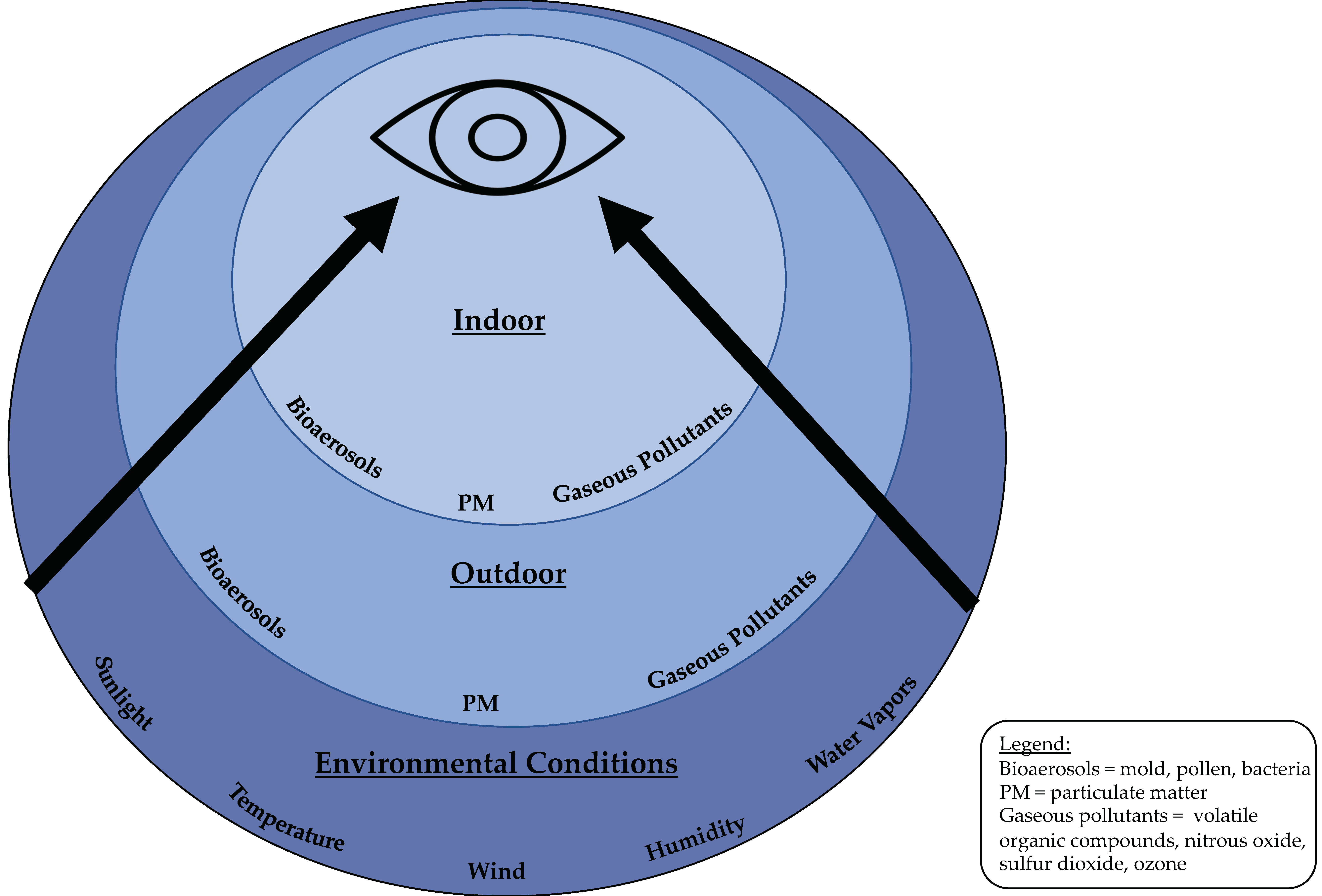
Figure 1. Concomitant environmental conditions affecting the ocular surface.
Jcm 09 03740 g001
Figure 2. Associations between various environmental exposures and dry eye (DE) diagnosis, symptoms, and signs with the size of each arrow indicating the strength of the existing data.
Jcm 09 03740 g002
Publisher’s Note: MDPI stays neutral with regard to jurisdictional claims in published maps and institutional affiliations.