Biatrial Remodeling in Patients with Cystic Fibrosis


Abstract
1. Introduction
2. Methodology
2.1. Echocardiography
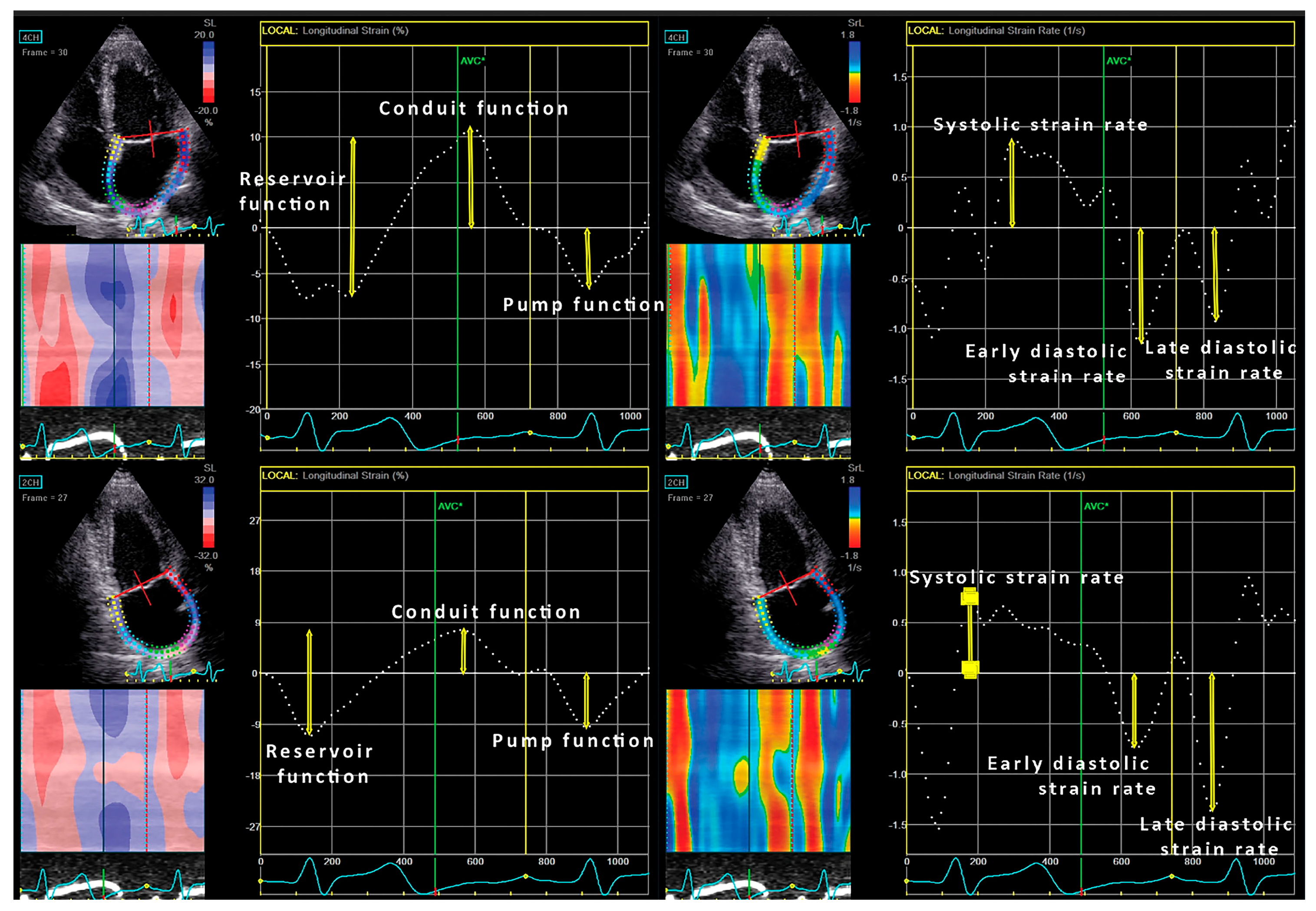
2.2. Assessment of Left Atrial Volumes and Strain
2.3. Right Ventricle and Atrium
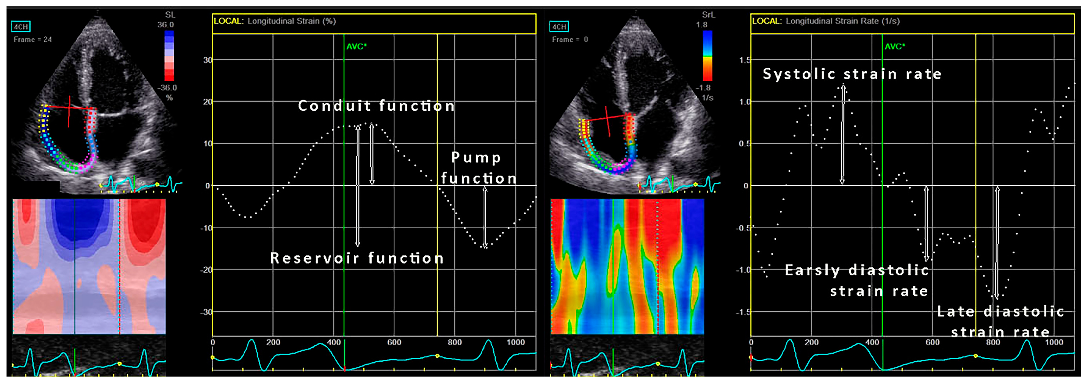
2.4. D Assessment of Right Atrial Volumes and Function
2.5. Statistical Analysis
3. Results
3.1. Left and Right Ventricle
3.2. LA and RA Strain Parameters
3.3. Predictors of Mortality
4. Discussion
5. Limitations
6. Conclusions
Author Contributions
Funding
Conflicts of Interest
References
- Royce, S.W. Cor pulmonale in infancy and early childhood: Report on 34 patients, with special reference to the occurrence of pulmonary heart disease in cystic fibrosis of the pancreas. Pediatrics 1951, 8, 255. [Google Scholar]
- Matthay, R.A.; Berger, H.J.; Loke, J.; Dolan, T.F.; Fagenholz, S.A.; Gottschalk, A.; Zaret, B.L. Right and left ventricular performance in ambulatory young adults with cystic fibrosis. Br. Heart J. 1980, 43, 474–480. [Google Scholar] [CrossRef] [PubMed]
- Fraser, K.L.; Tullis, D.E.; Sasson, Z.; Hyland, R.H.; Thornley, K.S.; Hanly, P.J. Pulmonary hypertension and cardiac function in adult cystic fibrosis: Role of hypoxemia. Chest 1999, 115, 1321–1328. [Google Scholar] [CrossRef] [PubMed]
- Ionescu, A.A.; Ionesc, A.A.; Payne, N.; Obieta-Fresnedo, I.; Fraser, A.G.; Shale, D.J. Subclinical right ventricular dysfunction in cystic fibrosis. A study using tissue Doppler echocardiography. Am. J. Respir. Crit. Care Med. 2001, 163, 1212–1218. [Google Scholar] [CrossRef] [PubMed]
- Panidis, I.P.; Ren, J.F.; Holsclaw, D.S.; Kotler, M.N.; Mintz, G.S.; Ross, J. Cardiac function in patients with cystic fibrosis: Evaluation by two-dimensional and Doppler echocardiography. J. Am. Coll. Cardiol. 1985, 6, 701–706. [Google Scholar] [CrossRef]
- Vizza, C.D.; Lynch, J.P.; Ochoa, L.L.; Richardson, G.; Trulock, E.P. Right and left ventricular dysfunction in patients with severe pulmonary disease. Chest 1998, 113, 576–583. [Google Scholar] [CrossRef] [PubMed]
- Eidt Rovedder, P.M.; Ziegler, B.; Furlan Pinotti, A.F.; Menna Barreto, S.S.; de Tarso Roth Dalcin, P. Prevalence of pulmonary hypertension evaluated by Doppler echocardiography in a population of adolescent and adult patients with cystic fibrosis. J. Bras. Pneumol. 2008, 34, 83–90. [Google Scholar]
- Henno, P.; Maurey, C.; Danel, C.; Bonnette, P.; Souilamas, R.; Stern, M.; Delclaux, C.; Levy, M.; Israel-Biet, D. Pulmonary vascular dysfunction in end-stage cystic fibrosis: Role of NF-kB and endothelin-1. Eur. Respir. J. 2009, 34, 1329–1337. [Google Scholar] [CrossRef] [PubMed]
- Giacchi, V.; Rotolo, N.; Amato, B.; Di Dio, G.; Betta, P.; La Rosa, M.; Leonardi, S.; Sciacca, P. Heart involvement in children and adults with cystic fibrosis: Correlation with pulmonary indexes and inflammation markers. Heart Lung Circ. 2015, 24, 1002–1010. [Google Scholar] [CrossRef] [PubMed]
- Baño-Rodrigo, A.; Salcedo-Posadas, A.; Villa-Asensi, J.R.; Tamariz-Martel, A.; Lopez-Neyra, A.; Blanco-Iglesias, E. Right ventricular dysfunction in adolescents with mild cystic fibrosis. J. Cyst. Fibros. 2012, 11, 274–278. [Google Scholar] [CrossRef] [PubMed]
- Eising, J.B.; van der Ent, C.K.; Teske, A.J.; Vanderschuren, M.M.; Uiterwaal, C.S.P.M.; Meijboom, F.J. Young patients with cystic fibrosis demonstrate subtle alterations of the cardiovascurrent recommendaticular system. J. Cyst. Fibros. 2018, 17, 643–649. [Google Scholar] [CrossRef] [PubMed]
- Sciatti, E.; Vizzardi, E.; Bonadei, I.; Valentini, F.; Menotti, E.; Prati, F.; Dallapellegrina, L.; Berlendis, M.; Poli, P.; Padoan, R.; et al. Focus on echocardiographic right ventricular strain analysis in cystic fibrosis adults without cardiovascular risk factors: A case-control study. Intern. Emerg. Med. 2019. [Google Scholar] [CrossRef] [PubMed]
- Sellers, Z.M.; McGlocklin, L.; Brasch, A. Strain rate echocardiography uncovers subclinical left ventricular dysfunction in cystic fibrosis. J. Cyst. Fibros. 2015, 14, 654–660. [Google Scholar] [CrossRef] [PubMed]
- Ozcelik, N.; Shell, R.; Holtzlander, M.; Cua, C. Decreased right ventricular function in healthy pediatric cystic fibrosis patients versus non-cystic fibrosis patients. Pediatr. Cardiol. 2013, 34, 159–164. [Google Scholar] [CrossRef] [PubMed]
- Labombarda, F.; Pellissier, A.; Ellafi, M.; Creveuil, C.; Ribault, V.; Laurans, M.; Guillot, M.; Bergot, E.; Grollier, G.; Milliez, P.; et al. Myocardial strain assessment in cystic fibrosis. J. Am. Soc. Echocardiogr. 2011, 24, 1037–1045. [Google Scholar] [CrossRef] [PubMed]
- Gupta, S.; Matulevicius, S.A.; Ayers, C.R.; Berry, J.D.; Patel, P.C.; Markham, D.W.; Levine, B.D.; Chin, K.M.; de Lemos, J.A.; Peshock, R.M.; et al. Left atrial structure and function and clinical outcomes in the general population. Eur. Heart J. 2013, 34, 278–285. [Google Scholar] [CrossRef] [PubMed]
- D’Alto, M.; D’Andrea, A.; Di Salvo, G.; Scognamiglio, G.; Argiento, P.; Romeo, E.; Di Marco, G.M.; Mattera Iacono, A.; Bossone, E.; Sarubbi, B.; et al. Right atrial function and prognosis in idiopathic pulmonary arterial hypertension. Int. J. Cardiol. 2017, 248, 320–325. [Google Scholar] [CrossRef] [PubMed]
- Lang, R.M.; Badano, L.P.; Mor-Avi, V.; Afilalo, J.; Armstrong, A.; Ernande, L.; Flachskampf, F.A.; Foster, E.; Goldstein, S.A.; Kuzentsova, T.; et al. Recommendations for cardiac chamber quantification by echocardiography in adults: An update from the American society of echocardiography and the European association of cardiovascular imaging. J. Am. Soc. Echocardiogr. 2015, 28, 1–39. [Google Scholar] [CrossRef] [PubMed]
- Quinones, M.A.; Otto, C.M.; Stoddard, M.; Waggoner, A.; Zoghbi, W.A. Recommendations for quantification of Doppler echocardiography: A report from the Doppler quantification task force of the nomenclature and standards committee of the American Society of Echocardiography. J. Am. Soc. Echocardiogr. 2002, 15, 167–184. [Google Scholar] [CrossRef] [PubMed]
- Badano, L.P.; Kolias, T.J.; Muraru, D.; Abraham, T.P.; Aurigemma, G.; Edvardsen, T.; D’Hooge, J.; Donal, E.; Fraser, A.G.; Marwick, T.; et al. Industry representatives. Standardization of left atrial, right ventricular, and right atrial deformation imaging using two-dimensional speckle tracking echocardiography: A consensus document of the EACVI/ASE/Industry Task Force to standardize deformation imaging. Eur. Heart J. Cardiovasc. Imaging 2018, 19, 591–600. [Google Scholar] [PubMed]
- Rudski, L.G.; Lai, W.W.; Afilalo, J.; Hua, L.; Handschumacher, M.D.; Chandrasekaran, K.; Solomon, S.D.; Louie, E.K.; Schiller, N.B. Guidelines for the echocardiographic assessment of the right heart in adults: A report from the American Society of Echocardiography endorsed by the European Association of Echocardiography, a registered branch of the European Society of Cardiology, and the Canadian Society of Echocardiography. J. Am. Soc. Echocardiogr. 2010, 23, 685–713. [Google Scholar] [PubMed]
- Labombarda, F.; Saloux, E.; Brouard, J.; Bergot, E.; Milliez, P. Heart involvement in cystic fibrosis: A specific cystic fibrosis-related myocardial changes? Respir. Med. 2016, 118, 31–38. [Google Scholar] [CrossRef] [PubMed]
- Blume, G.G.; Mcleod, C.J.; Barnes, M.E.; Seward, J.B.; Pellikka, P.A.; Bastiansen, P.M.; Tsang, T.S. Left atrial function: Physiology, assessment and clinical implications. Eur. J. Echocardiogr. 2011, 12, 421–430. [Google Scholar] [CrossRef] [PubMed]


| Controls (n = 32) | CF Survivors (n = 64) | CF Non-Survivors (n = 18) | p | |
|---|---|---|---|---|
| Age (years) | 36 ± 7 | 34 ± 11 | 34 ± 8 | 0.593 |
| Female (%) | 12 (38) | 32 (50) | 12 (67) | 0.138 |
| BMI (kg/m2) | 24.0 ± 3.3 | 19.9 ± 3.7 b | 18.1 ± 2.1 a,c | <0.001 |
| Plasma glucose (mg/dL) | 87 ± 13 | 133 ± 28 b | 146 ± 35 d | 0.011 |
| Diabetes (%) | 0 (0) | 27 (42) | 9 (50) | 0.372 |
| Urea (mg/dL) | 26 ± 6 | 30 ± 7 | 44 ± 8 a,c | 0.007 |
| Serum creatinine (mg/dL) | 0.87 ± 0.18 | 0.87 ± 0.28 | 1.33 ± 0.54 c,d | 0.066 |
| FVC (%) | - | 63 ± 21 | 43 ± 17 | <0.001 |
| FEV1 (%) | - | 45 ± 21 | 33 ± 13 | 0.033 |
| MEF 25 (%) | - | 21 ± 11 | 14 ± 7 | 0.310 |
| Controls (n = 32) | Cystic Fibrosis Survivors (n = 64) | Cystic Fibrosis Non-Survivors (n = 18) | p | |
|---|---|---|---|---|
| LV parameters | ||||
| LV end-diastolic diameter (mm) | 46.6 ± 0.5 | 41.2 ± 0.5 b | 42.8 ± 0.6 d | <0.001 |
| Interventricular septum thickness (mm) | 9.0 ± 1.2 | 9.2 ± 1.6 | 9.3 ± 2.0 | 0.819 |
| Posterior wall thickness (mm) | 8.3 ± 1.9 | 8.9 ± 1.5 | 9.0 ± 2.0 | 0.300 |
| Relative wall thickness | 0.36 ± 0.08 | 0.43 ± 0.09 b | 0.43 ± 0.1 a | <0.001 |
| LV mass index (g/m2) | 71.7 ± 19.7 | 73.8 ± 21.5 | 84.1 ± 35.1 | 0.202 |
| Ejection fraction (%) | 64 ± 6 | 63 ± 8 | 60 ± 11 | 0.257 |
| E/A ratio | 1.7 ± 0.7 | 1.3 ± 0.4 b | 1.2 ± 0.3 a | <0.001 |
| Deceleration time (ms) | 215 ± 74 | 198 ± 68 | 211 ± 65 | 0.543 |
| E/e’ | 5.9 ± 1.4 | 7.0 ± 1.8 | 8.8 ± 3.1 a,c | <0.001 |
| LV diastolic dysfunction (%) | 3 (9) | 7 (11) | 8 (44) a,e | 0.001 |
| LA volume index (mL/m2) | 26.5 ± 5.3 | 26.6 ± 8.1 | 33.7 ± 10.5 a,e | 0.004 |
| LA dilatation (%) | 1 (3) | 9 (14) | 10 (56) a,e | <0.001 |
| RV parameters | ||||
| RV basal diameter (mm) | 32.0 ± 4.6 | 29.9 ± 4.9 d | 29.6 ± 4.6 | 0.088 |
| RV end-diastolic area (cm2) | 16 ± 4 | 14 ± 4 d | 13 ± 3.5 f | 0.048 |
| RV end-systolic area (cm2) | 8.3 ± 2.5 | 7.6 ± 3.7 | 7.7 ± 3.6 | 0.686 |
| Fractional area change (%) | 48 ± 8 | 45 ± 10 | 47 ± 8 | 0.598 |
| s’ (cm/s) | 12.6 ± 1.4 | 11.6 ± 2.7 | 11.0 ± 1.5 | 0.191 |
| TAPSE (mm) | 24 ± 4 | 20 ± 4 b | 19 ± 3 a | <0.001 |
| RA volume index (mL/m2) | 21.2 ± 5.5 | 23.1 ± 9.0 d | 24.9 ± 11.8 e | 0.010 |
| RA dilatation (%) | 5 (16) | 11 (17) | 7 (39) | 0.096 |
| PAPs (mmHg) | 20 ± 7 | 31 ± 12 b | 35 ± 10 a | <0.001 |
| Pulmonary hypertension (%) | 1 (3) | 5 (8) | 7 (39) a,e | <0.001 |
| Controls (n = 32) | Cystic Fibrosis Survivors (n = 64) | Cystic Fibrosis Non-Survivors (n = 18) | p | |
|---|---|---|---|---|
| Global LA speckle tracking parameters | ||||
| LA global strain (%) | 38 ± 8 | 34 ± 9 | 33 ± 12 | 0.301 |
| LA positive strain (%) | 22 ± 8 | 19 ± 7 | 18 ± 8 | 0.228 |
| LA negative strain (%) | 15 ± 3 | 15 ± 5 | 15 ± 5 | 0.981 |
| LA early diastolic strain rate (cm/s) | 2.3 ± 0.7 | 1.9 ± 0.7 d | 1.7 ± 0.9 f | 0.047 |
| LA late diastolic strain rate (cm/s) | 2.3 ± 0.7 | 2.2 ± 0.9 | 2.0 ± 0.7 | 0.633 |
| LA systolic strain rate (cm/s) | 1.8 ± 0.4 | 1.9 ± 0.6 | 1.7 ± 0.5 | 0.638 |
| 4Ch LA speckle tracking parameters | ||||
| LA global strain (%) | 38 ± 9 | 33 ± 10 d | 30 ± 12 f | 0.040 |
| LA positive strain (%) | 23 ± 9 | 19 ± 8 d | 15 ± 7 a | 0.014 |
| LA negative strain (%) | 15 ± 4 | 14 ± 6 | 15 ± 5 | 0.839 |
| LA early diastolic strain rate (1/s) | 2.5 ± 0.8 | 1.9 ± 0.8 b | 1.6 ± 0.8 a | 0.002 |
| LA late diastolic strain rate (1/s) | 2.1 ± 0.7 | 2.0 ± 0.9 | 1.9 ± 0.7 | 0.800 |
| LA systolic strain rate (1/s) | 1.7 ± 0.4 | 1.8 ± 0.8 | 1.6 ± 0.6 | 0.570 |
| 2Ch LA speckle tracking parameters | ||||
| LA global strain (%) | 35 ± 7 | 34 ± 10 | 36 ± 12 | 0.544 |
| LA positive strain (%) | 20 ± 7 | 18 ± 8 | 21 ± 9.8 | 0.448 |
| LA negative strain (%) | 15 ± 4 | 16 ± 6 | 15 ± 5 | 0.998 |
| LA early diastolic strain rate (1/s) | 2.1 ± 0.7 | 1.8 ± 0.8 | 1.9 ± 0.9 | 0.398 |
| LA late diastolic strain rate (1/s) | 2.4 ± 0.9 | 2.4 ± 1.0 | 2.3 ± 0.8 | 0.876 |
| LA systolic strain rate (1/s) | 1.8 ± 0.5 | 1.9 ± 0.7 | 2.0 ± 0.6 | 0.831 |
| Controls (n = 32) | Cystic Fibrosis Survivors (n = 64) | Cystic Fibrosis Non-Survivors (n = 18) | p | |
|---|---|---|---|---|
| RA global strain (%) | 38 ± 9 | 35 ± 13 | 35 ± 10 | 0.518 |
| RA positive strain (%) | 24 ± 8 | 21 ± 10 | 19 ± 7 | 0.251 |
| RA negative strain (%) | 14 ± 5 | 14 ± 6 | 16 ± 6 | 0.567 |
| RA early diastolic strain rate (cm/s) | 1.9 ± 0.6 | 1.7 ± 0.7 | 1.6 ± 0.7 | 0.528 |
| RA late diastolic strain rate (cm/s) | 2.1 ± 0.7 | 2.1 ± 0.9 | 2.6 ± 1.2 | 0.194 |
| RA systolic strain rate (cm/s) | 2.1 ± 0.6 | 2.1 ± 0.7 | 2.3 ± 0.9 | 0.672 |
| Univariate | |||
|---|---|---|---|
| OR | 95% CI | p | |
| Age (years) | 0.52 | 0.17–1.50 | 0.215 |
| BMI (kg/m2) | 0.83 | 0.67–1.00 | 0.056 |
| FVC (%) | 0.94 | 0.91–0.98 | 0.001 |
| FEV1 (%) | 0.96 | 0.93–0.99 | 0.042 |
| LV mass index (g/m2) | 1.01 | 0.99–1.03 | 0.153 |
| LAVI (mL/m2) | 1.09 | 1.02–1.16 | 0.008 |
| LA longitudinal strain in 4Ch (%) | 0.97 | 0.92–1.03 | 0.362 |
| LA early diastolic strain rate 4Ch (cm/s) | 0.59 | 0.28–1.24 | 0.161 |
| LA positive strain 4Ch (%) | 0.95 | 0.88–1.02 | 0.149 |
| TAPSE (mm) | 0.94 | 0.80–1.09 | 0.400 |
| PAPs (mmHg) | 1.02 | 0.98–1.07 | 0.335 |
| RAVI (mL/m2) | 1.10 | 1.02–1.18 | 0.020 |
| RA longitudinal strain (%) | 0.99 | 0.95–1.05 | 0.808 |
© 2019 by the authors. Licensee MDPI, Basel, Switzerland. This article is an open access article distributed under the terms and conditions of the Creative Commons Attribution (CC BY) license (http://creativecommons.org/licenses/by/4.0/).
Share and Cite
Dordevic, A.; Genger, M.; Schwarz, C.; Cuspidi, C.; Tahirovic, E.; Pieske, B.; Düngen, H.-D.; Tadic, M.
Biatrial Remodeling in Patients with Cystic Fibrosis
. J. Clin. Med. 2019, 8, 1141.
https://doi.org/10.3390/jcm8081141
Dordevic A, Genger M, Schwarz C, Cuspidi C, Tahirovic E, Pieske B, Düngen H-D, Tadic M.
Biatrial Remodeling in Patients with Cystic Fibrosis
. Journal of Clinical Medicine. 2019; 8(8):1141.
https://doi.org/10.3390/jcm8081141
Dordevic, Aleksandar, Martin Genger, Carsten Schwarz, Cesare Cuspidi, Elvis Tahirovic, Burkert Pieske, Hans-Dirk Düngen, and Marijana Tadic.
2019. "Biatrial Remodeling in Patients with Cystic Fibrosis
" Journal of Clinical Medicine 8, no. 8: 1141.
https://doi.org/10.3390/jcm8081141
Dordevic, A., Genger, M., Schwarz, C., Cuspidi, C., Tahirovic, E., Pieske, B., Düngen, H.-D., & Tadic, M.
(2019). Biatrial Remodeling in Patients with Cystic Fibrosis
. Journal of Clinical Medicine, 8(8), 1141.
https://doi.org/10.3390/jcm8081141