Acute IgA-Dominant Glomerulonephritis Associated with Syphilis Infection in a Pregnant Teenager: A New Disease Association


 and
and
Abstract
1. Introduction
2. The Case
3. Discussion
4. Conclusions
Author Contributions
Funding
Acknowledgments
Conflicts of Interest
References
- Mol, B.W.J.; Roberts, C.T.; Thangaratinam, S.; Magee, L.A.; de Groot, C.J.M.; Hofmeyr, G.J. Pre-eclampsia. Lancet 2016, 387, 999–1011. [Google Scholar] [CrossRef]
- Trogstad, L.; Magnus, P.; Stoltenberg, C. Pre-eclampsia: Risk factors and causal models. Best Pract. Res. Clin. Obstet. Gynaecol. 2011, 25, 329–342. [Google Scholar] [CrossRef] [PubMed]
- Wagner, S.J.; Barac, S.; Garovic, V.D. Hypertensive pregnancy disorders: Current concepts. J. Clin. Hypertens. (Greenwich) 2007, 9, 560–566. [Google Scholar] [CrossRef]
- Garg, A.X.; Nevis, I.F.; McArthur, E.; Sontrop, J.M.; Koval, J.J.; Lam, N.N.; Hildebrand, A.M.; Reese, P.P.; Storsley, L.; Gill, J.S.; et al. Gestational hypertension and preeclampsia in living kidney donors. N. Engl. J. Med. 2015, 372, 124–133. [Google Scholar] [CrossRef]
- Davis, S.; Dylewski, J.; Shah, P.B.; Holmen, J.; You, Z.; Chonchol, M.; Kendrick, J. Risk of adverse maternal and fetal outcomes during pregnancy in living kidney donors: A matched cohort study. Clin. Transplant. 2018, e13453. [Google Scholar] [CrossRef] [PubMed]
- Kendrick, J.; Holmen, J.; You, Z.; Smits, G.; Chonchol, M. Association of Unilateral Renal Agenesis with Adverse Outcomes in Pregnancy: A Matched Cohort Study. Am. J. Kidney Dis. 2017, 70, 506–511. [Google Scholar] [CrossRef]
- Spotti, D. Pregnancy in women with diabetic nephropathy. J. Nephrol. 2018. [Google Scholar] [CrossRef] [PubMed]
- Piccoli, G.B.; Kooij, I.A.; Attini, R.; Montersino, B.; Fassio, F.; Gerbino, M.; Biolcati, M.; Cabiddu, G.; Versino, E.; Todros, T. A Systematic Review on Materno-Foetal Outcomes in Pregnant Women with IgA Nephropathy: A Case of "Late-Maternal" Preeclampsia? J. Clin. Med. 2018, 7, 212. [Google Scholar] [CrossRef]
- Attini, R.; Kooij, I.; Montersino, B.; Fassio, F.; Gerbino, M.; Biolcati, M.; Versino, E.; Todros, T.; Piccoli, G.B. Reflux nephropathy and the risk of preeclampsia and of other adverse pregnancy-related outcomes: A systematic review and meta-analysis of case series and reports in the new millennium. J. Nephrol. 2018, 31, 833–846. [Google Scholar] [CrossRef]
- Wu, M.; Wang, D.; Zand, L.; Harris, P.C.; White, W.M.; Garovic, V.D.; Kermott, C.A. Pregnancy outcomes in autosomal dominant polycystic kidney disease: A case-control study. J. Matern. Fetal Neonatal Med. 2016, 29, 807–812. [Google Scholar] [CrossRef]
- Piccoli, G.B.; Zakharova, E.; Attini, R.; Ibarra Hernandez, M.; Orozco Guillien, A.; Alrukhaimi, M.; Liu, Z.H.; Ashuntantang, G.; Covella, B.; Cabiddu, G.; et al. Pregnancy in Chronic Kidney Disease: Need for Higher Awareness. A Pragmatic Review Focused on What Could Be Improved in the Different CKD Stages and Phases. J. Clin. Med. 2018, 7, 415. [Google Scholar] [CrossRef] [PubMed]
- Webster, L.M.; Gill, C.; Seed, P.T.; Bramham, K.; Wiesender, C.; Nelson-Piercy, C.; Myers, J.E.; Chappell, L.C. Chronic hypertension in pregnancy: impact of ethnicity and superimposed preeclampsia on placental, endothelial, and renal biomarkers. Am. J. Physiol. Regul. Integr. Comp. Physiol. 2018, 315, R36–R47. [Google Scholar] [CrossRef]
- Acharya, A. Promising biomarkers for superimposed pre-eclampsia in pregnant women with established hypertension and chronic kidney disease. Kidney Int. 2016, 89, 743–746. [Google Scholar] [CrossRef] [PubMed]
- Bramham, K.; Seed, P.T.; Lightstone, L.; Nelson-Piercy, C.; Gill, C.; Webster, P.; Poston, L.; Chappell, L.C. Diagnostic and predictive biomarkers for pre-eclampsia in patients with established hypertension and chronic kidney disease. Kidney Int. 2016, 89, 874–885. [Google Scholar] [CrossRef] [PubMed]
- Costa, R.A.; Hoshida, M.S.; Alves, E.A.; Zugaib, M.; Francisco, R.P. Preeclampsia and superimposed preeclampsia: The same disease? The role of angiogenic biomarkers. Hypertens. Pregnancy 2016, 35, 139–149. [Google Scholar] [CrossRef] [PubMed]
- Rolfo, A.; Attini, R.; Tavassoli, E.; Neve, F.V.; Nigra, M.; Cicilano, M.; Nuzzo, A.M.; Giuffrida, D.; Biolcati, M.; Nichelatti, M.; et al. Is It Possible to Differentiate Chronic Kidney Disease and Preeclampsia by means of New and Old Biomarkers? A Prospective Study. Dis. Markers 2015. [Google Scholar] [CrossRef] [PubMed]
- Piccoli, G.B.; Cabiddu, G.; Castellino, S.; Gernone, G.; Santoro, D.; Moroni, G.; Spotti, D.; Giacchino, F.; Attini, R.; Limardo, M.; et al. A best practice position statement on the role of the nephrologist in the prevention and follow-up of preeclampsia: The Italian study group on kidney and pregnancy. J. Nephrol. 2017, 30, 307–317. [Google Scholar] [CrossRef]
- Robinson, B.M.; Akizawa, T.; Jager, K.J.; Kerr, P.G.; Saran, R.; Pisoni, R.L. Factors affecting outcomes in patients reaching end-stage kidney disease worldwide: Differences in access to renal replacement therapy, modality use, and haemodialysis practice. Lancet 2016, 388, 294–306. [Google Scholar] [CrossRef]
- Coresh, J.; Jafar, T.H. Disparities in worldwide treatment of kidney failure. Lancet 2015, 385, 1926–1928. [Google Scholar] [CrossRef]
- Leftwich, H.K.; Alves, M.V. Adolescent Pregnancy. Pediatr. Clin. N. Am. 2017, 64, 381–388. [Google Scholar] [CrossRef]
- Bakwa-Kanyinga, F.; Valério, E.G.; Bosa, V.L.; Alfama, C.O.; Sperb, M.; Capp, E.; Vettorazzi, J. Adolescent pregnancy: Maternal and fetal outcomes in patients with and without preeclampsia. Pregnancy Hypertens. 2017, 10, 96–100. [Google Scholar] [CrossRef] [PubMed]
- Silva, L.M.; Coolman, M.; Steegers, E.A.; Jaddoe, V.W.; Moll, H.A.; Hofman, A.; Mackenbach, J.P.; Raat, H. Low socioeconomic status is a risk factor for preeclampsia: the Generation R Study. J. Hypertens. 2008, 26, 1200–1208. [Google Scholar] [CrossRef] [PubMed]
- Ramesh, K.; Gandhi, S.; Rao, V. Socio-demographic and other risk factors of pre eclampsia at a tertiary care hospital, karnataka: Case control study. J. Clin. Diagn. Res. 2014, 8, JC01–JC04. [Google Scholar]
- Kim, M.K.; Lee, S.M.; Bae, S.H.; Kim, H.J.; Lim, N.G.; Yoon, S.J.; Lee, J.Y.; Jo, M.W. Socioeconomic status can affect pregnancy outcomes and complications, even with a universal healthcare system. Int. J. Equity Health 2018, 17, 2. [Google Scholar] [CrossRef] [PubMed]
- Han, Z.; Mulla, S.; Beyene, J.; Liao, G.; McDonald, S.D.; Knowledge Synthesis Group. Maternal underweight and the risk of preterm birth and low birth weight: A systematic review and meta-analyses. Int. J. Epidemiol. 2011, 40, 65–101. [Google Scholar] [CrossRef] [PubMed]
- Cipolla, M.J.; Kraig, R.P. Seizures in Women with Preeclampsia: Mechanisms and Management. Fetal Matern. Med. Rev. 2011, 22, 91–108. [Google Scholar] [CrossRef]
- Chapman Johnson, A. Mechanisms of Seizure during Pregnancy and Preeclampsia Graduate College Dissertations and Theses Dissertations and Theses, 2015 University of Vermont. Available online: https://scholarworks.uvm.edu/cgi/viewcontent.cgi?article=1335&context=graddis (accessed on 12 December 2018).
- Nevis, I.F.; Reitsma, A.; Dominic, A.; McDonald, S.; Thabane, L.; Akl, E.A.; Hladunewich, M.; Akbari, A.; Joseph, G.; Sia, W.; et al. Pregnancy outcomes in women with chronic kidney disease: A systematic review. Clin. J. Am. Soc. Nephrol. 2011, 6, 2587–2598. [Google Scholar] [CrossRef] [PubMed]
- Zhang, J.J.; Ma, X.X.; Hao, L.; Liu, L.J.; Lv, J.C.; Zhang, H. A Systematic Review and Meta-Analysis of Outcomes of Pregnancy in CKD and CKD Outcomes in Pregnancy. Clin. J. Am. Soc. Nephrol. 2015, 10, 1964–1978. [Google Scholar] [CrossRef]
- Feng, Z.; Minard, C.; Raghavan, R. Pregnancy outcomes in advanced kidney disease. Clin. Nephrol. 2015, 83, 272–278. [Google Scholar] [CrossRef] [PubMed]
- Piccoli, G.B.; Cabiddu, G.; Attini, R.; Gerbino, M.; Todeschini, P.; Perrino, M.L.; Manzione, A.M.; Piredda, G.B.; Gnappi, E.; Caputo, F.; et al. Working Group on Pregnancy in Renal Transplantation. Outcomes of Pregnancies After Kidney Transplantation: Lessons Learned From CKD. A Comparison of Transplanted, Nontransplanted Chronic Kidney Disease Patients and Low-Risk Pregnancies: A Multicenter Nationwide Analysis. Transplantation 2017, 101, 2536–2544. [Google Scholar] [PubMed]
- Buyon, J.P.; Kim, M.Y.; Guerra, M.M.; Laskin, C.A.; Petri, M.; Lockshin, M.D.; Sammaritano, L.; Branch, D.W.; Porter, T.F.; Sawitzke, A.; et al. Predictors of Pregnancy Outcomes in Patients with Lupus: A Cohort Study. Ann. Intern. Med. 2015, 163, 153–163. [Google Scholar] [CrossRef] [PubMed]
- Karumanchi, S.A.; Maynard, S.E.; Stillman, I.E.; Epstein, F.H.; Sukhatme, V.P. Preeclampsia: A renal perspective. Kidney Int. 2005, 67, 2101–2113. [Google Scholar] [CrossRef] [PubMed]
- Leduc, L.; Lederer, E.; Lee, W.; Cotton, D.B. Urinary sediment changes in severe preeclampsia. Obstet. Gynecol. 1991, 77, 186–189. [Google Scholar] [CrossRef] [PubMed]
- Fogazzi, G.B.; Saglimbeni, L.; Banfi, G.; Cantú, M.; Moroni, G.; Garigali, G.; Cesana, B.M. Urinary sediment features in proliferative and non-proliferative glomerular diseases. J. Nephrol. 2005, 18, 703–710. [Google Scholar] [PubMed]
- Petri, M. The Hopkins Lupus Pregnancy Center: Ten key issues in management. Rheum. Dis. Clin. N. Am. 2007, 33, 33227–33235. [Google Scholar] [CrossRef] [PubMed]
- Gallery, E.D.; Ross, M.; Györy, A.Z. Urinary red blood cell and cast excretion in normal and hypertensive human pregnancy. Am. J. Obstet. Gynecol. 1993, 168, 67–70. [Google Scholar] [CrossRef]
- Perazella, M.A. The urine sediment as a biomarker of kidney disease. Am. J. Kidney Dis. 2015, 66, 748–755. [Google Scholar] [CrossRef]
- Fogazzi, G.B. Urinary sediment: Still an important diagnostic tool. Clin. Chem. Lab. Med. 2015. [Google Scholar] [CrossRef]
- Caleffi, A.; Lippi, G. Cylindruria. Clin. Chem. Lab. Med. 2015. [Google Scholar] [CrossRef]
- Nasr, S.H.; Markowitz, G.S.; Stokes, M.B.; Said, S.M.; Valeri, A.M.; D’Agati, V.D. Acute postinfectious glomerulonephritis in the modern era: experience with 86 adults and review of the literature. Medicine (Baltimore) 2008, 87, 21–32. [Google Scholar] [CrossRef]
- Kambham, N. Postinfectious glomerulonephritis. Adv. Anat. Pathol. 2012, 19, 338–347. [Google Scholar] [CrossRef] [PubMed]
- Liu, Y.; Ma, X.; Zheng, J.; Liu, X.; Yan, T. A Systematic Review and Meta-Analysis of Kidney and Pregnancy Outcomes in IgA Nephropathy. Am. J. Nephrol. 2016, 44, 187–193. [Google Scholar] [CrossRef]
- Piccoli, G.B.; Attini, R.; Cabiddu, G.; Kooij, I.; Fassio, F.; Gerbino, M.; Maxia, S.; Biolcati, M.; Versino, E.; Todros, T. Maternal-foetal outcomes in pregnant women with glomerulonephritides. Are all glomerulonephritides alike in pregnancy? J. Autoimmun. 2017, 79, 91–98. [Google Scholar] [CrossRef] [PubMed]
- Sámano, R.; Martínez-Rojano, H.; Robichaux, D.; Rodríguez-Ventura, A.L.; Sánchez-Jiménez, B.; de la Luz Hoyuela, M.; Godínez, E.; Segovia, S. Family context and individual situation of teens before, during and after pregnancy in Mexico City. BMC Pregnancy Childbirth. 2017, 17, 382. [Google Scholar] [CrossRef]
- Ibarra-Hernández, M.; Orozco-Guillén, O.A.; de la Alcantar-Vallín, M.L.; Garrido-Roldan, R.; Jiménez-Alvarado, M.P.; Castro, K.B.; Villa-Villagrana, F.; Borbolla, M.; Gallardo-Gaona, J.M.; García-García, G.; et al. Acute kidney injury in pregnancy and the role of underlying CKD: A point of view from México. J. Nephrol. 2017, 30, 773–780. [Google Scholar] [CrossRef] [PubMed]
- Nasr, S.H.; Markowitz, G.S.; Whelan, J.D.; Albanese, J.J.; Rosen, R.M.; Fein, D.A.; Kim, S.S.; D’Agati, V.D. IgA-dominant acute poststaphylococcal glomerulonephritis complicating diabetic nephropathy. Hum. Pathol. 2003, 34, 1235–1241. [Google Scholar] [CrossRef]
- Bu, R.; Li, Q.; Duan, Z.Y.; Wu, J.; Chen, P.; Chen, X.M.; Cai, G.Y. Clinicopathologic features of IgA-dominant infection-associated glomerulonephritis: A pooled analysis of 78 cases. Am. J. Nephrol. 2015, 41, 98–106. [Google Scholar] [CrossRef]
- Handa, T.; Kakita, H.; Tateishi, Y.; Endo, T.; Suzuki, H.; Katayama, T.; Tsukamoto, T.; Muso, E. The features in IgA-dominant infection-related glomerulonephritis distinct from IgA nephropathy: A single-center study. Clin. Exp. Nephrol. 2018, 22, 1116–1127. [Google Scholar] [CrossRef]
- Kanno, M.; Tanaka, K.; Kimura, H.; Watanabe, K.; Hayashi, Y.; Asahi, K.; Nakayama, M.; Joh, K.; Watanabe, T. Garland-pattern postinfectious glomerulonephritis with IgA-dominant deposition. CEN Case Rep. 2014, 3, 56–62. [Google Scholar] [CrossRef]
- Nasr, S.H.; D’Agati, V.D. IgA-dominant postinfectious glomerulonephritis: A new twist on an old disease. Nephron Clin. Pract. 2011, 119, c18–c25. [Google Scholar] [CrossRef]
- Mascarenhas, R.; Fogo, A.B.; Steele, R.W.; Baliga, R. IgA-Dominant Postinfectious Glomerulonephritis. Clin. Pediatr.(Phila) 2016, 55, 873–876. [Google Scholar] [CrossRef] [PubMed]
- Braunstein, G.D.; Lewis, E.J.; Galvanek, E.G.; Hamilton, A.; Bell, W.R. The nephrotic syndrome associated with secondary syphilis. An immune deposit disease. Am. J. Med. 1970, 48, 643–648. [Google Scholar] [CrossRef]
- Bhorade, M.S.; Carag, H.B.; Lee, H.J.; Potter, E.V.; Dunea, G. Nephropathy of secondary syphilis. A clinical and pathological spectrum. JAMA 1971, 216, 1159–1166. [Google Scholar] [CrossRef] [PubMed]
- Tourville, D.R.; Byrd, L.H.; Kim, D.U.; Zajd, D.; Lee, I.; Reichman, L.B.; Baskin, S. Treponemal antigen in immunopathogenesis of syphilitic glomerulonephritis. Am. J. Pathol. 1976, 82, 479–492. [Google Scholar] [PubMed]
- Schillinger, F.; Montagnac, R.; Goclowski, C.; Dine, G.; Alessandri, E.; Hopfner, C.; Birembaut, P. Extramembranous glomerulonephritis of acquired syphilis in a patient recently infected by the hepatitis B virus. Demonstration of the treponemal antigen in the kidney by indirect immunofluorescence. Presse Med. 1983, 12, 153–156. [Google Scholar] [PubMed]
- Hunte, W.; al-Ghraoui, F.; Cohen, R.J. Secondary syphilis and the nephrotic syndrome. J. Am. Soc. Nephrol. 1993, 3, 1351–1355. [Google Scholar] [PubMed]
- Handoko, M.L.; Duijvestein, M.; Scheepstra, C.G.; de Fijter, C.W. Syphilis: A reversible cause of nephrotic syndrome. BMJ Case Rep. 2013. [Google Scholar] [CrossRef]
- Noutong, S.A.; Chornyy, V.; Alquadan, K.F.; Ejaz, A.A.; Koratala, A. A Red Herring in the Green Grass: Syphilitic Membranous Glomerulonephritis. Am. J. Med. 2017, 130, 285–287. [Google Scholar] [CrossRef]
- Havill, J.P.; Kuperman, M.B.; Bernardo, L.L.; Jaar, B.G. The Case ∣ An age-old enemy should not be forgotten. Kidney Int. 2011, 79, 924–925. [Google Scholar] [CrossRef]
- Mora Mora, M.T.; Gallego Domínguez, M.S.; Castellano Cerviño, M.I.; Novillo Santana, R.; Gómez-Martino Arroyo, J.R. Membranous glomerulonephritis in a patient with syphilis. Nefrologia 2011, 31, 372–373. [Google Scholar]
- Satoskar, A.A.; Kovach, P.; O’Reilly, K.; Nadasdy, T. An uncommon cause of membranous glomerulonephritis. Am. J. Kidney Dis. 2010, 55, 386–390. [Google Scholar] [CrossRef] [PubMed]
- Hartley, A.J.; Rajakariar, R.; Sheaff, M.; Buckland, M.; Goh, B.; O’Connell, R. Syphilis masquerading as focal segmental glomerulosclerosis. Int. J. STD AIDS 2014, 25, 529–531. [Google Scholar] [CrossRef]
- Krane, N.K.; Espenan, P.; Walker, P.D.; Bergman, S.M.; Wallin, J.D. Renal disease and syphilis: A report of nephrotic syndrome with minimal change disease. Am. J. Kidney Dis. 1987, 9, 176–179. [Google Scholar] [CrossRef]
- Stubanus, M.; Göbel, H.; Rieg, S.; Walz, G.; Gerke, P. Quiz page. Minimal change glomerulonephritis associated with secondary syphilis. Am. J. Kidney Dis. 2007, 49, A49–A50. [Google Scholar] [PubMed]
- Tognetti, L.; Cinotti, E.; Tripodi, S.; Garosi, G.; Rubegni, P. Unusual presentation of secondary syphilis: Membranoproliferative glomerulonephritis andmuco-cutaneous lesions. Int. J. STD AIDS 2018, 29, 410–413. [Google Scholar] [CrossRef] [PubMed]
- Hruby, Z.; Kuźniar, J.; Rabczyński, J.; Bogucki, J.; Steciwko, A.; Weyde, W. The variety of clinical and histopathologic presentations of glomerulonephritis associated with latent syphilis. Int. Urol. Nephrol. 1992, 24, 541–547. [Google Scholar] [CrossRef] [PubMed]


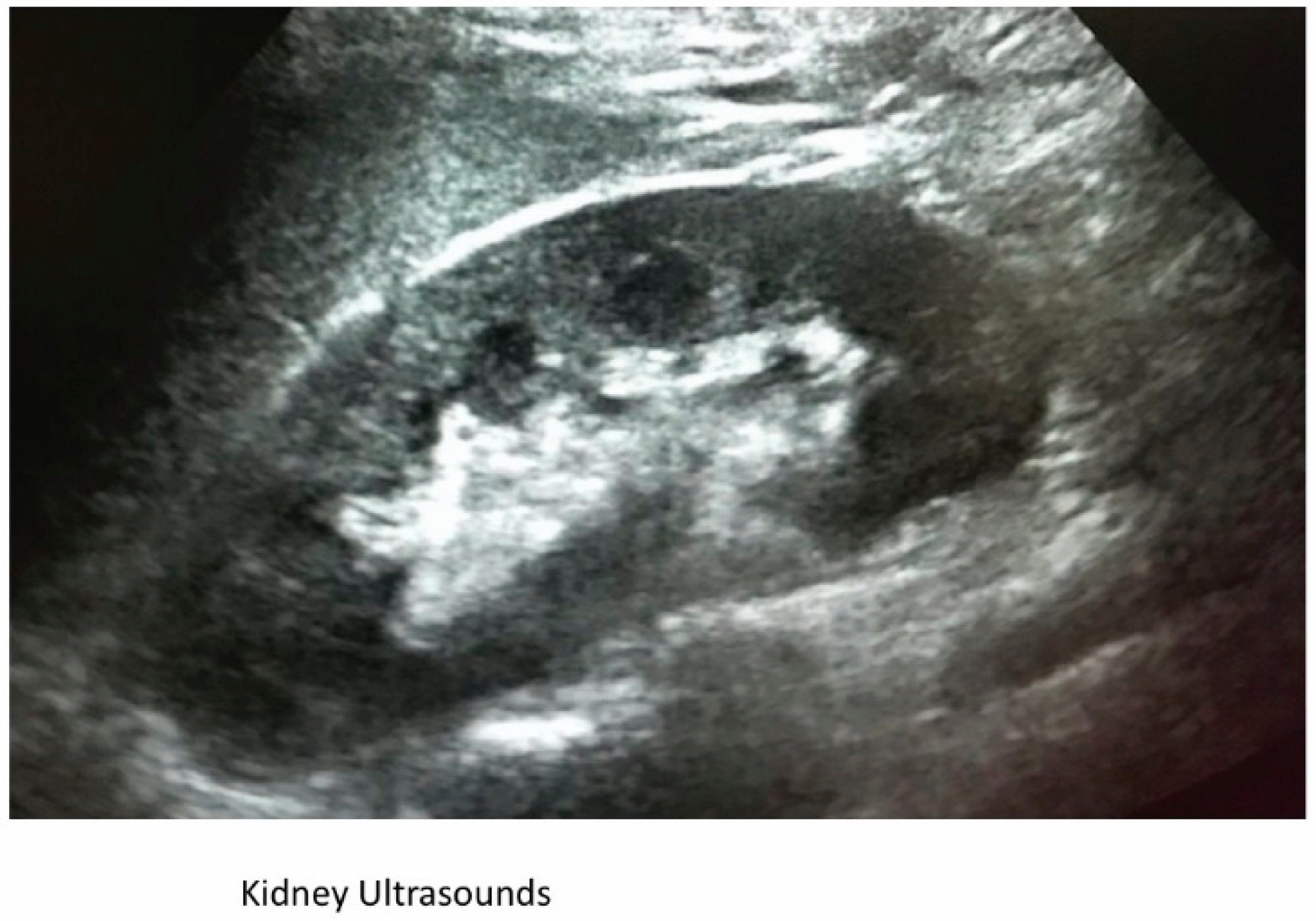
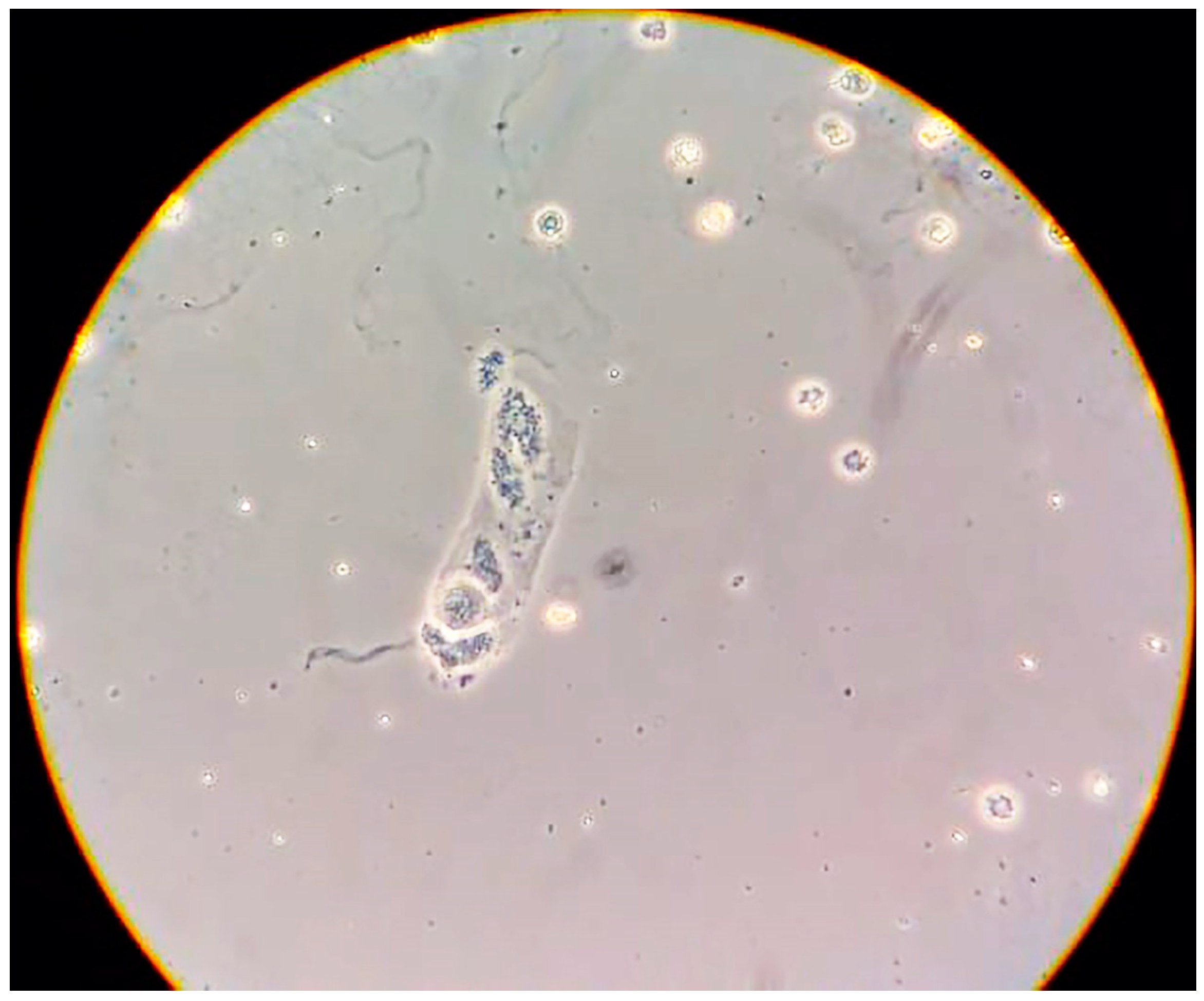
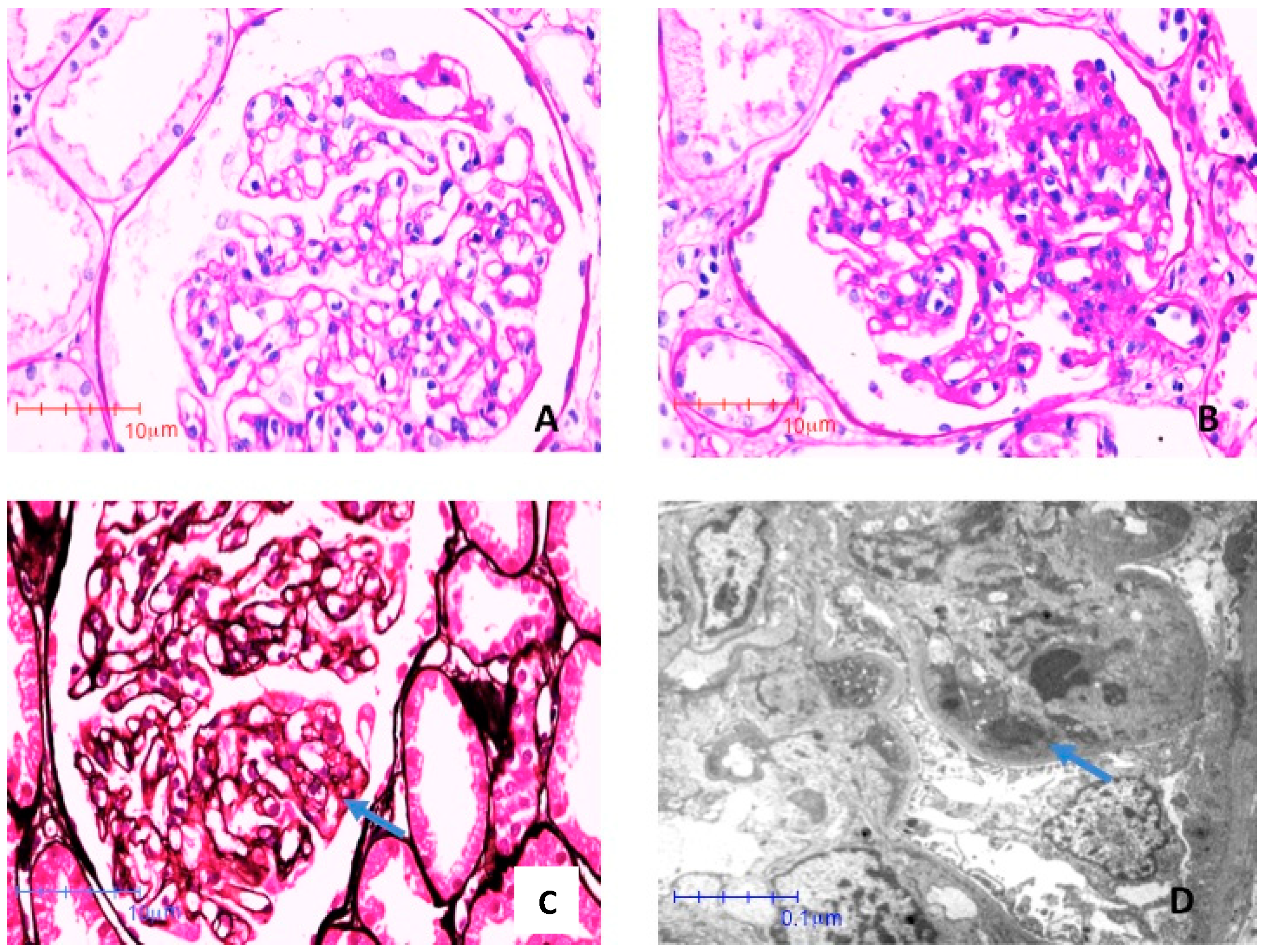
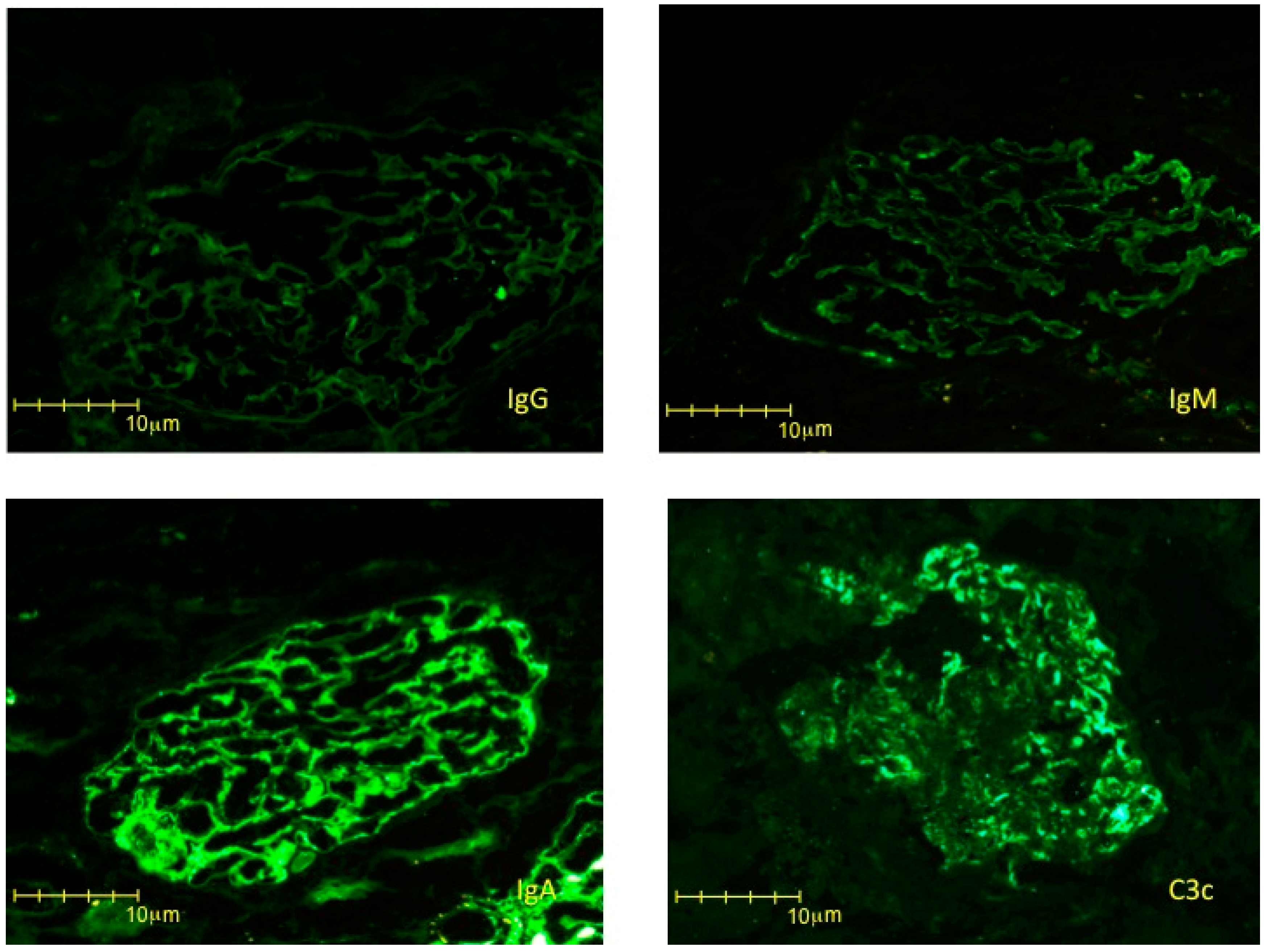
| Parameter | Initial Testing | 1 Week after Delivery (Kidney Biopsy) | 4 Months after Delivery |
|---|---|---|---|
| Albumin, g/dL | 2.3 | - | 4.7 |
| Creatinine, mg/dL | 0.8 | 0.8 | 0.7 |
| Urea, mg/dL | 43 | 32 | 26 |
| Hemoglobin, g/dL | 9.1 | 9.9 | 14.2 |
| LDH UI/L | 615 | 677 | 414 |
| AST, UI/L | 30 | 33 | 18 |
| ALT, UI/L | 40 | 46 | 27 |
| Glucose mg/dL | 73 | 71 | 72 |
| Uric acid mg/dL | 7.9 | 8 | 5.1 |
| Proteinuria/creatinuria g/g | 6.42 | 4.9 | 0.036 |
| Erythrocytes n /ml | 341 | - | 2 |
| VDRL | 1:128 | - | 1:32 |
| Syphilis IgM | Positive | - | - |
| Syphilis IgG | Positive | - | - |
© 2019 by the authors. Licensee MDPI, Basel, Switzerland. This article is an open access article distributed under the terms and conditions of the Creative Commons Attribution (CC BY) license (http://creativecommons.org/licenses/by/4.0/).
Share and Cite
Orozco Guillén, A.O.; Velazquez Silva, R.I.; Moguel González, B.; Guell, Y.A.; Garciadiego Fossas, P.; Custodio Gómez, I.G.; Miranda Araujo, O.; Soto Abraham, V.; Piccoli, G.B.; Madero, M. Acute IgA-Dominant Glomerulonephritis Associated with Syphilis Infection in a Pregnant Teenager: A New Disease Association. J. Clin. Med. 2019, 8, 114. https://doi.org/10.3390/jcm8010114
Orozco Guillén AO, Velazquez Silva RI, Moguel González B, Guell YA, Garciadiego Fossas P, Custodio Gómez IG, Miranda Araujo O, Soto Abraham V, Piccoli GB, Madero M. Acute IgA-Dominant Glomerulonephritis Associated with Syphilis Infection in a Pregnant Teenager: A New Disease Association. Journal of Clinical Medicine. 2019; 8(1):114. https://doi.org/10.3390/jcm8010114
Chicago/Turabian StyleOrozco Guillén, Alejandra Oralia, Ricardo Ivan Velazquez Silva, Bernardo Moguel González, Yubia Amaya Guell, Pamela Garciadiego Fossas, Iris Guadalupe Custodio Gómez, Osvaldo Miranda Araujo, Virgilia Soto Abraham, Giorgina Barbara Piccoli, and Magdalena Madero. 2019. "Acute IgA-Dominant Glomerulonephritis Associated with Syphilis Infection in a Pregnant Teenager: A New Disease Association" Journal of Clinical Medicine 8, no. 1: 114. https://doi.org/10.3390/jcm8010114
APA StyleOrozco Guillén, A. O., Velazquez Silva, R. I., Moguel González, B., Guell, Y. A., Garciadiego Fossas, P., Custodio Gómez, I. G., Miranda Araujo, O., Soto Abraham, V., Piccoli, G. B., & Madero, M. (2019). Acute IgA-Dominant Glomerulonephritis Associated with Syphilis Infection in a Pregnant Teenager: A New Disease Association. Journal of Clinical Medicine, 8(1), 114. https://doi.org/10.3390/jcm8010114