Review

Biobanking Organoids or Ground-State Stem Cells?

1
Institute of Molecular Medicine, McGovern Medical School of University of Texas Health Science Center, Houston, TX 77030, USA
2
Department of Biochemistry and Molecular Biology, University of Texas McGovern Medical School, Houston, TX 77030, USA
3
Department of Biology and Biochemistry, University of Houston, Houston, TX 77204, USA
4
Division of Molecular and Cellular Medicine, National Cancer Center Research Institute, Tokyo 1040045, Japan
5
Tract Pharmaceuticals, Inc., Marlborough, MA 01752, USA
*
Author to whom correspondence should be addressed.
J. Clin. Med. 2018, 7(12), 555; https://doi.org/10.3390/jcm7120555
Submission received: 12 November 2018 / Revised: 5 December 2018 / Accepted: 13 December 2018 / Published: 16 December 2018
(This article belongs to the Special Issue Biobanking and Regenerative Medicine)

Abstract

:
Autologous transplantation of human epidermal stem cells cultured in Green’s method is one of the first examples of utilizing adult stem cells in regenerative medicine. Using the same method, we cloned p63-expressing distal airway stem cells and showed their essential role in lung regeneration in a mouse model of acute respiratory distress syndrome. However, adult stem cells of columnar epithelial tissues had until recently evaded all attempts at cloning. To address this issue, we developed a novel technology that enabled cloning ground-state stem cells of the columnar epithelium. The adaption of this technology to clone stem cells of cancer precursors furthered our understanding of the dynamics of processes such as clonal evolution and dominance in Barrett’s esophagus, as well as for testing platforms for chemical screening. Taken together, the properties of these ground-state stem cells, including unlimited propagation, genomic stability, and regio-specificity, make them ideal for regenerative medicine, disease modeling and drug discovery.

1. Introduction

One of the most exciting directions in medicine is based on stem cell research, which holds the promise of treatments and cures for various diseases and conditions that have so far frustrated traditional pharmaceutical approaches. Both stem cell research and clinical trials in regenerative medicine are presently dominated by pluripotent stem cells: embryonic stem cells (ESC) and induced pluripotent stem cells (iPSCs). However, these remarkable cells face important and unresolved hurdles, including the risk of generating teratomas, the truly arduous and inefficient processes of directed commitment to desired lineages and the limited regenerative capacity of derived lineages [1,2,3,4,5,6]. The promise of pluripotent stem cells has largely overshadowed efforts to harness so-called “adult” or “somatic” stem cells intrinsic to regenerative tissues. Green and colleagues developed methods for cloning epidermal stem cells [7] in their most immature and clonogenic form, which could be differentiated at will to yield a stratified squamous epithelium. The Green strategy has also been applied to capture stem cells from other stratified epithelia, including those of the corneal, thymic and airway epithelia [8,9,10]. However, stem cells of columnar epithelial tissues, such as those of the gastrointestinal tract, liver and kidney, have resisted cloning in a manner that maintains their immaturity during proliferative expansion. Instead, many researchers maintain these columnar epithelia as regenerative “organoids” that contain a minor fraction of stem cells that drive their growth [11,12,13]. While their potential for regenerative medicine is obvious [14], organoids are labor-intensive, very slow to expand and mostly comprised of differentiated cells rather than regenerative stem cells. To overcome this significant barrier in adult stem cell biology, highly robust technologies have been developed to clone and propagate clonogenic, “ground-state” stem cells of the human intestine and colon [15]. These cultured stem cells show remarkable stability in their genomic integrity and epigenetic commitment programs, show unlimited replicative expansion and maintain high clonogenicity, suggesting tremendous potential in disease modeling and regenerative medicine.

2. Technologies for Adult Epithelial Stem Cell Culturing

Since 1975, the 3T3J2 feeder system generated in Green’s lab has been used widely to culture adult stem cells derived from p63-expressing stratified epithelium, including skin, thymus and lung [7,9,10]. Recently, a new technology developed in our lab, using a combination of 3T3J2 feeder cells and a specialized medium, was employed to clone ground-state stem cells from the columnar epithelium [15,16,17]. The common feature of these two feeder-dependent systems is their ability to keep highly clonogenic adult stem cells in culture so that stem cells can rapidly proliferate and reach sufficient numbers required for downstream applications, such as autologous transplantation, in a short time. In addition, the adult stem cells cultured in these feeder systems maintain the unipotent (e.g., skin) or multipotent differentiation ability (e.g., lung and intestine) and region-specificity despite long-term culturing [7,10,15,18]. The J2 strain of 3T3 cells, originally developed by Green, was used in both of these systems and cleared regulatory hurdles for clinical use thirty years ago, without reports of adverse effects [19,20,21,22,23,24]. Several clinical-grade cell culture methods have been developed under feeder-free conditions [20,21,22]. However, methods based on the Rheinwald and Green culture protocol remain the gold standard for clinical applications [23] due to the high clonogenicity and regenerative capacity.
Due to the different signaling requirements of the maintenance of stemness in the stratified epithelium and columnar epithelium, the growth medium of Green’s method and the method that we developed (herein Xian–McKeon method) are significantly distinct. In addition to the growth factors such as IGF and EGF that were included in the medium in Green’s method, the Xian–McKeon lab developed a media containing novel combinations of growth factors and regulators of TGF-β, Wnt/β-catenin, EGF, IGF, and Notch pathways [25,26] that supports the maintenance of human columnar epithelial stem cells, including intestinal stem cells, in a highly clonogenic, ground-state form. Importantly, single-cell derived, pedigree lines of human intestinal stem cells can be induced to differentiate into all cell types of the intestinal epithelium in air–liquid interface culture systems (Figure 1).
Regio-specificity of adult stem cells has been demonstrated in both of these systems [10,15]. In the Xian–McKeon method, the ground-state stem cells possessed robust epigenetic programs of commitment to regio-specific intestinal differentiation that are stable, despite more than a year of continuous propagation. This cell-autonomous regio-specificity of stem cells along the intestinal tract argues against a unitary “intestinal stem cell” or even one for each of the histologically recognized segments, and rather demonstrates a developmentally established spectrum of stem cells that ultimately maintain the histological and functional properties that define these segments. A heuristic deciphering of the commitment code from the regio-specific expression patterns described in Wang et al. [15] will guide parallel efforts with iPSCs to achieve appropriate lineage fates.
Despite the extensive studies on p63-expressing stem cells in the upper airways [27] and our own findings of p63-expressing cells contributing to lung regeneration following acute respiratory distress syndrome in mice [10,18], it was completely unclear whether we were dealing with one p63+ airway stem cell occupying different niches or many that were committed to regio-specific differentiation. Green’s method was used to clone tracheal airway stem cells (TASCs) and distal airway stem cells (DASCs). These regio-specific airway stem cells demonstrated a clear distinction in their differentiation ability in various in vitro assays and in vivo transplantation experiments. Thus, despite the morphological and gene expression similarities between the upper airway stem cells and the distal airway stem cells, they clearly possess distinct gene expression profiles and cell fate programs. However, in the final analysis, the identification of distinct stem cells in the airways is of critical importance for any future scheme in regenerative medicine or the testing of drugs or biologics for their ability to rally such cells for regenerative endpoints.
Human ESC and iPSC lines acquire, with successive passages, genomic structural variations, including some that confer a selective advantage [28,29]. To assess the genomic stability of ground-state stem cells, we examined the copy number (CNV) and single nucleotide variation (SNV) in our cloned ground-state intestinal stem cell pedigrees and showed that ground-state stem cells sustain few genomic changes within the first 100 days of proliferative expansion [15]. Due to their high clonogenicity, we estimated that more than one billion stem cells would be generated from a single stem cell following 60 days culturing in this system [17]. Previous research suggested that epithelial cells require the loss of the pRB/p16INK4a cell cycle control mechanisms, in addition to hTERT overexpression, to achieve immortality [30]. However, the remarkable proliferative potential of these ground-state stem cells, despite the absence of RB or p16 mutations suggests that their apparent immortality is a normal and intrinsic property of adult stem cells, rather than an indication of pathology.

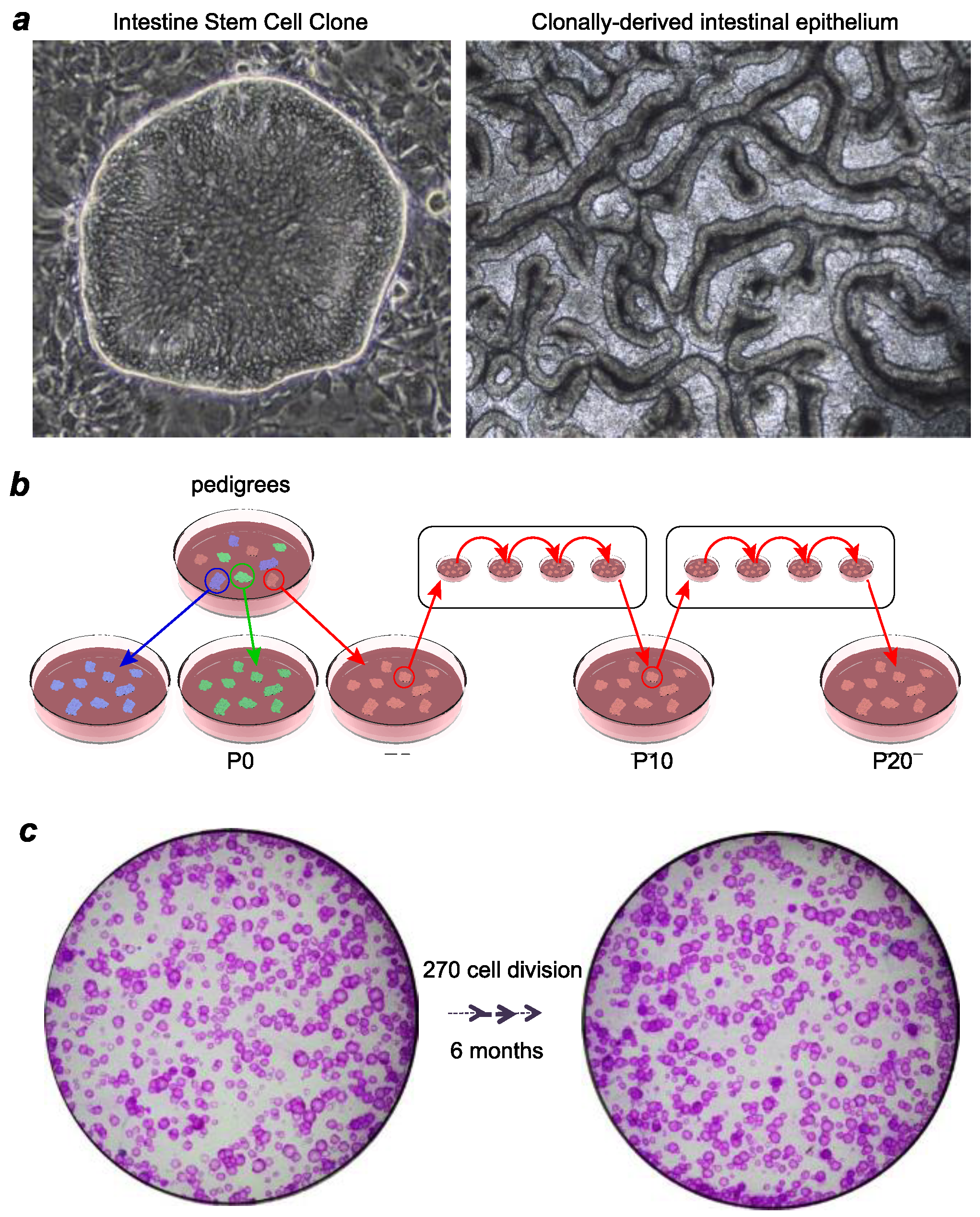
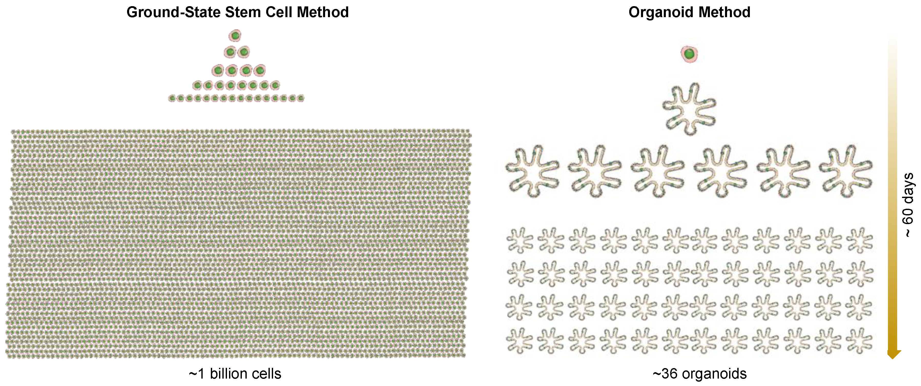
3. Ground-State Stem Cells versus Organoids

Mammalian cells have been cultured in collagen and laminin-rich matrices as organoids in 3D culture since at least 1980 [12,31,32,33,34,35]. An organoid is defined as a unit of function of a given organ that is able to reproduce, in vitro, a biological structure similar in architecture and function to its counterpart in vivo. There are multiple origins of organoids that include a fragment of tissue, a stem cell isolated in an adult organ, an embryonic stem cell, or an induced pluripotent stem cell. Importantly, organoids can also be generated from cells that have lost stem cell capacity—so-called “transit amplifying cells” with limited proliferative potential—indicating that organoids per se are not strictly assays for stem cells [36].
Despite fundamental advances in stem cells afforded by organoid technology [37], the organoid method does not support cloning the intestinal stem cells or any other adult stem cells in their highly immature, clonogenic state. We solved the problem of culturing populations of pure stem cells of the human gastrointestinal tract using a cocktail of factors impacting Wnt, Notch, and TGF-β signaling pathways among others and were able to clone ground-state stem cells from all regions of the intestine and colon from endoscopic biopsies [15,17]. In other words, the stem cells of columnar epithelia can now be captured in the same ground-state that Green showed for the stem cells of stratified epithelia such as the epidermis. We found the following properties of these ground-state stem cells that make this technology particularly advantageous compared with the organoid method: (1) a typical 1 mm endoscopic biopsy yields 100–300 independent stem cell clones; (2) each of these clones can be independently propagated while maintaining genomic integrity; (3) these clones uniformly express stem cell markers and have a clonogenicity rate of 60–90%; (4) each of these clones retain epigenetic memory of the region from which they were derived; and (5) single clones can be differentiated to yield 3-D intestinal epithelia with regionally-appropriate cell types.
It should be acknowledged that many of the features of these gastrointestinal stem cells were unknown prior to Wang et al. [15], Yamamoto et al. [16] and Duleba et al. [17], including the stability of the epigenetic programs underlying commitment, despite months of continuous in vitro propagation, the fact that these stem cells possess all of the information required to assemble into a higher order, and that regio-specific 3-D epithelia are nearly identical to those found in the mature intestine in vivo. As stem cells comprise only a minor component of organoids, perhaps less than 1% [38], the molecular features of the stem cells of columnar epithelia, such as the intestinal tract, have remained unclear (Figure 2). Therefore, the selective cloning and proliferative expansion of highly clonogenic, ground-state intestinal stem cells through the Xian–McKeon method offer a first glimpse into the molecular properties of these cells. Our recent success in adapting this technology to clone stem cells in precancerous lesions, such as Barrett’s esophagus, addressed the long-standing confusion in the cellular origin of Barrett’s esophagus. Furthermore, using this technology, we generated broad sets of patient-matched stem cells corresponding to all stages of precursor lesions both in-line and out-line with the progression to adenocarcinoma. We can anticipate that each of these clones can be highly annotated with genomics, gene expression and differentiation fate information, accessible through a dynamic biorepository and thus be valuable substrates for new investigations across the cancer biology community.

4. Biobanking of Ground-State Stem Cells and Personalized Regenerative Medicine

The onset of adult-stem-cell-based regenerative medicine started in the 1980s. Green and colleagues demonstrated the first example of cell therapy using cultured stem cells. They showed that human epidermis could be grown in the laboratory and transplanted to patients to reconstitute a functional epidermis [39,40]. Since then, transplantation of cultured epidermal stem cells has long been used to treat patients with burns, chronic wounds and stable vitiligo [41]. This is a life-saving procedure for patients with large area of burns. Moreover, the long-term effectiveness and safety of genetically-modified epidermal stem cells in correcting the severe skin blistering disease, epidermolysis bullosa, has been shown clinically [42].
In addition to skin, cultured stem cells from other epithelial tissues can be the source of stem-cell-based regenerative medicine. For example, a feature of lung regeneration that bodes well for regenerative medicine is that the underlying stem cell is highly clonogenic, which shows unlimited expansion capacity in vitro, and readily transplants to form functional alveoli in acutely damaged lungs [18]. We showed that a single p63+/Krt5+ DASC can be cloned, expanded and transplanted via intratracheal delivery to acutely damaged lungs, where they selectively inhabit damaged regions and differentiate to form Clara cells and alveoli composed of type I and type II pneumocytes. Importantly, these same p63+/Krt5+ DASCs showed no incorporation in mice without prior acute lung injury, suggesting that the efficient regenerative properties of these cells are not marred by “off-target” incorporation. Lastly, DASCs are readily cloneable from simple bronchoscopic biopsies, from bronchopulmonary lavage, or from transmural biopsies, providing good sources of autonomous stem cells that can be expanded to hundreds of billions of cells in weeks. Taken together, the established properties of DASCs, including clonogenicity, expandability, and facility for accurate transplantation obviate many theoretical objections that could have limited their use in regenerative medicine for either acute or chronic lung diseases.
It is also conceivable that the newfound ability to derive ground-state stem cells from columnar epithelium provides excellent sources for autologous transplantation to treat a wide range of disorders that current treatments are not able to help. For example, cultured ground-state intestinal stem cells may be of use to restore the intestinal epithelial functions following autologous transplantation in patients with severe forms of short bowel syndrome (SBS) [43], congenital disorders [44], or inflammatory bowel disease (IBD) [45,46].
In conclusion, we now have the technologies for cloning and culturing adult stem cells from nearly all types of epithelial tissues. Given the tremendous success of the pioneering work by Green and colleagues on the use of cultured adult stem cells in regenerative medicine, we should anticipate that more scientists and clinicians will recognize the potential of adult stem cells, appreciate the potential of biobanking various types of adult stem cells from individuals of diverse HLA haplotypes, and make attempts to use them for stem-cell-based personalized regenerative medicine.

Author Contributions

Conceptualization, W.X., M.V. and F.M.; Writing-Original Draft Preparation, W.X., M.D., and F.M; Writing-Review and Editing, W.X., Y.Y., M.V., M.D., and F.M.; Supervision, W.X. and F.M.; Funding Acquisition, W.X. and F.M.

Funding

This work was supported by grants from the Cancer Prevention Research Institute of Texas (CPRIT; RR15014 to WX and RR15088 to FM), the National Institutes of Health (1R01DK115445-01A1 to WX, and U24CA228550 to FM), the US Dept. of Defense (W81XWH-17-1-0123 to WX), the University of Texas Presidential Award (to WX) and the American Gastroenterology Association Research and Development Pilot Award in Technology (to WX).

Acknowledgments

We thank all the members in the Xian–McKeon laboratory for helpful discussions and support. We thank H. Green for advice and support.

Conflicts of Interest

W.X., F.M., M.D. and M.V. have filed a patent related to the technology used in the present work.

References

  1. Müller, A.M.; Dzierzak, E.A. ES cells have only a limited lymphopoietic potential after adoptive transfer into mouse recipients. Development 1993, 118, 1343–1351. Available online: http://www.ncbi.nlm.nih.gov/pubmed/8269860 (accessed on 11 November 2018).
  2. Helgason, C.D.; Sauvageau, G.; Lawrence, H.J.; Largman, C.; Humphries, R.K. Overexpression of HOXB4 enhances the hematopoietic potential of embryonic stem cells differentiated in vitro. Blood 1996, 87, 2740–2749. Available online: http://www.ncbi.nlm.nih.gov/pubmed/8639890 (accessed on 11 November 2018).
  3. Bonde, S.; Dowden, A.M.; Chan, K.-M.; Tabayoyong, W.B.; Zavazava, N. HOXB4 But Not BMP4 Confers Self-Renewal Properties to ES-Derived Hematopoietic Progenitor Cells. Transplantation 2008, 86, 1803–1809. [Google Scholar] [CrossRef] [PubMed] [Green Version]
  4. Iuchi, S.; Dabelsteen, S.; Easley, K.; Rheinwald, J.G.; Green, H. Immortalized keratinocyte lines derived from human embryonic stem cells. Proc. Natl. Acad. Sci. USA 2006, 103, 1792–1797. [Google Scholar] [CrossRef] [PubMed] [Green Version]
  5. Amabile, G.; Welner, R.S.; Nombela-Arrieta, C.; D’Alise, A.M.; di Ruscio, A.; Ebralidze, A.K.; Kraytsberg, Y.; Ye, M.; Kocher, O.; Neuberg, D.S.; et al. In vivo generation of transplantable human hematopoietic cells from induced pluripotent stem cells. Blood 2013, 121, 1255–1264. [Google Scholar] [CrossRef] [PubMed]
  6. Suzuki, N.; Yamazaki, S.; Yamaguchi, T.; Okabe, M.; Masaki, H.; Takaki, S.; Otsu, M.; Nakauchi, H. Generation of engraftable hematopoietic stem cells from induced pluripotent stem cells by way of teratoma formation., Molecular Therapy. J. Am. Soc. Gene Ther. 2013, 21, 1424–1431. [Google Scholar] [CrossRef] [PubMed]
  7. Rheinwald, J.G.; Green, H. Serial cultivation of strains of human epidermal keratinocytes: The formation of keratinizing colonies from single cells. Cell. 1975, 6, 331–343. Available online: http://www.ncbi.nlm.nih.gov/pubmed/1052771 (accessed on 11 November 2018). [CrossRef]
  8. Rama, P.; Matuska, S.; Paganoni, G.; Spinelli, A.; de Luca, M.; Pellegrini, G. Limbal Stem-Cell Therapy and Long-Term Corneal Regeneration. N. Engl. J. Med. 2010, 363, 147–155. [Google Scholar] [CrossRef] [PubMed]
  9. Senoo, M.; Pinto, F.; Crum, C.P.; McKeon, F. p63 Is Essential for the Proliferative Potential of Stem Cells in Stratified Epithelia. Cell 2007, 129, 523–536. [Google Scholar] [CrossRef] [Green Version]
  10. Kumar, P.A.; Hu, Y.; Yamamoto, Y.; Hoe, N.B.; Wei, T.S.; Mu, D.; Sun, Y.; Joo, L.S.; Dagher, R.; Zielonka, E.M.; et al. Distal Airway Stem Cells Yield Alveoli In Vitro and during Lung Regeneration following H1N1 Influenza Infection. Cell 2011, 147, 525–538. [Google Scholar] [CrossRef] [Green Version]
  11. Matsuura, R.; Kogo, H.; Ogaeri, T.; Miwa, T.; Kuwahara, M.; Kanai, Y.; Nakagawa, T.; Kuroiwa, A.; Fujimoto, T.; Torihashi, S. Crucial Transcription Factors in Endoderm and Embryonic Gut Development Are Expressed in Gut-Like Structures from Mouse ES Cells. Stem Cells 2006, 24, 624–630. [Google Scholar] [CrossRef] [Green Version]
  12. Sato, T.; Vries, R.G.; Snippert, H.J.; van de Wetering, M.; Barker, N.; Stange, D.E.; van Es, J.H.; Abo, A.; Kujala, P.; Peters, P.J.; et al. Single Lgr5 stem cells build crypt-villus structures in vitro without a mesenchymal niche. Nature 2009, 459, 262–265. [Google Scholar] [CrossRef]
  13. Fordham, R.P.; Yui, S.; Hannan, N.R.F.; Soendergaard, C.; Madgwick, A.; Schweiger, P.J.; Nielsen, O.H.; Vallier, L.; Pedersen, R.A.; Nakamura, T.; et al. Transplantation of Expanded Fetal Intestinal Progenitors Contributes to Colon Regeneration after Injury. Cell Stem Cell 2013, 13, 734–744. [Google Scholar] [CrossRef] [PubMed] [Green Version]
  14. Yin, X.; Farin, H.F.; van Es, J.H.; Clevers, H.; Langer, R.; Karp, J.M. Niche-independent high-purity cultures of Lgr5+ intestinal stem cells and their progeny. Nat. Methods 2014, 11, 106–112. [Google Scholar] [CrossRef] [PubMed]
  15. Wang, X.; Yamamoto, Y.; Wilson, L.H.; Zhang, T.; Howitt, B.E.; Farrow, M.A.; Kern, F.; Ning, G.; Hong, Y.; Khor, C.C.; et al. Cloning and variation of ground state intestinal stem cells. Nature 2015, 522, 173–178. [Google Scholar] [CrossRef] [PubMed] [Green Version]
  16. Yamamoto, Y.; Wang, X.; Bertrand, D.; Kern, F.; Zhang, T.; Duleba, M.; Srivastava, S.; Khor, C.C.; Hu, Y.; Wilson, L.H.; et al. Mutational spectrum of Barrett’s stem cells suggests paths to initiation of a precancerous lesion. Nat. Commun. 2016, 7, 10380. [Google Scholar] [CrossRef] [PubMed] [Green Version]
  17. Duleba, M.; Qi, Y.; Mahalingam, R.; Flynn, K.; Rinaldi, F.; Liew, A.-A.; Neupane, R.; Vincent, M.; Crum, C.P.; Ho, K.Y.; et al. An Efficient Method for Cloning Gastrointestinal Stem Cells from Patients via Endoscopic Biopsies. Gastroenterology 2018. [Google Scholar] [CrossRef] [PubMed]
  18. Zuo, W.; Zhang, T.; Wu, D.Z.; Guan, S.P.; Liew, A.-A.; Yamamoto, Y.; Wang, X.; Lim, S.J.; Vincent, M.; Lessard, M.; et al. p63(+)Krt5(+) distal airway stem cells are essential for lung regeneration. Nature 2015, 517, 616–620. [Google Scholar] [CrossRef] [PubMed]
  19. Llames, S.; García-Pérez, E.; Meana, Á.; Larcher, F.; del Río, M. Feeder Layer Cell Actions and Applications. Tissue Eng. Part B Rev. 2015, 21, 345–353. [Google Scholar] [CrossRef] [Green Version]
  20. Boyce, S.T.; Ham, R.G. Calcium-regulated differentiation of normal human epidermal keratinocytes in chemically defined clonal culture and serum-free serial culture. J. Investig. Dermatol. 1983, 81, 33s–40s. [Google Scholar] [CrossRef]
  21. de Corte, P.; Verween, G.; Verbeken, G.; Rose, T.; Jennes, S.; de Coninck, A.; Roseeuw, D.; Vanderkelen, A.; Kets, E.; Haddow, D.; et al. Feeder layer- and animal product-free culture of neonatal foreskin keratinocytes: Improved performance, usability, quality and safety. Cell Tissue Bank. 2012, 13, 175–189. [Google Scholar] [CrossRef]
  22. Lenihan, C.; Rogers, C.; Metcalfe, A.D.; Martin, Y.H. The effect of isolation and culture methods on epithelial stem cell populations and their progeny—Toward an improved cell expansion protocol for clinical application. Cytotherapy 2014, 16, 1750–1759. [Google Scholar] [CrossRef]
  23. de Luca, M.; Pellegrini, G.; Green, H. Regeneration of squamous epithelia from stem cells of cultured grafts. Regener. Med. 2006, 1, 45–57. [Google Scholar] [CrossRef] [PubMed]
  24. Green, H. The birth of therapy with cultured cells. BioEssays 2008, 30, 897–903. [Google Scholar] [CrossRef] [PubMed]
  25. Kim, K.-A.; Kakitani, M.; Zhao, J.; Oshima, T.; Tang, T.; Binnerts, M.; Liu, Y.; Boyle, B.; Park, E.; Emtage, P.; et al. Mitogenic Influence of Human R-Spondin1 on the Intestinal Epithelium. Science 2005, 309, 1256–1259. [Google Scholar] [CrossRef] [PubMed]
  26. Dreesen, O.; Brivanlou, A.H. Signaling pathways in cancer and embryonic stem cells. Stem Cell Rev. 2007, 3, 7–17. Available online: http://www.ncbi.nlm.nih.gov/pubmed/17873377 (accessed on 11 November 2018). [CrossRef] [PubMed]
  27. Rock, J.R.; Onaitis, M.W.; Rawlins, E.L.; Lu, Y.; Clark, C.P.; Xue, Y.; Randell, S.H.; Hogan, B.L.M. Basal cells as stem cells of the mouse trachea and human airway epithelium. Proc. Natl. Acad. Sci. USA 2009, 106, 12771–12775. [Google Scholar] [CrossRef] [PubMed] [Green Version]
  28. International Stem Cell Initiative; Amps, K.; Andrews, P.W.; Anyfantis, G.; Armstrong, L.; Avery, S.; Baharvand, H.; Baker, J.; Baker, D.; Munoz, M.B.; et al. Screening ethnically diverse human embryonic stem cells identifies a chromosome 20 minimal amplicon conferring growth advantage. Nat. Biotechnol. 2011, 29, 1132–1144. [Google Scholar] [CrossRef] [PubMed] [Green Version]
  29. Avery, S.; Hirst, A.J.; Baker, D.; Lim, C.Y.; Alagaratnam, S.; Skotheim, R.I.; Lothe, R.A.; Pera, M.F.; Colman, A.; Robson, P.; et al. BCL-XL mediates the strong selective advantage of a 20q11.21 amplification commonly found in human embryonic stem cell cultures. Stem Cell Rep. 2013, 1, 379–386. [Google Scholar] [CrossRef]
  30. Dickson, M.A.; Hahn, W.C.; Ino, Y.; Ronfard, V.; Wu, J.Y.; Weinberg, R.A.; Louis, D.N.; Li, F.P.; Rheinwald, J.G. Human keratinocytes that express hTERT and also bypass a p16(INK4a)-enforced mechanism that limits life span become immortal yet retain normal growth and differentiation characteristics. Mol. Cell. Biol. 2000, 20, 1436–1447. Available online: http://www.ncbi.nlm.nih.gov/pubmed/10648628 (accessed on 11 November 2018). [CrossRef]
  31. Lee, G.; Hynes, R.; Kirschner, M. Temporal and spatial regulation of fibronectin in early Xenopus development. Cell 1984, 36, 729–740. Available online: http://www.ncbi.nlm.nih.gov/pubmed/6697394 (accessed on 11 November 2018).
  32. Bissell, D.M.; Arenson, D.M.; Maher, J.J.; Roll, F.J. Support of cultured hepatocytes by a laminin-rich gel. Evidence for a functionally significant subendothelial matrix in normal rat liver. J. Clin. Investig. 1987, 79, 801–812. [Google Scholar] [CrossRef]
  33. Barcellos-Hoff, M.H.; Aggeler, J.; Ram, T.G.; Bissell, M.J. Functional differentiation and alveolar morphogenesis of primary mammary cultures on reconstituted basement membrane. Development 1989, 105, 223–235. [Google Scholar]
  34. Petersen, O.W.; Rønnov-Jessen, L.; Howlett, A.R.; Bissell, M.J. Interaction with basement membrane serves to rapidly distinguish growth and differentiation pattern of normal and malignant human breast epithelial cells. Proc. Natl. Acad. Sci. USA 1992, 89, 9064–9068. [Google Scholar] [CrossRef] [PubMed]
  35. Broeckx, S.Y.; Maes, S.; Martinello, T.; Aerts, D.; Chiers, K.; Mariën, T.; Patruno, M.; Franco-Obregón, A.; Spaas, J.H. Equine Epidermis: A Source of Epithelial-Like Stem/Progenitor Cells with In Vitro and In Vivo Regenerative Capacities. Stem Cells Dev. 2014, 23, 1134–1148. [Google Scholar] [CrossRef] [PubMed] [Green Version]
  36. Simian, M.; Bissell, M.J. Organoids: A historical perspective of thinking in three dimensions. J. Cell Biol. 2017, 216, 31–40. [Google Scholar] [CrossRef] [PubMed]
  37. Ootani, A.; Li, X.; Sangiorgi, E.; Ho, Q.T.; Ueno, H.; Toda, S.; Sugihara, H.; Fujimoto, K.; Weissman, I.L.; Capecchi, M.R.; et al. Sustained in vitro intestinal epithelial culture within a Wnt-dependent stem cell niche. Nat. Med. 2009, 15, 701–706. [Google Scholar] [CrossRef] [PubMed] [Green Version]
  38. Wang, F.; Scoville, D.; He, X.C.; Mahe, M.M.; Box, A.; Perry, J.M.; Smith, N.R.; Lei, N.Y.; Davies, P.S.; Fuller, M.K.; et al. Isolation and Characterization of Intestinal Stem Cells Based on Surface Marker Combinations and Colony-Formation Assay. Gastroenterology 2013, 145, 383–395. [Google Scholar] [CrossRef] [PubMed]
  39. Green, H.; Kehinde, O.; Thomas, J. Growth of cultured human epidermal cells into multiple epithelia suitable for grafting. Proc. Natl. Acad. Sci. USA 1979, 76, 5665–5668. Available online: http://www.ncbi.nlm.nih.gov/pubmed/293669 (accessed on 11 November 2018). [CrossRef] [PubMed]
  40. O’Connor, N.; Mulliken, J.; Banks-Schlegel, S.; Kehinde, O.; Green, H. Grafting of Burns with Cultured Epithelium Prepared from Autologous Epidermal Cells. Lancet 1981, 317, 75–78. [Google Scholar] [CrossRef]
  41. Gallico, G.G.; O’Connor, N.E.; Compton, C.C.; Kehinde, O.; Green, H. Permanent Coverage of Large Burn Wounds with Autologous Cultured Human Epithelium. N. Engl. J. Med. 1984, 311, 448–451. [Google Scholar] [CrossRef]
  42. Hirsch, T.; Rothoeft, T.; Teig, N.; Bauer, J.W.; Pellegrini, G.; de Rosa, L.; Scaglione, D.; Reichelt, J.; Klausegger, A.; Kneisz, D.; et al. Regeneration of the entire human epidermis using transgenic stem cells. Nature 2017, 551, 327–332. [Google Scholar] [CrossRef]
  43. Buchman, A.L.; Scolapio, J.; Fryer, J. AGA technical review on short bowel syndrome and intestinal transplantation. Gastroenterology 2003, 124, 1111–1134. [Google Scholar] [CrossRef]
  44. Hong, S.N.; Dunn, J.C.Y.; Stelzner, M.; Martín, M.G. Concise Review: The Potential Use of Intestinal Stem Cells to Treat Patients with Intestinal Failure. Stem Cells Transl. Med. 2017, 6, 666–676. [Google Scholar] [CrossRef] [PubMed]
  45. Salim, S.Y.; Söderholm, J.D. Importance of disrupted intestinal barrier in inflammatory bowel diseases. Inflamm. Bowel Dis. 2011, 17, 362–381. [Google Scholar] [CrossRef] [PubMed]
  46. Martini, E.; Krug, S.M.; Siegmund, B.; Neurath, M.F.; Becker, C. Mend Your Fences: The Epithelial Barrier and its Relationship With Mucosal Immunity in Inflammatory Bowel Disease. Cell. Mol. Gastroenterol. Hepatol. 2017, 4, 33–46. [Google Scholar] [CrossRef] [PubMed]
Figure 1. Xian–McKeon method of cloning ground-state intestinal stem cells. (a) Right, Representative image of a single-cell derived ISCGS colony. Left, Top view of in vitro intestinal epithelium generated from stem cells of a pedigree of ISCGS differentiated in an air–liquid interface setting. (b) Schematic diagram of establishing pedigrees from ISCGS. Long-term culturing does not affect the capacity of ISCGS to form single-cell derived pedigrees. (c) Clonogenicity assay revealing nearly unchanged number of Rhodamine red-stained colonies despite long-term culturing.
Jcm 07 00555 g001
Figure 2. Rapid expansion of a single cell to one billion cells in approximately 60 days using the Xian–McKeon method. In comparison, one cell can become 36 organoids in the organoid method.
Jcm 07 00555 g002

