Article

Distinct Expression and Clinical Significance of Zinc Finger AN-1-Type Containing 4 in Oral Squamous Cell Carcinomas

by
Julián Suárez-Canto
1,
Faustino Julián Suárez-Sánchez
2,
Francisco Domínguez-Iglesias
2,
Gonzalo Hernández-Vallejo
3,
Juana M. García-Pedrero
4,5,6,* and
Juan C. De Vicente
5,7,8,*
1
395 Los Prados Cabueñes, 33394 Gijón, Asturias, Spain
2
Department of Pathology, Hospital Universitario de Cabueñes, 395 Los Prados Cabueñes, 33394 Gijón, Asturias, Spain
3
School of Dentristry, Complutense University of Madrid, Pza. Ramón y Cajal, s/n, Ciudad Universitaria, 28040 Madrid, Spain
4
Department of Otolaryngology, Hospital Universitario Central de Asturias (HUCA), C/Carretera de Rubín, s/n, 33011 Oviedo, Asturias, Spain
5
Instituto de Investigación Sanitaria del Principado de Asturias (ISPA), Instituto Universitario de Oncología del Principado de Asturias (IUOPA), Universidad de Oviedo, C/Carretera de Rubín, s/n, 33011 Oviedo, Asturias, Spain
6
Ciber de Cáncer (CIBERONC), Instituto de Salud Carlos III, Av. Monforte de Lemos, 3-5, 28029 Madrid, Spain
7
Department of Oral and Maxillofacial Surgery, Hospital Universitario Central de Asturias (HUCA), C/Carretera de Rubín, s/n, 33011 Oviedo, Asturias, Spain
8
Department of Surgery, University of Oviedo, Av. Julián Clavería, 6, 33006 Oviedo, Asturias, Spain
*
Authors to whom correspondence should be addressed.
J. Clin. Med. 2018, 7(12), 534; https://doi.org/10.3390/jcm7120534
Submission received: 27 October 2018 / Revised: 12 November 2018 / Accepted: 7 December 2018 / Published: 10 December 2018

Abstract

:
Zinc finger AN1-type containing 4 (ZFAND4) has emerged as a promising prognostic marker and predictor of metastasis for patients with oral squamous cell carcinoma (OSCC). However, further validation is fundamental before clinical implementation. Hence, this study evaluated the expression pattern of ZFAND4 protein expression by immunohistochemistry using an independent cohort of 125 patients with OSCC, and correlations with the clinicopathologic parameters and disease outcome. Remarkably, ZFAND4 expression, while negligible in normal epithelium, exhibited two distinct expression patterns in tumors that did not overlap. A gross granular staining was characteristic of the undifferentiated cells at the invasive front of tumors, whereas the most differentiated cells located at the center of the tumor nests showed diffuse non-granular staining. ZFAND4 staining was higher in undifferentiated than in differentiated areas of tumors. High ZFAND4 expression in differentiated cells was significantly associated to well-differentiated (p = 0.04) and non-recurrent tumors (p = 0.04), whereas ZFAND4 expression in undifferentiated cells correlated with tumor location (p = 0.005). No correlations between the ZFAND4 expression and patient survival were found. These data question the clinical relevance of ZFAND4 expression as a prognostic biomarker in OSCC, and also reveal distinct ZFAND4 expression patterns depending on the differentiation areas of tumors that should be evaluated separately.

1. Introduction

Head and neck squamous cell carcinoma that includes, among others, oral squamous cell carcinoma (OSCC) is the sixth most common cancer in the world, with an annual prevalence of nearly 600,000 new cases worldwide [1,2]. It is generally accepted that OSCC initiates and progresses through a series of multiple genetic alterations caused by chronic exposure to carcinogens, such as alcohol, smoking, and human papilloma virus [3]. Multiple genetic and molecular studies have improved our understanding of the molecular basis of this disease. Indeed, several cellular signaling pathways have been found dysregulated in these tumors through genetic and epigenetic alterations. However, despite major advances in diagnosis and treatment, the survival rate of patients with OSCC has modestly improved over the past 40 years, and it remains at approximately 50% [4].
Sasahira et al. [5] investigated the transcriptional profiles of primary and recurrent OSCC, and found that one of the most upregulated genes identified in recurrent OSCC was zinc finger AN1-type containing 4 (ZFAND4), also known as AN1 and ubiquitin-like homolog (ANUBL1). Although the functional role of ZFAND4 in cancer is still unknown, Kurihara-Shimomura et al. [6] evaluated its prognostic utility in OSCC. They concluded that ZFAND4 could be a useful marker for predicting metastasis and poor prognosis in patients with OSCC. In addition, Tang et al. [7] demonstrated that ZFAND4 expression is upregulated in gastric cancer and positively associated with the grading of this disease.
In the light of these data, ZFAND4 emerges as a promising prognostic biomarker; however, further validation in independent study cohorts is fundamental for implementation to the clinic. Therefore, the objective of this study was to investigate the expression pattern and clinical relevance of ZFAND4 protein expression using an independent cohort of 125 patients with OSCC, and to establish correlations with the clinicopathologic parameters and disease outcome.

2. Experimental Section

2.1. Patients and Tissue Specimens

A retrospective study was designed. Surgical tissue specimens from 125 patients with OSCC who underwent surgical treatment with curative purposes at the Hospital Universitario Central de Asturias between 1996 and 2007 were retrospectively collected, in accordance to approved institutional review board guidelines. All experimental procedures were conducted in accordance to the Declaration of Helsinki and approved by the Institutional Ethics Committee of the Hospital Universitario Central de Asturias and by the Regional CEIC from Principado de Asturias. Informed consent was obtained from all patients. Clinicopathologic data were collected from medical records. Tissue specimens were obtained from the Biobanco del Principado de Asturias, and representative tissue sections from archival, formalin-fixed paraffin-embedded blocks.

2.2. Tissue Microarray Construction

The original archived hematoxylin- and eosin-stained slides were reviewed by an experienced pathologist (FDI) to confirm histological diagnosis. Three representative tissue cores (1 mm diameter) were selected from each tumor block, and transferred to a recipient ‘Master’ block in a grid-like manner using a manual tissue microarray instrument. In addition, each tissue microarray also contained three cores of normal epithelium as an internal control. A section from each microarray was stained with hematoxylin and eosin, and examined by light microscopy to check the adequacy of tissue sampling.

2.3. Immunohistochemistry (IHC)

TMA sections (4 μm) were cut and dried and dried on Flex IHC microscope slides (Dako). The sections were deparaffinized with standard xylene, hydrated through graded alcohols into water, and pretreated by hydrogen peroxide to quench the endogenous peroxidase activity. Antigen retrieval was performed using Envision Flex Target Retrieval solution (Dako), at room temperature on an automatic staining workstation (DakoAutostainer Plus, Dako, Glostrup, Denmark). Staining was carried out at room temperature on an automatic staining workstation (Dako Autostainer Plus) with anti-ZFAND4 antibody (Atlas Antibodies, Stockholm, Sweden) diluted to 0.5 Nµg/mL using the Dako EnVision detection system (Dako, Glostrup, Denmark). Sections were counterstained with hematoxylin, dehydrated with ethanol, and permanently coverslipped. For negative control purposes, DakoCytomation mouse serum diluted at the same concentration as the primary antibody was used.
Staining was scored blinded to clinical data by two independent observers. ZFAND4 expression was evaluated according to the percentage of stained tumor cells and the staining intensity using the Allred score, as previously described [8]. The proportion of ZFAND4-positive cells was evaluated in both undifferentiated and differentiated areas of the tumors. In all these groups, proportional scores were categorized as: 0, no cells were stained; 1, 1/100 cells were stained; 2, 1/10 cells were stained; 4, 2/3 cells were stained; 5, all cells were stained. Staining intensity was scored as: 0, negative; 1, weak; 2, intermediate; and 3, strong. The total score was calculated by the sum of the proportional and intensity scores, ranging from 0 to 8. Similar to Kurihara-Shimomura et al. [6], the optimal cut-off score for ZFAND4 expression was selected using the receiver operating characteristic (ROC) curve according to the survival status. ZFAND4 staining was independently evaluated in undifferentiated areas of tumors mainly located at the invasive front, and in differentiated areas at the center of the tumor islands.

2.4. Statistical Analysis

χ2 and the Fisher’s exact test were used for comparison between categorical variables. Disease-specific survival (DSS) was determined for the date of treatment completion to death for the tumor. For time-to-event analysis, survival curves were plotted using the Kaplan-Meier method. Differences between survival times were analyzed by the log-rank test. Hazard ratios (HR) with their 95% confidence intervals (CI) for clinicopathologic variables were calculated using the univariate Cox proportional hazards model analysis. All tests were two-sided and p values less than 0.05 were considered statistically significant. All statistical analyses were performed using SPSS version 21 (IBM Co., Armonk, NY, USA).

3. Results

3.1. Patient Characteristics

The cohort of 125 OSCC patients was composed of 82 men and 43 women, ranging from 28 to 91 years, with a median age of 57 years. Forty-one patients (33%) were never-smokers and 56 (45%) never-drinkers. The main clinicopathologic characteristics are summarized in Table 1. Forty-nine cases (39%) showed neck lymph node metastasis, more than 50% were well-differentiated tumors and advanced clinical stages (III or IV), and the most common site was the tongue (41%) followed by the floor of the mouth (30%). Adjuvant radiotherapy was administered to 75 patients (60%), and adjuvant chemotherapy to 14 patients (11.2%). Fifty-four cases (43%) showed loco-regional recurrence, and 19 (15%) suffered from a second primary carcinoma. Over a median follow-up of 61 months (range, 1 to 230 months) 53 deaths occurred.

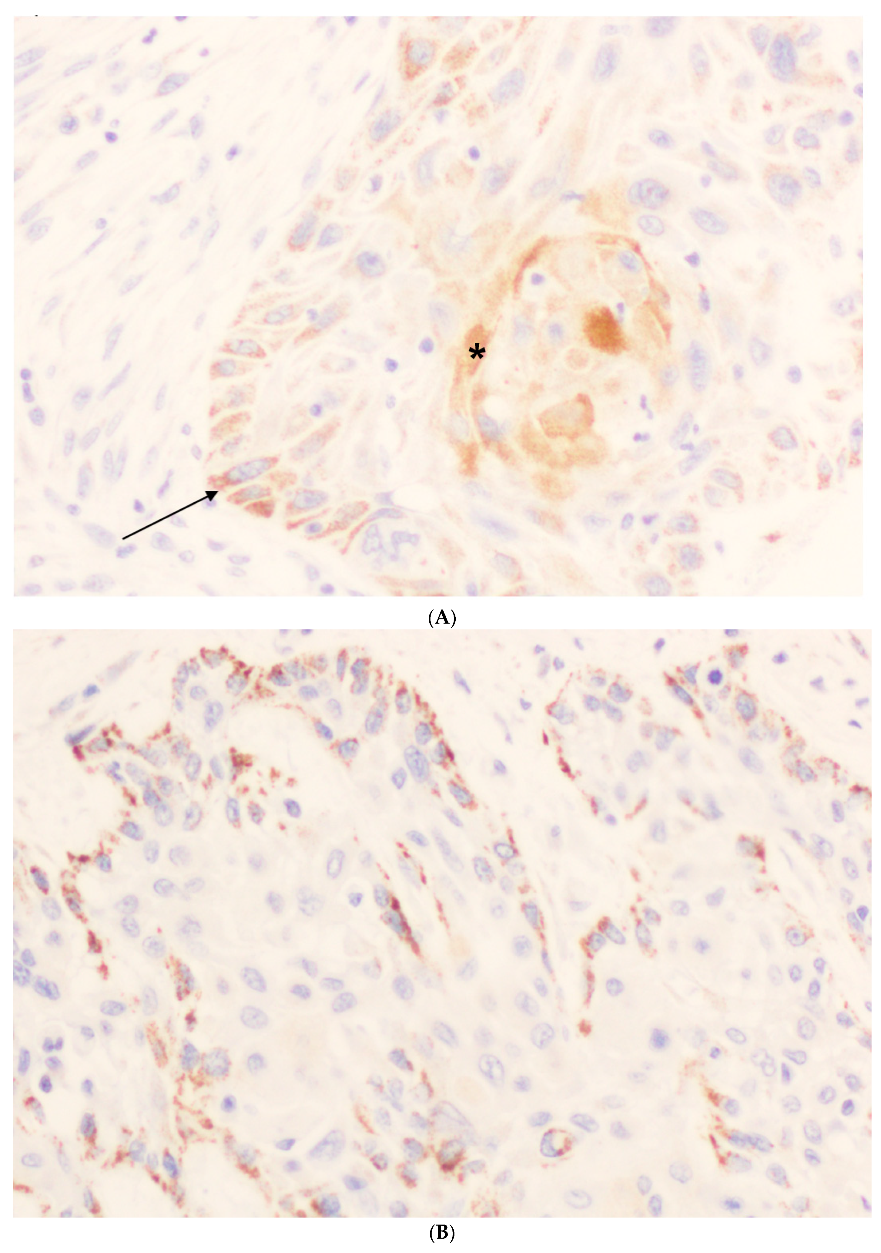
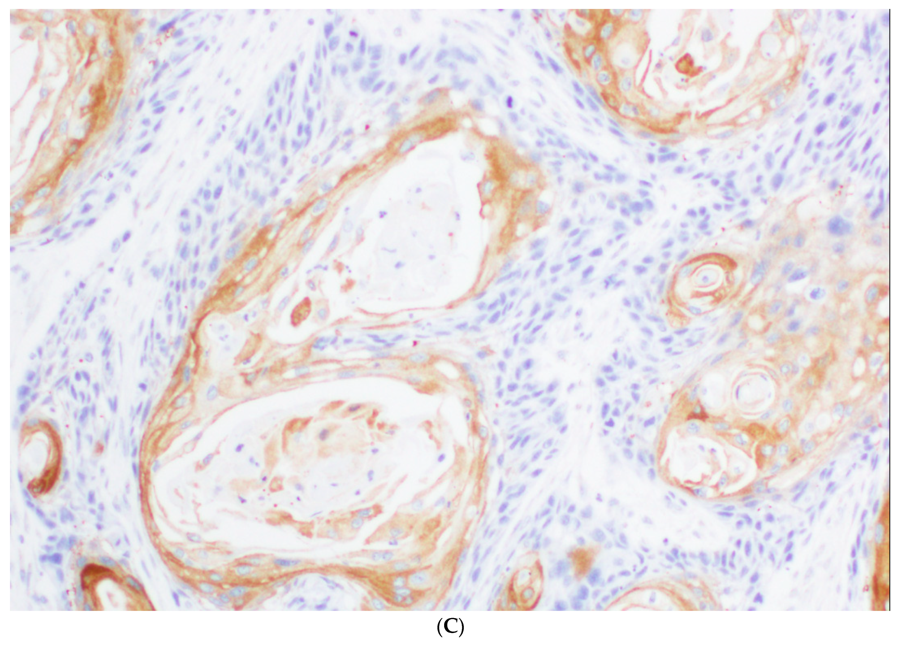
3.2. Immunohistochemical Analysis of ZFAND4 Expression in OSCC Tissue Specimens

ZFAND4 staining was not valuable in two (1.6%) of 125 OSCC specimens. While ZFAND4 expression was negligible in normal epithelium, two distinct expression patterns were noted in the tumors that did not overlap in any of the samples (Figure 1). A gross granular staining was characteristic of the undifferentiated cells at the invasive front of tumors, whereas the most differentiated cells located at the center of the tumor nests showed diffuse non-granular staining. The mean percentages of positive ZFAND4 staining were 44.98 (standard deviation –SD-35.38) in undifferentiated cells and 17.18 (SD, 20.61) in differentiated cells. ZFAND4 staining intensity was also evaluated in both undifferentiated and differentiated areas. In undifferentiated cells, there were 13 (11%) negative cases, 37 (30%) weak, 43 (35%) intermediate, and 30 (24%) cases with strong staining. In differentiated cells, 47 (38%) cases were scored negative, 11 (9%) weak, 47 (38%) intermediate, and 18 (15%) had strong staining. Since each tumor was represented by three different tissue cores in the OSCC TMAs, the percentages of stained cells frequently varied in the three tumor areas assessed. Taking this into consideration, the Allred score was determined in two different ways: Either considering the maximum value of ZFAND4 positivity or the mean value of the three tumor cores. Regarding the intensity of immunostaining, the maximum value was always used for all calculations. Finally, the total score was calculated by the sum of the percentages of staining and intensity scores. The resulting indexes ranged between 0 and 8. The receiver operating characteristics (ROC) curve was used to determine the best cut-off score to predict patients’ survival, and this value was 4. Accordingly, those cases with an Allred score above 4 were considered as high ZFAND4 expression.

3.3. Associations of ZFAND4 with Clinicopathologic Characteristics

We next assessed the correlations of high ZFAND4 expression with the clinical data. Table 2 shows the associations of high ZFAND4 expression determined by using the maximum value of the percentage of stained cells to calculate the Allred score. In differentiated areas, high ZFAND4 expression was significantly associated with well-differentiated (p = 0.04) and non-recurrent tumors (p = 0.04), whereas ZFAND4 expression in undifferentiated cells was significantly correlated with tumor location in the tongue (p = 0.005).
On the other hand, when the Allred score was calculated using the mean value of percentage of stained cells (Table 3), ZFAND4 expression in differentiated cells was found to be significantly associated with N status (p = 0.02), being more frequently detected in pN0 and pN1 cases compared to pN2. However, no significant relationship was found between ZFAND4 expression in undifferentiated cells and any clinicopathologic variable.

3.4. ZFAND4 Expression and Patients’ Survival

Over a median follow-up period of 61 months, 27 patients (42.1%) harboring high ZFAND4 expression in undifferentiated cells calculated by using the mean Allred score died due to the index cancer, and 15 patients (41.6%) with high ZFAND4 expression in the differentiated cells. When the maximum Allred score was used, 31 patients (40.7%) with high ZFAND4 expression in the undifferentiated cells, and 20 patients (36.3%) with high ZFAND4 expression in the differentiated cells died due to the index cancer. Kaplan-Meier analysis showed that there were no statistically significant differences in disease-specific survival (DSS) between patients with high versus low ZFAND4 expression in either differentiated or undifferentiated cells (Table 4).

4. Discussion

This study aimed to investigate the clinical relevance and prognostic significance of ZFAND4 in OSCC. The prevalence of OSCC is estimated at 264,000 cases and 128,000 deaths annually worldwide [3]. Since the completion of the Human Genome Project in 2003, the subsequent progress in understanding the biology of cancer has led to the development of personalized therapies based on the patient’s unique molecular and genetic profile to target defective signaling pathways of tumor cells. It is generally accepted that OSCC arises from multiple genetic alterations, although the molecular basis of carcinogenesis is not fully understood. DNA sequencing technologies coupled with advances in algorithms have enormously contributed to the molecular and functional characterization of mutations, genes, and pathways altered in multiple cancers, including OSCC [9,10,11,12]. Furthermore, the majority of tumors showed alterations in multiple targetable genes that are candidates for combination therapy [11]. It is of paramount importance to identify molecular alterations involved in the development of recurrent and metastatic disease, which remains the main cause of morbidity and mortality in OSCC patients.
In a recent paper, Sasahira et al. [5] conducted a cDNA microarray analysis in order to compare the gene expression profile of primary and recurrent OSCC. Ten genes were found to be upregulated in recurrent OSCC compared with the primary tumors. Among these genes, ZFAND4 showed a 100-fold recurrent/primary, thus suggesting a possible role for this gene in tumorigenesis. Tang et al. [7] reported that ZFAND4 expression was consistently highly expressed in gastric cancer compared to normal tissue, and positively associated with increased stage. Functionally, ZFAND4 was found to downregulate the expression of the anti-proliferative miRNAs, miR-148b, miR-375, and miR-182, in SGC-7901 cells, thereby promoting cell proliferation by activation of cyclin-dependent kinase and downregulation of p21 and p53 [7], which supports the notion that ZFAND4 may act as an oncogene in gastric cancer.
In this study, we assumed the same methodology used by Kurihara-Shimomura et al. [6] in order to validate their results in an independent cohort of OSCC patients, and more importantly, the utility of ZFAND4 as a predictor of metastasis and poor prognostic marker. Interestingly, ZFAND4 staining consistently showed distinct expression patterns and distribution in our cohort of 125 OSCC, i.e., granular staining in undifferentiated areas and diffuse staining in differentiated areas of the tumors. These two expression patterns were analyzed separately to evaluate possible correlations with the clinical and follow-up data. Moreover, Allred scores were calculated using both maximum value and mean value of ZFAND4-positive cells for the three tissue cores selected from each tumor.
We found that ZFAND4 staining was higher in undifferentiated than in differentiated areas of tumors. However, ZFAND4 expression in differentiated cells showed the most relevant and significant associations with well-differentiated (p = 0.04) and non-recurrent tumors (p = 0.04). Nevertheless, ZFAND4 expression did not show a major impact on patient survival. Only a trend was observed between low ZFAND4 expression in differentiated cells and tumor-associated deaths (Table 4). Therefore, the prognostic relevance of ZFAND4 described by Kurihara-Shimomura et al. [6] has not been replicated in our series. Furthermore, Kurihara-Shimomura et al. [6] reported that ZFAND4 is essential for distant metastasis in OSCC, and hypothesized that ZFAND4 could facilitate metastasis to the lymph nodes and distant organs by promoting angiogenesis and/or lymphangiogenesis. In marked contrast, our results only proved a marginal (if any) relationship between ZFAND4 expression and the presence of lymph node metastasis. The limitations of our study are the retrospective design and the use of tissue microarrays to evaluate ZFAND4 immunostaining. Several factors could contribute to the discrepant results between these two studies. On one hand, etiological, clinical, and epidemiological differences in the patient cohorts as well as molecular/biological differences among tumors depending on the geographic areas. Moreover, ZFAND4 expression exhibited a highly heterogeneous pattern within the tumors depending on the differentiation status, which could certainly have a major contribution on the varying results. Particularly since we evaluated separately the clinical significance and correlations of the two distinct ZFAND4 expression patterns in undifferentiated and differentiated areas of tumors, while Kurihara-Shimomura et al. [6] did not distinguish expression patterns within the different tumor areas. Thus, positive ZFAND4 immunostaining was scored in the whole tumor as the diffuse staining in tumor islands, cords, or sheets, irrespective of the cell differentiation status and the grade of keratinization. Since ZFAND4 expression showed two distinct non-overlapping expression patterns depending on the differentiation areas of tumors, each with a clearly distinct clinical significance, evaluation of ZFAND4 expression in the whole tumor could therefore be misleading.

5. Conclusions

The herein presented data question the clinical relevance of ZFAND4 expression as a prognostic biomarker in OSCC, and also revealed distinct ZFAND4 expression patterns depending on the differentiation areas of tumors that should be evaluated separately. Further studies are necessary to fully elucidate the pathobiological role of ZFAND4 in OSCC and the potential clinical implications.

Author Contributions

Conceptualization, J.C.d.V.; Funding acquisition, J.C.d.V. and J.M.G.-P.; Investigation, J.S.C., F.J.S.S. and F.D.I.; Methodology, J.S.C., F.J.S.S.; Resources, F.D.I., G.H.V. and J.C.d.V.; Writing—original draft, J.C.d.V.; Writing—review & editing, J.M.G.-P.

Funding

This study was supported by grants from the Plan Nacional de I + D + I ISCIII PI16/00280 and CIBERONC (CB16/12/00390), the Instituto de Investigación Sanitaria del Principado de Asturias (ISPA), Fundación Bancaria Caja de Ahorros de Asturias-IUOPA and the FEDER Funding Program from the European Union.

Acknowledgments

We thank the samples and technical assistance kindly provided by the Principado de Asturias BioBank (PT13/0010/0046), financed jointly by Servicio de Salud del Principado de Asturias, Instituto de Salud Carlos III and Fundación Bancaria Cajastur and integrated in the Spanish National Biobanks Network. This study was supported by grants from the Plan Nacional de I + D + I ISCIII PI16/00280 and CIBERONC (CB16/12/00390), the Instituto de Investigación Sanitaria del Principado de Asturias (ISPA), Fundación Bancaria Caja de Ahorros de Asturias-IUOPA and the FEDER Funding Program from the European Union.

Conflicts of Interest

The authors declare no conflict of interest.

References

  1. Rautava, J.; Luukkaa, M.; Heikinheimo, K.; Alin, J.; Grenman, R.; Happonen, R.P. Squamous cell carcinomas arising from different types of oral epithelia differ in their tumor and patient characteristics and survival. Oral Oncol. 2007, 43, 911–919. [Google Scholar] [CrossRef] [PubMed]
  2. Ferlay, J.; Shin, H.R.; Bray, F.; Forman, D.; Mathers, C.; Parkin, D.M. Estimates of worldwide burden of cancer in 2008: GLOBOCAN 2008. Int. J. Cancer 2010, 127, 2893–2917. [Google Scholar] [CrossRef] [PubMed] [Green Version]
  3. Sasahira, T.; Kirita, T.; Kuniyasu, H. Update of molecular pathobiology in oral cancer: A review. Int. J. Clin. Oncol. 2014, 19, 431–436. [Google Scholar] [CrossRef] [PubMed]
  4. Leemans, C.R.; Snijders, P.J.F.; Brakenhoff, R.H. The molecular landscape of head and neck cancer. Nat. Rev. Cancer 2018, 18, 269–282. [Google Scholar] [CrossRef] [PubMed]
  5. Sasahira, T.; Kurihara, M.; Nishiguchi, Y.; Fujiwara, R.; Kirita, T.; Kuniyasu, H. NEDD 4 binding protein 2-like 1 promotes cancer cell invasion in oral squamous cell carcinoma. Virchows Arch. 2016, 469, 163–172. [Google Scholar] [CrossRef] [PubMed]
  6. Kurihara-Shimomura, M.; Sasahira, T.; Nakamura, H.; Nakashima, C.; Kuniyasu, H.; Kirita, T. Zinc finger AN1-type containing 4 is a novel marker for predicting metastasis and poor prognosis in oral squamous cell carcinoma. J. Clin. Pathol. 2018, 71, 436–441. [Google Scholar] [CrossRef] [PubMed]
  7. Tang, L.; Chen, F.; Pang, E.J.; Zhang, Z.Q.; Jin, B.W.; Dong, W.F. MicroRNA-182 inhibits proliferation through targeting oncogenic ANUBL1 in gastric cancer. Oncol. Rep. 2015, 33, 1707–1716. [Google Scholar] [CrossRef] [PubMed]
  8. Allred, D.C.; Harvey, J.M.; Berardo, M.; Clark, G.M. Prognostic and predictive factors in breast cancer by immunohistochemical analysis. Mod. Pathol. 1998, 11, 155–168. [Google Scholar] [PubMed]
  9. Kandoth, C.; McLellan, M.D.; Vandin, F.; Ye, K.; Nium, B.; Lu, C.; Xie, M.; Zhang, Q.; McMichael, J.F.; Wyczalkowski, M.A.; et al. Mutational landscape and significance across 12 major cancer types. Nature 2013, 502, 333–339. [Google Scholar] [CrossRef] [PubMed]
  10. Agrawal, N.; Frederick, M.J.; Pickering, C.R.; Bettegowda, C.; Chang, K.; Li, R.J.; Fakhry, C.; Xie, T.X.; Zhang, J.; Wang, J.; et al. Exome sequencing of head and neck squamous cell carcinoma reveals inactivating mutations in NOTCH1. Science 2011, 333, 1154–1157. [Google Scholar] [CrossRef] [PubMed]
  11. Pickering, C.R.; Zhang, J.; Yoo, S.Y.; Bengtsson, L.; Moorthy, S.; Neskey, D.M.; Zhao, M.; Ortega Alves, M.V.; Chang, K.; Drummond, J.; et al. Integrative genomic characterization of oral squamous cell carcinoma identifies frequent somatic drivers. Cancer Discov. 2013, 3, 770–781. [Google Scholar] [CrossRef] [PubMed]
  12. Stransky, N.; Egloff, A.M.; Tward, A.D.; Kostic, A.D.; Cibulskis, K.; Sivachenko, A.; Kryukov, G.V.; Lawrence, M.S.; Sougnez, C.; McKenna, A.; et al. The mutational landscape of head and neck squamous cell carcinoma. Science 2011, 333, 1157–1160. [Google Scholar] [CrossRef] [PubMed]
Figure 1. Immunoexpression of ZFAND4 in oral squamous cell carcinoma. (A) Staining in undifferentiated areas (arrow) and differentiated areas (*). (B) Staining in undifferentiated cells, mainly located in the invasive front of tumor tissue. (C) Staining in differentiated cells located at the center of the tumor islands.
Jcm 07 00534 g001aJcm 07 00534 g001b
Table 1. Clinical and pathological characteristics of 123 patients with oral squamous cell carcinoma and where zinc finger AN1-type containing 4 (ZFAND4) was valuable.
VariableNumber (%)
Age (year) (mean ± SD; median; range)58.6 ± 14.3; 57; 28–91
Gender
Men81 (66)
Women42 (34)
Tobacco use
Smoker83 (67)
Non-smoker40 (33)
Alcohol use
Drinker68 (55)
Non-drinker55 (45)
Location of oral squamous oral cell carcinoma
Tongue51 (41)
Floor of the mouth35 (28)
Other sites within te oral cavity37 (31)
Tumor status
pT127 (22)
pT253 (43)
pT315 (12)
pT428 (23)
Nodal status
pN074 (60)
pN125 (20)
pN224 (20)
Clinical stage
Stage I20 (16)
Stage II31 (25)
Stage III25 (20)
Stage IV47 (39)
G status
G178 (63)
G241 (33)
G34 (4)
Second primary carcinoma
No104 (85)
Yes19 (15)
Local recurrence
No69 (56)
Yes54 (44)
Clinical status at the end of the follow-up
Live and without recurrence51 (41)
Dead of index cancer53 (43)
Lost or died of other causes (censored)19 (16)
Table 2. Relationships between clinical and pathological variables and high ZFAND4 expression determined by using the maximum value of stained cells to calculate the Allred score.
VariableNumber of CasesHigh ZFAND4 Expression in Undifferentiated Cells (%)pHigh ZFAND4 Expression in Differentiated Cells (%)
Gender0.98
Men8150 (62)38 (47)
Women4226 (62)17 (40)
Tobacco use0.77
Smoker8352 (63)34 (41)
Non-smoker4024 (60)21 (52)
Alcohol use0.99
Drinker6842 (62)31 (46)
Non-drinker5534 (62)24 (44)
pT0.44
pT12719 (70)16 (59)
pT25333 (62)25 (47)
pT31510 (67)4 (27)
pT42814 (50)10 (36)
pN0.75
pN07445 (61)34 (46)
pN12517 (68)13 (52)
pN22414 (58)8 (33)
Clinical stage0.69
Stage I2013 (65)9 (45)
Stage II3120 (64)16 (52)
Stage III2517 (68)11 (44)
Stage IV4726 (55)19 (40)
G status0.14
G1 (Well)7852 (67)40 (51)
G2 + G3 (Moderate + poor)4524 (53)15 (33)
Tumor location0.005
Tongue5139 (76)23 (45)
Rest7237 (51)32 (44)
Tumor location0.5
Floor of the mouth3520 (57)16 (46)
Rest8856 (64)39 (44)
Tumor recurrence0.61
No6944 (64)36 (52)
Yes5432 (59)19 (35)
Second primary carcinoma0.89
No10464 (61)48 (46)
Yes1912 (63)7 (37)
Clinical status at the end of the follow-up0.73
Live and without recurrence5132 (63)26 (51)
Dead of index cancer5331 (58)20 (38)
Lost or died of other causes1913 (68)9 (47)
Table 3. Relationships between clinical and pathological variables and high ZFAND4 expression determined by using the mean value of stained cells to calculate the Allred score.
VariableNumber of CasesHigh ZFAND4 Expression in Undifferentiated Cells (%)pHigh ZFAND4 Expression in Differentiated Cells (%)p
Gender0.950.33
Men8142 (52)26 (32)
Women4222 (52)10 (24)
Tobacco use0.750.58
Smoker8344 (53)23 (28)
Non-smoker4020 (50)13 (32)
Alcohol use0.890.21
Drinker6835 (51)23 (34)
Non-drinker5529 (53)13 (24)
pT0.290.33
pT12716 (59)11 (41)
pT25327 (51)15 (28)
pT31510 (67)2 (13)
pT42811 (39)8 (29)
pN0.850.02
pN07437 (50)24 (32)
pN12514 (56)10 (40)
pN22413 (54)2 (8)
Clinical stage0.460.81
Stage I2010 (50)6 (30)
Stage II3117 (55)11 (35)
Stage III2516 (64)9 (36)
Stage IV4721 (45)10 (21)
G status0.360.08
G1 (Well)7843 (55)27 (35)
G2 + G3 (Moderate + poor)4521 (47)9 (20)
Tumor location0.10.43
Tongue5131 (61)13 (25)
Rest7233 (46)23 (32)
Tumor location0.750.44
Floor of the mouth3519 (54)12 (34)
Rest8845 (51)24 (27)
Tumor recurrence0.970.47
No6936 (52)22 (32)
Yes5428 (52)14 (26)
Second primary carcinoma0.290.75
No10452 (50)31 (30)
Yes1912 (63)5 (26)
Clinical status at the end of the follow-up0.850.59
Live and without recurrence5126 (51)17 (33)
Dead of index cancer5327 (51)15 (28)
Lost or died of other causes1911 (58)4 (21)
Table 4. Univariate Kaplan-Meier and Cox analysis to assess the association of ZFAND4 expression on disease-specific survival in oral squamous cell carcinoma patients.
ZFAND4 ExpressionCensored Patients (%)Mean Survival Time (95% CI)HR (95% CI)p
Undifferentiated cells0.7
calculated by using the mean Allred score
Low33 (56)107.72 (86.32–129.12)Reference
High37 (58)135.20 (108.87–161.52)0.90 (0.52–1.55)
Differentiated cells0.8
calculated by using the mean Allred score
Low49 (56)126.62 (104.38–148.85)Reference
High21 (58)135.11 (99.60–170.62)0.93 (0.51–1.69)
Undifferentiated cells0.53
calculated by using the maximum Allred score
Low25 (53)104.14 (80.46–127.82)Reference
High45 (59)136.44 (111.81–161.06)0.84 (0.48–1.45)
Differentiated cells0.25
calculated by using the maximum Allred score
Low35 (51)105.55 (85.11–126.00)Reference
High35 (64)144.08 (114.95–173.21)0.72 (0.41–1.26)

