Are AI Neuroimaging Models Ready for Clinical Use? A Systematic Methodological Review
 , , , , , , , , , , and
Abstract
1. Introduction
2. Materials and Methods
2.1. Study Identification
2.2. Eligibility Criteria
2.3. Study Selection Process
2.4. Data Extraction
2.5. Data Analysis
2.6. Risk of Bias Assessment
3. Results
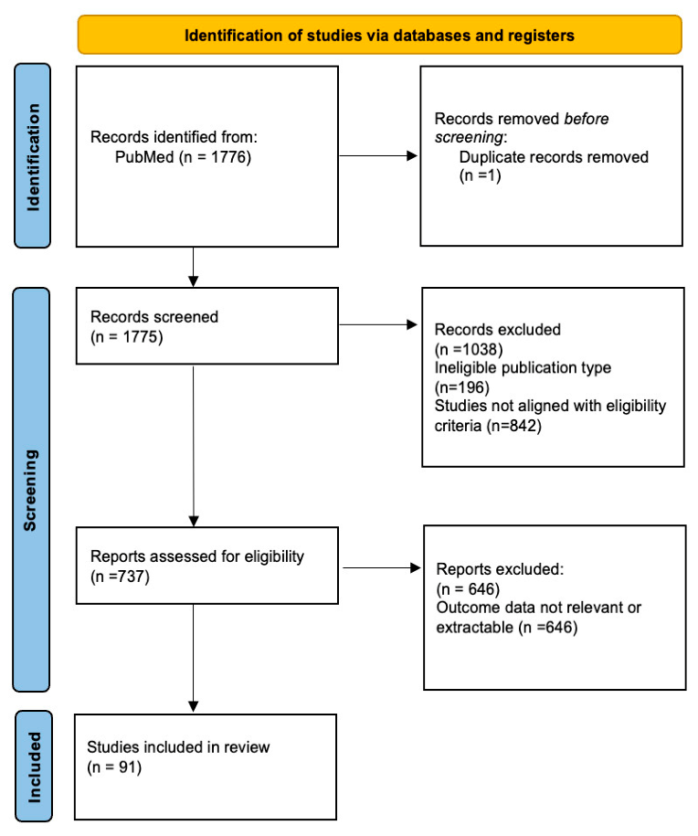
3.1. Study Selection
3.2. General Study Characteristics
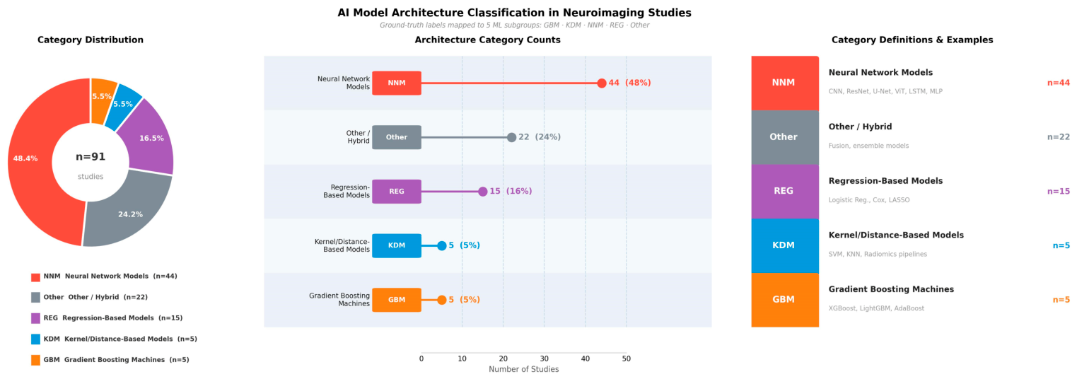
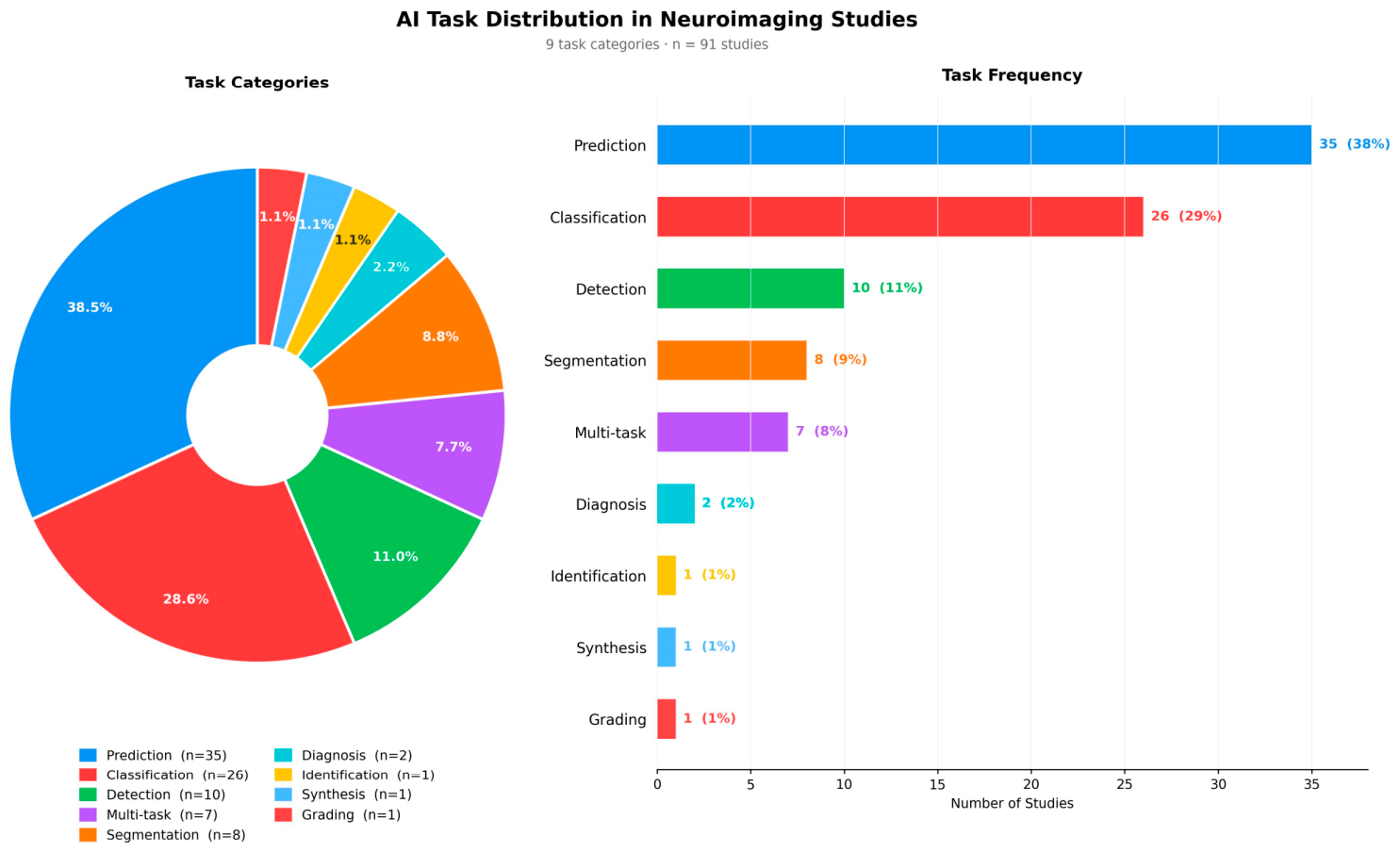
3.3. Artificial Intelligence Methodological Characteristics
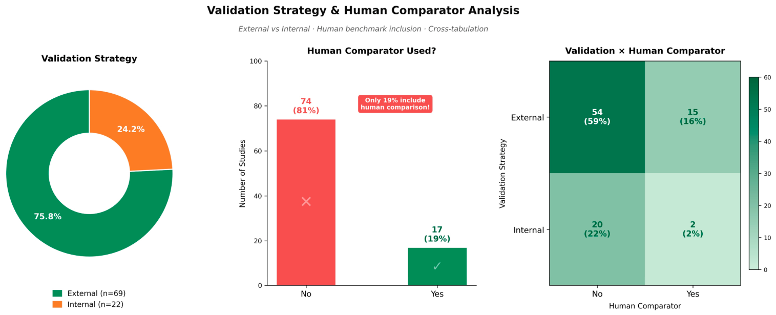
3.4. Validation and Dataset Characteristics
3.5. Methodological Transparency and Bias Indicators
4. Discussion
4.1. Scope of Included Studies
4.2. External Validation and Generalizability
4.3. Data Leakage and Methodological Transparency
4.4. Clinical Benchmarking and Calibration
4.5. Incomplete Adherence to Standardized Reporting Frameworks
4.6. Translational Maturity and Future Directions
4.7. Strengths and Limitations
4.8. Future Directions for AI Methodology in Medical Imaging
5. Conclusions
Supplementary Materials
Author Contributions
Funding
Institutional Review Board Statement
Informed Consent Statement
Data Availability Statement
Conflicts of Interest
Abbreviations
| AI | Artificial intelligence |
| AUC | Area under the receiver operating characteristic curve |
| C-index | Concordance index |
| CLAIM | Checklist for Artificial Intelligence in Medical Imaging |
| CONSORT-AI | Consolidated Standards of Reporting Trials for Artificial Intelligence |
| CT | Computed tomography |
| CTA | Computed tomography angiography |
| DCA | Decision-curve analysis |
| DL | Deep learning |
| ML | Machine learning |
| MRA | Magnetic resonance angiography |
| MRI | Magnetic resonance imaging |
| PRISMA | Preferred Reporting Items for Systematic Reviews and Meta-Analyses |
| PROBAST+AI | Prediction model Risk of Bias Assessment Tool for Artificial Intelligence |
| PROSPERO | International Prospective Register of Systematic Reviews |
| TRIPOD-AI | Transparent Reporting of a multivariable prediction model for Individual Prognosis or Diagnosis using Artificial Intelligence |
References
- Wong, A.; Otles, E.; Donnelly, J.P.; Krumm, A.; McCullough, J.; DeTroyer-Cooley, O.; Pestrue, J.; Phillips, M.; Konye, J.; Penoza, C.; et al. External Validation of a Widely Implemented Proprietary Sepsis Prediction Model in Hospitalized Patients. JAMA Intern. Med. 2021, 181, 1065–1070. [Google Scholar] [CrossRef]
- Ötleş, E.; Denton, B.T.; Qu, B.; Murali, A.; Merdan, S.; Auffenberg, G.B.; Hiller, S.C.; Lane, B.R.; George, A.K.; Singh, K. Development and Validation of Models to Predict Pathological Outcomes of Radical Prostatectomy in Regional and National Cohorts. J. Urol. 2022, 207, 358–366. [Google Scholar] [CrossRef]
- Nagendran, M.; Chen, Y.; Lovejoy, C.A.; Gordon, A.C.; Komorowski, M.; Harvey, H.; Topol, E.J.; Ioannidis, J.P.A.; Collins, G.S.; Maruthappu, M. Artificial intelligence versus clinicians: Systematic review of design, reporting standards, and claims of deep learning studies. BMJ 2020, 368, m689. [Google Scholar] [CrossRef] [PubMed]
- Koçak, B.; Köse, F.; Keleş, A.; Şendur, A.; Meşe, İ.; Karagülle, M. Adherence to the Checklist for Artificial Intelligence in Medical Imaging (CLAIM): An umbrella review with a comprehensive two-level analysis. Diagn. Interv. Radiol. 2025, 31, 440–455. [Google Scholar] [CrossRef]
- Chen, Z.; Liu, X.; Yang, Q.; Wang, Y.J.; Miao, K.; Gong, Z.; Yu, Y.; Leonov, A.; Liu, C.; Feng, Z.; et al. Evaluation of Risk of Bias in Neuroimaging-Based Artificial Intelligence Models for Psychiatric Diagnosis: A Systematic Review. JAMA Netw. Open 2023, 6, e231671. [Google Scholar] [CrossRef]
- Tejani, A.S.; Klontzas, M.E.; Gatti, A.A.; Mongan, J.T.; Moy, L.; Park, S.H.; Kahn, C.E., Jr.; Panel, C.U. Checklist for Artificial Intelligence in Medical Imaging (CLAIM): 2024 Update. Radiol. Artif. Intell. 2024, 6, e240300. [Google Scholar] [CrossRef]
- Collins, G.S.; Moons, K.G.M.; Dhiman, P.; Riley, R.D.; Beam, A.L.; Van Calster, B.; Ghassemi, M.; Liu, X.; Reitsma, J.B.; van Smeden, M.; et al. TRIPOD+AI statement: Updated guidance for reporting clinical prediction models that use regression or machine learning methods. BMJ 2024, 385, e078378. [Google Scholar] [CrossRef] [PubMed]
- Martindale, A.P.L.; Llewellyn, C.D.; de Visser, R.O.; Ng, B.; Ngai, V.; Kale, A.U.; di Ruffano, L.F.; Golub, R.M.; Collins, G.S.; Moher, D.; et al. Concordance of randomised controlled trials for artificial intelligence interventions with the CONSORT-AI reporting guidelines. Nat. Commun. 2024, 15, 1619. [Google Scholar] [CrossRef]
- Page, M.J.; McKenzie, J.E.; Bossuyt, P.M.; Boutron, I.; Hoffmann, T.C.; Mulrow, C.D.; Shamseer, L.; Tetzlaff, J.M.; Akl, E.A.; Brennan, S.E.; et al. The PRISMA 2020 statement: An updated guideline for reporting systematic reviews. BMJ 2021, 372, n71. [Google Scholar] [CrossRef]
- Moons, K.G.M.; Damen, J.A.A.; Kaul, T.; Hooft, L.; Andaur Navarro, C.; Dhiman, P.; Beam, A.L.; Van Calster, B.; Celi, L.A.; Denaxas, S.; et al. PROBAST+AI: An updated quality, risk of bias, and applicability assessment tool for prediction models using regression or artificial intelligence methods. BMJ 2025, 388, e082505. [Google Scholar] [CrossRef] [PubMed]
- Chen, J.; Wang, Z.; Yang, B. mpMRI-based MGMT methylation status prediction for glioblastoma through off-the-shelf deep features: A multi-dataset feasibility study. J. Appl. Clin. Med. Phys. 2025, 26, e70373. [Google Scholar] [CrossRef]
- Chen, Y.; Rivier, C.A.; Mora, S.A.; Torres Lopez, V.; Payabvash, S.; Sheth, K.N.; Harloff, A.; Falcone, G.J.; Rosand, J.; Mayerhofer, E.; et al. Deep learning survival model predicts outcome after intracerebral hemorrhage from initial CT scan. Eur. Stroke J. 2025, 10, 225–235. [Google Scholar] [CrossRef]
- Dai, Y.; Zhong, Z.; Qin, Y.; Wang, Y.; Yu, G.; Kobets, A.; Swenson, D.W.; Boxerman, J.L.; Li, G.; Robinson, S.; et al. AI Model Integrating Imaging and Clinical Data for Predicting CSF Diversion in Neonatal Hydrocephalus: A Preliminary Study. Hum. Brain Mapp. 2025, 46, e70363. [Google Scholar] [CrossRef]
- Dong, Y.; Pachade, S.; Roberts, K.; Jiang, X.; Sheth, S.A.; Giancardo, L. Generalizable self-supervised learning for brain CTA in acute stroke. Comput. Biol. Med. 2025, 184, 109337. [Google Scholar] [CrossRef]
- Gui, Y.; Hu, W.; Ren, J.; Tang, F.; Wang, L.; Zhang, F.; Zhang, J. Preoperative diagnosis of meningioma sinus invasion based on MRI radiomics and deep learning: A multicenter study. Cancer Imaging 2025, 25, 20. [Google Scholar] [CrossRef]
- Hamon, G.; Legrand, L.; Hmeydia, G.; Turc, G.; Hassen, W.B.; Charron, S.; Debacker, C.; Naggara, O.; Thirion, B.; Chen, B.; et al. Multicenter validation of synthetic FLAIR as a substitute for FLAIR sequence in acute ischemic stroke. Eur. Stroke J. 2025, 10, 161–171. [Google Scholar] [CrossRef] [PubMed]
- Jeon, E.T.; Kim, S.M.; Jung, J.M. Automated rating of Fazekas scale in fluid-attenuated inversion recovery MRI for ischemic stroke or transient ischemic attack using machine learning. Sci. Rep. 2025, 15, 32219. [Google Scholar] [CrossRef] [PubMed]
- Kamel, P.; Kanhere, A.; Kulkarni, P.; Khalid, M.; Steger, R.; Bodanapally, U.; Gandhi, D.; Parekh, V.; Yi, P.H. Optimizing Acute Stroke Segmentation on MRI Using Deep Learning: Self-Configuring Neural Networks Provide High Performance Using Only DWI Sequences. J. Imaging Inform. Med. 2025, 38, 717–726. [Google Scholar] [CrossRef] [PubMed]
- Kesari, A.; Maurya, S.; Sheikh, M.T.; Gupta, R.K.; Singh, A. Large blood vessel segmentation in quantitative DCE-MRI of brain tumors: A Swin UNETR approach. Magn. Reson. Imaging 2025, 118, 110342. [Google Scholar] [CrossRef]
- Ketabi, S.; Wagner, M.W.; Hawkins, C.; Tabori, U.; Ertl-Wagner, B.B.; Khalvati, F. Multimodal contrastive learning for enhanced explainability in pediatric brain tumor molecular diagnosis. Sci. Rep. 2025, 15, 10943. [Google Scholar] [CrossRef]
- Krag, C.H.; Muller, F.C.; Gandrup, K.L.; Plesner, L.L.; Sagar, M.V.; Andersen, M.B.; Nielsen, M.; Kruuse, C.; Boesen, M. Impact of spectrum bias on deep learning-based stroke MRI analysis. Eur. J. Radiol. 2025, 188, 112161. [Google Scholar] [CrossRef]
- Kulathilake, C.D.; Udupihille, J.; Abeysundara, S.P.; Senoo, A. Deep learning-driven multi-class classification of brain strokes using computed tomography: A step towards enhanced diagnostic precision. Eur. J. Radiol. 2025, 187, 112109. [Google Scholar] [CrossRef]
- Liang, X.; Ke, X.; Hu, W.; Jiang, J.; Li, S.; Xue, C.; Liu, X.; Dend, J.; Yan, C.; Gao, M.; et al. Deep learning radiomic nomogram outperforms the clinical model in distinguishing intracranial solitary fibrous tumors from angiomatous meningiomas and can predict patient prognosis. Eur. Radiol. 2025, 35, 2670–2680. [Google Scholar] [CrossRef]
- Lilhore, U.K.; Sunder, R.; Simaiya, S.; Alsafyani, M.; Monish Khan, M.D.; Alroobaea, R.; Alsufyani, H.; Baqasah, A.M. AG-MS3D-CNN multiscale attention guided 3D convolutional neural network for robust brain tumor segmentation across MRI protocols. Sci. Rep. 2025, 15, 24306. [Google Scholar] [CrossRef]
- Lin, X.; Zou, E.; Chen, W.; Chen, X.; Lin, L. Advanced multi-label brain hemorrhage segmentation using an attention-based residual U-Net model. BMC Med. Inform. Decis. Mak. 2025, 25, 286. [Google Scholar] [CrossRef]
- Liu, J.; Xing, F.; Elkhill, C.; Linguraru, M.G.; Miles, R.C.; Cruz-Guerrero, I.A.; Porras, A.R. Population-Driven Synthesis of Personalized Cranial Development From Cross-Sectional Pediatric CT Images. IEEE Trans. Biomed. Eng. 2025, 72, 2732–2741. [Google Scholar] [CrossRef]
- Lv, C.; Shu, X.J.; Qiu, J.; Xiong, Z.C.; Bo Ye, J.; Bo Li, S.; Chen, S.B.; Rao, H. AI-enabled precise brain tumor segmentation by integrating Refinenet and contour-constrained features in MRI images. Med. Phys. 2025, 52, e17958. [Google Scholar] [CrossRef] [PubMed]
- Pelcat, A.; Le Berre, A.; Ben Hassen, W.; Debacker, C.; Charron, S.; Thirion, B.; Legrand, L.; Turc, G.; Oppenheim, C.; Benzakoun, J. Generative T2*-weighted images as a substitute for true T2*-weighted images on brain MRI in patients with acute stroke. Diagn. Interv. Imaging 2025, 106, 264–271. [Google Scholar] [CrossRef]
- Rastogi, D.; Johri, P.; Donelli, M.; Kadry, S.; Khan, A.A.; Espa, G.; Feraco, P.; Kim, J. Deep learning-integrated MRI brain tumor analysis: Feature extraction, segmentation, and Survival Prediction using Replicator and volumetric networks. Sci. Rep. 2025, 15, 1437. [Google Scholar] [CrossRef] [PubMed]
- Ruhling, S.; Petzsche, M.R.H.; Loffler, M.T.; Sollmann, N.; Baum, T.; Bodden, J.; Schwarting, J.; Lange, N.; Aftahy, K.; Wostrack, M.; et al. Opportunistic osteoporosis screening in intraoperative CT can accurately identify patients with low volumetric bone mineral density and osteoporosis during spine surgery. Eur. Spine J. 2025, 34, 1461–1469. [Google Scholar] [CrossRef] [PubMed]
- Tu, J.; Shen, C.; Liu, J.; Hu, B.; Chen, Z.; Yan, Y.; Li, C.; Xiong, J.; Daoud, A.M.; Wang, X.; et al. Detection of Microscopic Glioblastoma Infiltration in Peritumoral Edema Using Interactive Deep Learning With DTI Biomarkers: Testing via Stereotactic Biopsy. J. Magn. Reson. Imaging 2025, 62, 1802–1811. [Google Scholar] [CrossRef]
- Wang, B.; Wei, J.; Wang, Z.; Niu, P.; Yang, L.; Hu, Y.; Shao, D.; Zhao, W. Development of a deep learning-based MRI diagnostic model for human Brucella spondylitis. BioMed. Eng. Online 2025, 24, 87. [Google Scholar] [CrossRef]
- Wang, K.; Lin, F.; Liao, Z.; Wang, Y.; Zhang, T.; Wang, R. Development of a Dual-Plane MRI-Based Deep Learning Model to Assess the 1-Year Postoperative Outcomes in Lumbar Disc Herniation After Tubular Microdiscectomy. J. Magn. Reson. Imaging 2025, 61, 2294–2307. [Google Scholar] [CrossRef]
- Wang, T.; Jiang, C.; Ding, W.; Chen, Q.; Shen, D.; Ding, Z. Deep-Learning Generated Synthetic Material Decomposition Images Based on Single-Energy CT to Differentiate Intracranial Hemorrhage and Contrast Staining Within 24 Hours After Endovascular Thrombectomy. CNS Neurosci. Ther. 2025, 31, e70235. [Google Scholar] [CrossRef]
- Wang, Y.; Wen, Z.; Bao, S.; Huang, D.; Wang, Y.; Yang, B.; Li, Y.; Zhou, P.; Zhang, H.; Pang, H. Diffusion-CSPAM U-Net: A U-Net model integrated hybrid attention mechanism and diffusion model for segmentation of computed tomography images of brain metastases. Radiat. Oncol. 2025, 20, 50. [Google Scholar] [CrossRef]
- Xing, L.P.; Liu, G.; Zhang, H.C.; Wang, L.; Zhu, S.; Bao, M.D.H.; Wang, Y.N.; Chen, C.; Wang, Z.; Liu, X.Y.; et al. Evaluating CNN Architectures for the Automated Detection and Grading of Modic Changes in MRI: A Comparative Study. Orthop. Surg. 2025, 17, 233–243. [Google Scholar] [CrossRef] [PubMed]
- Zahoora, U.; Shahid, A.R.; Gondal, F.F. A bias-resilient client selection analysis for federated brain tumor segmentation. Sci. Rep. 2025, 15, 37670. [Google Scholar] [CrossRef] [PubMed]
- Jia, X.; Chen, Y.; Zheng, K.; Chen, C.; Liu, J. Deep Learning-Driven Multimodal Fusion Model for Prediction of Middle Cerebral Artery Aneurysm Rupture Risk. Acad. Radiol. 2025, 32, 6114–6124. [Google Scholar] [CrossRef]
- Harper, J.P.; Lee, G.R.; Pan, I.; Nguyen, X.V.; Quails, N.; Prevedello, L.M. External Validation of a Winning Artificial Intelligence Algorithm from the RSNA 2022 Cervical Spine Fracture Detection Challenge. AJNR Am. J. Neuroradiol. 2025, 46, 1852–1858. [Google Scholar] [CrossRef] [PubMed]
- Dai, M.; Tiu, B.C.; Schlossman, J.; Ayobi, A.; Castineira, C.; Kiewsky, J.; Avare, C.; Chaibi, Y.; Chang, P.; Chow, D.; et al. Validation of a Deep Learning Tool for Detection of Incidental Vertebral Compression Fractures. J. Comput. Assist. Tomogr. 2025, 49, 669–674. [Google Scholar] [CrossRef]
- Kang, D.W.; Kim, M.; Park, G.H.; Kim, Y.S.; Han, M.K.; Lee, M.; Kim, D.; Ryu, W.S.; Jeong, H.G. Deep learning-assisted detection of intracranial hemorrhage: Validation and impact on reader performance. Neuroradiology 2025, 67, 1511–1519. [Google Scholar] [CrossRef] [PubMed]
- Hossain, M.M.; Ahmed, M.M.; Nafi, A.A.N.; Islam, M.R.; Ali, M.S.; Haque, J.; Miah, M.S.; Rahman, M.M.; Islam, M.K. A novel hybrid ViT-LSTM model with explainable AI for brain stroke detection and classification in CT images: A case study of Rajshahi region. Comput. Biol. Med. 2025, 186, 109711. [Google Scholar] [CrossRef]
- Chen, R.; Lu, Y.; Tian, Z.; Chen, J.; Li, W.; Wang, C.; Zhang, Z.; Huang, X.; Ding, C.; Liu, X.; et al. DWI-based deep learning radiomics nomogram for predicting the impaired quality of life in patients with unruptured intracranial aneurysm developing new iatrogenic cerebral infarcts following stent placement: A multicenter cohort study. Neurosurg. Rev. 2025, 48, 508. [Google Scholar] [CrossRef]
- Felefly, T.; Francis, Z.; Roukoz, C.; Fares, G.; Achkar, S.; Yazbeck, S.; Nasr, A.; Kordahi, M.; Azoury, F.; Nasr, D.N.; et al. A 3D Convolutional Neural Network Based on Non-enhanced Brain CT to Identify Patients with Brain Metastases. J. Imaging Inform. Med. 2025, 38, 858–864. [Google Scholar] [CrossRef] [PubMed]
- Nalentzi, K.; Gerogiannis, K.; Bougias, H.; Stogiannos, N.; Papavasileiou, P. Comparative analysis of transformer-based deep learning models for glioma and meningioma classification. J. Med. Imaging Radiat. Sci. 2025, 56, 102008. [Google Scholar] [CrossRef]
- Liao, L.; Puel, U.; Sabardu, O.; Harsan, O.; Medeiros, L.L.; Loukoul, W.A.; Anxionnat, R.; Kerrien, E. AI-assisted detection of cerebral aneurysms on 3D time-of-flight MR angiography: User variability and clinical implications. J. Neuroradiol. 2025, 52, 101388. [Google Scholar] [CrossRef]
- Pettersson, S.D.; Filo, J.; Liaw, P.; Skrzypkowska, P.; Klepinowski, T.; Szmuda, T.; Fodor, T.B.; Ramirez-Velandia, F.; Zielinski, P.; Chang, Y.M.; et al. Addressing Limited Generalizability in Artificial Intelligence-Based Brain Aneurysm Detection for Computed Tomography Angiography: Development of an Externally Validated Artificial Intelligence Screening Platform. Neurosurgery 2025, 97, 1388–1396. [Google Scholar] [CrossRef] [PubMed]
- Ryu, W.S.; Schellingerhout, D.; Park, J.; Chung, J.; Jeong, S.W.; Gwak, D.S.; Kim, B.J.; Kim, J.T.; Hong, K.S.; Lee, K.B.; et al. Deep learning-based automatic segmentation of cerebral infarcts on diffusion MRI. Sci. Rep. 2025, 15, 13214. [Google Scholar] [CrossRef]
- Yang, H.; Wang, W.; Zhao, X.; Xuan, Q.; Jiang, C.; Zhao, B. Deep learning models based on DWI-MRI for prognosis prediction in acute ischemic stroke receiving intravenous thrombolysis: Development and validation. J. Neuroradiol. 2025, 52, 101391. [Google Scholar] [CrossRef]
- Kong, C.; Yan, D.; Liu, K.; Yin, Y.; Ma, C. Multiple deep learning models based on MRI images in discriminating glioblastoma from solitary brain metastases: A multicentre study. BMC Med. Imaging 2025, 25, 171. [Google Scholar] [CrossRef]
- Topff, L.; Petrychenko, L.; Jain, N.; Lingier, S.; Bertels, J.; Astudillo, P.; Prosec, M.; Menendez Fernandez-Miranda, P.; Gevaert, O.; Smits, M.; et al. A Data-Centric Approach to Deep Learning for Brain Metastasis Analysis at MRI. Radiology 2025, 315, e242416. [Google Scholar] [CrossRef]
- Mahootiha, M.; Tak, D.; Ye, Z.; Zapaishchykova, A.; Likitlersuang, J.; Climent Pardo, J.C.; Boyd, A.; Vajapeyam, S.; Chopra, R.; Prabhu, S.P.; et al. Multimodal deep learning improves recurrence risk prediction in pediatric low-grade gliomas. Neuro-Oncology 2025, 27, 277–290. [Google Scholar] [CrossRef] [PubMed]
- Zeng, L.; Wen, L.; Jing, Y.; Xu, J.X.; Huang, C.C.; Zhang, D.; Wang, G.X. Assessment of the stability of intracranial aneurysms using a deep learning model based on computed tomography angiography. Radiol. Medica 2025, 130, 248–257. [Google Scholar] [CrossRef]
- Tuxunjiang, P.; Huang, C.; Zhou, Z.; Zhao, W.; Han, B.; Tan, W.; Wang, J.; Kukun, H.; Zhao, W.; Xu, R.; et al. Prediction of NIHSS Scores and Acute Ischemic Stroke Severity Using a Cross-attention Vision Transformer Model with Multimodal MRI. Acad. Radiol. 2025, 32, 5453–5467. [Google Scholar] [CrossRef]
- Sina, E.M.; Limage, K.; Anisman, E.; Pudik, N.; Tam, E.; Kahn, C.; Daggumati, S.; Evans, J.J.; Rabinowitz, M.R.; Rosen, M.R.; et al. Automated Machine Learning Differentiation of Pituitary Macroadenomas and Parasellar Meningiomas Using Preoperative Magnetic Resonance Imaging. Otolaryngol.–Head Neck Surg. 2025, 173, 1376–1384. [Google Scholar] [CrossRef] [PubMed]
- Wang, H.; Schwirtlich, T.; Houskamp, E.; Hutch, M.; Murphy, J.; Nascimento, J.; Zini, A.; Brancaleoni, L.; Giacomozzi, S.; Luo, Y.; et al. Automated Detection of the Black Hole Sign for Patients with Intracerebral Hemorrhage Using Self-Supervised Learning. AJNR Am. J. Neuroradiol. 2025, 46, 2300–2309. [Google Scholar] [CrossRef]
- Nada, A.; Sayed, A.A.; Hamouda, M.; Tantawi, M.; Khan, A.; Alt, A.; Hassanein, H.; Sevim, B.C.; Altes, T.; Gaballah, A. External validation and performance analysis of a deep learning-based model for the detection of intracranial hemorrhage. Neuroradiol. J. 2025, 38, 312–321. [Google Scholar] [CrossRef]
- Hao, M.; Yan, J.; Wang, X.; Tan, Y.; Zhang, H.; Yang, G. Survival prediction in gliomas based on MRI radiomics combined with clinical factors and molecular biomarkers. PeerJ 2025, 13, e19906. [Google Scholar] [CrossRef]
- Akbari, H.; Bakas, S.; Sako, C.; Fathi Kazerooni, A.; Villanueva-Meyer, J.; Garcia, J.A.; Mamourian, E.; Liu, F.; Cao, Q.; Shinohara, R.T.; et al. Machine learning-based prognostic subgrouping of glioblastoma: A multicenter study. Neuro-Oncol. 2025, 27, 1102–1115. [Google Scholar] [CrossRef] [PubMed]
- Belke, M.; Zahnert, F.; Steinbrenner, M.; Halimeh, M.; Miron, G.; Tsalouchidou, P.E.; Linka, L.; Keil, B.; Jansen, A.; Möschl, V.; et al. Automatic detection of hippocampal sclerosis in patients with epilepsy. Epilepsia 2025, 66, 3852–3864. [Google Scholar] [CrossRef]
- Demirel, E.; Dilek, O. Utilizing Radiomics of Peri-Lesional Edema in T2-FLAIR Subtraction Digital Images to Distinguish High-Grade Glial Tumors From Brain Metastasis. J. Magn. Reson. Imaging 2025, 61, 1728–1737. [Google Scholar] [CrossRef] [PubMed]
- Fatania, K.; Frood, R.; Mistry, H.; Short, S.C.; O’Connor, J.; Scarsbrook, A.F.; Currie, S. Impact of intensity standardisation and ComBat batch size on clinical-radiomic prognostic models performance in a multi-centre study of patients with glioblastoma. Eur. Radiol. 2025, 35, 3354–3366. [Google Scholar] [CrossRef]
- Foltyn-Dumitru, M.; Mahmutoglu, M.A.; Brugnara, G.; Kessler, T.; Sahm, F.; Wick, W.; Heiland, S.; Bendszus, M.; Vollmuth, P.; Schell, M. Shape matters: Unsupervised exploration of IDH-wildtype glioma imaging survival predictors. Eur. Radiol. 2025, 35, 1351–1360. [Google Scholar] [CrossRef] [PubMed]
- Roh, Y.H.; Cheong, E.N.; Jung, S.C.; Yun, J.; Ko, J.S.; Cho, S.J.; Choi, K.M.; Park, S.I.; Jeong, S.Y.; Lee, D.H.; et al. Prediction of hemorrhagic transformation in acute ischemic stroke patients using clinico-radiomics models. Sci. Rep. 2025, 15, 38628. [Google Scholar] [CrossRef] [PubMed]
- Yang, Q.; Wang, Y.; Wu, J.; Hu, H.; He, Y.; Wang, Y.; Yang, B. Preoperative prediction of pituitary neuroendocrine tumor consistency based on multiparametric MRI radiomics: A multicenter study. BMC Cancer 2025, 25, 1501. [Google Scholar] [CrossRef]
- Fan, Y.; Zhang, W.; Mou, A.; Fang, H.; Guo, S.; Feng, M. Development and Validation of Multiparametric MRI-based Clini-radiomic Model for Preoperative Prediction of Somatotroph Adenomas Subtypes: A Multicenter Study. Acad. Radiol. 2025, 32, 7471–7485. [Google Scholar] [CrossRef] [PubMed]
- Huang, L.; Xu, X.; Tian, B.; Liao, A.; Wang, L.; Shen, X.; Cao, Z.; Liu, X.; Lu, S.; Li, J.; et al. Development and validation of a radiomics model based on the ASPECTS framework using CT imaging for predicting malignant cerebral edema. Eur. J. Radiol. 2025, 192, 112410. [Google Scholar] [CrossRef]
- Liang, Q.; Duan, X.; Yan, H.; Li, X.; Li, Z.; Niu, W.; Liu, X.; Tan, Y.; Wang, X.; Yang, G.; et al. Development and validation of radiopathomics models for predicting molecular subtypes and WHO grades in adult-type diffuse gliomas: A multicenter study. J. Transl. Med. 2025, 23, 1120. [Google Scholar] [CrossRef]
- Xia, X.; Liu, J.; Cui, J.; You, Y.; Huang, C.; Li, H.; Zhang, D.; Ren, Q.; Jiang, Q.; Meng, X. A nomogram incorporating CT-based peri-hematoma radiomics features to predict functional outcome in patients with intracerebral hemorrhage. Eur. J. Radiol. 2025, 183, 111871. [Google Scholar] [CrossRef]
- Liu, J.; Tu, J.; Hu, B.; Li, C.; Piao, S.; Lu, Y.; Li, A.; Ding, T.; Xiong, J.; Zhu, F.; et al. Prognostic Assessment in Patients With Primary Diffuse Large B-Cell Lymphoma of the Central Nervous System Using MRI-Based Radiomics. J. Magn. Reson. Imaging 2025, 61, 1142–1152. [Google Scholar] [CrossRef]
- Song, D.; Wei, Q.; Zhao, S.; Lou, Y.; Zhang, K.; Duan, C.; Wang, F.; Gao, Q.; Yan, J.; Yan, D.; et al. Exploring a recurrence model for atypical meningioma based on multiparametric MRI radiomic and clinical characteristics: A multicenter retrospective cohort study. Radiat. Oncol. 2025, 20, 30. [Google Scholar] [CrossRef] [PubMed]
- Patel, B.K.; Zohdy, Y.M.; Lohana, S.; Tariciotti, L.; Rodas, A.; Alawieh, A.; Jahangiri, A.; Faraj, R.R.; Maldonado, J.; Uribe-Pacheco, R.; et al. Predictive Modeling of Nonfunctioning Giant Pituitary Neuroendocrine Tumor Resection: A Multi-Planar Perspective. World Neurosurg. 2025, 195, 123653. [Google Scholar] [CrossRef]
- Zheng, B.; Zhu, Z.; Ma, K.; Liang, Y.; Liu, H. Three-Dimensional Radiomics and Machine Learning for Predicting Postoperative Outcomes in Laminoplasty for Cervical Spondylotic Myelopathy: A Clinical-Radiomics Model. World Neurosurg. 2025, 203, 124464. [Google Scholar] [CrossRef]
- Hu, W.; Lin, G.; Chen, W.; Wu, J.; Zhao, T.; Xu, L.; Qian, X.; Shen, L.; Yan, Z.; Chen, M.; et al. Radiomics based on dual-energy CT virtual monoenergetic images to identify symptomatic carotid plaques: A multicenter study. Sci. Rep. 2025, 15, 10415. [Google Scholar] [CrossRef]
- Cai, Z.Y.; Hu, K.; Linli, Z.Q. Sexual dimorphism of white-matter functional connectome in healthy young adults. Prog. Neuro-Psychopharmacol. Biol. Psychiatry 2025, 142, 111486. [Google Scholar] [CrossRef]
- Feng, L.; Han, H.; Mo, J.; Huang, Y.; Huang, K.; Zhou, C.; Wang, X.; Zhang, J.; Yang, Z.; Liu, D.; et al. Individualized structural network deviations predict surgical outcome in mesial temporal lobe epilepsy: A multicenter validation study. Int. Surg. J. 2025, 111, 7594–7605. [Google Scholar] [CrossRef]
- Ma, Z.; Zhang, C.; Guo, Y.; Li, Y.; Wang, Z.; Hu, Y.; Zhang, X.; Duan, M.; Wang, W.; Yan, D.; et al. Improving radiomics-based differentiation of supratentorial malignant brain tumors preoperatively with diffusion-weighted imaging: A three-class machine learning algorithm. Eur. J. Surg. Oncol. 2025, 51, 110533. [Google Scholar] [CrossRef]
- Sun, K.; Shi, R.; Yu, X.; Wang, Y.; Zhang, W.; Yang, X.; Zhang, M.; Wang, J.; Jiang, S.; Li, H.; et al. Noninvasive imaging biomarker reveals invisible microscopic variation in acute ischaemic stroke (</= 24 h): A multicentre retrospective study. Sci. Rep. 2025, 15, 3743. [Google Scholar] [CrossRef] [PubMed]
- Sunavsky, A.; Hashmi, M.A.; Robertson, J.W.; Veinot, J.; Hashmi, J.A. The nucleus accumbens-prefrontal connectivity as a predictor of chronic low back pain. Pain 2025, 166, e363–e377. [Google Scholar] [CrossRef]
- Wang, G.; Zhang, Y.; Xu, L.; Ni, J.; Shen, Y.; Jin, Q. Radiomics-based diagnosis of carotid artery stenosis using non-contrast CT: Model development and validation. Eur. J. Med. Res. 2025, 30, 1237. [Google Scholar] [CrossRef] [PubMed]
- Wang, H.; Kong, J.F.; Wen, L.; Wang, X.J.; Zhang, W.T.; Wang, Z.Q.; Zeng, L.; Huang, Y.T.; Yang, S.H.; Li, M.; et al. Development of predictive models to identify the intracranial aneurysm responsible for subarachnoid hemorrhage in patients with multiple saccular aneurysms. Eur. J. Radiol. 2025, 193, 112466. [Google Scholar] [CrossRef]
- Xia, X.; Wu, W.; Tan, Q.; Gou, Q. Interpretable Machine Learning Models for Differentiating Glioblastoma From Solitary Brain Metastasis Using Radiomics. Acad. Radiol. 2025, 32, 5388–5400. [Google Scholar] [CrossRef] [PubMed]
- Xu, W.; Li, Y.; Zhang, J.; Zhang, Z.; Shen, P.; Wang, X.; Yang, G.; Du, J.; Zhang, H.; Tan, Y. Predicting the molecular subtypes of 2021 WHO grade 4 glioma by a multiparametric MRI-based machine learning model. BMC Cancer 2025, 25, 1171. [Google Scholar] [CrossRef]
- Xu, X.; Zhou, Y.; Sun, S.; Cui, L.; Chen, Z.; Guo, Y.; Jiang, J.; Wang, X.; Sun, T.; Yang, Q.; et al. Risk prediction for elderly cognitive impairment by radiomic and morphological quantification analysis based on a cerebral MRA imaging cohort. Eur. Radiol. 2025, 35, 4300–4314. [Google Scholar] [CrossRef]
- Ye, B.; Sun, Y.; Chen, G.; Wang, B.; Meng, H.; Shan, L. Development and validation of machine learning models to predict vertebral artery injury by C2 pedicle screws. Eur. Spine J. 2025, 34, 3950–3961. [Google Scholar] [CrossRef]
- Zeng, Q.; Jia, F.; Tang, S.; He, H.; Fu, Y.; Wang, X.; Zhang, J.; Tan, Z.; Tang, H.; Wang, J.; et al. Ensemble learning-based radiomics model for discriminating brain metastasis from glioblastoma. Eur. J. Radiol. 2025, 183, 111900. [Google Scholar] [CrossRef] [PubMed]
- Zhai, D.; Wu, Y.; Cui, M.; Liu, Y.; Zhou, X.; Hu, D.; Wang, Y.; Ju, S.; Fan, G.; Cai, W. Combinations of Clinical Factors, CT Signs, and Radiomics for Differentiating High-Density Areas after Mechanical Thrombectomy in Patients with Acute Ischemic Stroke. AJNR Am. J. Neuroradiol. 2025, 46, 66–74. [Google Scholar] [CrossRef]
- Zhao, K.; Chen, C.; Zhang, Y.; Huang, Z.; Zhao, Y.; Yue, Q.; Xu, J. Preoperative Assessment of Ki-67 Labeling Index in Pituitary Adenomas Using Delta-Radiomics Based on Dynamic Contrast-Enhanced MRI. J. Magn. Reson. Imaging 2025, 62, 508–518. [Google Scholar] [CrossRef]
- Zhao, K.; Deng, Y.; Su, X.; Hu, W.; Yin, T.; Yang, X.; Zhang, D.; Sun, J.; Li, Y.; Xu, J.; et al. Differential Diagnosis of Early-Stage Atypical Primary Central Nervous System Lymphoma and Low-Grade Glioma Using Magnetic Resonance Imaging-Based Radiomics. World Neurosurg. 2025, 196, 123740. [Google Scholar] [CrossRef]
- Zhuang, X.; Wang, J.; Kang, J.; Lin, Z. Diagnosis of Acute Versus Chronic Thoracolumbar Vertebral Compression Fractures Using CT Radiomics Based on Machine Learning: A Preliminary Study. J. Imaging Inform. Med. 2025, 38, 2183–2193. [Google Scholar] [CrossRef] [PubMed]
- Sun, Y.; Wang, Y.; Jiang, M.; Jia, W.; Chen, H.; Wang, H.; Ding, Y.; Wang, X.; Yang, C.; Sun, B.; et al. Habitat-based MRI radiomics to predict the origin of brain metastasis. Med. Phys. 2025, 52, 3075–3087. [Google Scholar] [CrossRef]
- Albadr, R.J.; Sur, D.; Yadav, A.; Rekha, M.M.; Jain, B.; Jayabalan, K.; Kubaev, A.; Taher, W.M.; Alwan, M.; Jawad, M.J.; et al. Optimizing meningioma grading with radiomics and deep features integration, attention mechanisms, and reproducibility analysis. Eur. J. Med. Res. 2025, 30, 808. [Google Scholar] [CrossRef]
- Chen, Y.; Hu, X.; Fan, T.; Zhou, Y.; Yu, C.; Yu, J.; Zhou, X.; Wang, B. Predicting Postoperative Prognosis in Pediatric Malignant Tumor With MRI Radiomics and Deep Learning Models: A Retrospective Study. J. Craniofacial Surg. 2025, 36, 1929–1935. [Google Scholar] [CrossRef] [PubMed]
- Choi, J.H.; Sobisch, J.; Kim, M.; Park, J.C.; Ahn, J.S.; Kwun, B.D.; Spiclin, Z.; Bizjak, Z.; Park, W. Prediction of intracranial aneurysm rupture from computed tomography angiography using an automated artificial intelligence framework. Comput. Biol. Med. 2025, 197, 110965. [Google Scholar] [CrossRef]
- Li, D.; Hu, W.; Ma, L.; Yang, W.; Liu, Y.; Zou, J.; Ge, X.; Han, Y.; Gan, T.; Cheng, D.; et al. Deep learning radiomics nomograms predict Isocitrate dehydrogenase (IDH) genotypes in brain glioma: A multicenter study. Magn. Reson. Imaging 2025, 117, 110314. [Google Scholar] [CrossRef]
- Li, Z.; Gao, Y.; Zhang, K.; Wang, J.; Han, L.; Xie, B.; Sun, Y.; Yan, R.; Li, Y.; Cui, H. Predicting the prognosis of symptomatic intracranial atherosclerotic stenosis (sICAS) patients using deep learning models: A multicenter study based on high-resolution magnetic resonance vessel wall imaging. Clin. Radiol. 2025, 91, 107092. [Google Scholar] [CrossRef] [PubMed]
- Liu, J.; Jiang, S.; Wu, Y.; Zou, R.; Bao, Y.; Wang, N.; Tu, J.; Xiong, J.; Liu, Y.; Li, Y. Deep learning-based radiomics and machine learning for prognostic assessment in IDH-wildtype glioblastoma after maximal safe surgical resection: A multicenter study. Int. Surg. J. 2025, 111, 4576–4585. [Google Scholar] [CrossRef] [PubMed]
- Saadh, M.J.; Albadr, R.J.; Sur, D.; Yadav, A.; Roopashree, R.; Sangwan, G.; Krithiga, T.; Aminov, Z.; Taher, W.M.; Alwan, M.; et al. Reproducible meningioma grading across multi-center MRI protocols via hybrid radiomic and deep learning features. Neuroradiology 2025, 67, 2741–2761. [Google Scholar] [CrossRef]
- Yin, L.; Wang, J. Enhancing brain tumor classification by integrating radiomics and deep learning features: A comprehensive study utilizing ensemble methods on MRI scans. J. X-Ray Sci. Technol. 2025, 33, 47–57. [Google Scholar] [CrossRef]
- Yin, S.; Ming, J.; Chen, H.; Sun, Y.; Jiang, C. Integrating deep learning and radiomics for preoperative glioma grading using multi-center MRI data. Sci. Rep. 2025, 15, 36756. [Google Scholar] [CrossRef]
- Yonar, A. A swarm intelligence-driven hybrid framework for brain tumor classification with enhanced deep features. Sci. Rep. 2025, 15, 37543. [Google Scholar] [CrossRef]
- Senthil, R.; Anand, T.; Somala, C.S.; Saravanan, K.M. Bibliometric analysis of artificial intelligence in healthcare research: Trends and future directions. Future Healthc. J. 2024, 11, 100182. [Google Scholar] [CrossRef]
- Liu, X.; Cruz Rivera, S.; Moher, D.; Calvert, M.J.; Denniston, A.K. Reporting guidelines for clinical trial reports for interventions involving artificial intelligence: The CONSORT-AI extension. Nat. Med. 2020, 26, 1364–1374. [Google Scholar] [CrossRef]
- Haibe-Kains, B.; Adam, G.A.; Hosny, A.; Khodakarami, F.; Waldron, L.; Wang, B.; McIntosh, C.; Goldenberg, A.; Kundaje, A.; Greene, C.S.; et al. Transparency and reproducibility in artificial intelligence. Nature 2020, 586, E14–E16. [Google Scholar] [CrossRef]
- Kim, D.W.; Jang, H.Y.; Kim, K.W.; Shin, Y.; Park, S.H. Design Characteristics of Studies Reporting the Performance of Artificial Intelligence Algorithms for Diagnostic Analysis of Medical Images: Results from Recently Published Papers. Korean J. Radiol. 2019, 20, 405–410. [Google Scholar] [CrossRef]
- Yu, A.C.; Mohajer, B.; Eng, J. External Validation of Deep Learning Algorithms for Radiologic Diagnosis: A Systematic Review. Radiol. Artif. Intell. 2022, 4, e210064. [Google Scholar] [CrossRef] [PubMed]
- Spaanderman, D.J.; Marzetti, M.; Wan, X.; Scarsbrook, A.F.; Robinson, P.; Oei, E.H.G.; Visser, J.J.; Hemke, R.; van Langevelde, K.; Hanff, D.F.; et al. AI in radiological imaging of soft-tissue and bone tumours: A systematic review evaluating against CLAIM and FUTURE-AI guidelines. EBioMedicine 2025, 114, 105642. [Google Scholar] [CrossRef]
- Xu, H.L.; Gong, T.T.; Song, X.J.; Chen, Q.; Bao, Q.; Yao, W.; Xie, M.M.; Li, C.; Grzegorzek, M.; Shi, Y.; et al. Artificial Intelligence Performance in Image-Based Cancer Identification: Umbrella Review of Systematic Reviews. J. Med. Internet Res. 2025, 27, e53567. [Google Scholar] [CrossRef] [PubMed]
- Bramer, W.M.; Rethlefsen, M.L.; Kleijnen, J.; Franco, O.H. Optimal database combinations for literature searches in systematic reviews: A prospective exploratory study. Syst. Rev. 2017, 6, 245. [Google Scholar] [CrossRef]
- Wenderott, K.; Krups, J.; Zaruchas, F.; Weigl, M. Effects of artificial intelligence implementation on efficiency in medical imaging—A systematic literature review and meta-analysis. npj Digit. Med. 2024, 7, 265. [Google Scholar] [CrossRef] [PubMed]
- Mongan, J.; Moy, L.; Kahn, C.E., Jr. Checklist for Artificial Intelligence in Medical Imaging (CLAIM): A Guide for Authors and Reviewers. Radiol. Artif. Intell. 2020, 2, e200029. [Google Scholar] [CrossRef]




| Author | Country of Origin | Medical Field | Specific Field | AI Type | AI Task During Research |
|---|---|---|---|---|---|
| Akbari H. et al. [59] | Multinational | Oncology | Glioblastoma prognostic subgrouping | ML | Survival prediction and prognostic subgrouping |
| Albadr R.J. et al. [92] | Multinational | Oncology | Meningioma grading | Hybrid (DL/ML) | Preoperative classification/grading |
| Belke M. et al. [60] | Germany | Neurology | Epilepsy imaging/hippocampal sclerosis detection | ML | Detection/diagnosis |
| Cai Z.Y. et al. [75] | China | Neuroscience | White-matter functional connectomics/sex classification | ML | Classification |
| Chen J. et al. [11] | China | Oncology | Glioblastoma molecular marker prediction (MGMT) | DL | Classification/biomarker prediction |
| Chen R. et al. [43] | China | Neurosurgery | Intracranial aneurysm outcome prediction | DL | Prediction/risk modeling |
| Chen Y. et al. [93] | China | Pediatrics | Pediatric brain tumor prognosis | Hybrid (DL/ML) | Prognosis prediction |
| Chen Y. et al. [12] | USA | Neurology | Intracerebral hemorrhage outcome prediction | DL | Functional outcome prediction |
| Choi J.H. et al. [94] | S. Korea | Neurosurgery | Intracranial aneurysm rupture prediction | Hybrid (DL/ML) | Classification/rupture risk prediction |
| Dai M. et al. [40] | Multinational | Radiology | Vertebral compression fracture detection | DL | Detection |
| Dai Y. et al. [13] | Multinational | Pediatrics | Neonatal hydrocephalus/CSF diversion prediction | DL | Prediction |
| Demirel E. et al. [61] | Turkey | Oncology | Brain tumor differential diagnosis | ML | Classification |
| Dong Y. et al. [14] | USA | Neurology/Radiology | Generalizable CTA representation learning for acute stroke tasks | DL | Detection/classification/prediction |
| Fan Y. et al. [66] | China | Radiology | Pituitary adenoma subtype prediction | ML | Preoperative classification |
| Fatania K. et al. [62] | UK | Radiology | Glioblastoma radiomics survival modeling | ML | Prognosis modeling |
| Felefly T. et al. [44] | Multinational | Oncology | Brain metastasis detection on CT | DL | Detection/classification |
| Feng L. et al. [76] | China | Neurology | Epilepsy surgery outcome prediction | ML | Prediction |
| Foltyn-Dumitru M. et al. [63] | Germany | Neuroradiology | Glioma imaging phenotyping/survival prediction | ML | Unsupervised clustering/prognosis |
| Gui Y. et al. [15] | China | Oncology | Meningioma sinus invasion diagnosis | DL | Preoperative classification |
| Hamon G. et al. [16] | France | Neurology | Synthetic MRI/DWI-FLAIR mismatch assessment | DL | Image synthesis/diagnostic support |
| Hao M. et al. [58] | China | Oncology | MGMT promoter methylation prediction in glioblastoma | ML | Survival prediction/risk stratification |
| Harper J.P. et al. [39] | USA | Radiology | Cervical spine fracture detection | DL | Detection |
| Hossain M.M. et al. [42] | Bangladesh | Neurology | Brain stroke classification on CT | DL | Classification |
| Hu W. et al. [74] | China | Radiology | Carotid plaque symptom classification | ML | Identification |
| Huang L. et al. [67] | China | Neurology | Malignant cerebral edema prediction | ML | Prediction |
| Jeon E.T. et al. [17] | S. Korea | Neurology | White matter hyperintensity/Fazekas grading | DL | Segmentation/grading |
| Jia X. et al. [38] | China | Neuroradiology | Middle cerebral artery aneurysm rupture risk prediction | DL | Prediction |
| Kamel P. et al. [18] | USA | Neurology | Ischemic stroke infarct segmentation on MRI | DL | Segmentation |
| Kang D.W. et al. [41] | S. Korea | Radiology | Intracranial hemorrhage detection | DL | Detection |
| Kesari A. et al. [19] | India | Oncology | Brain tumor blood-vessel segmentation | DL | Segmentation |
| Ketabi S. et al. [20] | Canada | Oncology | Pediatric low-grade glioma genetic marker classification | DL | Classification |
| Kong C. et al. [50] | China | Radiation Oncology | Glioblastoma versus solitary brain metastasis differentiation | DL | Classification |
| Krag C.H. et al. [21] | Denmark | Neurology | Acute ischemic stroke lesion detection on MRI | DL | Classification |
| Kulathilake C.D. et al. [22] | Multinational | Neurology | Brain stroke CT classification | DL | Classification |
| Li D. et al. [95] | China | Oncology | IDH mutation prediction from MRI | Hybrid (DL/ML) | Prediction |
| Li Z. et al. [96] | China | Radiology | Prediction of stroke recurrence in symptomatic intracranial atherosclerotic stenosis | Hybrid (DL/ML) | Prediction |
| Liang Q. et al. [68] | China | Oncology | Adult diffuse glioma grading/molecular subtyping | ML | Prediction |
| Liang X. et al. [23] | China | Oncology | Intracranial solitary fibrous tumor (ISFT) versus angiomatous meningioma differentiation | DL | Classification |
| Liao L. et al. [46] | France | Neuroradiology | Cerebral aneurysm detection on TOF-MRA | DL | Detection |
| Lilhore U.K. et al. [24] | India | Oncology | Brain tumor segmentation on multimodal MRI | DL | Segmentation |
| Lin X. et al. [25] | China | Radiology | Intracranial hemorrhage segmentation on CT | DL | Segmentation |
| Liu J. et al. [26] | USA | Pediatrics | Prediction of normative pediatric brain development from MRI | DL | Prediction |
| Liu J. et al. [70] | China | Oncology | MRI-based survival prediction in primary CNS lymphoma | ML | Survival prediction |
| Liu J. et al. [97] | China | Oncology | Glioblastoma prognostic stratification | Hybrid (DL/ML) | Survival prediction/risk stratification |
| Lv C. et al. [27] | China | Oncology/Radiology | Brain tumor MRI segmentation | DL | Segmentation |
| Ma Z. et al. [77] | China | Oncology | MRI radiomics-based classification of malignant brain tumors | ML | Classification |
| Mahootiha M. et al. [52] | USA | Oncology | Pediatric low-grade glioma recurrence prediction | DL | Prediction/risk modeling |
| Nada A. et al. [57] | USA | Radiology | Intracranial hemorrhage detection | DL | Detection |
| Nalentzi K. et al. [45] | Greece | Oncology | Brain tumor MRI classification (glioma versus meningioma) | DL | Classification |
| Patel B.K. et al. [72] | USA | Oncology | Prediction of extent of resection in giant pituitary neuroendocrine tumors | ML | Prediction |
| Pelcat A. et al. [28] | France | Neurology | MRI hemorrhage detection in acute stroke | DL | Synthesis |
| Petterson S. et al. [47] | USA | Neuroradiology | Brain aneurysm detection on CTA | DL | Detection/screening |
| Rastogi D. et al. [29] | Multinational | Oncology/Radiology | Brain tumor segmentation and survival prediction from MRI | DL | Segmentation/prediction |
| Roh Y.H. et al. [64] | S. Korea | Neurology | Hemorrhagic transformation prediction in acute ischemic stroke | ML | Prediction/risk modeling |
| Rühling S. et al. [30] | Germany | Radiology | Osteoporosis screening/bone mineral density analysis | DL | Detection |
| Ryu W.S. et al. [48] | S. Korea | Neurology | Acute infarct segmentation on MRI | DL | Segmentation |
| Saadh M.J. et al. [98] | Multinational | Oncology | Meningioma grading | Hybrid (DL/ML) | Classification/grading |
| Sina E.M. et al. [55] | USA | Otolaryngology | Pituitary macroadenoma vs. parasellar meningioma MRI differentiation | DL | Classification |
| Song D. et al. [71] | China | Oncology | Atypical meningioma recurrence prediction | ML | Prediction |
| Sun K. et al. [78] | China | Neurology | Acute ischemic stroke CT radiomics | ML | Radiomics-based detection/classification of MRI-occult ischemic stroke lesions on non-contrast CT |
| Sun Y. et al. [91] | China | Oncology | Brain metastasis primary tumor origin prediction | ML | Prediction |
| Sunavsky A. et al. [79] | Canada | Neurology | Chronic low back pain classification using fMRI connectivity | ML | Classification |
| Topff L. et al. [51] | Netherlands | Oncology | Detection, segmentation, and longitudinal tracking of brain metastases on MRI | DL | Detection, segmentation, and longitudinal tracking |
| Tu J. et al. [31] | China | Oncology | Glioblastoma infiltration detection in peritumoral edema | DL | Detection/segmentation |
| Tuxunjiang P. et al. [54] | China | Neurology | Stroke severity prediction using multimodal MRI | DL | Prediction/severity estimation |
| Wang B. et al. [32] | China | Infectious Disease | MRI differentiation of Brucella and tuberculosis spondylitis | DL | Classification/diagnosis |
| Wang G. et al. [80] | China | Radiology | Carotid artery stenosis detection on non-contrast CT | ML | Classification/diagnosis |
| Wang H. et al. [81] | China | Neuroradiology/Stroke | Responsible aneurysm identification in SAH patients with multiple aneurysms | ML | Prediction |
| Wang H et al. [56] | China | Radiology | ICH black hole sign identification on CT | ML | Prediction |
| Wang K. et al. [33] | China | Orthopedics | Postoperative outcome prediction after tubular microdiscectomy for lumbar disc herniation | DL | Prediction/outcome classification |
| Wang T. et al. [34] | China | Radiology/Stroke | Post-thrombectomy intracranial hemorrhage CT differentiation | DL | Image generation/diagnostic classification |
| Wang Y. et al. [35] | China | Oncology | Brain metastasis segmentation | DL | Segmentation |
| Xia X. et al. [82] | China | Oncology | Glioblastoma versus solitary brain metastasis differentiation | ML | Classification/diagnosis |
| Xia X. et al. [69] | China | Neurology | Functional outcome prediction after ICH | ML | Prediction/prognosis |
| Xing L. et al. [36] | China | Orthopedics/Spine | Modic changes detection and grading on lumbar spine MRI | DL | Detection/grading |
| Xu W. et al. [83] | China | Oncology | Grade 4 glioma molecular subtyping with MRI radiomics | ML | Preoperative molecular subtype classification and prognostic stratification |
| Xu X. et al. [84] | China | Neurology | Prediction of cerebrovascular disease related cognitive impairment | ML | Prediction/risk stratification |
| Yang H. et al. [49] | China | Neurology | Prognostic prediction in acute ischemic stroke after thrombolysis | DL | Prediction/prognosis |
| Yang Q. et al. [65] | China | Oncology | Pituitary neuroendocrine tumor consistency prediction using mpMRI radiomics | ML | Classification/prediction |
| Ye B. et al. [85] | China | Orthopedics/Spine | Prediction of vertebral artery injury during C2 pedicle screw placement | ML | Risk prediction/classification |
| Yin L. et al. [99] | China | Oncology/Radiology | Preoperative glioma grading using MRI | Hybrid (DL/ML) | Classification/diagnosis |
| Yin S. et al. [100] | China | Oncology | Preoperative glioma grading | Hybrid (DL/ML) | Classification/grading |
| Yonar A. et al. [101] | Turkey | Oncology | Brain tumor type classification using MRI | Hybrid (DL/ML) | Classification/diagnosis |
| Zahoora U. et al. [37] | Pakistan | Oncology | Brain tumor segmentation on MRI | DL | Segmentation |
| Zeng L. et al. [53] | China | Neuroradiology | Intracranial aneurysm stability prediction on CTA | DL | Classification/risk prediction |
| Zeng Q. et al. [86] | China | Oncology | Glioblastoma versus solitary brain metastasis differentiation | ML | Classification/diagnosis |
| Zhai D. et al. [87] | China | Neuroradiology/Stroke | Hemorrhagic transformation versus contrast extravasation differentiation after mechanical thrombectomy | ML | Classification/diagnosis |
| Zhao K. et al. [89] | China | Oncology | Differential analysis between PCNSL versus low grade glioma | ML | Classification |
| Zhao K. et al. [88] | China | Oncology | Pituitary adenoma Ki-67 prediction | ML | Prediction |
| Zheng B. et al. [73] | China | Neurosurgery | Cervical spondylotic myelopathy prognosis prediction | ML | Prediction |
| Zhuang X. et al. [90] | China | Orthopedics | Spine fracture imaging analysis | ML | Classification |
| AI Task Category | Total (n) | With Human Comparator (n) | % | Clinical Interpretation |
|---|---|---|---|---|
| Classification | 26 | 1 | 3.8% | Critical gap—benchmarking against clinicians essential for diagnostic AI |
| Prediction | 35 | 5 | 14.3% | Substantial gap—benchmarking against clinical scores/experts needed |
| Detection | 10 | 6 | 60.0% | Adequate—most detection systems benchmarked against radiologists |
| Segmentation | 8 | 0 | 0.0% | Methodologically appropriate—Dice coefficient vs. expert ground-truth |
| Multi-task | 7 | 4 | 57.1% | Adequate—mostly driven by detection sub-components |
| Other * | 5 | 1 | 20.0% | - |
| Total | 91 | 17 | 18.7% |
Disclaimer/Publisher’s Note: The statements, opinions and data contained in all publications are solely those of the individual author(s) and contributor(s) and not of MDPI and/or the editor(s). MDPI and/or the editor(s) disclaim responsibility for any injury to people or property resulting from any ideas, methods, instructions or products referred to in the content. |
© 2026 by the authors. Licensee MDPI, Basel, Switzerland. This article is an open access article distributed under the terms and conditions of the Creative Commons Attribution (CC BY) license.
Share and Cite
Sulaimanov, U.; Sanlier, N.; Moniri, A.; Demir, B.; Serikkanov, Y.; Bayramoglu, A.R.; Al-Jebur, M.S.; Uslu, I.; Ozturk, O.; Nizzola, M.; et al. Are AI Neuroimaging Models Ready for Clinical Use? A Systematic Methodological Review. J. Clin. Med. 2026, 15, 3441. https://doi.org/10.3390/jcm15093441
Sulaimanov U, Sanlier N, Moniri A, Demir B, Serikkanov Y, Bayramoglu AR, Al-Jebur MS, Uslu I, Ozturk O, Nizzola M, et al. Are AI Neuroimaging Models Ready for Clinical Use? A Systematic Methodological Review. Journal of Clinical Medicine. 2026; 15(9):3441. https://doi.org/10.3390/jcm15093441
Chicago/Turabian StyleSulaimanov, Umid, Nafiye Sanlier, Ariorad Moniri, Behman Demir, Yerkebulan Serikkanov, Ahmed Rasim Bayramoglu, Maryam Sabah Al-Jebur, Irem Uslu, Oyku Ozturk, Mariagrazia Nizzola, and et al. 2026. "Are AI Neuroimaging Models Ready for Clinical Use? A Systematic Methodological Review" Journal of Clinical Medicine 15, no. 9: 3441. https://doi.org/10.3390/jcm15093441
APA StyleSulaimanov, U., Sanlier, N., Moniri, A., Demir, B., Serikkanov, Y., Bayramoglu, A. R., Al-Jebur, M. S., Uslu, I., Ozturk, O., Nizzola, M., Ötleş, E., Ammanuel, S. G., Keles, A., Erginoglu, U., & Baskaya, M. K. (2026). Are AI Neuroimaging Models Ready for Clinical Use? A Systematic Methodological Review. Journal of Clinical Medicine, 15(9), 3441. https://doi.org/10.3390/jcm15093441