


Predictive and Prognostic Biomarkers in Pediatric Intussusception—A Systematic Review


Abstract
1. Introduction
2. Materials and Methods
2.1. Study Design and Search Strategy
2.2. Inclusion and Exclusion Criteria
2.3. Screening Process, Critical Appraisal and Data Extraction
2.4. Assessment of the Methodological Quality and the Risk of Bias of Studies
3. Results
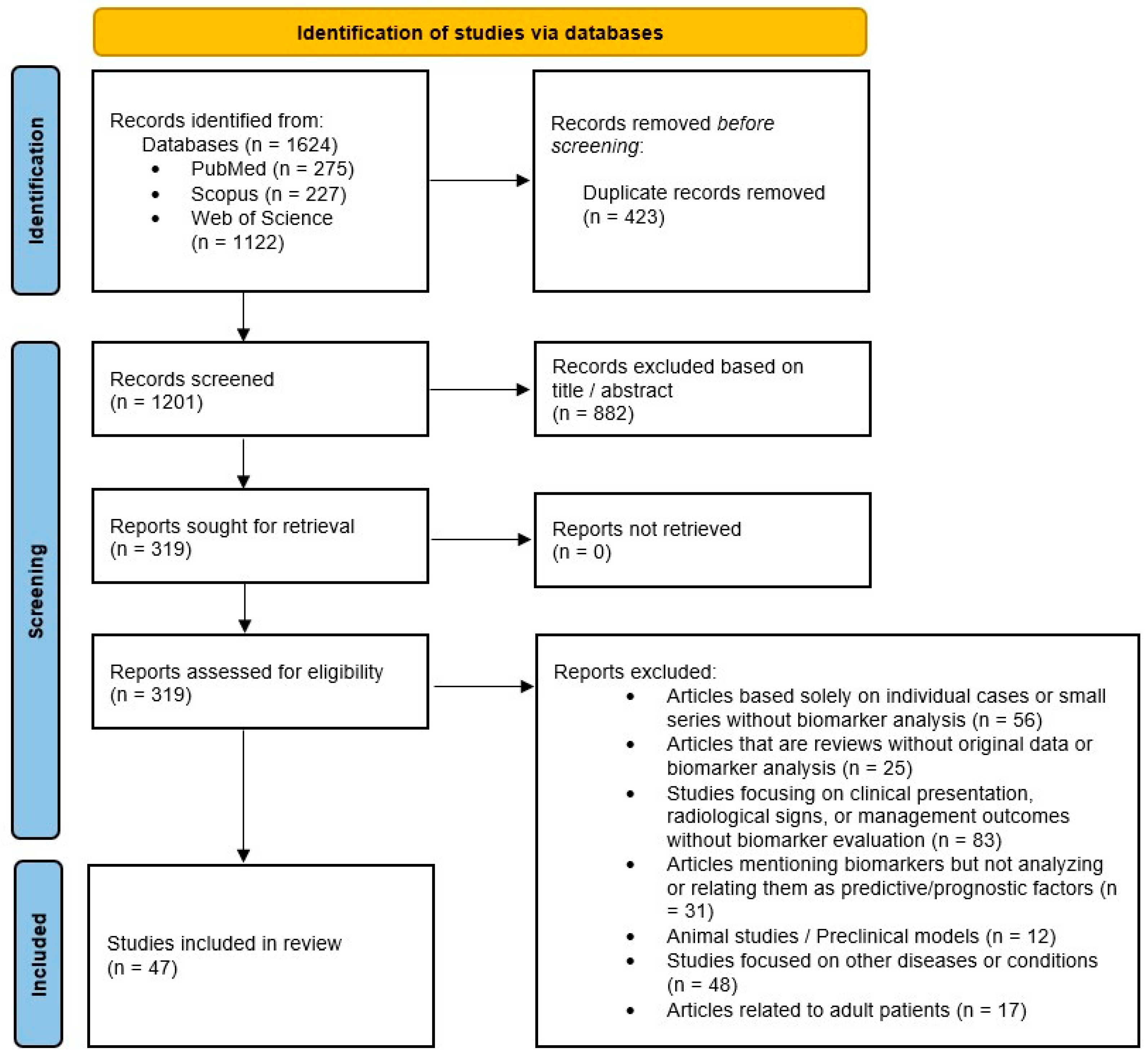
3.1. Study Selection
3.2. Study Characteristics, Risk of Bias, and Summary of Included Studies
3.3. Hematological and Blood Parameters
3.4. Markers of Inflammation and Immune Response
3.5. Derived Ratios and Inflammatory Indices
3.6. Biochemical and Metabolic Parameters
3.7. Oxidative Stress and Lipid Peroxidation Markers
3.8. Serum Proteins and Peptides
3.9. Acid-Base Balance and Gas Exchange Parameters
3.10. Other Biomarkers and Novel Markers
4. Discussion
5. Conclusions
Supplementary Materials
Author Contributions
Funding
Institutional Review Board Statement
Informed Consent Statement
Data Availability Statement
Acknowledgments
Conflicts of Interest
Abbreviations
| PRISMA | Preferred Reporting Items for Systematic Reviews and Meta-Analysis |
| JBI | Joanna Briggs Institute |
| CRP | C-reactive protein |
| NEC | Necrotizing enterocolitis |
| IBD | Inflammatory bowel disease |
| RNA | Ribonucleic acid |
| ELISA | Enzyme-linked immunosorbent assay |
| VIP | Vasoactive intestinal peptide |
| SP | Substance P |
| tRNA | Transfer ribonucleic acid |
| tRFs | Transfer ribonucleic acid-derived fragments |
| qRT-PCR | Quantitative reverse-transcription polymerase chain reaction |
| yrs | Years |
| mth | Month |
| OR | Odds ratio |
| WBC | White blood cells |
| TBA | Total bile acid |
| HSP | Henoch–Schönlein purpura |
| AI | Acute intussusception |
| ROC | Receiver operating characteristic |
| Mdn | Median |
| ICU | Intensive care unit |
| GAS | Gastrin |
| MTL | Motilin |
| IL | Interleukin |
| TNF-α | Tumor necrosis factor-α |
| ESR | Erythrocyte sedimentation rate |
| TLC | Total leukocyte counts |
| CBC | Complete blood count |
| PLR | Platelet-to-lymphocyte ratio |
| CAR | C-reactive protein/albumin ratio |
| NLR | Neutrophil-to-lymphocyte ratio |
| LCR | Lymphocyte-to-C-reactive protein ratio |
| IgAV | IgA vasculitis |
| CK-MB | Creatine kinase-MB |
| I-FABP | Intestinal fatty-acid-binding protein |
| SII | Systemic immune inflammatory index |
| SIRI | Systemic inflammation response index |
| PNR | Platelet-to-neutrophil ratio |
| LMR | Lymphocyte-to-monocyte ratio |
| alpha-GST | Alpha-glutathione S-transferase |
| Na | Sodium |
| K | Potassium |
| USGHR | Ultrasound-guided hydrostatic reduction |
| Ig | Immunoglobulin |
| EndoCAb | Antiendotoxin core antibody |
| HPLC | High-performance liquid chromatography |
| MCP-1 | Monocyte chemoattractant protein-1 |
| IMA | Ischemia-modified albumin |
| BUN | Blood urea nitrogen |
References
- Marsicovetere, P.; Ivatury, S.J.; White, B.; Holubar, S.D. Intestinal Intussusception: Etiology, Diagnosis, and Treatment. Clin. Colon Rectal Surg. 2017, 30, 30–39. [Google Scholar] [CrossRef] [PubMed]
- Alnamshan, M.; Almatroudi, D.; ALmutairi, D.; Almagushi, N.A.; Almadhi, L.; Alenazi, A.M. Idiopathic Intussusception in Infants and Children: Different Outcomes in Relation to Interventions. Cureus 2023, 15, e48026. [Google Scholar] [CrossRef] [PubMed]
- Attoun, M.A.; Albalawi, S.M.D.; Ayoub, A.; Alnasser, A.K.; Alkaram, E.H.; Khubrani, F.A.; Alzahrani, K.J.; Alatawi, K.A.; Almutairi, N.; Alnami, A.G. The Management of Intussusception: A Systematic Review. Cureus 2023, 15, e49481. [Google Scholar] [CrossRef]
- Wang, L.; Zhang, H.; Wang, D.; Shen, Q.; Huang, L.; Liu, T. Clinical features of intussusception in children secondary to small bowel tumours: A retrospective study of 31 cases. BMC Pediatr. 2024, 24, 227. [Google Scholar] [CrossRef]
- Rahmani, E.; Amani-Beni, R.; Hekmatnia, Y.; Fakhre Yaseri, A.; Ahadiat, S.A.; Talebi Boroujeni, P.; Kiani, M.; Tavakoli, R.; Shafagh, S.G.; Shirazinia, M.; et al. Diagnostic Accuracy of Ultrasonography for Detection of Intussusception in Children; a Systematic Review and Meta-Analysis. Arch. Acad. Emerg. Med. 2023, 11, e24. [Google Scholar]
- Rice-Townsend, S.; Chen, C.; Barnes, J.N.; Rangel, S.J. Variation in practice patterns and resource utilization surrounding management of intussusception at freestanding Children’s Hospitals. J. Pediatr. Surg. 2013, 48, 104–110. [Google Scholar] [CrossRef]
- Hwang, J.; Yoon, H.M.; Kim, P.H.; Jung, A.Y.; Lee, J.S.; Cho, Y.A. Current diagnosis and image-guided reduction for intussusception in children. Clin. Exp. Pediatr. 2023, 66, 12–21. [Google Scholar] [CrossRef]
- Zhao, J.; Sun, J.; Li, D.; Xu, W.J. Laparoscopic versus open reduction of idiopathic intussusception in children: An updated institutional experience. BMC Pediatr. 2022, 22, 44. [Google Scholar] [CrossRef]
- Li, S.M.; Wu, X.Y.; Luo, C.F.; Yu, L.J. Laparoscopic approach for managing intussusception in children: Analysis of 65 cases. World J. Clin. Cases 2022, 10, 830–839. [Google Scholar] [CrossRef]
- Kelley-Quon, L.I.; Arthur, L.G.; Williams, R.F.; Goldin, A.B.; St Peter, S.D.; Beres, A.L.; Hu, Y.Y.; Renaud, E.J.; Ricca, R.; Slidell, M.B.; et al. Management of intussusception in children: A systematic review. J. Pediatr. Surg. 2021, 56, 587–596. [Google Scholar] [CrossRef] [PubMed]
- Gluckman, S.; Karpelowsky, J.; Webster, A.C.; McGee, R.G. Management for intussusception in children. Cochrane Database Syst. Rev. 2017, 6, CD006476. [Google Scholar] [CrossRef] [PubMed]
- Ahmad, A.; Imran, M.; Ahsan, H. Biomarkers as Biomedical Bioindicators: Approaches and Techniques for the Detection, Analysis, and Validation of Novel Biomarkers of Diseases. Pharmaceutics 2023, 15, 1630. [Google Scholar] [CrossRef] [PubMed]
- Clough, J.; Colwill, M.; Poullis, A.; Pollok, R.; Patel, K.; Honap, S. Biomarkers in inflammatory bowel disease: A practical guide. Therap Adv. Gastroenterol. 2024, 17, 17562848241251600. [Google Scholar] [CrossRef]
- Ciurea, N.A.; Pantea, C.M.; Grama, P.; Kosovski, I.B.; Bataga, S. Fecal Zonulin as a Non-Invasive Marker of Intestinal Permeability: Findings from a Prospective Cohort Study. Medicina 2025, 61, 1527. [Google Scholar] [CrossRef]
- Guo, H.; Li, Y.; Wang, L. Assessment of inflammatory biomarkers to identify surgical/death necrotizing enterocolitis in preterm infants without pneumoperitoneum. Pediatr. Surg. Int. 2024, 40, 191. [Google Scholar] [CrossRef]
- Hu, H.; Chen, G.; Bai, D.; Wu, G.; Wu, Y.; Guo, S.; Tang, Y.; Liu, Q.; Hu, J.; Tian, Y. Diagnostic accuracy of routine hematological biomarkers for complications and prognosis in bowel obstruction: A systematic review and meta-analysis. World J. Emerg. Surg. 2025, 20, 80. [Google Scholar] [CrossRef]
- Zafirovski, A.; Zafirovska, M.; Kuhelj, D.; Pintar, T. The Impact of Biomarkers on the Early Detection of Acute Mesenteric Ischemia. Biomedicines 2023, 12, 85. [Google Scholar] [CrossRef]
- Sarshar, M.; Scribano, D.; Ambrosi, C.; Palamara, A.T.; Masotti, A. Fecal microRNAs as Innovative Biomarkers of Intestinal Diseases and Effective Players in Host-Microbiome Interactions. Cancers 2020, 12, 2174. [Google Scholar] [CrossRef]
- Rashid, H.; Hossain, B.; Siddiqua, T.; Kabir, M.; Noor, Z.; Ahmed, M.; Haque, R. Fecal MicroRNAs as Potential Biomarkers for Screening and Diagnosis of Intestinal Diseases. Front. Mol. Biosci. 2020, 7, 181. [Google Scholar] [CrossRef]
- Page, M.J.; McKenzie, J.E.; Bossuyt, P.M.; Boutron, I.; Hoffmann, T.C.; Mulrow, C.D.; Shamseer, L.; Tetzlaff, J.M.; Akl, E.A.; Brennan, S.E.; et al. The PRISMA 2020 statement: An updated guideline for reporting systematic reviews. BMJ 2021, 372, n71. [Google Scholar] [CrossRef] [PubMed]
- Munn, Z.; Barker, T.H.; Moola, S.; Tufanaru, C.; Stern, C.; McArthur, A.; Stephenson, M.; Aromataris, E. Methodological quality of case series studies: An introduction to the JBI critical appraisal tool. JBI Evid. Synth. 2020, 18, 2127–2133. [Google Scholar] [CrossRef]
- Soleimanpour, Z.; Memarian, S.; Rajabi, M.M.; Zamani, Z.; Alimadadi, H.; Gharib, B. Clinical and paraclinical differences between pediatric patients requiring surgical versus non-surgical treatment for intussusception: A retrospective study at a referral center in Iran. BMC Surg. 2025, 25, 373. [Google Scholar] [CrossRef]
- Ammar, S.; Sellami, I.; Krichen, E.; Rhaiem, W.; Sellami, S.; Hbaieb, M.; Kammoun, M.; Jarraya, A.; Zitouni, H.; Mhiri, R. Predicting surgical intervention in pediatric intussusception using machine learning model. Tunis. Med. 2025, 103, 792–797. [Google Scholar] [CrossRef]
- Kamffer, C.M.; du Preez, H.; van Rensburg, J.J. Risk factors associated with the outcomes of fluoroscopy-guided pneumatic enema reductions of intussusceptions. SA J. Radiol. 2025, 29, 3155. [Google Scholar] [CrossRef]
- Liu, D.; Cai, H.; He, Q.Q.; Wang, S.; Li, F.; Zhang, J.; Fu, Y.J.; Su, Z.L.; Jiang, Y.D.; Jing, L. Systemic immune inflammatory index (SII) as a predictive marker for the failure of air enema treatment in children with Ileocolic intussusception: A case-control study. BMC Pediatr. 2025, 26, 92. [Google Scholar] [CrossRef]
- Xu, X.X.; Cai, Y.J.; Liu, J.; Zhuang, Y.; Ma, Q. Predicting bowel necrosis in pediatric acute intussusception using roundness and other related factors. BMC Pediatr. 2025, 25, 843. [Google Scholar] [CrossRef]
- Elhadidi, M.; Elghazaly, M.; El-Saied, A.W.; Awad, M.; Elayyouti, M. Bypassing the Delay: Directing Pediatric Intussusception Cases to the OR Through Inflammatory Marker Assessment. World J. Surg. 2025, 49, 1519–1525. [Google Scholar] [CrossRef]
- Ulusoy, E.; Ulusoy, O.; Sütçüoğlu, N.; Küme, T.; Şık, N.; Yılmaz, D.; Duman, M. Is it possible to predict the spontaneous reduction of pediatric intussusception using biomarkers? Pediatr. Int. 2025, 67, e70141. [Google Scholar] [CrossRef] [PubMed]
- Chang, C.Y.; Chen, S.C.; Kuo, H.C.; Huo, C.H.; Tsai, C.F.; Yang, H.Y. Beyond hyponatremia: The crucial role of hypernatremia and hypokalemia in pediatric intussusception surgical outcomes. J. Pediatr. Surg. 2025, 162835. [Google Scholar] [CrossRef] [PubMed]
- Tuşat, M.; Memiş, S. Evaluation of inflammatory markers and HALP score in childhood intussusceptions. Ann. Clin. Anal. Med. 2025, 16, 530–534. [Google Scholar] [CrossRef]
- Yu, Y.Y.; Zhang, J.J.; Xu, Y.T.; Lin, Z.X.; Guo, S.K.; Li, Z.R.; Huang, H.Y.; Huang, X.Z. Developing and validating a nomogram for early predicting the need for intestinal resection in pediatric intussusception. Front. Pediatr. 2024, 12, 1409046. [Google Scholar] [CrossRef]
- Xia, B.; Chen, G.; Liu, Q.; Yan, C.; Lu, P.; Guo, C. A comprehensive scoring system for the better prediction of bowel resection in pediatric intussusception. BMC Gastroenterol. 2024, 24, 180. [Google Scholar] [CrossRef]
- Wei, X.Y.; Huo, H.C.; Li, X.; Sun, S.L.; Zhang, J. Relationship between postoperative rehabilitation style, gastrointestinal function, and inflammatory factor levels in children with intussusception. World J. Gastrointest. Surg. 2024, 16, 2640–2648. [Google Scholar] [CrossRef]
- Shah, J.Y.; Banday, I.; Hamdani, H.Z.; Haq, M.F.; Parray, F.Q.; Banday, M.; Bhat, G.A. A Study of Predictors of Failure of Nonoperative Management of Ileocolic Intussusception in Children. Euroasian J. Hepatogastroenterol. 2024, 14, 81–85. [Google Scholar]
- Budiananti, A.; Hariastawa, I.A.; Matulatan, F. Correlation between pathology indicators and intestinal necrosis in pediatric intussusception. Chirurgia 2024, 37, 183–187. [Google Scholar] [CrossRef]
- Mu, J.; Hao, J.; Zhen, X.; Mo, X.; Sheng, Y. Surgical complications in children with IgA vasculitis: Clinical analysis of 28 cases. Investig. Clín. 2024, 65, 134–142. [Google Scholar] [CrossRef]
- Liu, J.; Wang, Y.; Jiang, Z.; Duan, G.; Mao, X.; Zeng, D. Developing a Nomogram for Predicting Surgical Intervention in Pediatric Intussusception After Pneumatic Reduction: A Multicenter Study from China. Ther. Clin. Risk. Manag. 2024, 20, 313–323. [Google Scholar] [CrossRef]
- Liu, J.; Zeng, D.; Jiang, Z.; Xiu, W.; Mao, X.; Li, H. Developing a nomogram to predict recurrent intussusception after pneumatic reduction of primary intussusception in children. BMC Surg. 2024, 24, 275. [Google Scholar] [CrossRef]
- Mu, J. Clinical characteristics and risk factors of IgA vasculitis with intussusception and intestinal perforation. J. Paediatr. Child. Health 2024, 60, 5–11. [Google Scholar] [CrossRef] [PubMed]
- Kocaoğlu, Ç.; Kocaoğlu, C.; Madenci, H. Can Ischemia-Modified Albumin Be a Helpful Marker in the Diagnosis and Follow-Up of Childhood Intussusception? Pediatr. Emerg. Care 2024, 40, e105–e107. [Google Scholar] [CrossRef]
- Yu, S.; Feng, W.; Wang, Y.; Zhao, M.; Tu, Y.; Guo, Z. Serum total bile acid levels assist in the prediction of acute intussusception with abdominal type Henoch-Schonlein purpura in children. Front. Pediatr. 2023, 11, 1183470. [Google Scholar] [CrossRef] [PubMed]
- Yang, M.; Xie, Y.; Zhuang, Y.; Chen, Y.; Lin, X.; Liu, Z.; Zhang, P.; Xiao, W.; Chen, Y.; Chen, C.; et al. Risk factors and predictive models for early recurrent intussusception in children: A retrospective cohort study. Transl. Pediatr. 2023, 12, 1800–1809. [Google Scholar] [CrossRef]
- Zhuang, Y.; Wang, X.; Fan, X.; Li, F.; He, G.; Luo, M.; Tang, Y. Developing a nomogram for predicting surgical intervention in pediatric intussusception after hydrostatic reduction. Front. Pediatr. 2023, 11, 1092548. [Google Scholar] [CrossRef]
- Delgado-Miguel, C.; García, A.; Delgado, B.; Muñoz-Serrano, A.J.; Miguel-Ferrero, M.; Camps, J.; Lopez-Santamaria, M.; Martinez, L. Neutrophil-to-Lymphocyte Ratio as a Predictor of the Need for Surgical Treatment in Children’s Intussusception. Eur. J. Pediatr. Surg. 2023, 33, 422–427. [Google Scholar] [CrossRef]
- Zhang, J.; Dong, Q.; Su, X.; Long, J. Factors associated with in-hospital recurrence of intestinal intussusception in children. BMC Pediatr. 2023, 23, 428. [Google Scholar] [CrossRef]
- Wu, T.H.; Huang, G.S.; Wu, C.T.; Lai, J.Y.; Chen, C.C.; Hu, M.H. Clinical characteristics of pediatric intussusception and predictors of bowel resection in affected patients. Front. Surg. 2022, 9, 926089. [Google Scholar] [CrossRef] [PubMed]
- Liu, Z.; Yu, M.; Li, N.; Hu, M.; Su, F.; Hong, L.; Deng, X.; Zhang, S.; Tao, Q. Predictors of Surgical Treatment of Intussusception and Intussusception Complicated with Intestinal Necrosis in Children. Inn. J. Pediatr. 2022, 32, e117708. [Google Scholar] [CrossRef]
- Zhu, D.; Xu, X.; Zhang, M.; Wang, T.; Zhu, H. Significance of MCP-1 in predicting the short-term recurrence of primary intussusception in children: An observational study. Medicine 2022, 101, e30743. [Google Scholar] [CrossRef]
- Zhao, L.; Chen, W.; Guo, W.L. Serum tRNA-derived fragments as potential biomarkers in children with acute intussusception. Arch. Med. Sci. 2021, 19, 1889–1900. [Google Scholar] [CrossRef]
- Huang, H.Y.; Lin, X.K.; Guo, S.K.; Bao, X.Z.; Lin, Z.X.; Li, Z.R.; Huang, X.Z. Haemostatic indexes for predicting intestinal necrosis in children with intussusception. ANZ J. Surg. 2021, 91, 1485–1490. [Google Scholar] [CrossRef] [PubMed]
- Hou, J.; Hou, J.; Die, X.; Sun, J.; Zhang, M.; Liu, W.; Wang, Y. Impact of overweight/obesity on clinical outcomes after reduction for intussusception in children. Pediatr. Surg. Int. 2021, 37, 887–895. [Google Scholar] [CrossRef] [PubMed]
- Chen, B.; Cao, J.; Yan, C.; Zheng, C.; Chen, J.; Guo, C. A promising new predictive factor for detecting bowel resection in childhood intussusception: The lymphocyte-C-reactive protein ratio. BMC Pediatr. 2021, 21, 577. [Google Scholar] [CrossRef]
- Zhao, Q.; Yang, Y.; He, S.W.; Wang, X.T.; Liu, C. Risk factors for intussusception in children with Henoch-Schönlein purpura: A case-control study. World J. Clin. Cases 2021, 9, 6244–6253. [Google Scholar] [CrossRef]
- Younes, A.; Lee, S.; Lee, J.I.; Seo, J.M.; Jung, S.M. Factors Associated with Failure of Pneumatic Reduction in Children with Ileocolic Intussusception. Children 2021, 8, 136. [Google Scholar] [CrossRef]
- Lee, J.Y.; Byun, Y.H.; Park, J.S.; Lee, J.S.; Ryu, J.M.; Choi, S.J. Lactic acid level as an outcome predictor in pediatric patients with intussusception in the emergency department. BMC Pediatr. 2020, 20, 184. [Google Scholar] [CrossRef] [PubMed]
- Zhu, Z.C.; Wang, H.B.; Yan, X.Q.; Yang, J.; Duan, X.F.; Bian, H.Q.; Kuang, H.F. Serum substance P and vasoactive intestinal peptide levels in infants with acute intussusception. J. Int. Med. Res. 2019, 47, 2446–2451. [Google Scholar] [CrossRef]
- Xiaolong, X.; Yang, W.; Qi, W.; Yiyang, Z.; Bo, X. Risk factors for failure of hydrostatic reduction of intussusception in pediatric patients: A retrospective study. Medicine 2019, 98, e13826. [Google Scholar] [CrossRef] [PubMed]
- Lee, D.H.; Kim, S.J.; Lee, H.J.; Jang, H.J. Identifying Predictive Factors for the Recurrence of Pediatric Intussusception. Pediatr. Gastroenterol. Hepatol. Nutr. 2019, 22, 142–151. [Google Scholar] [CrossRef]
- Ademuyiwa, A.; Alakaloko, F.; Elebute, O.; Bode, C.; Udenze, I. Serum intestinal fatty-acid binding protein: Predictor of bowel necrosis in pediatric intussusception. J. Pediatr. Surg. 2018, 53, 335–338. [Google Scholar] [CrossRef]
- Lim, R.Z.M.; Lee, T.; Ng, J.Y.Z.; Quek, K.F.; Abdul Wahab, N.; Amansah, S.L.; Vellusamy, V.M.A.M.; Ngim, C.F. Factors associated with ultrasound-guided water enema reduction for pediatric intussusception in resource-limited setting: Potential predictive role of thrombocytosis and anemia. J. Pediatr. Surg. 2018, 53, 2312–2317. [Google Scholar] [CrossRef]
- Tamas, V.; Ishimine, P. Comparison of Lactic Acid Levels in Children with Suspected and Confirmed Intussusception. J. Emerg. Med. 2017, 53, 815–818. [Google Scholar] [CrossRef]
- Carapinha, C.; Truter, M.; Bentley, A.; Welthagen, A.; Loveland, J. Factors determining clinical outcomes in intussusception in the developing world: Experience from Johannesburg, South Africa. S. Afr. Med. J. 2016, 106, 177–180. [Google Scholar] [CrossRef]
- Karabulut, B.; Erdoğan, D.; Bostancı, I.; Onde, U.; Karakoç, A.E. Are interleukin-6, body mass index and atopy crucial in infantile intussusception? Indian J. Pediatr. 2010, 77, 1257–1260. [Google Scholar] [CrossRef]
- Fragoso, A.C.; Campos, M.; Tavares, C.; Costa-Pereira, A.; Estevão-Costa, J. Pneumatic reduction of childhood intussusception. Is prediction of failure important? J. Pediatr. Surg. 2007, 42, 1504–1508. [Google Scholar] [CrossRef] [PubMed]
- Willetts, I.E.; Kite, P.; Barclay, G.R.; Banks, R.E.; Rumley, A.; Allgar, V.; Stringer, M.D. Endotoxin, cytokines and lipid peroxides in children with intussusception. Br. J. Surg. 2001, 88, 878–883. [Google Scholar] [CrossRef] [PubMed]
- McDermott, V.G.; Taylor, T.; Mackenzie, S.; Hendry, G.M. Pneumatic reduction of intussusception: Clinical experience and factors affecting outcome. Clin. Radiol. 1994, 49, 30–34. [Google Scholar] [CrossRef]
- Frey, B.; Kistler, W. Reactive lymphocytes and intussusception in childhood. Pediatr. Surg. Int. 1994, 9, 360–361. [Google Scholar] [CrossRef]
- Reijnen, J.A.; Festen, C.; van Roosmalen, R.P. Intussusception: Factors related to treatment. Arch. Dis. Child. 1990, 65, 871–873. [Google Scholar] [CrossRef] [PubMed]
- Guo, H.; Lei, H.; Luo, J.; Yang, J.; Bian, H.; Yang, H.; Guo, Q. Clinical manifestation and treatment of intussusception in children aged 3 months and under: A single centre analysis of 38 cases. BMC Pediatr. 2025, 25, 233. [Google Scholar] [CrossRef]
- Alsabri, M.; Rath, S.; Elkarargy, M.A.; Aboali, A.A.; Abouelmagd, K.; Ramadan, A.A.A.; Gamboa, L.L.; Yoo, P.; Cheng, Y. Accuracy of ultrasound for intussusception in pediatric emergency presentations: A systematic review and diagnostic meta-analysis. Int. J. Emerg. Med. 2026, 19, 41. [Google Scholar] [CrossRef] [PubMed]
- Sandhu, A.I.; Abdullah, M.F.; Habib, T.; Arif, H. Diagnostic Accuracy of Sonographic Findings of Intestinal Intussusception in Paediatric Population with Surgical Outcomes as Gold Standard. Indus J. Biosci. Res. 2025, 3, 49–54. [Google Scholar] [CrossRef]
- Pušnik, L.; Slak, P.; Nikšić, S.; Winant, A.J.; Lee, E.Y.; Plut, D. Ultrasound-guided hydrostatic reduction of intussusception: Comparison of success rates between subspecialized pediatric radiologists and non-pediatric radiologists or radiology residents. Eur. J. Pediatr. 2023, 182, 3257–3264. [Google Scholar] [CrossRef]
- Bencardino, S.; D’aMico, F.; Zilli, A.; Parigi, T.L.; Allocca, M.; Fiorino, G.; Danese, S.; Furfaro, F. Fecal, Blood, and Urinary Biomarkers in Inflammatory Bowel Diseases. J. Transl. Gastroenterol. 2024, 2, 61–75. [Google Scholar] [CrossRef]
- Abdelrahman, S.T.; Omran, J.A.; Del Castillo Miranda, J.C.; Onyejesi, C.D.; Hamam, M.; Ebraheem, E.A.; Alsabri, M. Point-of-Care Biomarkers in Pediatric Emergency Medicine: Advancing Rapid Diagnostics, Overcoming Limitations, and Shaping Future Innovations. Curr. Treat. Options Peds. 2025, 11, 19. [Google Scholar] [CrossRef]
- Yan, Q.; Xu, L.; Chao, J.; Zhang, Z.; Wang, H. Characteristics of intestinal-related lymphoid hyperplasia in children and its correlation with intussusception of children. BMC Pediatr. 2022, 22, 641. [Google Scholar] [CrossRef]
- Jain, K.K. Role of Biomarkers in Health Care. In The Handbook of Biomarkers; Humana Press: Totowa, NJ, USA, 2010; pp. 115–188. [Google Scholar]
- Chen, L.; Zhang, X.; Shi, P. Recent advances in biomarkers for detection and diagnosis of sepsis and organ dysfunction: A comprehensive review. Eur. J. Med. Res. 2025, 30, 1081. [Google Scholar] [CrossRef]
- Mouliou, D.S. C-Reactive Protein: Pathophysiology, Diagnosis, False Test Results and a Novel Diagnostic Algorithm for Clinicians. Diseases 2023, 11, 132. [Google Scholar] [CrossRef]
- Singh, B.; Goyal, A.; Patel, B.C. C-Reactive Protein: Clinical Relevance and Interpretation. In StatPearls; StatPearls Publishing: Treasure Island, FL, USA, 2025. [Google Scholar]
- Alhazmi, H.; Alzahrani, A.; Alshaikh, S.; Azzhary, L.; Alhaddad, F.; Alshamrani, Z.; Alwagdani, R. The Significance of Inflammatory Markers in Pediatric Patients with Acute Gastroenteritis Presenting to the Emergency Department. Children 2025, 12, 617. [Google Scholar] [CrossRef] [PubMed]
- Tan, S.; Yang, X.; Mu, X.; Liu, S.; Wang, Y.; Li, Y.; Bian, Y.; Xu, C. The predictive role of peripheral serum inflammatory markers NLR, PLR, and LMR in ulcerative colitis and Crohn’s disease: A systematic review and meta-analysis. Front. Immunol. 2025, 16, 1623899. [Google Scholar] [PubMed]
- Alshuweishi, Y.; Alfaifi, M.; Almoghrabi, Y.; Al-Sheikh, Y.A.; Alfhili, M.A. A Retrospective Analysis of the Association of Neutrophil-Lymphocyte Ratio (NLR) with Anemia in the Saudi Population. Medicina 2023, 59, 1592. [Google Scholar] [CrossRef]
- Lin, B.D.; Hottenga, J.J.; Abdellaoui, A.; Dolan, C.V.; de Geus, E.J.C.; Kluft, C.; Boomsma, D.I.; Willemsen, G. Causes of variation in the neutrophil-lymphocyte and platelet-lymphocyte ratios: A twin-family study. Biomark. Med. 2016, 10, 1061–1072. [Google Scholar] [CrossRef]
- Honore, P.M.; Jacobs, R.; Hendrickx, I.; De Waele, E.; Spapen, H.D. Lactate: The Black Peter in high-risk gastrointestinal surgery patients. J. Thorac. Dis. 2016, 8, E440-2. [Google Scholar] [CrossRef]
- Montagnana, M.; Danese, E.; Lippi, G. Biochemical markers of acute intestinal ischemia: Possibilities and limitations. Ann. Transl. Med. 2018, 6, 341. [Google Scholar] [CrossRef]
- Belu, A.; Filip, N.; Trandafir, L.M.; Spoială, E.L.; Țarcă, E.; Zamosteanu, D.; Ghiga, G.; Bernic, J.; Jehac, A.; Cojocaru, E. Lactate, an Essential Metabolic Marker in the Diagnosis and Management of Pediatric Conditions. Diagnostics 2025, 15, 816. [Google Scholar] [CrossRef]
- Lingayat, N.; Jani, P. Early Lactate Clearance vs. Initial Lactate Levels: Which Predicts Mortality Better? A Systematic Review. Vasc. Endovasc. Rev. 2025, 8, 4–8. [Google Scholar]
- Esmailian, M.; Vakili, Z.; Nasr-Esfahani, M.; Heydari, F.; Masoumi, B. D-dimer Levels in Predicting Severity of Infection and Outcome in Patients with COVID-19. Tanaffos 2022, 21, 419–433. [Google Scholar]
- Liu, S.; Liu, Y.; Lai, S.; Xie, Y.; Xiu, W.; Yang, C. Values of serum intestinal fatty acid-binding protein, fecal calprotectin, and fecal human β-defensin 2 for predicting necrotizing enterocolitis. BMC Pediatr. 2024, 24, 183. [Google Scholar] [CrossRef] [PubMed]
- Voth, M.; Duchene, M.; Auner, B.; Lustenberger, T.; Relja, B.; Marzi, I. I-FABP is a Novel Marker for the Detection of Intestinal Injury in Severely Injured Trauma Patients. World J. Surg. 2017, 41, 3120–3127. [Google Scholar] [CrossRef] [PubMed]
- Khurana, S.; Corbally, M.T.; Manning, F.; Armenise, T.; Kierce, B.; Kilty, C. Glutathione S-transferase: A potential new marker of intestinal ischemia. J. Pediatr. Surg. 2002, 37, 1543–1548. [Google Scholar] [CrossRef]
- Ztriva, E.; Moschonas, I.C.; Tselepis, A.; Lambrou, D.; Ntaios, G.; Savopoulos, C.; Kaiafa, G. Monocyte Chemoattractant Protein-1 as a Biomarker in Acute Ischemic Stroke: A Prospective Pilot Study. J. Clin. Med. 2025, 14, 3295. [Google Scholar] [CrossRef]
- Zhang, Y.; Xiao, G.; Wang, R. Clinical significance of systemic immune-inflammation index (SII) and C-reactive protein-to-albumin ratio (CAR) in patients with esophageal cancer: A meta-analysis. Cancer Manag. Res. 2019, 11, 4185–4200. [Google Scholar] [CrossRef] [PubMed]
- Coşkun, S.; Güngörer, V.; Öner, N.; Sezer, M.; Karagöl, C.; Tekin, Z.E.; Tekgöz, P.N.; Kaplan, M.M.; Polat, M.C.; Çelikel, E.; et al. The role of indices in predicting disease severity and outcomes of multisystem inflammatory syndrome in children. Pediatr. Int. 2023, 65, e15609. [Google Scholar] [CrossRef] [PubMed]
- Jeong, H.R.; Hwang, I.T. MicroRNAs as novel biomarkers for the diagnosis and treatment of pediatric diseases. Clin. Exp. Pediatr. 2024, 67, 119–125. [Google Scholar] [CrossRef] [PubMed]
- Baldassarre, A.; Felli, C.; Prantera, G.; Masotti, A. Circulating microRNAs and Bioinformatics Tools to Discover Novel Diagnostic Biomarkers of Pediatric Diseases. Genes 2017, 8, 234. [Google Scholar] [CrossRef]
- Obeagu, E.I. Integrative bioinformatics approaches for early detection biomarkers in ovarian cancer. Ann. Med. Surg. 2026, 88, 1687–1694. [Google Scholar] [CrossRef]
- Servian, J.E.; Brady, B.; Biswas, P.; Sukumar, T.K.; King, S.E. Pharmacoepigenetic Biomarkers in Inflammatory Bowel Diseases: A Narrative Review. Yale J. Biol. Med. 2025, 98, 171–186. [Google Scholar] [CrossRef]
- Kumar, D.R.; Banaś, A.; Krukiewicz, K. Challenges and Advances in Biomarker Detection for Rapid and Accurate Sepsis Diagnosis: An Electrochemical Approach. Biosensors 2024, 14, 309. [Google Scholar] [CrossRef]

| Author(s), Year of Publication | Country | Study Design | Sample Size, Male: Female, Patients’ Age | Type of Intussusception | Cause | Type of Biomarker | Method of Biomarker Measurement | Timing of Biomarker Assessment | Predictive/Prognostic Outcomes | Treatment Modalities Used | Association Between Biomarkers and Outcomes | Threshold or Cutoff Values Used for Positivity |
|---|---|---|---|---|---|---|---|---|---|---|---|---|
| Soleimanpour et al., 2025 [22] | Iran | Retrospective cohort study | 165 (55—underwent surgery, 110—treated non-surgically), 116:49, 2.68 ± 1.65 yrs/3.88 ± 3.51 yrs | Location of the intussusception: ileoileal (n = 8), ileocolic (n = 112), colocolic (n = 2), right lower quadrant (n = 12), right upper quadrant (n = 19), left lower quadrant (n = 3), left upper quadrant (n = 1), jejunojenunal (n = 3), jejuno-ileal (n = 1), multiple (n = 4) | NA | WBC, ESR, CRP | Blood tests | Pre-treatment | Comparison of groups, comparison of clinical and paraclinical characteristics | Hydrostatic or pneumatic reduction, surgery | Laboratory test results showed no significant differences between groups (p > 0.05). The study found that younger age, bloody stools, and ileocolic location were associated with a higher likelihood of needing surgery | NA |
| Ammar et al., 2025 [23] | Tunisia | Retrospective cohort study | 156 (109—training group, 47—validation group) | Ileocolic | NA | WBC, CRP, sodium, potassium, chloride | Blood tests | At diagnosis | Likelihood of requiring surgical intervention | Hydrostatic or pneumatic reduction, surgery | The univariate analysis showed that the elevated WBC (p = 0.029) was associated with surgical treatment. Bloody stools (p = 0.033; OR = 2.61), the duration of symptoms (p = 0.028; OR = 1.02), and the length of the intussusception (p = 0.014; OR = 1.265) were identified as independent risk factors for surgical treatment | NA |
| Kamffer et al., 2025 [24] | South Africa | Retrospective cohort study | 110 (73—attempts air enema, 37—primarily surgically managed), NA, Mdn 7 mth | Ileocolic | In three of the patients who were part of the unsuccessful group, a pathological lead point was found, including Meckel’s diverticulum and Wauch’s syndrome | CRP, white cell counts | Blood tests | At diagnosis | Identification of factors associated with fluoroscopy-guided air enema reduction outcomes in pediatric intussusception | Air enema, surgery | Unsuccessful fluoroscopy-guided air enema was significantly associated with younger age (p = 0.0249), dehydration (p = 0.0299), ascites (p = 0.0172), and increased outer wall intussusception diameter on ultrasound (p = 0.0026). No predictive value of elevated levels of CRP and white cell counts regarding the success of pneumatic reduction (p > 0.05) | NA |
| Liu et al., 2025 [25] | China | Retrospective cohort study | 574 (469—air enema success group, 105—air enema failure group), 396:178, mean 22.13 mth | Ileocolic | Idiopathic | WBC, absolute neutrophil, absolute lymphocyte, absolute monocyte, platelet counts, SII, SIRI, NLR, PLR, PNR, LMR | Blood tests | Pre-treatment | Identification of SII as a predictor of air enema failure | Air enema, surgery | Threshold analysis revealed that, to the right of this inflection point (332.3), there was a significant 25% increase in the risk of air enema treatment failure for ICI per 100-unit increase in the SII (OR 1.25). In the ROC curve analysis, SII demonstrated the highest AUC of 0.8665 compared to other inflammation indices | The critical point for SII was estimated to be 721.5 (sensitivity, 86.67%; specificity, 71.00%) |
| Xu et al., 2025 [26] | China | Retrospective cohort study | 87 (23—intestinal necrosis group, 64—control group), 58:29, NA | NA | NA | NLR, CRP | Automatic blood analyzer | Pre-treatment | Investigation of the risk factors of intestinal necrosis in children with intussusception | Air enema, surgery | NLRs were all higher in the bowel necrosis group (p < 0.05). The logistic regression analysis indicated that roundness (×100) (OR = 1.397) and blood flow signal (<grade 4) (OR = 0.099) were independent predictors of bowel necrosis in intussusception | NA |
| Elhadidi et al., 2025 [27] | Egypt | Retrospective cohort study | 100 (60—nonresection group, 40—resection group), 46:54, 13.29 ± 11.31 8.4 ± 4.09 mth | NA | NA | CBC, albumin, CRP, NLR, PLR, LCR, CAR | Blood tests | Pre-treatment | The effectiveness of different combinations of inflammatory markers in predicting intestinal necrosis and the need for intestinal resection in cases of intussusception | Surgery | A statistically significantly higher mean CAR was observed among cases with resection. Conversely, the mean LCR was significantly lower in the resection group. CAR was a statistically significant predictor of the need for resection, with each unit increase in CAR increasing the risk by 1.42 | LCR—cutoff point of 0.1233 (sensitivity of 85.7% and specificity of 90%), CAR—cutoff point of 7.73 (sensitivity of 92.6% and specificity of 90%). If a patient’s CAR exceeds 7.73 and if LCR is below 0.1233, they are more likely to need surgery due to necrosis |
| Ulusoy et al., 2025 [28] | Turkey | Prospective case–control | 78 (52—intussusception group, 26—control group), 47:31, 24 mth | Ileocolic | NA | Alpha-GST, I-FABP, inflammatory markers | ELISA, blood tests | Pre-treatment | Alpha-glutathione S-transferase (alpha-GST), intestinal fatty-acid-binding protein (I-FABP), and inflammatory markers may predict spontaneous reduction in ileocolic intussusceptions | Hydrostatic reduction, surgery | Alpha-GST, NLR, and CRP levels were significantly higher in the group requiring intervention than in the spontaneous reduction group | Alpha-GST: 3.29 ng/mL (AUC: 0.917, sensitivity: 88.9%, specificity: 85.7%), CRP: 5.5 ng/mL (AUC: 0.808, sensitivity: 64.3%, specificity: 85.7%), NLR: 1.87 (AUC: 0.739, sensitivity: 75.0%, specificity: 71.4%) |
| Chang et al., 2025 [29] | Taiwan | Retrospective cohort study | 11,111, 5348:3252, 0–3 years group—7562 cases (1.0 ± 1.0 yrs)/3–18 years group—3485 cases (8.8 ± 4.6 yrs) | NA | NA | Sodium, potassium | Blood tests | 24 h before or on the day of diagnosis | Quantification of the association between dysnatremia and dyskalemia at presentation and major surgical complications in children with intussusception | Air or contrast enema, surgery | Electrolyte disturbances were prevalent: hyponatremia (9.3%), hypernatremia (1.8%), hypokalemia (4.7%), and hyperkalemia (2.8%). After adjustment, hypernatremia demonstrated the most potent association across all outcomes. Hyponatremia was independently associated with all three surgical outcomes, with more modest effect sizes (open reduction, bowel perforation or resection, peritonitis or sepsis) | Hyponatremia < 135 mmol/L, hypernatremia > 145 mmol/L, hypokalemia < 3.5 mmol/L, hyperkalemia > 5.0 mmol/L |
| Tuşat & Memiş, 2025 [30] | Turkey | Retrospective cohort study | 78 (30—surgical group, 48—non-surgical group), 49:29, Mdn 19 mth | Surgical group: ileoileal (n = 3), ileocolic (n = 27) | NA | CBC, NLR, PLR, HALP score | Blood tests | Pre-treatment | Determination of whether the HALP score and the inflammatory markers scores differ between cases requiring surgical reduction and those not requiring surgical reduction in patients diagnosed with intussusception | Hydrostatic reduction, surgery | Higher NLR and PLR, lower HALP scores associated with surgical cases (p < 0.0001). However, no statistically significant difference was detected for CRP levels (p = 0.095) | NA |
| Yu et al., 2024 [31] | China | Retrospective cohort study | 547 (414—non-intestinal necrosis, 133—intestinal necrosis and underwent resection), 365/182, training set = 15.81 ± 23.91 mth, validation set = 17.6 ± 26.98 mth | Ileo-colic (n = 263), ileo-cecal (n = 117), compound/complex (n = 95), multiple (n = 2), small intestinal (n = 70) | NA | A series of parameters from the blood | Blood tests | Pre-treatment | Predicting the risk of intestinal resection | Failed air enema, intestinal resection | Duration of symptoms (OR = 1.050, p < 0.001), CRP (OR = 1.021, p = 0.004), WBCs (OR = 1.110, p < 0.001), ascites (OR = 3.781, p < 0.001) | Duration of symptoms, C-reactive protein, white blood cells, and ascites were selected for inclusion in the nomogram, with a concordance index of 0.871 |
| Xia et al., 2024 [32] | China | Retrospective cohort study | 660 (442—no resection group, 218—bowel resection group), 302:140/143:65, Mdn 21.88 mth/15.73 mth | Location of intussusception: ascending colon (n = 483), transverse colon (n = 141), descending colon (n = 20), sigmoid colon (n = 16) | NA | Blood-based biomarkers | Blood tests | Pre-treatment | The necessity of bowel resection | Surgery | Bowel resection occurrence was linked to an extended duration of symptoms (OR = 2.14, p = 0.0015), the presence of gross bloody stool (OR = 8.98, p < 0.001), elevated C-reactive protein levels (OR = 4.79, p = 0.0072), lactate clearance rate (OR = 17.25, p < 0.001), and the intussusception location (OR = 12.65, p < 0.001) | A scoring system (totaling 14.02 points) was developed from the cumulative β coefficients, with a threshold of 5.22 effectively differentiating infants requiring bowel resection, CRP > 8.0 mg/L, LCR < 0.121 |
| Wei et al., 2024 [33] | China | Retrospective cohort study | 18 (6—routine nursing group, 12—rehabilitation training group), 11:7, 5.24 ± 3.05 yrs/5.16 ± 3.14 yrs | NA | NA | GAS, MTL, IL-2, IL-4, IL-6, IL-10, CRP, TNF-α (serum) | Radioimmunoassay, ELISA | Post-treatment—after 5 days of rehabilitation | Effects of different rehabilitation methods on gastrointestinal function and inflammatory factor levels | Surgery (laparoscopic), rehabilitation | GAS (r = 0.490) and MTL (r = 0.714) levels were positively correlated with postoperative rehabilitation (p < 0.05). IL-2 (r = −0.782), IL-4 (r = −0.871), IL-6 (r = −0.971), IL-10 (r = −0.979), CRP (r = −0.981), and TNF-α (r = −0.921) levels were negatively correlated with postoperative rehabilitation (p < 0.05) | NA |
| Shah et al., 2024 [34] | India | Prospective cohort study | 110 (reduced and not reduced group), 72:38, mean age 13.54 months | Ileocolic | NA | CBC, CRP, blood gas analysis with serum electrolytes | Blood tests | At diagnosis | Assessment of the clinical and radiological predictors of success or failure of nonoperative management of intussusception | Hydrostatic reduction, surgery | Elevated TLC, CRP, lactate, and low potassium were significantly associated with increased failure risk (p < 0.001) | NA |
| Budiananti et al., 2024 [35] | Indonesia | Cross-sectional study | 36, 24:12, mean 7 mth (no intestinal necrosis), 8 mth (intestinal necrosis) | NA | NA | WBC, lymphocytes, neutrophils, platelets, albumin, CRP, PLR, CAR, NLR, LCR | Blood tests | At diagnosis | Prediction of intestinal ischemia and necrosis | Surgery | Significantly related markers were albumin (p = 0.00; AUC 0.888), CRP (p = 0.00; AUC 0.948), CAR (p = 0.00; AUC 0.914), NLR (p = 0.032; AUC 0.714), and LCR (p < 0.001; AUC 0.906). | Albumin ≤ 3.5 g/dL CRP ≥ 3.37 mg/dL CAR > 1 NLR > 1.2 LCR ≤ 0.9 |
| Mu et al., 2024 [36] | China | Retrospective cohort study | 28 (21—intussusception, 7—intestinal perforation), 10:18, mean 7.2 yrs | Ileoileal (n = 12), ileo-colonic (n = 6), jejunal-jejunal (n = 2), colo-colonic (n = 1) | Associated with IgA vasculitis | Leukocytes, CRP, D-dimer | Blood tests | At diagnosis | Surgical complications in children with IgAV | Conservative treatment methods, surgery | Increased leukocytes were observed in 60.7% of children. CRP and D-dimer were elevated in 53.3% and 75% of children, respectively | NA |
| Liu et al., 2024 [37] | China | Retrospective cohort study | 1041 (852—successful reduction group, 189—failed reduction group, 728—training set, 313—validation set), 696:345, 33.50 ± 20.98 mth/26.55 ± 27.56 mth | NA | Nested position: right colon (n = 818), left colon (n = 223) | WBC, CRP | Blood tests | At diagnosis | Development and validation of a nomogram for predicting the need for surgical intervention in pediatric intussusception after pneumatic reduction | Pneumatic reduction, surgery | Logistic regression analysis of the training set identified age, time of abdominal pain, presence or absence of hematochezia, C-reactive protein value from blood test on admission (OR 1.034, p < 0.001), and nested position indicated by B-ultrasound as independent predictors of intussusception intervention | NA |
| Liu et al., 2024 [38] | China | Retrospective cohort study | 2406 (208—recurrent intussusception group, 2198—control group), 1620:786, mean 30.12 ± 19.97 mth | Excluded ileal intussusception from analysis | NA | WBC, CRP | Blood tests | Pre-treatment | Development and validation of a nomogram for predicting recurrent intussusception in children within 48 h after pneumatic reduction in primary intussusception | Pneumatic reduction | Age, abdominal pain time, white blood cell counts (OR = 1.12), and hypersensitive C-reactive protein levels (OR = 1.16) were identified as predictors and incorporated into the nomogram | NA |
| Mu, 2024 [39] | China | Retrospective case–control study | 192 (32, 25—intussusception, 7—intestinal perforation, 160—control group), 99:93, mean 6.44 ± 2.21 yrs | Ileum-ileal (n = 16), ileo-colonic (n = 6), jejunum-jejunum (n = 2), colon-colic (n = 1) | Small bowel polyps (n = 2), Meckel’s diverticulum (n = 1), malrotation (n = 1) | WBC, CRP, D-dimer, erythrocyte sedimentation rate | Blood tests | At diagnosis | A summary of the clinical features of IgAV complicated by intussusception and intestinal perforation, and explore its risk factors | Enema reduction, surgery | Higher WBC, CRP, and D-dimer levels are associated with surgical complications. Multivariate logistic regression analysis indicated that age ≤ 7 years, GI symptoms prior to skin purpura, abdominal pain intensity, and timing of glucocorticoid treatment were independent risk factors of IgAV with intussusception and intestinal perforation | WBC > 10 × 109/L CRP > 8 mg/L D-dimer > 0.24 mg/L |
| Kocaoğlu et al., 2024 [40] | Turkey | Case–control study | 70 (36—intussusception group, 34—control group), 36:34, Mdn 34 mth/30 mth | Ileoileal (n = 9), ileocecal (n = 24), colocolic (n = 3) | NA | IMA | ELISA | Pre-treatment | Determination of the sensitivity of IMA and the correlation between IMA and the severity of intestinal ischemia in intussusception cases | Hydrostatic reduction, surgery | The mean IMA level of the intussusception group was 179.13 ± 220.33 ng/mL, whereas the mean level was found as 89 ± 70.9 ng/mL in the control group (p = 0.023). When the patients were categorized as ileoileal, ileocecal, and colocolic, the mean IMA levels were detected as 235.65 ± 268.14 ng/mL, 174.46 ± 212.8 ng/mL, and 46.95 ± 19.56 ng/mL, respectively | The sensitivity and specificity rates were determined as 55.6% and 55.9% using a cutoff value of 60.1 ng/mL for IMA |
| Yu et al., 2023 [41] | China | Retrospective cohort study | 708 (613—HSP, 95—HSP with AI), HSP without AI group—357/256, Mdn 5 yrs 8 mth, HSP with AI group—56/39, Mdn 6 yrs 5 mth | According to imaging manifestations - ileocolic (n = 45), small intestinal (n = 37) | NA | Total bile acid (serum) | Blood tests | Pre-treatment (at diagnosis during GI symptoms) | High serum TBA predicts AI; higher TBA levels were associated with increased operative treatment, intestinal necrosis, and longer hospital stays | Air enema, intestinal resection | Vomiting (OR = 396.492, p < 0.001), haematochezia (OR = 87.436, p = 0.001), TBA (OR = 16.287, p < 0.001), and D-dimer (OR = 5.987, p = 0.003) were independent risk factors for abdominal-type HSP with AI | TBA > 3 μmol/L for predicting AI in children with abdominal-type HSP, TBA ≥ 6.98 μmol/L associated with an increased incidence of operative treatment, intestinal necrosis, and length of hospital stay |
| Yang et al., 2023 [42] | China | Retrospective cohort study | 869 (787—with no relapse, 82—recurrent intussusception), 591/278, <1 year old = 312, ≥1 year old = 557 | NA | NA | Blood-based biomarkers | Blood tests | Pre-treatment | Likelihood of early recurrence of intussusception (<48 h) | Enema therapy, surgery | Age (OR 7.67, p = 0.001), vomiting (OR 0.17, p < 0.001), bloody stool (OR 0.14, p = 0.01), and monocyte ratio (OR 9.52, p = 0.003) were independently associated with the clinical endpoints | Children older than 1 year in age, who lacked vomiting and bloody stool symptoms, and who exhibited an elevated ratio of monocytes, were more likely to relapse early |
| Zhuang et al., 2023 [43] | China | Retrospective cohort study | 199 (139—training group, 60—validation group), 146:53, | Location: right side (n = 145), left side (n = 54) | NA | WBCs, platelets, CRP, fibrinogen, CK-MB, potassium, sodium, chloride | Blood tests | At diagnosis | Development and validation of a nomogram for predicting surgical intervention in pediatric intussusception after hydrostatic reduction | Hydrostatic reduction, surgery | Duration of symptoms, bloody stools, WBCs (OR = 1.203, p = 0.026), CK-MB (OR = 1.034, p = 0.040), long-axis diameter, poor prognostic signs by ultrasound and mental state were identified as the independent predictors of surgical intervention for intussusception | A model that incorporated the independent predictors was developed and presented as a nomogram. The C-index of the nomogram in the validation set was 0.948 |
| Delgado-Miguel et al., 2023 [44] | Spain | Retrospective case–control study | 511 (410—effective enema group, 101—need for surgery group), 333:178, Mdn 16 mth/16.5 mth | Ileocolic | NA | CBC, ionogram, glucose, urea, fibrinogen, CRP | Blood tests | Pre-treatment | Identification of predictors of the need for surgical treatment in ileocolic intussusception | Hydrostatic enema, surgical treatment | The surgery group presented higher median laboratory inflammatory markers: NLR (6.8 vs. 1.8; p < 0.001), neutrophils (10,148 vs. 7468; p < 0.001), and CRP (28.2 vs. 4.7; p < 0.001). In ROC curve analysis, NLR had an AUC of 0.925. | It was estimated that a cutoff point of NLR greater than 4.52 (sensitivity: 73.2%; specificity: 94.5%), neutrophils greater than 9420 (sensitivity: 55.4%, specificity: 82.8%), and CRP greater than 4.8 (sensitivity: 65.7%, specificity: 60.4%) |
| Zhang et al., 2023 [45] | China | Retrospective cohort study | 624 (73—recurrence of intussusception after successful reduction with air enema), 400:224; Mdn 1.8 yrs | Site of intussusception: hepatic flexure (n = 237), transverse colon (n = 184), Ileocecal junction (n = 92), ascending colon (n = 63), splenic flexure (n = 31), other (n = 17) | NA | WBC count, neutrophil percentage, lymphocyte Percentage, CRP | Blood tests | Pre-treatment | Investigation of the factors associated with in-hospital Recurrence of intussusception | Air enema reduction | Multivariate logistic regression analysis identified age > 1 year old (OR = 7.65), secondary intestinal intussusception (OR = 14.40), and mesenteric lymph node enlargement (OR = 1.90) as factors independently associated with in-hospital recurrence of intussusception | NA |
| Wu et al., 2022 [46] | Taiwan | Retrospective observational study | 584, 379/205, 27.2 ± 20.3 mth | Operative findings: ileocolic (n = 90), ileoileal (n = 9), ileo-ileocolic or ileo-colocolic (n = 39) | 38 patients had pathological lead points—Meckel diverticulum (n = 8), bands (n = 8), polyp (n = 5), enlarged lymph nodes (n = 5), lymphoid mass (n = 5), Burkitt lymphoma (n = 1), enteric duplication cyst (n = 2), Peutz-Jeghers syndrome (n = 3), and cecal serosal lesions (n = 1) | Blood-based biomarkers | Blood tests | Pre-treatment | Prediction of the need for bowel resection | Enema reduction, surgery | Abdominal pain (OR = 0.372, p = 0.013), bloody stool (OR = 3.553, p = 0.044), and hyponatremia (OR = 4.12, p = 0.003) were independent predictors for surgery. Prolonged time to surgery (OR = 6.863, p = 0.009), long intussusception (OR = 5.088, p = 0.014), pathological lead point (OR = 6.926, p = 0.003), and ICU admission (OR = 11.777, p = 0.001) were associated with bowel resection | Hyponatremia (<135 mEq/L), hypochloremia (≤100 mEq/L), hyperglycemia (≥100 mg/dL) |
| Liu et al., 2022 [47] | China | Retrospective cohort study | 8235 (5743—hydraulic enema group (HE), 2492—surgical group (SM)/398—with necrosis subgroup (SN+), 2094—without necrosis subgroup (SN−)), 3986:1757/1690:802, 1.54 ± 0.02 yrs/1.17 ± 0.04 yrs | NA | NA | 74 clinical and biochemical parameters | Blood tests | Pre-treatment | Likelihood of successful reduction with an enema versus the need for surgery. Likelihood of intestinal necrosis. | Hydrostatic reduction, surgery (+/− resection) | The prediction model composed of seven variables (NEUT#, platelet, albumin, β-2MG macroglobulin, glucose, uric acid, and chlorine was suitable for HE/SM. The prediction model composed of six variables (NEUT#, average hemoglobin concentration, PLT, the total protein, CRP, and urea) was suitable for SN−/SN+ | NA |
| Zhu et al., 2022 [48] | China | Retrospective observational study | 412 (375—nonrecurring cases, 37—cases of short-term recurrence), 293:119, NA | Mass location: ascending colon (n = 280), transverse colon (n = 132) | NA | MCP-1, IL-6 | ELISA | Pre-treatment | Evaluation of the relationship between the expression level of MCP-1 in peripheral blood and the short-term recurrence of primary intussusception in children | Hydrostatic reduction | Logistic regression analysis found that increased MCP-1 was a risk factor for recurrence | ROC showed that 23.24 ng/mL was used as a cutoff value (sensitivity—82.14%, specificity—75.67%) |
| Zhao et al., 2021 [49] | China | Cross-sectional study | 40 (intussusception—20/healthy controls—20), 10:10/12:8, 30.97 ± 34.94 mth/35.21 ± 40.78 mth | Ileum colon (n = 16), ileum cecum (n = 2), ileum ileum (n = 2), | Predominantly idiopathic; some cases with lead points (e.g., duplication) | Transfer ribonucleic acid (tRNA)-derived fragments (tRFs) (serum) | qRT-PCR | Immediately after the final diagnosis by air enema, before reduction | Comparison of groups, focus on diagnosis rather than prognosis | Aair enema (n = 18), surgery (n = 2) | tRF-Leu-TAA-006 (0.984, p < 0.05), tRF-Gln-TTG-033 (0.970, p < 0.05), tRF-Lys-TTT-028 (0.837, p < 0.05) correlated with diagnosis | tRFs with AUC > 0.5, p < 0.05 were considered appropriate biomarkers |
| Huang et al., 2021 [50] | China | Retrospective Cohort study | 540 (113—intestinal necrosis group, 427—non-intestinal necrosis groups), 355:185, Mdn 5.23/7.52 | NA | Most cases are idiopathic; 37 cases had pathological lead points | Fibrinogen, paletets, D-dimer | Blood tests | Pre-treatment | Determination of the risk factors for intestinal necrosis among children with failed non-surgical reduction for intussusception | Surgery | Multivariable analysis revealed that duration of symptom (OR 1.12, p = 0.000), fibrinogen (OR 1.26, p = 0.010) and D-dimer (OR 2.07, p = 0.000) independently predicted intestinal necrosis in individuals undergoing surgical reduction for intussusception | Intestinal necrosis in intussusception patients was more likely with D-dimer levels > 1.005 mg/L |
| Hou et al., 2021 [51] | China | Retrospective cohort study | 564 (132—overweight and obesity group, 432—no overweight and obesity group), 405:159, <24 mth = 353, >24 mth = 211) | Ileocolic | Idiopathic (patients with identified lead points were excluded) | WBC, CRP | Blood tests | Pre-treatment | Comparison of clinical outcomes after primary air enema reduction for intussusception in grouped overweight and obese (body mass index-for-age percentile ≥ 85) pediatric patients compared with non-overweight and obese patients | Pneumatic reduction, surgical reduction | After multivariate logistic regression analysis, overweight and obesity (OR = 8.045) and WBC count ≥ 20 × 109/L were risk factors for both surgical reduction (OR = 6.151) and the recurrence (OR = 3.357) of intussusception | WBC count ≥ 20 × 109/L (predefined threshold) |
| Chen et al., 2021 [52] | China | Retrospective cohort study | 115 (47—underwent intestinal resection, 68—did not undergo intestinal resection), NA, 1.79 ± 0.88 yrs/1.87 ± 0.92 yrs | NA | NA | Neutrophils, platelets, CRP, lymphocytes, albumin | Blood tests | Pre-treatment (within 2 days prior to surgery) | Investigation of the value of various combinations of inflammatory factors to predict intestinal necrosis and resection | Surgery | A combination of lymphocytic count along with C-reactive protein levels demonstrated the highest correlation with intestinal resection due to intussusception compared with other parameters in the patients, with a sensitivity of 0.82 and specificity of 0.80 for the diagnosis of strangulation | CRP > 11.26, albumin > 29.4, PLR > 188.5, LCR < 0.121, NLR > 5.72, CAR = 0.286 |
| Zhao et al., 2021 [53] | China | Retrospective case–control study | 160 (60—children with HSP, 100—control group), 71:89, mean 6.6 ± 2 yrs | Surgery: small bowel intussusception (n = 29), ileocolic (n = 19) | NA | WBC count, neutrophil count, platelet count, CRP, erythrocyte sedimentation rate, D-dimer | Blood tests | Pre-treatment | Investigation of the risk factors for intussusception in children with Henoch–Schönlein purpura | Air enema reduction, surgery | Univariate and multiple regression analyses revealed age at onset, not receiving glucocorticoid therapy within 72 h of emergence of GI symptoms, hematochezia, and increased D-dimer levels (OR = 7.193) as independent risk factors for intussusception in children with HSP (p < 0.05) | D-dimer levels ≥ 1 mg/L |
| Younes et al., 2021 [54] | South Korea | Retrospective cohort study | 145 (124—pneumatic reduction, 21—surgical reduction), 95:50, mean 24.6 ± 15.2 mth | Ileocolic | Idiopathic | WBC with differential, hemoglobin, and CRP | Blood tests | At diagnosis | Identification of factors that can lead to pneumatic reduction failure in children with ileocolic intussusception | Pneumatic reduction, surgery | Multivariate analysis showed that a high segmented neutrophil count, low hemoglobin level, and higher weight percentile were significantly associated with pneumatic reduction failure | Segmented neutrophil > 67.3% hemoglobin < 12.2 g/dL |
| Lee et al., 2020 [55] | South Korea | Retrospective cohort study | 249 (199—good outcome group, 50—poor outcome group/intussusception recurrence or required surgical reductions), 159:90, Mdn 1.8 yrs | Ileocolic | Idiopathic | pH, lactic acid, bicarbonate | Venous blood gas analysis | At diagnosis | Determination of whether lactic acid levels are associated with pediatric intussusception outcomes | Air enema, surgery | The poor and good outcome groups showed significant differences in their respective blood gas analyses for pH (7.39 vs. 7.41, p = 0.001), lactic acid (1.70 vs. 1.30 mmol/L, p < 0.001), and bicarbonate (20.70 vs. 21.80 mmol/L, p = 0.036). Multivariable logistic regression analyses showed that pH (OR 0.000, p = 0.003) and lactic acid (OR 3.066, p < 0.001) levels were the two factors significantly associated with poor outcomes. | When the lactic acid level cutoff values were ≥1.5, ≥2.0, ≥2.5, and ≥3.0 mmol/L, the positive predictive values for poor outcomes were 30.0, 34.6, 50.0, and 88.9%, respectively |
| Zhu et al., 2019 [56] | China | Retrospective cohort study | 60 (acute intussusception—30/indirect inguinal hernia, control group—30), 18:12/20:10, 1.2 ± 0.6 yrs/1.3 ± 0.5 yrs | NA | NA | Vasoactive intestinal peptide, substance P (serum) | ELISA | Pre-treatment and 1, 2, and 4 weeks post-treatment | Comparison of groups, monitoring of biomarkers over time after treatment | Air enema (symptoms < 48 h, n = 27), surgery (abdominal distension and peritonitis, or those younger than 3 months, n = 3) | In a comparison of groups, the intussusception group had significantly lower serum VIP levels before treatment and significantly higher SP levels. After treatment, VIP levels gradually increased, and SP levels decreased (p < 0.05) | NA |
| Xiaolong et al., 2019 [57] | China | Retrospective cohort study | 621 (62—failed group, 559—successful group), 2:1, Mdn 22 mth | Failed group: ileocolic (n = 58), ileoileocolic (n = 4) | Failed group: idiopathic (n = 39), polyp (n = 5), Meckel (n = 17), angioma (n = 1) | White blood cell counts, neutrophils, and electrolytes | Blood tests | Pre-treatment | Exploration of the risk factors associated with the failure of hydrostatic reduction | Hydrostatic reduction, surgery | Significant risk factors for failure of hydrostatic reduction in intussusception were an age of under 1-year old (OR = 3.915), duration of symptoms more than or equal to 48 h (OR = 0.056), rectal bleeding (OR = 0.283), constipation (OR = 0.086), palpable abdominal mass (OR = 0.370), and location of mass (left over right side) (OR = 13.782) | NA |
| Lee et al., 2019 [58] | South Korea | Retrospective cohort study | 137 (23—recurrent intussusception), 80:57, mean 2.17 ± 1.36 yrs | Ileocolic (n = 136), small bowel type (n = 1) | No specific anatomical lead points were identified during surgical reduction | WBC, erythrocyte sedimentation rate, CRP, albumin | Blood tests | At diagnosis | Identification of factors related to the recurrence of intussusception in pediatric patients | Enema reduction, surgery | Patients in the recurrence group had higher levels of CRP (2.68 ± 1.49 mg/dL vs. 1.49 ± 1.03 mg/dL, p = 0.024). On regression analysis, age > 1 year at the time of presentation (OR = 4.79) and no history of infection (OR = 0.18) were retained as predictors of recurrence | CRP > 0.5 mg/dL |
| Ademuyiwa et al., 2018 [59] | Nigeria | Prospective study | 75 (25—with necrotic bowel, 25—without bowel necrosis, 25—controls), 51:24, 7 ± 3.16 mth | NA | Idiopathic | Intestinal fatty-acid-binding protein (serum) | ELISA | Pre-treatment | Likelihood of bowel resection due to necrosis | Surgery (intestinal resection) | Twenty-five children were diagnosed with necrotic intussusception whose serum I-FABP immunoassay has significantly higher median compared with those without necrosis and controls (2056 ng/mL vs. 943 ng/mL and 478 ng/mL, p = 0.0002). Length of necrosed bowel correlates with I-FABP levels (r = 0.62) | Using a cutoff value of 1538 ng/mL, the sensitivity, specificity, PPV, and NPV were 64%, 88%, 84%, and 71%, respectively. I-FABP titer greater than 1538 ng/mL was found to have a higher likelihood of necrotic bowel (OR = 13.04, p = 0.002) |
| Lim et al., 2018 [60] | Malaysia | Retrospective observational study | 172, 107:65, Mdn 2.2 yrs | NA | Failed USGHR group: lymphoma (n = 3), neuroendocrine tumor of the appendix (n = 1), Meckel’s diverticulum (n = 1), Henoch–Schönlein purpura hamartoma (n = 1), enteric duplication cyst (n = 1) | Hemoglobin, platelet count, and leukocytes | Blood tests | Pre-treatment | Examination of the experience and factors associated with the success or failure of ultrasound-guided hydrostatic reduction using water | USGHR, surgery | Age more than 3 years old (OR = 7.16), anemia (OR = 10.12), thrombocytosis (OR = 11.21), ultrasound findings of free fluid (OR = 9.39), and left-sided intussusception (OR = 8.18) were independently associated with USGHR irreducibility | NA |
| Tamas et al., 2017 [61] | USA | Prospective cohort study | 39 (16—intussusception), 27:12, mean 25/20 mth | NA | NA | Lactic acid | VITROS LAC slide method | Pre-treatment | Evaluation of lactic acid levels to determine if they can predict the presence of intussusception | Enema reduction | Mean (± standard deviation) lactic acid levels were not significantly different between children with suspected (1.7 ± 0.69 mmol/L) and confirmed intussusception (1.93 ± 1.13 mmol/L) (p = 0.29) | NA |
| Carapinha et al., 2016 [62] | South Africa | Prospective observational study | 97, NA, between 3 mths and 3 yrs | NA | Idiopathic | CRP | Latex immunoassay | Pre-treatment | Determination of the impact of revised protocols to better select patients for pneumatic reduction, documentation of the associated morbidity and mortality, and the factors that affect the above | Pneumatic reduction, surgery | Prolonged duration of symptoms and a raised CRP level predicted a poor outcome. A raised CRP is predictive of failure of pneumatic reduction (OR = 1.01, p = 0.043) and relook laparotomy (OR = 1.01, p = 0.025) | NA |
| Karabulut et al., 2010 [63] | Turkey | Case–control study | 42 (22—study group, 20—control group), 28:14, mean 13 ± 5.66 mth/12.6 ± 5.1 mth | Ileocolic | Idiopathic | Hemoglobin, WBC, IL-6, CRP, BUN | Human IL-6 assay, blood tests | At diagnosis | Determination of the role of inflammation related to body mass index and atopy in the etiology of idiopathic intussusception | Enema reduction, surgery | When binary logistic regression analysis with the cutoff value of IL-6 set as 1.6 pg/mL was applied to all data, statistically significant values were obtained only when the case was in the study group and when CRP levels were increased | IL-6 = 1.6 pg/mL |
| Fragoso et al., 2007 [64] | Portugal | Cross-sectional study | 164, 122:42, mean 11.6 ± 10.7 mth | NA | NA | WBC, neutrophils (%) | Blood tests | At diagnosis | Determination of risk factors and design and evaluation of a predictive model of air enema failure | Pneumatic reduction, manual reduction | Multivariable analysis adjusted for age and sex revealed that delayed diagnosis (evolution > 24 h) (OR = 11.52) and raised neutrophils (%) (OR = 1.06) were associated with failure | The area under the receiver operating characteristic curve was 0.826. At the best cutoff (0.15), the positive predictive value was 35% and the negative 93%. At the cutoff of 0.50, the positive predictive value was 70% and the negative 87%; the sensitivity was 29% |
| Willetts et al., 2001 [65] | United Kingdom | Prospective observational study | 32, 23:9, Mdn 4 mth | Ileocolic | NA | Malondialdehyde, CRP, IL 6, neopterin, tumor necrosis factor alpha, endotoxin, Ig G, and IgM EndoCAb | ELISA, HPLC, immunoturbidimetric assay, colorimetric assay | pre-treatment | Investigation of selected inflammatory mediators in children with acute intussusception and to identify potentially useful plasma markers of clinical outcome | Air enema reduction, surgery | Acute levels of plasma IL-6, neopterin and CRP were significantly raised in comparison to both normal laboratory ranges and convalescent samples. Using stepwise discriminant analysis, CRP was identified as the best variable at distinguishing | NA |
| McDermott et al., 1994 [66] | Scotland | Retrospective cohort study | 54, 32:22, 12.5 mth | Surgical findings: ileo-ileo-colic (n = 2), ileoileal (n = 1), ileocolic (n = 3) | Meckel diverticulum (n = 2), pinworm (n = 1) | WBC | Blood tests | Pre-treatment | Success or failure of the air enema | Air enema, surgery | The mean leucocytosis was 14,635/cubic centimeter (cc), and of the three patients with a white cell count greater than 20 000/cc, only one needed surgery. Among patients with failed reduction, leucocytosis was present in 64% (9/14) | NA |
| Frey & Kistler, 1994 [67] | Switzerland | Retrospective and prospective case–control study | 38 (15—idiopathic intussusception group, 23 control group), NA, 9.5 ± 9.5 mth/13.5 ± 7 mth | Origin in the ileum | Idiopathic | Leucocytes, lymphocytes, reactive lymphocytes | Blood smears | At diagnosis | Investigation of whether the presence of reactive lymphocytes may guide careful supervision in atypical cases lacking classic clinical signs | Surgery | Increased absolute number of reactive lymphocytes in the infants with intussusception compared to the controls (0.14 × 10⁹/1 ± 0.08 and 0.07 × 10⁹/L ± 0.07, respectively) | NA |
| Reijnen et al., 1990 [68] | Netherlands | Retrospective cohort study | 130—group A (n = 65)—hydrostatic reduction, group B (n = 36)—laparotomy after hydrostatic reduction had failed, group C (n = 21)—primary laparotomy, group D (n = 8)—bowel resection, 90:40, from 1 day to 14.3 yrs | Colonic components to their intussusceptions | NA | WBC | Blood tests | At diagnosis | Prediction of failure of hydrostatic reduction | Hydrostatic reduction, surgery | Rectal bleeding and duration of symptoms of more than 48 h contributed significantly to the prediction of failure of hydrostatic reduction. WBC > 20 × 109/L associated with intestinal resection in univariate analysis (p = 0.03). | WBC > 20 × 109/L (mentioned but not central to the main predictive model)—group A compared with groups B, C, and D |
Disclaimer/Publisher’s Note: The statements, opinions and data contained in all publications are solely those of the individual author(s) and contributor(s) and not of MDPI and/or the editor(s). MDPI and/or the editor(s) disclaim responsibility for any injury to people or property resulting from any ideas, methods, instructions or products referred to in the content. |
© 2026 by the authors. Licensee MDPI, Basel, Switzerland. This article is an open access article distributed under the terms and conditions of the Creative Commons Attribution (CC BY) license.
Share and Cite
Jurković, K.; Pehar, K.; Jurić, D.; Bašković, M. Predictive and Prognostic Biomarkers in Pediatric Intussusception—A Systematic Review. J. Clin. Med. 2026, 15, 3114. https://doi.org/10.3390/jcm15083114
Jurković K, Pehar K, Jurić D, Bašković M. Predictive and Prognostic Biomarkers in Pediatric Intussusception—A Systematic Review. Journal of Clinical Medicine. 2026; 15(8):3114. https://doi.org/10.3390/jcm15083114
Chicago/Turabian StyleJurković, Kristina, Karla Pehar, Danijela Jurić, and Marko Bašković. 2026. "Predictive and Prognostic Biomarkers in Pediatric Intussusception—A Systematic Review" Journal of Clinical Medicine 15, no. 8: 3114. https://doi.org/10.3390/jcm15083114
APA StyleJurković, K., Pehar, K., Jurić, D., & Bašković, M. (2026). Predictive and Prognostic Biomarkers in Pediatric Intussusception—A Systematic Review. Journal of Clinical Medicine, 15(8), 3114. https://doi.org/10.3390/jcm15083114

