From Plaque to Perfusion: A Narrative Review of Multimodality Imaging in Acute Coronary Syndromes
 , , and
Abstract
1. Introduction: The Need for Deep Phenotyping in ACS
2. Objectives
- To summarise the current evidence base for each imaging modality within this framework, including CCTA, FFR-CT, PET-CT, OCT, IVUS, and CMR.
- To evaluate the role of artificial intelligence as an enabling technology that enhances diagnostic precision, automates quantification, and supports scalable decision-making across the ACS continuum.
- To assess the application of multimodality imaging in the specific clinical scenario of Myocardial Infarction with Non-Obstructive Coronary Arteries (MINOCA).
- To identify current limitations and future research directions, including ongoing trials.
3. Methodology
4. Results
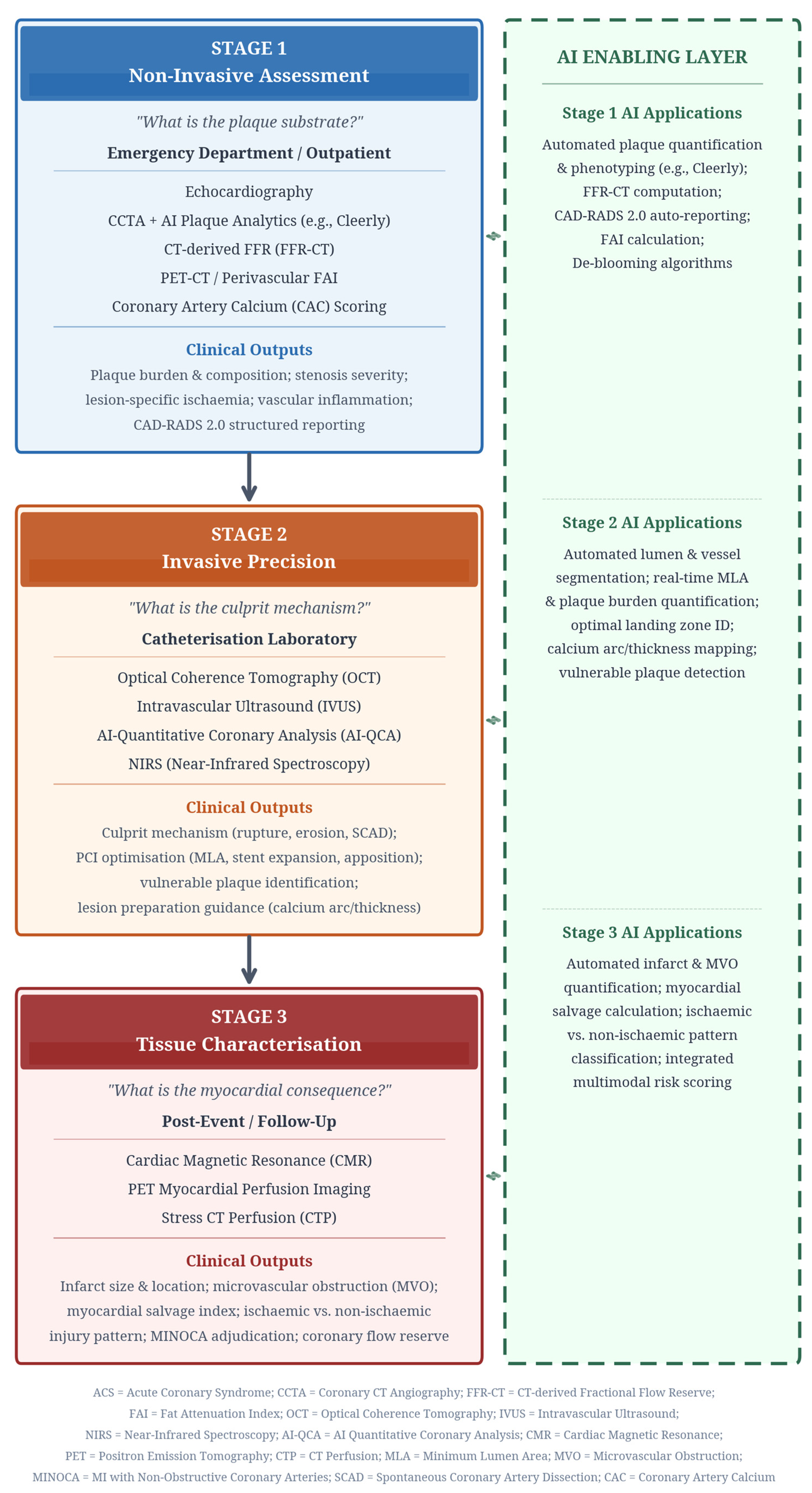
4.1. The “From Plaque to Perfusion” Framework
4.2. Summary of Key Evidence
4.3. Advanced Non-Invasive Assessment: AI-Powered Coronary CT Angiography
4.4. The Shift Beyond Angiography: Intelligent Intravascular Imaging
4.5. Special Clinical Scenario: MINOCA
4.6. Aligning the Framework with Clinical Practice Guidelines
5. Discussion
5.1. Comparative Analysis of Non-Invasive Assessment Strategies
5.2. Comparative Analysis of Intravascular Imaging Strategies
5.3. From Vulnerable Plaque to Guided Intervention:
5.4. Multimodality Imaging in MINOCA: An Integrated Diagnostic Algorithm
6. Strengths, Limitations, and Future Directions
6.1. Strengths
6.2. Limitations
6.3. Future Directions
7. Conclusions
Author Contributions
Funding
Institutional Review Board Statement
Informed Consent Statement
Data Availability Statement
Acknowledgments
Conflicts of Interest
References
- Byrne, R.A.; Rossello, X.; Coughlan, J.J.; Barbato, E.; Berry, C.; Chieffo, A.; Claeys, M.J.; Dan, G.A.; Dweck, M.R.; Galbraith, M.; et al. 2023 ESC Guidelines for the management of acute coronary syndromes: Developed by the task force on the management of acute coronary syndromes of the European Society of Cardiology (ESC). Eur. Heart J. 2023, 44, 3720–3826. [Google Scholar] [CrossRef]
- Collet, J.P.; Thiele, H.; Barbato, E.; Barthélémy, O.; Bauersachs, J.; Bhatt, D.L.; Dendale, P.; Dorobantu, M.; Edvardsen, T.; Folliguet, T.; et al. 2020 ESC Guidelines for the management of acute coronary syndromes in patients presenting without persistent ST-segment elevation: The Task Force for the management of acute coronary syndromes in patients presenting without persistent ST-segment elevation of the European Society of Cardiology (ESC). Eur. Heart J. 2021, 42, 1289–1367. [Google Scholar] [CrossRef]
- Agewall, S.; Beltrame, J.F.; Reynolds, H.R.; Niessner, A.; Rosano, G.; Caforio, A.L.; De Caterina, R.; Zimarino, M.; Roffi, M.; Kjeldsen, K.; et al. ESC working group position paper on myocardial infarction with non-obstructive coronary arteries. Eur. Heart J. 2017, 38, 143–153. [Google Scholar] [CrossRef]
- Cury, R.C.; Leipsic, J.; Abbara, S.; Achenbach, S.; Berman, D.; Bittencourt, M.; Budoff, M.; Chinnaiyan, K.; Choi, A.D.; Ghoshhajra, B.; et al. CAD-RADSTM 2.0: 2022 Coronary Artery Disease-Reporting and Data System. JACC Cardiovasc. Imaging 2022, 15, 1972–1986. [Google Scholar] [CrossRef]
- Knuuti, J.; Wijns, W.; Saraste, A.; Capodanno, D.; Barbato, E.; Funck-Brentano, C.; Prescott, E.; Storey, R.F.; Deaton, C.; Cuisset, T.; et al. 2019 ESC Guidelines for the diagnosis and management of chronic coronary syndromes: The Task Force for the diagnosis and management of chronic coronary syndromes of the European Society of Cardiology (ESC). Eur. Heart J. 2020, 41, 407–477. [Google Scholar] [CrossRef]
- Dey, D.; Slomka, P.J.; Leeson, P.; Comaniciu, D.; Shrestha, S.; Sengupta, P.P.; Marwick, T.H. Artificial intelligence in cardiovascular imaging: JACC state-of-the-art review. J. Am. Coll. Cardiol. 2019, 73, 1317–1335. [Google Scholar] [CrossRef] [PubMed]
- Gulati, M.; Levy, P.D.; Mukherjee, D.; Amsterdam, E.; Bhatt, D.L.; Birtcher, K.K.; Blankstein, R.; Boyd, J.; Bullock-Palmer, R.P.; Conejo, T.; et al. 2021 AHA/ACC/ASE/CHEST/SAEM/SCCT/SCMR Guideline for the Evaluation and Diagnosis of Chest Pain. J. Am. Coll. Cardiol. 2021, 78, e187–e285. [Google Scholar] [CrossRef] [PubMed]
- Sandoval, Y.; Leipsic, J.; Collet, C.; Ali, Z.A.; Azzalini, L.; Barbato, E.; Cavalcante, J.L.; Costa, R.A.; Garcia-Garcia, H.M.; Jones, D.A.; et al. Coronary computed tomography angiography to guide percutaneous coronary intervention: Expert opinion from a SCAI/SCCT roundtable. J. Soc. Cardiovasc. Angiogr. Interv. 2025, 4, 103664. [Google Scholar] [CrossRef] [PubMed]
- Beneki, E.; Dimitriadis, K.; Pyrpyris, N.; Antonopoulos, A.; Aznaouridis, K.; Antiochos, P.; Fragoulis, C.; Lu, H.; Meier, D.; Tsioufis, K.; et al. Computed tomography angiography in the catheterization laboratory: A guide towards optimizing coronary interventions. J. Cardiovasc. Dev. Dis. 2025, 12, 28. [Google Scholar] [CrossRef]
- Ali, Z.A.; Landmesser, U.; Maehara, A.; Matsumura, M.; Shlofmitz, R.A.; Guagliumi, G.; Price, M.J.; Hill, J.M.; Akasaka, T.; Prati, F.; et al. Optical coherence tomography-guided versus angiography-guided PCI (ILUMIEN IV: OPTIMIZE PCI). N. Engl. J. Med. 2023, 389, 1466–1476. [Google Scholar] [CrossRef] [PubMed]
- Holm, N.R.; Andreasen, L.D.; Neghabat, O.; Laanmets, P.; Kumsars, I.; Bennett, J.; Olsen, N.T.; Odenstedt, J.; Hoffmann, P.; Dens, J.; et al. OCT or Angiography Guidance for PCI in complex bifurcation lesions (OCTOBER). N. Engl. J. Med. 2023, 389, 1477–1487. [Google Scholar] [CrossRef]
- Kang, D.Y.; Ahn, J.M.; Yun, S.C.; Hur, S.H.; Cho, Y.K.; Lee, C.H.; Hong, S.J.; Lim, S.; Kim, S.W.; Won, H.; et al. Optical coherence tomography-guided or intravascular ultrasound-guided percutaneous coronary intervention: The OCTIVUS randomized clinical trial. Circulation 2023, 148, 1195–1206. [Google Scholar] [CrossRef]
- Carvalho, P.E.P.; Antunes, V.L.J.; Bittar de Pontes, V.; Gomes, W.F.; Polachini Assunes Goncalves, B.; Caixeta, A.; Strepkos, D.; Alexandrou, M.; Mutlu, D.; Gibson, C.M.; et al. IVUS, OCT, or Angiography as Guidance for PCI in Complex Coronary Artery Lesions: Network Meta-Analysis of Randomized Controlled Trials. JACC Cardiovasc. Interv. 2026, 19, 31–43. [Google Scholar] [CrossRef]
- Sörensson, P.; Ekenbäck, C.; Lundin, M.; Agewall, S.; Bacsovics Brolin, E.; Caidahl, K.; Cederlund, K.; Collste, O.; Daniel, M.; Jensen, J.; et al. Early comprehensive cardiac magnetic resonance imaging in patients with myocardial infarction with nonobstructive coronary arteries. JACC Cardiovasc. Imaging 2021, 14, 1774–1783. [Google Scholar] [CrossRef]
- Pelliccia, F.; Pasceri, V.; Niccoli, G.; Tanzilli, G.; Speciale, G.; Gaudio, C.; Crea, F.; Camici, P.G. Predictors of Mortality in Myocardial Infarction and Nonobstructed Coronary Arteries: A Systematic Review and Meta-Regression. Am. J. Med. 2020, 133, 73–83.e4. [Google Scholar] [CrossRef] [PubMed]
- Pontone, G.; Rossi, A.; Guglielmo, M.; Dweck, M.R.; Gaemperli, O.; Nieman, K.; Pugliese, F.; Maurovich-Horvat, P.; Gimelli, A.; Cosyns, B.; et al. Clinical applications of cardiac computed tomography: A consensus paper of the European Association of Cardiovascular Imaging—Part II. Eur. Hear. J.-Cardiovasc. Imaging 2022, 23, e136–e161. [Google Scholar] [CrossRef] [PubMed]
- Hoffmann, U.; Truong, Q.A.; Schoenfeld, D.A.; Chou, E.T.; Woodard, P.K.; Nagurney, J.T.; Pope, J.H.; Hauser, T.H.; White, C.S.; Weiner, S.G.; et al. Coronary CT angiography versus standard evaluation in acute chest pain (ROMICAT-II). N. Engl. J. Med. 2012, 367, 299–308. [Google Scholar] [CrossRef]
- Litt, H.I.; Gatsonis, C.; Snyder, B.; Singh, H.; Miller, C.D.; Entrikin, D.W.; Leaming, J.M.; Gavin, L.J.; Pacella, C.B.; Hollander, J.E. CT angiography for safe discharge of patients with possible acute coronary syndromes. N. Engl. J. Med. 2012, 366, 1393–1403. [Google Scholar] [CrossRef]
- Goldstein, J.A.; Chinnaiyan, K.M.; Abidov, A.; Achenbach, S.; Berman, D.S.; Hayes, S.W.; Hoffmann, U.; Lesser, J.R.; Mikati, I.A.; O’Neil, B.J.; et al. The CT-STAT (Coronary Computed Tomographic Angiography for Systematic Triage of Acute Chest Pain Patients to Treatment) trial. J. Am. Coll. Cardiol. 2011, 58, 1414–1422. [Google Scholar] [CrossRef] [PubMed]
- Douglas, P.S.; De Bruyne, B.; Pontone, G.; Patel, M.R.; Norgaard, B.L.; Byrne, R.A.; Curzen, N.; Purcell, I.; Gutberlet, M.; Rioufol, G.; et al. 1-Year outcomes of FFRCT-guided care in patients with suspected coronary disease: The PLATFORM study. J. Am. Coll. Cardiol. 2016, 68, 435–445. [Google Scholar] [CrossRef]
- Patel, M.R.; Nørgaard, B.L.; Fairbairn, T.A.; Nieman, K.; Akasaka, T.; Berman, D.S.; Raff, G.L.; Hurwitz Koweek, L.M.; Pontone, G.; Kawasaki, T.; et al. 1-Year impact on medical practice and clinical outcomes of FFRCT: The ADVANCE Registry. JACC Cardiovasc. Imaging 2020, 13, 97–105. [Google Scholar] [CrossRef] [PubMed]
- Curzen, N.; Nicholas, Z.; Stuart, B.; Wilding, S.; Hill, K.; Shambrook, J.; Eminton, Z.; Ball, D.; Barrett, C.; Johnson, L.; et al. Fractional flow reserve derived from computed tomography coronary angiography in the assessment and management of stable chest pain: The FORECAST randomized trial. Eur. Heart J. 2021, 42, 3844–3852. [Google Scholar] [CrossRef] [PubMed]
- Stone, G.W.; Maehara, A.; Lansky, A.J.; de Bruyne, B.; Cristea, E.; Mintz, G.S.; Mehran, R.; McPherson, J.; Farhat, N.; Marso, S.P.; et al. A prospective natural-history study of coronary atherosclerosis (PROSPECT). N. Engl. J. Med. 2011, 364, 226–235. [Google Scholar] [CrossRef]
- Erlinge, D.; Maehara, A.; Ben-Yehuda, O.; Bøtker, H.E.; Maeng, M.; Kjøller-Hansen, L.; Engstrøm, T.; Matsumura, M.; Crowley, A.; Dressler, O.; et al. Identification of vulnerable plaques and patients by intracoronary near-infrared spectroscopy and ultrasound (PROSPECT II): A prospective natural history study. Lancet 2021, 397, 985–995. [Google Scholar] [CrossRef]
- Prati, F.; Romagnoli, E.; Gatto, L.; La Manna, A.; Burzotta, F.; Ozaki, Y.; Marco, V.; Boi, A.; Fineschi, M.; Fabbiocchi, F.; et al. Relationship between coronary plaque morphology of the left anterior descending artery and 12 months clinical outcome: The CLIMA study. Eur. Heart J. 2020, 41, 383–391. [Google Scholar] [CrossRef]
- AStone, G.W.; Maehara, A.; Ali, Z.A.; Held, C.; Matsumura, M.; Kjøller-Hansen, L.; Bøtker, H.E.; Maeng, M.; Engstrøm, T.; Wiseth, R.; et al. Percutaneous coronary intervention for vulnerable coronary atherosclerotic plaque. J. Am. Coll. Cardiol. 2020, 76, 2289–2301. [Google Scholar] [CrossRef]
- Oikonomou, E.K.; Marwan, M.; Desai, M.Y.; Mancio, J.; Alashi, A.; Hutt Centeno, E.; Thomas, S.; Herdman, L.; Kotanidis, C.P.; Thomas, K.E.; et al. Non-invasive detection of coronary inflammation using computed tomography and prediction of residual cardiovascular risk (the CRISP-CT study): A post-hoc analysis of prospective outcome data. Lancet 2018, 392, 929–939. [Google Scholar] [CrossRef]
- Chan, K.; Wahome, E.; Tsiachristas, A.; Antonopoulos, A.S.; Patel, P.; Lyasheva, M.; Kingham, L.; West, H.; Oikonomou, E.K.; Volpe, L.; et al. Inflammatory risk and cardiovascular events in patients without obstructive coronary artery disease: The ORFAN multicentre, longitudinal cohort study. Lancet 2024, 403, 2606–2618. [Google Scholar] [CrossRef]
- Ali, Z.A.; Maehara, A.; Généreux, P.; Shlofmitz, R.A.; Fabbiocchi, F.; Nazif, T.M.; Guagliumi, G.; Meraj, P.M.; Alfonso, F.; Samady, H.; et al. Optical coherence tomography compared with intravascular ultrasound and with angiography to guide coronary stent implantation (ILUMIEN III: OPTIMIZE PCI): A randomised controlled trial. Lancet 2016, 388, 2618–2628. [Google Scholar] [CrossRef]
- Zhang, J.; Gao, X.; Kan, J.; Ge, Z.; Han, L.; Lu, S.; Tian, N.; Lin, S.; Lu, Q.; Wu, X.; et al. Intravascular ultrasound versus angiography-guided drug-eluting stent implantation: The ULTIMATE Trial. J. Am. Coll. Cardiol. 2018, 72, 3126–3137. [Google Scholar] [CrossRef] [PubMed]
- Reynolds, H.R.; Maehara, A.; Kwong, R.Y.; Sedlak, T.; Saw, J.; Smilowitz, N.R.; Mahmud, E.; Wei, J.; Marzo, K.; Matsumura, M.; et al. Coronary optical coherence tomography and cardiac magnetic resonance imaging to determine underlying causes of myocardial infarction with nonobstructive coronary arteries in women. Circulation 2021, 143, 624–640. [Google Scholar] [CrossRef]
- Taqueti, V.R.; Solomon, S.D.; Shah, A.M.; Desai, A.S.; Groarke, J.D.; Osborne, M.T.; Hainer, J.; Bibbo, C.F.; Dorbala, S.; Blankstein, R.; et al. Coronary microvascular dysfunction and future risk of heart failure with preserved ejection fraction. Eur. Heart J. 2018, 39, 840–849. [Google Scholar] [CrossRef]
- Nagumo, S.; Collet, C.; Norgaard, B.L.; Otake, H.; Ko, B.; Koo, B.K.; Leipsic, J.; Andreini, D.; Heggermont, W.; Jensen, J.M.; et al. Rationale and design of the precise percutaneous coronary intervention plan (P3) study: Prospective evaluation of a virtual computed tomography-based percutaneous intervention planner. Clin. Cardiol. 2021, 44, 446–454. [Google Scholar] [CrossRef] [PubMed]
- Precise Procedural and PCI Plan (P4). ClinicalTrials.gov. 2022. Available online: https://clinicaltrials.gov/study/NCT05253677 (accessed on 5 January 2026).
- Maroules, C.D.; Rybicki, F.J.; Ghoshhajra, B.B.; Batlle, J.C.; Branch, K.; Chinnaiyan, K.; Hamilton-Craig, C.; Hoffmann, U.; Litt, H.; Meyersohn, N.; et al. 2022 Use of coronary CT angiography for patients presenting with acute chest pain to the emergency department: Expert consensus document. J. Cardiovasc. Comput. Tomogr. 2023, 17, 146–163. [Google Scholar] [CrossRef] [PubMed]
- Kurogi, K.; Ishii, M.; Sakamoto, K.; Komaki, S.; Kusaka, H.; Yamamoto, N.; Takashio, S.; Arima, Y.; Yamamoto, E.; Kaikita, K.; et al. Optical coherence tomography-guided percutaneous coronary intervention with low-molecular-weight dextran—Effect on renal function. Circ. J. 2020, 84, 917–925. [Google Scholar] [CrossRef]
- Sakai, K.; Ikari, Y.; Nanasato, M.; Umetsu, H.; Okutsu, M.; Takikawa, T.; Sumitsuji, S.; Sadamatsu, K.; Takada, M.; Kato, Y.; et al. Impact of intravascular ultrasound-guided minimum-contrast coronary intervention on 1-year clinical outcomes in patients with stage 4 or 5 advanced chronic kidney disease. Cardiovasc. Interv. Ther. 2019, 34, 234–241. [Google Scholar] [CrossRef] [PubMed]
- Räber, L.; Mintz, G.S.; Koskinas, K.C.; Johnson, T.W.; Holm, N.R.; Onuma, Y.; Radu, M.D.; Joner, M.; Yu, B.; Jia, H.; et al. Clinical use of intracoronary imaging. Part 1: Guidance and optimization of coronary interventions. An expert consensus document of the European Association of Percutaneous Cardiovascular Interventions. Eur. Heart J. 2018, 39, 3281–3300. [Google Scholar] [CrossRef]


| Research Direction | Key Study/Trial | Main Author | Year | Primary Objective | Key Result/Finding |
|---|---|---|---|---|---|
| CCTA in acute chest pain | ROMICAT-II | Hoffmann U | 2012 | Compare CCTA vs. standard evaluation in ED | CCTA reduced time to diagnosis and length of stay without increasing downstream costs [17] |
| CCTA in acute chest pain | CT-ACS | Litt HI | 2012 | Assess CCTA for safe discharge of ACS patients | CCTA safely identified low-risk patients for early discharge [18] |
| CCTA in acute chest pain | CT-STAT | Goldstein JA | 2011 | Evaluate CCTA vs. MPI for acute chest pain | CCTA reduced diagnostic time and was cost-effective compared to MPI [19] |
| FFR-CT clinical utility | PLATFORM | Douglas PS | 2016 | Assess if FFR-CT reduces unnecessary ICA | FFR-CT strategy reduced unnecessary ICA and lowered costs at 1 year [20] |
| FFR-CT clinical utility | ADVANCE | Patel MR | 2020 | Relate FFR-CT to downstream management | Revascularization more frequent when FFR-CT ≤ 0.80; low overall MACE [21] |
| FFR-CT clinical utility | FORECAST | Curzen N | 2021 | Compare CCTA + FFR-CT vs. standard care (UK) | ICA reduced in CCTA + FFR-CT arm; QoL and major events similar [22] |
| Vulnerable plaque identification | PROSPECT | Stone GW | 2011 | Natural history of coronary atherosclerosis | VH-IVUS identified high-risk non-culprit plaques predicting future events [23] |
| Vulnerable plaque identification | PROSPECT II | Erlinge D | 2021 | Identify predictors of future non-culprit MACE | High plaque burden (IVUS) + large lipid core (NIRS) enriched for future MACE [24] |
| Vulnerable plaque identification | CLIMA | Prati F | 2020 | Assess OCT plaque features and outcomes | Clustered OCT features (thin cap, large lipid arc, macrophages, small MLA) predicted events [25] |
| Prophylactic plaque intervention | PROSPECT ABSORB | Stone GW | 2020 | Evaluate bioresorbable scaffold for vulnerable plaques | Demonstrated feasibility; plaque-directed intervention remains investigational [26] |
| Coronary inflammation | CRISP-CT | Oikonomou EK | 2018 | Assess if perivascular FAI predicts cardiac mortality | High FAI independently associated with increased cardiac mortality [27] |
| Coronary inflammation | ORFAN | Chan K | 2024 | Assess FAI in patients without obstructive CAD | Elevated FAI Score strengthened prediction of cardiac mortality and MACE [28] |
| OCT-guided PCI | ILUMIEN III | Ali ZA | 2016 | Compare OCT vs. IVUS vs. angiography for stent implantation | OCT achieved similar stent expansion to IVUS; both superior to angiography [29] |
| OCT-guided PCI | ILUMIEN IV | Ali ZA | 2023 | Compare OCT-guided vs. angiography-guided PCI | OCT guidance improved minimum stent area but did not reduce 2-year TVF [10] |
| OCT-guided PCI | OCTOBER | Holm NR | 2023 | Evaluate OCT guidance in complex bifurcation PCI | OCT guidance resulted in superior procedural outcomes [11] |
| OCT vs. IVUS guidance | OCTIVUS | Kang DY | 2023 | Compare OCT vs. IVUS guidance for PCI | OCT was non-inferior to IVUS for 1-year target-vessel failure [12] |
| IVUS-guided PCI | ULTIMATE | Zhang J | 2018 | Compare IVUS-guided vs. angiography-guided DES implantation | IVUS guidance superior to angiography alone for clinical outcomes [30] |
| Imaging-guided PCI (network) | Network meta-analysis | Carvalho PEP | 2026 | Compare IVUS, OCT, and angiography guidance | Both IVUS and OCT superior to angiography; comparable to each other [13] |
| CMR in MINOCA | Sörensson P | Sörensson P | 2021 | Early CMR in MINOCA patients | CMR identified a definitive diagnosis in the majority of MINOCA cases [14] |
| MINOCA outcomes | Meta-analysis | Pustjens T | 2020 | Systematic review of MINOCA outcomes | MINOCA carries significant morbidity; multimodality imaging improves diagnosis [15] |
| MINOCA in women | Reynolds HR | Reynolds HR | 2021 | OCT + CMR to determine MINOCA causes in women | Combined OCT and CMR identified the underlying cause in most women [31] |
| PET in INOCA | Taqueti VR | Taqueti VR | 2018 | Assess PET for microvascular dysfunction | PET-detected microvascular dysfunction predicted heart failure hospitalisation [32] |
| CT-guided PCI planning | P3 study design | Sonck J | 2021 | Design of CT-guided PCI planning study | Established rationale for CCTA-based procedural planning [33] |
| CT-guided PCI planning | P4 trial | ClinicalTrials.gov | 2022 | Compare CCTA-guided vs. IVUS-guided PCI | Ongoing; definitive outcomes awaited [34] |
| Platform | Advance | Forecast | |
|---|---|---|---|
| Clinical question | Prospective, multicenter comparative-effectiveness study (patients managed by usual care vs. CCTA with selective FFR-CT; includes a “planned invasive” stratum) | In real-world practice, how does FFR-CT (≤0.80 vs. >0.80) relate to downstream management (revascularisation) and outcomes after CCTA shows atherosclerosis? | In UK rapid access chest pain clinics, does CCTA + selective FFR-CT improve resource utilization/costs vs. NICE-guided standard care? |
| Primary endpoint | At 90 days: rate of ICA showing no obstructive CAD (i.e., “unnecessary” cath) | Relationship of FFR-CT to downstream care + 1-year clinical outcomes (MACE, death/MI, etc.) | Total cardiac costs at 9 months |
| Results | In the planned invasive stratum, lower costs at 1 year with CCTA + selective FFR-CT and similarly low MACE/QoL vs. usual care | Revascularization much more frequent when FFR-CT ≤ 0.80; event rates are overall low, with higher CV death/MI in abnormal vs. normal FFR-CT | No significant cost reduction, but ICA was reduced in the CCTA + selective FFR-CT arm; QoL/angina and major events were similar |
| Guideline Recommendation | Class | Framework Stage | Clinical Application |
|---|---|---|---|
| CCTA for ACS Rule-Out | I (ESC/ACC) | Stage 1: Non-Invasive Assessment | In low-to-intermediate risk patients with suspected ACS and no ischaemic ECG changes or elevated troponins, CCTA is recommended to rule out obstructive CAD [1,2,7]. |
| Intravascular Imaging for PCI Optimization | IIa (ESC) | Stage 2: Invasive Precision | Intravascular imaging (IVUS or OCT) should be considered to optimize stent implantation, assess stent expansion, malapposition, and edge dissections, particularly in complex lesions [1,38]. |
| CMR for MINOCA Evaluation | I (ESC) | Stage 3: Tissue Characterization | In patients with MINOCA, CMR is recommended to establish the underlying cause by differentiating ischaemic from non-ischaemic myocardial injury [1,3]. |
| FFR-CT for Functional Assessment | IIa (ESC) | Stage 1: Non-Invasive Assessment | FFR-CT may be considered in patients with intermediate-stenosis lesions on CCTA to determine haemodynamic significance and guide decisions regarding invasive angiography [7]. |
Disclaimer/Publisher’s Note: The statements, opinions and data contained in all publications are solely those of the individual author(s) and contributor(s) and not of MDPI and/or the editor(s). MDPI and/or the editor(s) disclaim responsibility for any injury to people or property resulting from any ideas, methods, instructions or products referred to in the content. |
© 2026 by the authors. Licensee MDPI, Basel, Switzerland. This article is an open access article distributed under the terms and conditions of the Creative Commons Attribution (CC BY) license.
Share and Cite
Shahin, A.; Agamy, S.; Zaghloul, S.; ElShafey, R.; Molda, M.; Khan, Z.; Candilio, L. From Plaque to Perfusion: A Narrative Review of Multimodality Imaging in Acute Coronary Syndromes. J. Clin. Med. 2026, 15, 2905. https://doi.org/10.3390/jcm15082905
Shahin A, Agamy S, Zaghloul S, ElShafey R, Molda M, Khan Z, Candilio L. From Plaque to Perfusion: A Narrative Review of Multimodality Imaging in Acute Coronary Syndromes. Journal of Clinical Medicine. 2026; 15(8):2905. https://doi.org/10.3390/jcm15082905
Chicago/Turabian StyleShahin, Ahmed, Salaheldin Agamy, Sheref Zaghloul, Ranin ElShafey, Maha Molda, Zahid Khan, and Luciano Candilio. 2026. "From Plaque to Perfusion: A Narrative Review of Multimodality Imaging in Acute Coronary Syndromes" Journal of Clinical Medicine 15, no. 8: 2905. https://doi.org/10.3390/jcm15082905
APA StyleShahin, A., Agamy, S., Zaghloul, S., ElShafey, R., Molda, M., Khan, Z., & Candilio, L. (2026). From Plaque to Perfusion: A Narrative Review of Multimodality Imaging in Acute Coronary Syndromes. Journal of Clinical Medicine, 15(8), 2905. https://doi.org/10.3390/jcm15082905