Systematic Review

Platelet-Rich Plasma in Anterior Cruciate Ligament Reconstruction: An Updated Systematic Review and Meta-Analysis of Clinical and Radiological Outcomes


1
Department of Orthopaedic Surgery, Lebanese University, Beirut P.O. Box 6573/14, Lebanon
2
Department of Anesthesiology, Lebanese American University Medical Center, Beirut P.O. Box 13-5053, Lebanon
3
Department of Orthopaedic Surgery, Lebanese American University Medical Center, Beirut P.O. Box 13-5053, Lebanon
4
Department of Orthopaedic Surgery, Saint George Hospital University Medical Center, Beirut P.O.Box 166-378, Lebanon
*
Author to whom correspondence should be addressed.
J. Clin. Med. 2026, 15(7), 2526; https://doi.org/10.3390/jcm15072526
Submission received: 5 February 2026 / Revised: 4 March 2026 / Accepted: 13 March 2026 / Published: 26 March 2026
(This article belongs to the Section Orthopedics)

Abstract

Background/Objectives: To evaluate the efficacy of platelet-rich plasma (PRP) as an adjunctive treatment in anterior cruciate ligament reconstruction (ACLR) and its impact on key clinical outcomes. Methods: A systematic search was conducted across five databases until 11 November 2024, including 33 randomized controlled trials (RCTs) that investigated PRP in ACLR. Outcomes analyzed included ligamentization (MRI hypointensity grades), pain VAS scores, functional scores (IKDC, Lysholm, Tegner), knee stability (KT-1000 arthrometer), and tunnel characteristics. Subgroup analyses were performed based on PRP application site, graft type, risk of bias, and follow-up duration. Results: PRP significantly enhanced ligamentization, particularly at 12 months, with marked reductions in MRI hypointensity grades. Patellar tendon grafts demonstrated the most substantial benefits. PRP also significantly reduced postoperative pain, with effects most pronounced in the early recovery period (1–9 months). However, the analgesic benefits diminished over time. Improvements in IKDC scores were observed only in studies with a high risk of bias, while Lysholm and Tegner scores showed no consistent differences between PRP and controls. Knee stability improved significantly with PRP, but this effect was limited to early follow-up periods (3 months). The heterogeneity in PRP preparation methods, application protocols, and patient populations limited the generalizability of the findings. Conclusions: PRP enhances ligamentization and provides short-term pain relief and stability benefits in ACLR. However, its impact on long-term functional recovery and other clinical outcomes remains limited and inconsistent. Standardization of PRP protocols and further high-quality research are necessary to refine its application and therapeutic potential.

1. Introduction

Anterior cruciate ligament (ACL) injuries represent a significant challenge in orthopedic and sports medicine due to their high prevalence and the long recovery periods they entail [1,2]. ACL reconstruction (ACLR) is widely recognized as the gold standard for restoring knee stability and function in active individuals, particularly athletes [3]. Despite advances in surgical techniques and postoperative rehabilitation protocols, a substantial proportion of patients fail to achieve optimal outcomes, including a full return to pre-injury activity levels and long-term joint health [4,5]. Persistent issues such as graft maturation, integration, and postoperative pain remain critical barriers to recovery [6,7].
Ligamentization—the biological remodeling of graft tissue into ligament-like structures—plays a pivotal role in the success of ACLR [8]. This process encompasses several stages, including necrosis, cellular infiltration, collagen synthesis, and revascularization. While this natural progression is essential for graft incorporation, it is inherently slow and subject to variability influenced by patient factors, graft type, and surgical technique [9]. Enhancing this process has therefore become a key focus of research, with the aim of improving clinical outcomes and reducing recovery times.
Platelet-rich plasma (PRP) has emerged as a promising biological adjunct in ACLR [10]. Derived from autologous blood, PRP is enriched with growth factors such as platelet-derived growth factor (PDGF), transforming growth factor-beta (TGF-β), and vascular endothelial growth factor (VEGF), which are known to facilitate tissue repair and regeneration [11]. Preclinical and early clinical studies have suggested that PRP may enhance graft maturation, promote bone–tunnel healing, and modulate inflammatory responses [12,13]. However, the evidence remains inconsistent, with conflicting findings regarding its efficacy in improving ligamentization, functional outcomes, and pain relief [14,15].
The clinical application of PRP in ACLR is further complicated by variations in its preparation, dosing, and administration. Differences in patient populations, graft types, and outcome measures have contributed to the heterogeneity of results observed across studies, limiting the generalizability of findings and precluding definitive clinical recommendations [15,16]. Consequently, there is a pressing need for a systematic evaluation of the available evidence to better understand PRP’s role in ACLR and its potential to address the unmet challenges of graft healing and functional recovery.
A major challenge in interpreting PRP-related outcomes is the lack of standardized biological characterization. PRP is not a uniform product; its cellular composition, platelet concentration, leukocyte content, activation method, injected volume, and total platelet dose vary considerably depending on preparation protocols. Contemporary classification systems, such as the PAW and MARSPILL classifications, emphasize that the total platelet dose and biological composition may represent critical determinants of clinical efficacy. However, many clinical trials fail to report sufficient details regarding the platelet concentration or dosing, limiting reproducibility and cross-study comparisons. This heterogeneity complicates the interpretation of PRP efficacy in ACLR and may partially explain inconsistent findings across trials.
This study aims to systematically review and synthesize the evidence on the efficacy of PRP in ACLR, focusing on its impact on ligamentization, pain, functional scores, knee stability, and other clinically relevant outcomes. By integrating data from randomized controlled trials and leveraging advanced meta-analytic techniques, this research seeks to provide a comprehensive and nuanced understanding of PRP’s therapeutic potential.

2. Materials and Methods

2.1. Design and Literature Search

This systematic review and meta-analysis was conducted in line with the recent PRISMA [17] and AMSTAR [18] guidelines. The study protocol was not registered on PROSPERO given the presence of an already registered protocol at the time of registration (CRD420251142319). The PRISMA checklist is provided as Supplementary File S1.
We searched PubMed, Scopus, Web of Science, Cochrane Library, and Google Scholar (first 200 records) [19] up to 11 November 2024. The search strategy included the following keywords: “anterior cruciate ligament”, platelet, and random*. The full search query can be found in Table S1. Citations were filtered based on their titles and abstracts. No restrictions were applied regarding the original language of publication. Manual searches included reviewing reference lists and related articles on PubMed [20] as well as on Google Software.

2.2. Selection Strategy

The study selection was carried out following the PICOS (Population, Intervention, Comparison, Outcomes, and Study Design) theme [21].
The inclusion criteria were as follows:
  • Population: Patients who underwent ACL reconstruction.
  • Intervention: PRP.
  • Comparison: Control.
  • Outcome: Pain (as primary endpoint) and clinical/radiological measures (secondary endpoints).
  • Study Design: Only randomized controlled trials (RCTs) were considered.
The exclusion criteria included the following:
  • Non-original research.
  • Non-randomized studies of intervention.
  • Abstract-only publications.
  • Case reports and case series.
  • Case–control and cohort studies.
  • Duplicate studies or studies with overlapping datasets (as in post hoc studies or subgroup analytic studies).
  • Meniscal reconstruction (not ACL reconstruction).
  • Animal studies plus in vivo or in vitro studies.

2.3. Data Collection and Outcomes

The data extraction sheet was formatted using Microsoft Excel, which is made up of four parts. The first part included citation-specific data (last name of first author, year of publication and investigation, country, study design, and total sample). The second part was related to patients’ characteristics (i.e., age, gender, graft type, intervention groups, and follow-up). When available, data regarding PRP preparation protocol, platelet concentration, and reported biological characteristics were extracted. Total platelet dose could not be calculated because most trials did not report both baseline platelet counts and final PRP concentrations. Consequently, formal subclassification of PRP formulations (e.g., PAW or MARSPILL systems) was not feasible.
For the purposes of this review, PRP was defined broadly as any autologous platelet-derived concentrate applied locally during ACL reconstruction with the intention of enhancing graft healing. This included products labeled as platelet-rich plasma (PRP), plasma rich in growth factors (PRGFs), platelet-rich fibrin (PRF), autologous platelet concentrate (APC), and platelet concentrate (PC). Because detailed biological characterization (e.g., leukocyte content, fibrin architecture, platelet concentration, activation method, total platelet dose) was inconsistently reported across trials, subclassification into biologically distinct categories was not feasible.
The third part included the outcome data. The primary outcome was postoperative pain, measured by the visual analogue scale (VAS). Secondary endpoints included clinical and radiological measures. Clinical measures included Lysholm score, Tegner score, IKDC score, and knee intensity (measured by KT-1000 arthrometry), while radiological measures included tibial/femoral tunnel diameter, ligamentization (MRI hypointensity grades 1–4: mild–diffuse), and graft maturation, as measured by the signal noise quotient (SNQ). The fourth part included the methodological quality assessment part.

2.4. Risk-of-Bias Assessment

For all included RCTs, the revised version of the Cochrane’s risk-of-bias tool was used. The methodological quality was assessed over five domains: randomization, deviation from intended interventions, missing outcome data, measurement bias, and selective reporting. Overall, a study was deemed as high risk, low risk, or some concerns.

2.5. Statistical Analysis

All statistical analyses were conducted using STATA version 18 (StataCorp, USA). Because the studies included in the quantitative synthesis were highly heterogeneous, a random-effects model was applied, with pooled estimates calculated using the restricted maximum likelihood (REML) method to reduce potential bias related to incomplete data [22]. Owing to substantial variability in outcome reporting—such as differences in follow-up intervals, graft types, and injection sites—deriving a single overall pooled estimate was considered inappropriate. Accordingly, subgroup analyses were performed based on follow-up duration, graft type, PRP injection site, and risk of bias.
Heterogeneity was quantified using the I2 statistic, with significant heterogeneity defined as I2 > 40% [23]. The robustness of the findings was further examined through sensitivity analyses, in which Galbraith plots were used to detect potential outliers. Publication bias was assessed using funnel plots and tests for asymmetry [24]. These analyses showed that the results remained stable after sensitivity testing, and no significant evidence of publication bias was detected.

3. Results

3.1. Literature Search Results

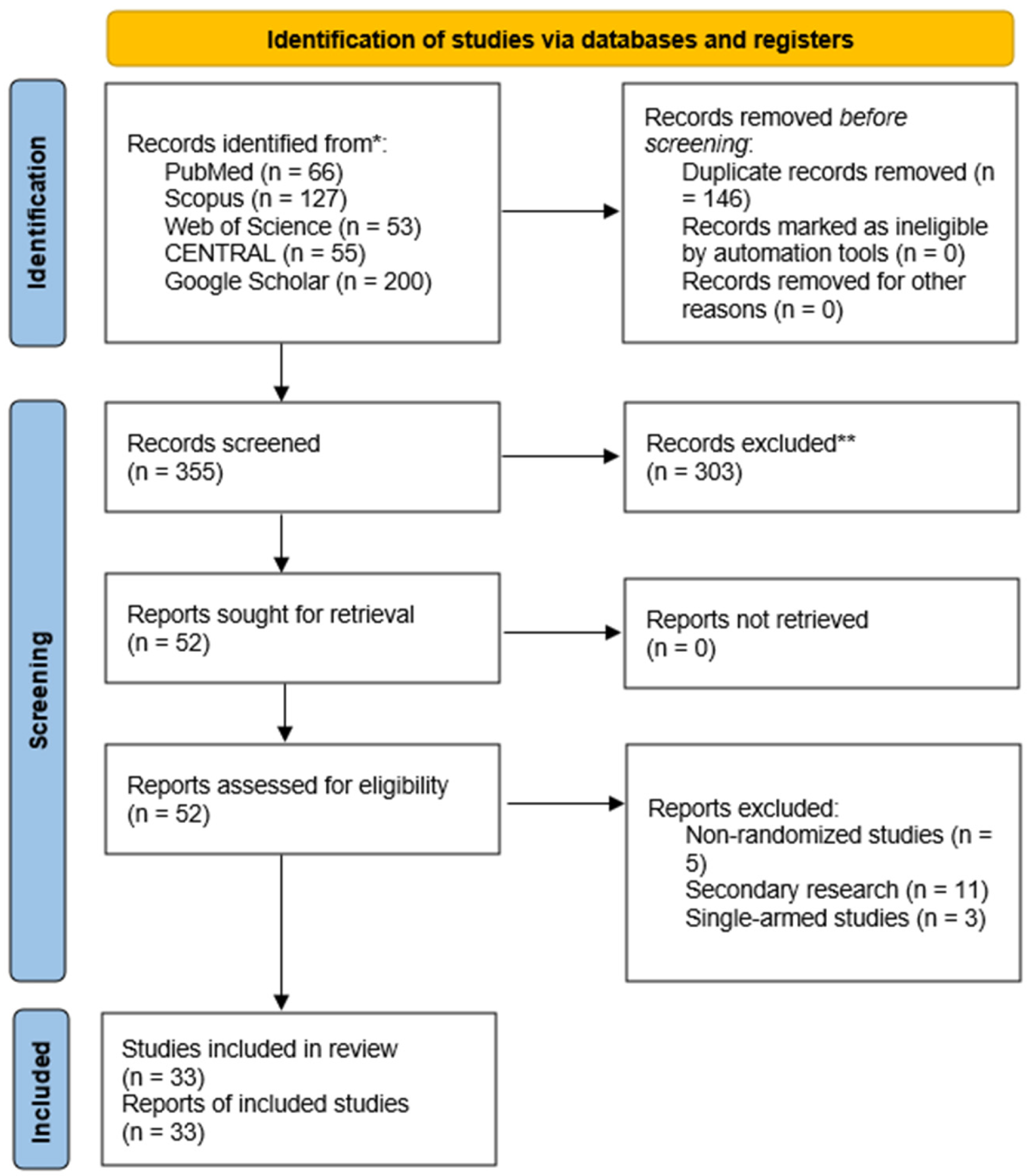
The systematic search across multiple databases yielded 501 records (Figure 1). After removing 146 duplicates, 355 unique articles remained for screening. During the title and abstract review, 303 records were excluded. Subsequently, 52 full-text articles were retrieved and assessed for eligibility, all of which were successfully obtained. Among these, 19 studies were excluded for the following reasons: non-randomized study design (n = 5), secondary research articles (n = 11), and single-arm studies (n = 3). Ultimately, 33 randomized controlled trials (RCTs) fulfilled the eligibility criteria and were included in the meta-analysis [25,26,27,28,29,30,31,32,33,34,35,36,37,38,39,40,41,42,43,44,45,46,47,48,49,50,51,52,53,54,55,56,57].

3.2. Baseline Characteristics of Included Studies

The baseline clinicodemographic characteristics of the included studies and examined patients are presented in Table 1. Most evidence came from Spain (6 RCTs, 17.65%) and China (4 RCTs, 11.76%). A total of 1913 patients who underwent ACL reconstruction were allocated to either the PRP (981 patients) or control group (932 patients). The follow-up ranged from as short as 1 day to 24 months. The types of grafts used in ACL reconstruction included hamstring tendon (18 RCTs), patellar tendon (9 RCTs), peroneus longus tendon (1 RCT), quadriceps tendon (1 RCT), and semitendinosus tendon (1 RCT). A description of patients’ age and gender is provided in Table 1.
The included RCTs utilized various platelet-derived formulations, including platelet-rich plasma (PRP), plasma rich in growth factors (PRGFs), platelet-rich fibrin (PRF), autologous platelet concentrate (APC), and platelet concentrate (PC). Terminology and preparation protocols varied considerably across studies. Detailed reporting of leukocyte content, red blood cell contamination, fibrin matrix characteristics, and activation protocols was inconsistent. Due to insufficient biological characterization and limited numbers within each product category, separate quantitative pooling by product type was not statistically feasible.

3.3. Risk-of-Bias Assessment

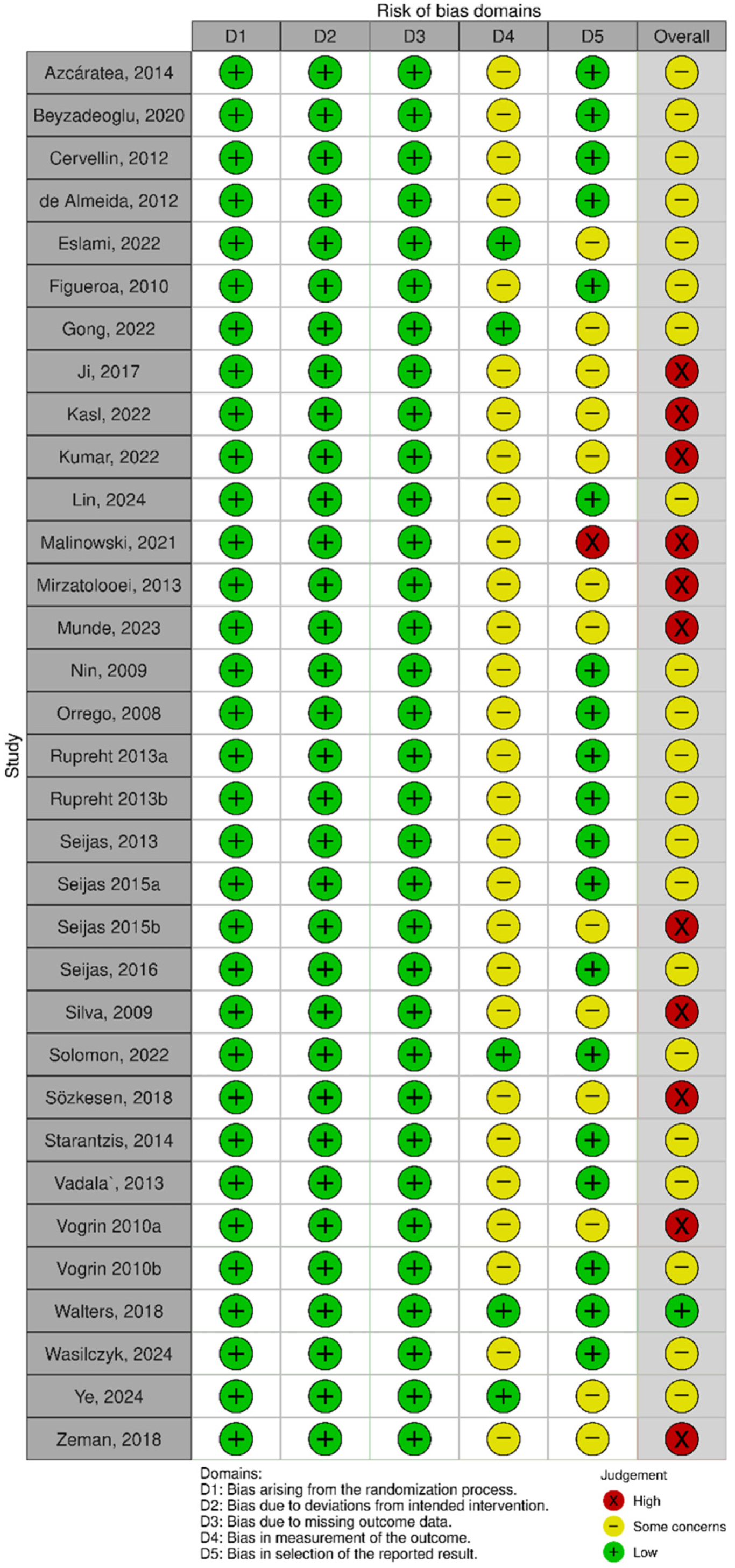
The overall risk-of-bias assessment is summarized in Figure 2. Among the 33 included RCTs, most were judged as having some concerns (21/33, 63.6%), while 11 (33.3%) were at high risk of bias. Only one study (3.0%) was considered low risk.

3.4. Primary Endpoint (VAS Score)

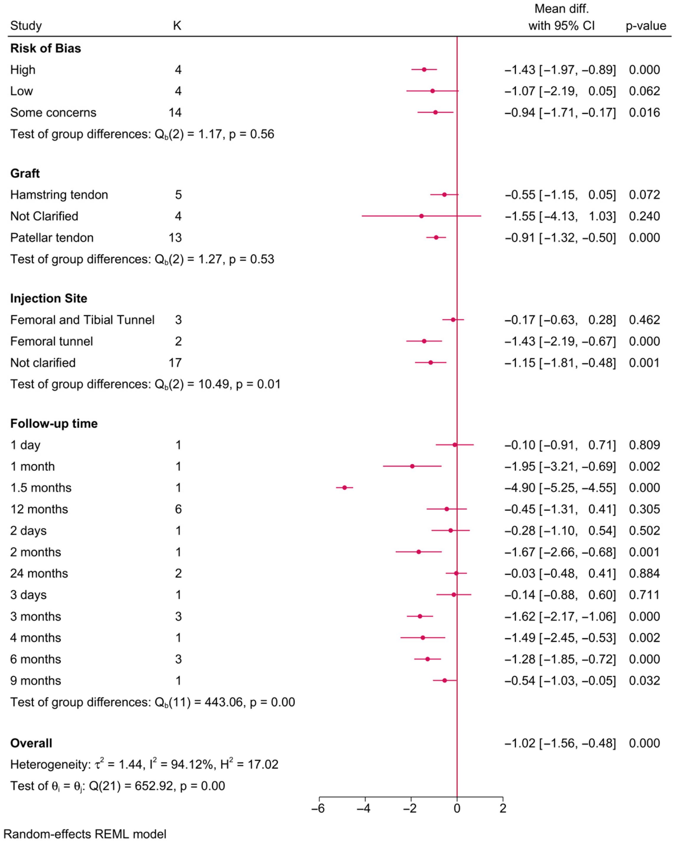
Twenty-two RCTs investigated the impact of PRP on pain scores (Figure 3). The meta-analysis showed that the injection site (p = 0.01) and follow-up time (p = 0.001) had a significant effect modification on postoperative pain. For instance, in studies where PRP was injected into the femoral tunnel, a significant reduction in pain score was observed compared to the control (MD = −1.43; 95% CI: −2.19; −0.67]. However, no change was observed with PRP injected in both the femoral and tibial tunnels simultaneously. Additionally, the observed reduction in pain score in favor of PRP was observed from the first month (MD = −1.95; 95% CI: −3.21, −0.69] till the ninth month [MD = −0.54; 95% CI: −1.03, −0.05] of follow-up. However, this difference was diminishing over time.
Of note, the reduction in pain score in favor of PRP was observed only in RCTs with a high risk of bias or those with some concerns, while RCTs of a low risk of bias exhibited no significant difference between both groups (MD = −1.07; 95% CI: −2.19, 0.05]. Although differences in pain were observed with different graft types, this factor did not play a significant effect-modifying role on postoperative pain (p = 0.53). That being said, the reduction in pain scores was noted only with the use of the patellar tendon graft (MD = −0.91; 95% CI: −1.32, −0.50), while the hamstring tendons graft showed no difference in postoperative pain.

3.5. Secondary Endpoints

3.5.1. IKDC Score

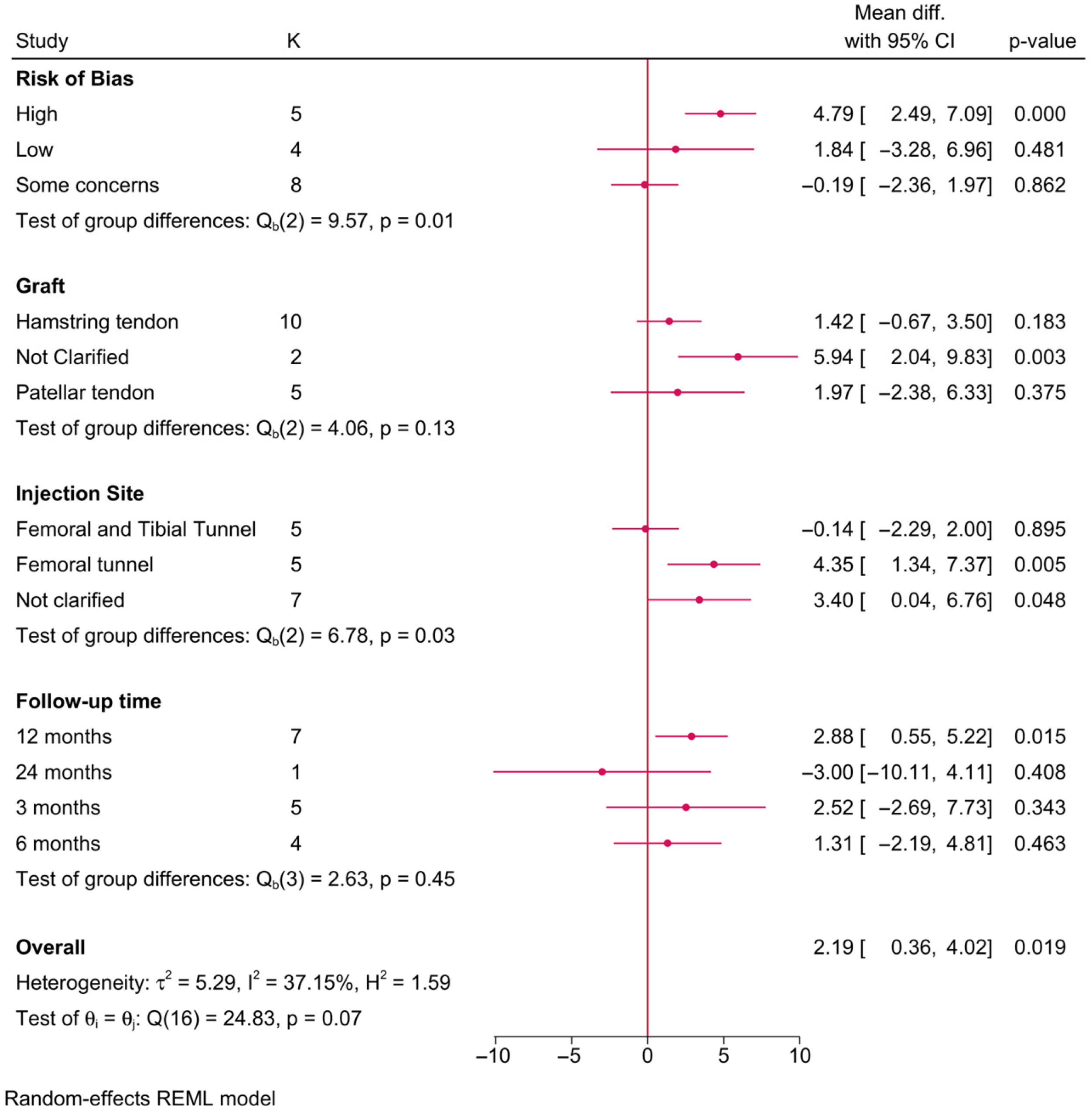
Seventeen RCTs investigated the impact of PRP on postoperative IKDC scores (Figure 4). The risk of bias (p = 0.01) and injection site (p = 0.03) exhibited a significant effect-modifying role on the postoperative IKDC score. For instance, a significant increase in the postoperative IKDC score in favor of PRP was only observed in RCTs of a high risk of bias (MD = 4.79; 95% CI: 2.49, 7.09), while those with a low risk or some concerns exhibited no difference. Additionally, this increase in the IKDC score of PRP over the control group was observed in those who received PRP injection into the femoral tunnel alone (MD = 4.35; 95%CI: 1.34, 7.37), while those injected into both the femoral and tibial tunnels simultaneously exhibited no difference. Noteworthy, the observed difference in the IKDC score between PRP and the control was only observed at 12 months of follow-up (MD = 2.88; 95% CI: 0.55, 5.22) and not before that time (3–6 months).

3.5.2. Lysholm Score

Sixteen RCTs investigated the impact of PRP on postoperative Lysholm scores. None of the investigated variables showed a significant effect-modifying role on the postoperative Lysholm score, including the risk of bias (p = 0.14), graft type (p = 0.82), PRP injection site (p = 0.77), or follow-up time (p = 0.63) (Supplementary Figure S1). No significant difference in the postoperative Lysholm score was observed between the PRP and control groups across all investigated subgroups/comparisons.

3.5.3. Tegner Score

Six RCTs investigated the impact of PRP on postoperative Tegner scores. None of the investigated variables showed a significant effect-modifying role on the postoperative Tegner score, including the risk of bias (p = 0.30), graft type (p = 0.72), PRP injection site (p = 0.72), or follow-up time (p = 0.25) (Supplementary Figure S2). No significant difference in the postoperative Tegner score was observed between the PRP and control groups across all investigated subgroups/comparisons.

3.5.4. Graft Maturation (SNQ)

Two RCTs investigated the impact of PRP on graft maturation as measured by the SNQ (Supplementary Figure S3). None of the examined variables showed a significant effect-modifying role on the SNQ. A significant reduction in the postoperative SNQ was observed in favor of PRP compared to the control only in those who had a hamstring tendon graft (MD = −0.63; 95% CI: −0.99, −0.27) and at 12 months of follow-up.

3.5.5. Tunnel Diameters (mm)

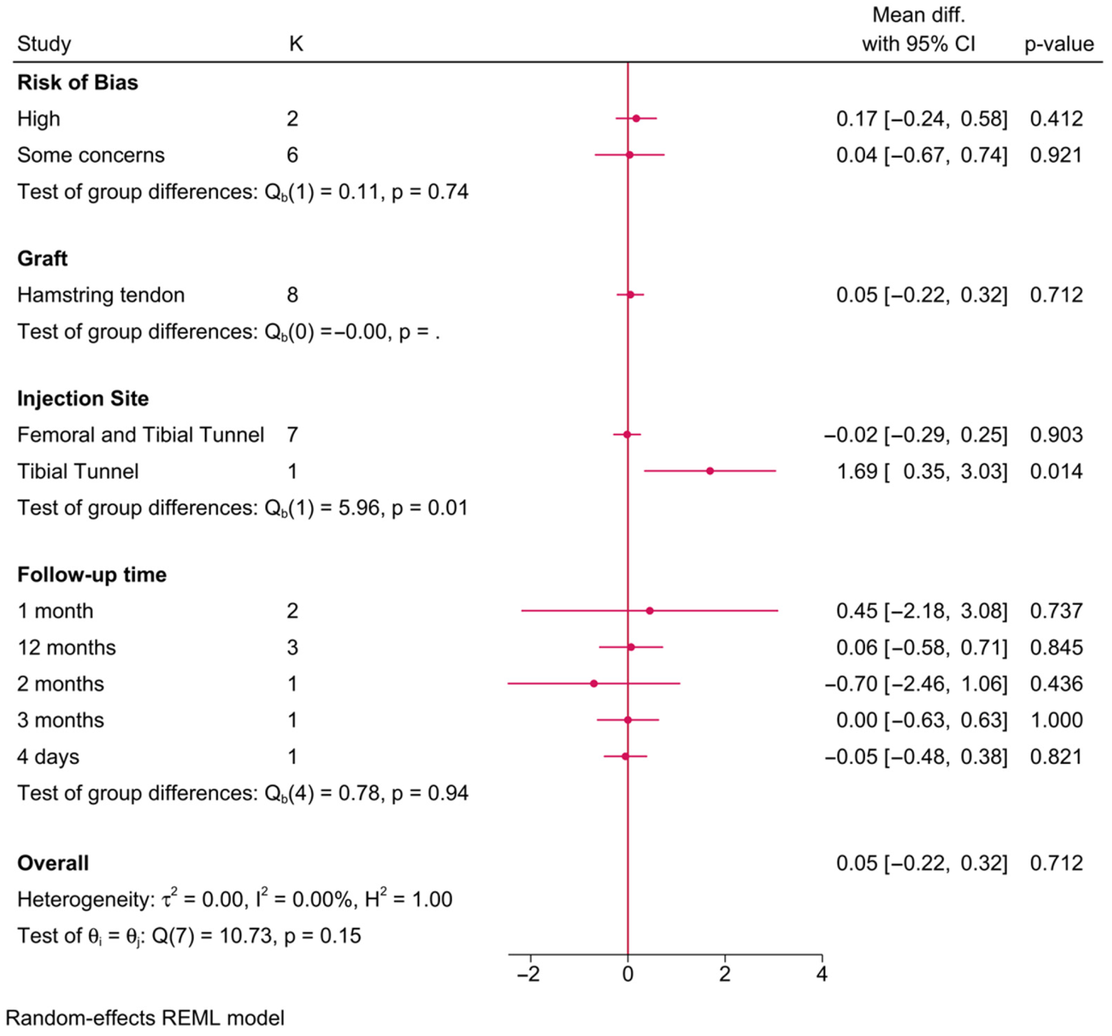
Eight RCTs investigated the association between PRP and tibial tunnel diameter (Figure 5). The injection site was the only significant effect modifier of the tibial tunnel diameter (p = 0.01). PRP showed a significant increase in tibial tunnel diameter compared to the control group, only where PRP was injected into the tibial tunnel alone (MD = 1.69; 95% CI: 0.35, 3.03), with no change when injected into both the femoral and tibial tunnels simultaneously.
Seven RCTs investigated the association between PRP and femoral tunnel diameter (Supplementary Figure S4). Risk of bias was barely a significant effect modifier of the effect of PRP on the postoperative femoral tunnel diameter (p = 0.05). That being said, no significant difference between PRP and the control was observed across all subgroups.

3.5.6. Knee Stability (KT-1000 Arthrometric Measure—mm)

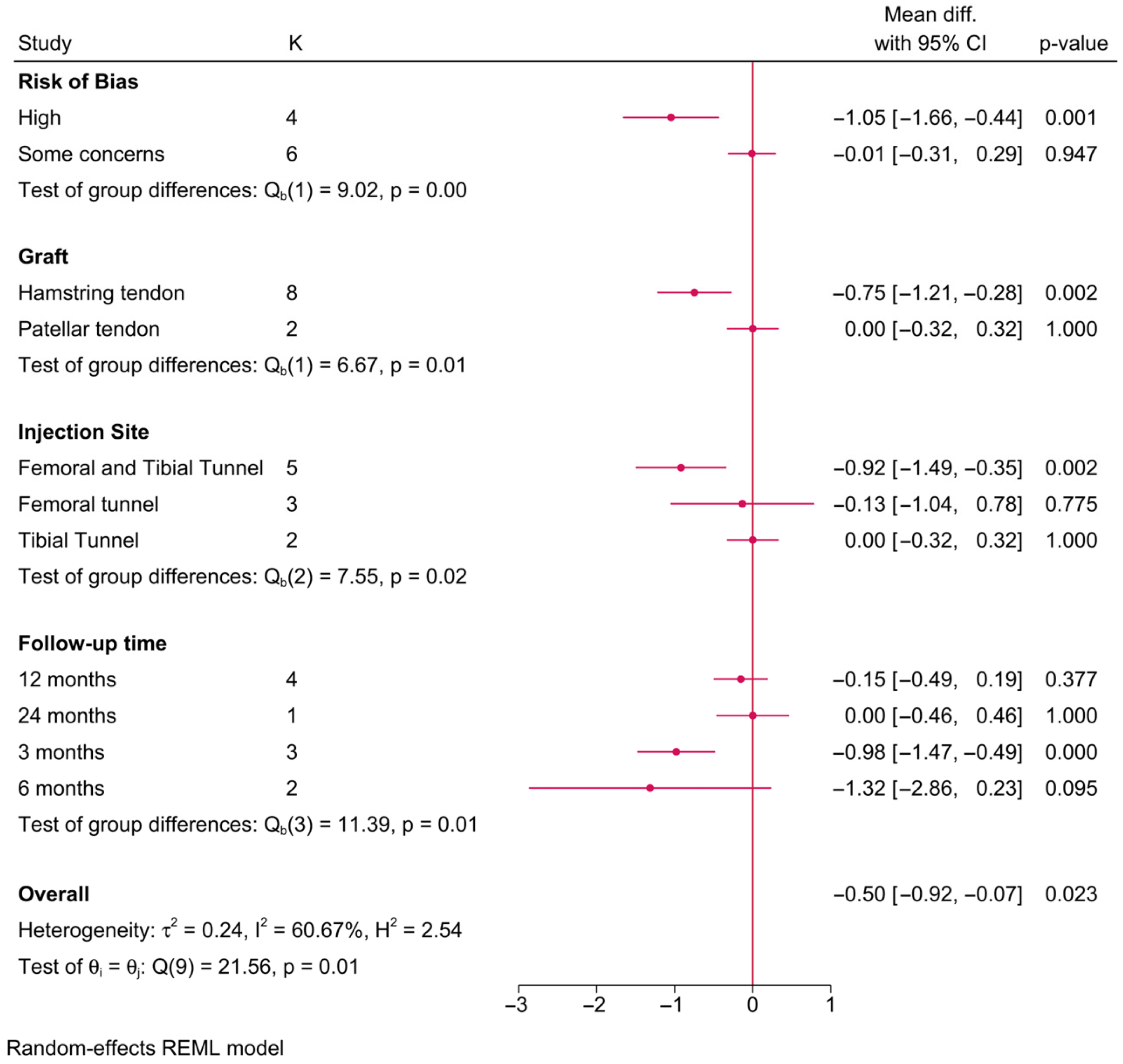
Ten RCTs investigated the impact of PRP on knee stability, as measured by the KT-1000 arthrometric measure (Figure 6). The risk of bias (p = 0.001), graft type (p = 0.01), injection site (p = 0.02), and follow-up time (p = 0.01) significantly modified the effect of PRP on postoperative knee stability. For instance, PRP was associated with a significantly lower KT-1000 score compared to the control; however, this difference was only observed in RCTs with a high risk of bias (MD = −1.05; 95% CI: −1.66, −0.44), surgeries utilizing hamstring tendon grafts (MD = −0.75; 95% CI: −1.21, −0.28), and when PRP was injected into both the femoral and tibial tunnels simultaneously (MD = −0.92; 95% CI: −1.49, −0.35). This effect was observable only at 3 months of follow-up (MD = −0.98; 95% CI: −1.47, −0.49) and not beyond or before that time point.

3.5.7. Ligamentization (MRI Hypointensity Grade)

PRP had a significant impact on promoting ligamentization and enhancing graft maturation (Table 2). Specifically, PRP was associated with a marked reduction in the odds of achieving higher grades of hypointensity, which correspond to less mature grafts. The odds ratio (OR) for achieving Grade 4 (diffusely hypointense) outcomes was notably reduced in the PRP group compared to the controls, suggesting a positive role of PRP in accelerating graft integration and maturation.
Subgroup analyses provided additional insights. Studies utilizing patellar tendon grafts demonstrated a significant improvement in ligamentization outcomes in the PRP group, with an OR of 2.59 (95% CI: 1.21–5.52) for achieving better MRI hypointensity grades compared to the controls. In contrast, outcomes for hamstring tendon grafts were less conclusive, highlighting a potential graft-specific effect of PRP. Further, when PRP was injected into the femoral and tibial tunnels, the results consistently showed improved ligamentization metrics.
Temporal analysis revealed that the benefits of PRP on ligamentization were more pronounced at longer follow-up durations, particularly at 12 months. This suggests that PRP may exert its greatest effect during the later stages of graft remodeling. However, variability in the findings was observed across studies, influenced by factors such as the risk of bias, follow-up time, and PRP preparation and application protocols.

4. Discussion

The findings of our systematic review and meta-analysis underscore the complex and heterogeneous nature of the impact of PRP on ACLR. The analysis revealed nuanced insights into the role of PRP in ligamentization, graft maturation, and other clinical outcomes, while highlighting persistent gaps in the literature. Here, we contextualize these findings within the broader landscape of ACLR research.

4.1. Pain Reduction

PRP demonstrated a significant, albeit transient, reduction in postoperative pain. This effect was most pronounced in studies with femoral tunnel injections and at earlier follow-up points (1 to 9 months). However, the magnitude of pain reduction diminished over time, suggesting that PRP primarily accelerates early healing rather than providing sustained analgesic effects. This aligns with the hypothesized mechanisms of PRP, wherein growth factors like platelet-derived growth factor and vascular endothelial growth factor attenuate early inflammatory responses and promote tissue repair [11,15]. Notably, the association between PRP and pain reduction was predominantly observed in studies with a high or moderate risk of bias, raising questions about its clinical reliability [14,16].

4.2. Functional Scores (Lysholm, Tegner, and IKDC)

The impact of PRP on functional scores revealed a complex picture. While PRP significantly improved IKDC scores at 12 months of follow-up, these effects were limited to studies with a higher risk of bias and injections targeting femoral tunnels. Similarly, the Lysholm and Tegner scores showed no meaningful differences between the PRP and control groups across most studies. These findings suggest that PRP’s functional benefits, if present, may be context-dependent, influenced by factors such as the graft type, injection site, and study design [16,58]. This is consistent with prior meta-analyses, which have highlighted PRP’s limited role in enhancing patient-reported outcomes [12,59].

4.3. Knee Stability (KT-1000)

Knee stability, as measured by KT-1000 arthrometry, showed significant improvement with PRP in studies that utilized hamstring tendon grafts and employed dual-site injections (femoral and tibial tunnels). However, these effects were only evident at the 3-month follow-up and were not sustained over time. This transient improvement in stability may reflect PRP’s early modulation of graft integration, but the lack of long-term benefits raises concerns about its durability [16,59].

4.4. PRP and Ligamentization

Our analysis demonstrated that PRP was significantly associated with enhanced ligamentization as measured by MRI hypointensity grades, particularly at later stages of follow-up (12 months). This effect was most pronounced when PRP was used in conjunction with patellar tendon grafts, which may be attributable to the unique biological environment created by these grafts. The odds ratios favoring PRP for improved ligamentization outcomes corroborate earlier reports suggesting that growth factors within PRP, such as platelet-derived growth factor and transforming growth factor-beta, enhance fibroblast activity, angiogenesis, and collagen synthesis [11,12].
Despite these promising results, the effect of PRP on ligamentization varied based on the injection site and follow-up duration. Studies where PRP was administered to the femoral and tibial tunnels reported more consistent benefits, aligning with preclinical evidence that PRP enhances graft–bone integration [58,59]. However, variability in PRP preparation, application techniques, and patient populations across studies likely contributes to inconsistent results, emphasizing the need for standardized protocols [15,16].
A notable divergence in the literature pertains to the impact of PRP on clinical outcomes such as pain and functional scores. While our study observed significant pain reduction in the early postoperative period, particularly with injections targeting specific sites, the benefits diminished over time. These findings parallel prior reviews suggesting that PRP may accelerate early recovery but fails to provide sustained long-term benefits [16,59].

4.5. Temporal Pattern of Effects and Heterogeneity

An important observation across outcomes was the predominantly early and time-dependent nature of PRP-associated benefits. Reductions in postoperative pain were most evident within the first 1–9 months, while improvements in knee stability were primarily observed at 3 months and were not sustained thereafter. Several explanations may account for this pattern. Firstly, PRP-derived growth factors such as PDGF and TGF-β are thought to exert their greatest influence during the early inflammatory and proliferative phases of graft healing. During this period, enhanced angiogenesis, cellular infiltration, and collagen synthesis may accelerate initial graft integration. However, as graft remodeling progresses into the maturation phase, the biological contribution of exogenous platelet-derived factors may diminish, leading to convergence between the PRP and control groups over time. Secondly, standardized postoperative rehabilitation protocols may reduce early between-group differences as mechanical loading and neuromuscular adaptation become dominant determinants of functional recovery. Thus, any biologically mediated acceleration in early healing may not necessarily translate into sustained clinical superiority. Thirdly, patient-reported outcome measures such as IKDC, Lysholm, and Tegner scores may exhibit ceiling effects at later follow-up intervals, limiting their sensitivity to detect subtle long-term differences. Finally, heterogeneity in patient populations, graft types, and PRP preparation methods likely contributed to the variability in early responses. While certain subgroups (e.g., specific graft types or injection sites) demonstrated transient benefits, these effects were not consistently reproducible across broader populations.

4.6. Clinical Implications

The clinical relevance of PRP in ACL reconstruction should be interpreted cautiously. Although PRP was associated with improved ligamentization and reductions in postoperative pain, these effects were largely time-dependent and most evident during early follow-up. Similarly, improvements in knee stability (KT-1000 measurements) were observed primarily at 3 months and were not sustained at later time points. Moreover, several favorable findings were restricted to specific subgroups or studies with a higher risk of bias.
Importantly, consistent long-term improvements in functional outcomes such as Lysholm, Tegner, and IKDC scores were not demonstrated across the broader evidence base. Therefore, current data do not support the routine use of PRP as a standard adjunct in ACL reconstruction.
PRP may represent a biologically plausible adjunct with potential short-term benefits in selected contexts; however, its role in enhancing durable clinical recovery remains uncertain. Future high-quality randomized trials with standardized PRP characterization and extended follow-up are necessary before definitive clinical recommendations can be made.

4.7. Limitations and Future Directions

Several limitations must be acknowledged. The heterogeneity in the trials’ risk-of-bias degree, PRP preparation methods, and outcome assessments complicates the synthesis of evidence. Additionally, the predominance of studies with a high risk of bias underscores the need for rigorous randomized controlled trials with standardized methodologies. Another important limitation relates to pooling across biologically heterogeneous platelet-derived products. Although labeled differently (PRP, PRGF, PRF, APC, PC), these products were consistently applied as autologous platelet-enriched adjuncts during ACL reconstruction. However, the preparation techniques, fibrin structure, leukocyte content, and activation status likely differed across trials. Because most studies did not report sufficient laboratory characterization, biologically stratified meta-analysis was not possible.
A critical limitation of the included literature is the absence of standardized PRP characterization. Contemporary orthobiologic concepts emphasize that the total platelet dose, leukocyte content, activation status, and preparation protocol may represent key determinants of therapeutic efficacy. However, most RCTs failed to report the platelet concentration relative to the baseline, leukocyte content, or injected platelet mass. Without these parameters, formal classification using systems such as PAW or MARSPILL was not feasible. The inability to account for biological variability likely contributed to the heterogeneity and may partially explain the inconsistent clinical findings observed across studies.
Another limitation relates to the limited availability of direct mechanistic data within the included trials. Most randomized studies focused on clinical, radiological, and functional outcomes without incorporating histological analysis, quantitative vascular assessment, biomarker evaluation, or standardized biological profiling of PRP formulations. Consequently, interpretations regarding the underlying biological mechanisms of PRP in ligamentization and graft maturation remain indirect and largely extrapolated from preclinical research. This limits the ability to establish definitive causal pathways between PRP application and observed clinical effects.
Future research should focus on elucidating the mechanisms underlying PRP’s variable efficacy, optimizing its preparation and application, and identifying patient subgroups most likely to benefit. Comparative trials investigating different PRP formulations and dosing regimens could further refine its clinical utility.

5. Conclusions

PRP holds promise as an adjunctive therapy in ACLR, particularly for enhancing ligamentization and early postoperative recovery. However, its role in improving long-term functional outcomes remains limited and contentious. Standardization of PRP preparation protocols, including systematic reporting of the platelet concentration, leukocyte composition, activation methods, injected volume, and total platelet dose, is essential to enable reproducibility and clarify its therapeutic role in ACLR. Our findings provide a foundation for future investigations aimed at bridging the gap between experimental promise and clinical applicability in ACLR.

Supplementary Materials

The following supporting information can be downloaded at: https://www.mdpi.com/article/10.3390/jcm15072526/s1, Supplementary File S1: a PRISMA checklist. Figure S1: a forest plot showing the difference in postoperative Lysholm score between PRP and control stratified by risk-of-bias level, graft type, injection site, and follow-up time; Figure S2: a Forest plot showing the difference in postoperative Tegner score between PRP and control stratified by risk-of-bias level, graft type, injection site, and follow-up time; Figure S3: a Forest plot showing the difference in graft maturation (SNQ score) between PRP and control stratified by risk-of-bias level, graft type, injection site, and follow-up time; Figure S4: a Forest plot showing the difference in postoperative femoral tunnel diameter between PRP and control stratified by risk-of-bias level, graft type, injection site, and follow-up time. Table S1: the detailed database search query employed in this systematic review.

Author Contributions

Conceptualization, A.A.; methodology, G.A., C.C. (Caroline Chahine), G.A.O., J.B.C., A.C., H.S., C.C. (Charbel Chaiban), A.G.; software, C.C. (Caroline Chahine); validation, A.A., S.J.; formal analysis, C.C. (Caroline Chahine); investigation, A.A., G.A.O., A.C., H.S., M.G., C.C. (Charbel Chaiban); resources, G.A.O.; data curation, G.A., C.C. (Caroline Chahine), A.C., J.B.C., H.S., C.C. (Charbel Chaiban), A.G.; writing—original draft preparation, G.A., S.J., M.G.; writing—review and editing, J.B.C., A.G.; visualization, S.J.; supervision, A.A. and M.G.; project administration, M.G. All authors have read and agreed to the published version of the manuscript.

Funding

This research received no external funding.

Institutional Review Board Statement

Not applicable.

Informed Consent Statement

Not applicable.

Data Availability Statement

Data are available within the manuscript and its Supplementary Materials.

Acknowledgments

We would like to thank Abdelaziz Abdelaal for helping with the statistical analysis and write-up of this manuscript.

Conflicts of Interest

The authors declare no conflicts of interest.

Abbreviations

The following abbreviations are used in this manuscript:
ACLAnterior Cruciate Ligament
ACLRAnterior Cruciate Ligament Reconstruction
AMSTARAssessing the Methodological Quality of Systematic Reviews
APCAutologous Platelet Concentrate
BTBBone–Tendon–Bone
CIConfidence Interval
IKDCInternational Knee Documentation Committee
I2Inconsistency Index
KT-1000KT-1000 Arthrometer
MDMean Difference
MRIMagnetic Resonance Imaging
NRNot Reported
OROdds Ratio
PCPlatelet Concentrate
PDGFPlatelet-Derived Growth Factor
PICOSPopulation, Intervention, Comparison, Outcomes, Study design
PRGFPlasma Rich in Growth Factors
PRISMAPreferred Reporting Items for Systematic Reviews and Meta-Analyses
PRPPlatelet-Rich Plasma
PRFPlatelet-Rich Fibrin
RCTRandomized Controlled Trial
REMLRestricted Maximum Likelihood
SDStandard Deviation
SNQSignal-to-Noise Quotient
TGF-βTransforming Growth Factor Beta
VASVisual Analogue Scale
VEGFVascular Endothelial Growth Factor
YOIYear of Investigation
YOPYear of Publication

References

  1. Cimino, F.; Volk, B.S.; Setter, D. Anterior cruciate ligament injury: Diagnosis, management, and prevention. Am. Fam. Physician 2010, 82, 917–922. [Google Scholar]
  2. Kiapour, A.; Murray, M. Basic science of anterior cruciate ligament injury and repair. Bone Jt. Res. 2014, 3, 20–31. [Google Scholar] [CrossRef]
  3. Chambat, P.; Guier, C.; Sonnery-Cottet, B.; Fayard, J.-M.; Thaunat, M. The evolution of ACL reconstruction over the last fifty years. Int. Orthop. 2013, 37, 181–186. [Google Scholar] [CrossRef]
  4. Eckenrode, B.J.; Carey, J.L.; Sennett, B.J.; Zgonis, M.H. Prevention and management of post-operative complications following ACL reconstruction. Curr. Rev. Musculoskelet. Med. 2017, 10, 315–321. [Google Scholar] [CrossRef] [PubMed]
  5. Papakonstantinou, O.; Chung, C.B.; Chanchairujira, K.; Resnick, D.L. Complications of anterior cruciate ligament reconstruction: MR imaging. Eur. Radiol. 2003, 13, 1106–1117. [Google Scholar] [CrossRef] [PubMed]
  6. Okoroha, K.R.; Keller, R.A.; Jung, E.K.; Khalil, L.; Marshall, N.; Kolowich, P.A.; Moutzouros, V. Pain assessment after anterior cruciate ligament reconstruction: Bone–patellar tendon–bone versus hamstring tendon autograft. Orthop. J. Sports Med. 2016, 4, 2325967116674924. [Google Scholar] [CrossRef] [PubMed]
  7. Tripp, D.A.; Stanish, W.D.; Reardon, G.; Coady, C.; Sullivan, M.J. Comparing postoperative pain experiences of the adolescent and adult athlete after anterior cruciate ligament surgery. J. Athl. Train. 2003, 38, 154. [Google Scholar]
  8. Claes, S.; Verdonk, P.; Forsyth, R.; Bellemans, J. The “ligamentization” process in anterior cruciate ligament reconstruction: What happens to the human graft? A systematic review of the literature. Am. J. Sports Med. 2011, 39, 2476–2483. [Google Scholar] [CrossRef]
  9. Pauzenberger, L.; Syré, S.; Schurz, M. “Ligamentization” in hamstring tendon grafts after anterior cruciate ligament reconstruction: A systematic review of the literature and a glimpse into the future. Arthroscopy 2013, 29, 1712–1721. [Google Scholar] [CrossRef]
  10. Davey, M.S.; Hurley, E.T.; Withers, D.; Moran, R.; Moran, C.J. Anterior cruciate ligament reconstruction with platelet-rich plasma: A systematic review of randomized control trials. Arthroscopy 2020, 36, 1204–1210. [Google Scholar] [CrossRef]
  11. Bissell, L.; Tibrewal, S.; Sahni, V.; Khan, W.S. Growth factors and platelet rich plasma in anterior cruciate ligament reconstruction. Curr. Stem Cell Res. Ther. 2015, 10, 19–25. [Google Scholar] [CrossRef] [PubMed]
  12. Delcogliano, M.; Sangiorgio, A.; Bensa, A.; Andriolo, L.; Boffa, A.; Filardo, G. Platelet-rich plasma augmentation in anterior cruciate ligament reconstruction: Evidence is still too scattered. A scoping review of randomised controlled trials. Knee Surg. Sports Traumatol. Arthrosc. 2024, 32, 1143–1159. [Google Scholar] [CrossRef] [PubMed]
  13. Figueroa, D.; Figueroa, F.; Calvo, R.; Vaisman, A.; Ahumada, X.; Arellano, S. Platelet-rich plasma use in anterior cruciate ligament surgery: Systematic review of the literature. Arthroscopy 2015, 31, 981–988. [Google Scholar] [CrossRef] [PubMed]
  14. de Andrade, A.L.L.; Sardeli, A.V.; Garcia, T.A.; Livani, B.; Belangero, W.D. PRP does not improve the objective outcomes of anterior cruciate ligament reconstruction: A systematic review and meta-analysis. Knee Surg. Sports Traumatol. Arthrosc. 2021, 29, 3049–3058. [Google Scholar] [CrossRef]
  15. McRobb, J.; Kamil, K.H.; Ahmed, I.; Dhaif, F.; Metcalfe, A. Influence of platelet-rich plasma (PRP) analogues on healing and clinical outcomes following anterior cruciate ligament (ACL) reconstructive surgery: A systematic review. Eur. J. Orthop. Surg. Traumatol. 2023, 33, 225–253. [Google Scholar] [CrossRef]
  16. Zhu, T.; Zhou, J.; Hwang, J.; Xu, X. Effects of platelet-rich plasma on clinical outcomes after anterior cruciate ligament reconstruction: A systematic review and meta-analysis. Orthop. J. Sports Med. 2022, 10, 23259671211061535. [Google Scholar] [CrossRef]
  17. Page, M.J.; McKenzie, J.E.; Bossuyt, P.M.; Boutron, I.; Hoffmann, T.C.; Mulrow, C.D.; Shamseer, L.; Tetzlaff, J.M.; Akl, E.A.; Brennan, S.E. The PRISMA 2020 statement: An updated guideline for reporting systematic reviews. BMJ 2021, 372, n71. [Google Scholar] [CrossRef]
  18. Shea, B.J.; Hamel, C.; Wells, G.A.; Bouter, L.M.; Kristjansson, E.; Grimshaw, J.; Henry, D.A.; Boers, M. AMSTAR is a reliable and valid measurement tool to assess the methodological quality of systematic reviews. J. Clin. Epidemiol. 2009, 62, 1013–1020. [Google Scholar] [CrossRef]
  19. Muka, T.; Glisic, M.; Milic, J.; Verhoog, S.; Bohlius, J.; Bramer, W.; Chowdhury, R.; Franco, O.H. A 24-step guide on how to design, conduct, and successfully publish a systematic review and meta-analysis in medical research. Eur. J. Epidemiol. 2020, 35, 49–60. [Google Scholar] [CrossRef]
  20. Abdelaal, A.; Eltaras, M.M.; Katamesh, B.E.; Serhan, H.A.; Farahat, R.A.; Badr, H.; Abdelazeem, B. The prevalence and presentation patterns of microcystic macular oedema: A systematic review and meta-analysis of 2128 glaucomatous eyes. Eye 2023, 37, 3322–3333. [Google Scholar] [CrossRef]
  21. Amir-Behghadami, M.; Janati, A. Population, Intervention, Comparison, Outcomes and Study (PICOS) design as a framework to formulate eligibility criteria in systematic reviews. Emerg. Med. J. 2020, 37, 387. [Google Scholar] [CrossRef] [PubMed]
  22. Mavridis, D.; Salanti, G.; Furukawa, T.A.; Cipriani, A.; Chaimani, A.; White, I.R. Allowing for uncertainty due to missing and LOCF imputed outcomes in meta-analysis. Stat. Med. 2019, 38, 720–737. [Google Scholar] [CrossRef] [PubMed]
  23. Sedgwick, P. Meta-analyses: Heterogeneity and subgroup analysis. Bmj 2013, 346, f4040. [Google Scholar] [CrossRef]
  24. Lin, L.; Chu, H. Quantifying publication bias in meta-analysis. Biometrics 2018, 74, 785–794. [Google Scholar] [CrossRef]
  25. Azcárate, A.V.; Lamo-Espinosa, J.; Beola, J.D.A.; Gonzalez, M.H.; Gasque, G.M.; Nin, J.R.V. Comparison between two different platelet-rich plasma preparations and control applied during anterior cruciate ligament reconstruction. Is there any evidence to support their use? Injury 2014, 45, S36–S41. [Google Scholar] [CrossRef]
  26. Beyzadeoglu, T.; Pehlivanoglu, T.; Yildirim, K.; Buldu, H.; Tandogan, R.; Tuzun, U. Does the application of platelet-rich fibrin in anterior cruciate ligament reconstruction enhance graft healing and maturation? A comparative MRI study of 44 cases. Orthop. J. Sports Med. 2020, 8, 2325967120902013. [Google Scholar] [CrossRef]
  27. Cervellin, M.; De Girolamo, L.; Bait, C.; Denti, M.; Volpi, P. Autologous platelet-rich plasma gel to reduce donor-site morbidity after patellar tendon graft harvesting for anterior cruciate ligament reconstruction: A randomized, controlled clinical study. Knee Surg. Sports Traumatol. Arthrosc. 2012, 20, 114–120. [Google Scholar] [CrossRef]
  28. de Almeida, A.M.; Demange, M.K.; Sobrado, M.F.; Rodrigues, M.B.; Pedrinelli, A.; Hernandez, A.J. Patellar tendon healing with platelet-rich plasma: A prospective randomized controlled trial. Am. J. Sports Med. 2012, 40, 1282–1288. [Google Scholar] [CrossRef]
  29. Eslami, S.; Fattah, S.; Taher, S.A.; Rezasoltani, Z. Platelet-rich plasma therapy or arthroscopic surgery on repair of anterior cruciate ligament rupture. Eur. J. Transl. Myol. 2022, 32, 10538. [Google Scholar] [CrossRef]
  30. Figueroa, D.; Melean, P.; Calvo, R.; Vaisman, A.; Zilleruelo, N.; Figueroa, F.; Villalón, I. Magnetic resonance imaging evaluation of the integration and maturation of semitendinosus-gracilis graft in anterior cruciate ligament reconstruction using autologous platelet concentrate. Arthroscopy 2010, 26, 1318–1325. [Google Scholar] [CrossRef]
  31. Gong, H.; Huang, B.; Zheng, Z.; Fu, L.; Chen, L. Clinical Use of Platelet-Rich Plasma to Promote Tendon–Bone Healing and Graft Maturation in Anterior Cruciate Ligament Reconstruction—A Randomized Controlled Study. Indian J. Orthop. 2022, 56, 805–811. [Google Scholar] [CrossRef]
  32. Ji, Q.; Yang, Y.; Chen, H.; Geng, W.; Dong, H.; Yu, Q. Clinical evaluations of anterior cruciate ligament reconstruction with platelet rich plasma. Zhongguo Xiu Fu Chong Jian Wai Ke Za Zhi 2017, 31, 410–416. [Google Scholar] [PubMed]
  33. Kasl, J.; Zeman, P.; Pavelka, T.; Cibulková, J.; Pilný, J.; Matějka, J. Clinical Outcomes of the Application of Platelet-Rich Fibrin during Anatomic Anterior Cruciate Ligament Reconstruction: Results of Prospective Randomised Study. Acta Chir. Orthop. Traumatol. Cechoslov. 2022, 89, 279–285. [Google Scholar] [CrossRef] [PubMed]
  34. Kumar, A.; Kushwaha, N.S.; Kumar, D.; Singh, A.; Gupta, V.; Kumar, S. Prospective Comparison of Functional and Radiological Outcomes of Arthroscopic Anterior Cruciate Ligament Reconstruction by Hamstring Graft Alone and Platelet-Rich Plasma Added to the Hamstring Graft. Cureus 2022, 14, e23017. [Google Scholar] [CrossRef] [PubMed]
  35. Lin, Y.-C.; Chen, Y.-J.; Fan, T.-Y.; Chou, P.-H.; Lu, C.-C. Effect of bone marrow aspiration concentrate and platelet-rich plasma combination in anterior cruciate ligament reconstruction: A randomized, prospective, double-blinded study. J. Orthop. Surg. Res. 2024, 19, 4. [Google Scholar] [CrossRef]
  36. Malinowski, K.; Ebisz, M.; LaPrade, R.F.; Mostowy, M. Platelet-Rich Plasma in Anterior Cruciate Ligament Quadriceps Tendon Bone Reconstruction—Impact of PRP Administration on Pain, Range of Motion Restoration, Knee Stability, Tibial Tunnel Widening and Functional Results. Appl. Sci. 2021, 11, 3993. [Google Scholar] [CrossRef]
  37. Mirzatolooei, F.; Alamdari, M.; Khalkhali, H. The impact of platelet-rich plasma on the prevention of tunnel widening in anterior cruciate ligament reconstruction using quadrupled autologous hamstring tendon: A randomised clinical trial. Bone Jt. Res. 2013, 95, 65–69. [Google Scholar] [CrossRef]
  38. Munde, K.; Banerjee, S.; Gahlot, N.; Elhence, A.; Mandal, S.; Yadav, T. Effect of platelet-rich plasma on healing of autologous graft after anterior cruciate ligament reconstruction: A randomized control trial. Regen. Med. 2023, 18, 601–610. [Google Scholar] [CrossRef]
  39. Nin, J.R.V.; Gasque, G.M.; Azcarate, A.V.; Beola, J.D.A.; Gonzalez, M.H. Has platelet-rich plasma any role in anterior cruciate ligament allograft healing? Arthroscopy 2009, 25, 1206–1213. [Google Scholar]
  40. Orrego, M.; Larrain, C.; Rosales, J.; Valenzuela, L.; Matas, J.; Durruty, J.; Sudy, H.; Mardones, R. Effects of platelet concentrate and a bone plug on the healing of hamstring tendons in a bone tunnel. Arthroscopy 2008, 24, 1373–1380. [Google Scholar] [CrossRef]
  41. Rupreht, M.; Jevtič, V.; Serša, I.; Vogrin, M.; Jevšek, M. Evaluation of the tibial tunnel after intraoperatively administered platelet-rich plasma gel during anterior cruciate ligament reconstruction using diffusion weighted and dynamic contrast-enhanced MRI. J. Magn. Reson. Imaging 2013, 37, 928–935. [Google Scholar] [CrossRef] [PubMed]
  42. Rupreht, M.; Vogrin, M.; Hussein, M. MRI evaluation of tibial tunnel wall cortical bone formation after platelet-rich plasma applied during anterior cruciate ligament reconstruction. Radiol. Oncol. 2013, 47, 119–124. [Google Scholar] [CrossRef] [PubMed]
  43. Seijas, R.; Ares, O.; Catala, J.; Alvarez-Diaz, P.; Cusco, X.; Cugat, R. Magnetic resonance imaging evaluation of patellar tendon graft remodelling after anterior cruciate ligament reconstruction with or without platelet-rich plasma. J. Orthop. Surg. 2013, 21, 10–14. [Google Scholar] [CrossRef] [PubMed]
  44. Seijas, R.; Ares, O.; Català, J.; Cuscó, X.; Álvarez, P.; García-Balletbó, M.; Cugat, R. Role of Growth Factors in Bone-Tendon-Bone ACL Surgery: Time for Maturation of the ACL Graft and the Patellar Tendon Donor Site. Int. J. Orthop. 2015, 2, 196–201. [Google Scholar]
  45. Seijas, R.; Cuscó, X.; Sallent, A.; Serra, I.; Ares, O.; Cugat, R. Pain in donor site after BTB-ACL reconstruction with PRGF: A randomized trial. Arch. Orthop. Trauma. Surg. 2016, 136, 829–835. [Google Scholar] [CrossRef]
  46. Seijas, R.; Rius, M.; Ares, O.; García-Balletbó, M.; Serra, I.; Cugat, R. Healing of donor site in bone-tendon-bone ACL reconstruction accelerated with plasma rich in growth factors: A randomized clinical trial. Knee Surg. Sports Traumatol. Arthrosc. 2015, 23, 991–997. [Google Scholar] [CrossRef]
  47. Silva, A.; Sampaio, R. Anatomic ACL reconstruction: Does the platelet-rich plasma accelerate tendon healing? Knee Surg. Sports Traumatol. Arthrosc. 2009, 17, 676–682. [Google Scholar] [CrossRef]
  48. Solomon, R.; Hommen, J.P.; Travascio, F. Effects of Platelet-Rich Osteoconductive–Osteoinductive Allograft Compound on Tunnel Widening of ACL Reconstruction: A Randomized Blind Analysis Study. Pathophysiology 2022, 29, 394–404. [Google Scholar] [CrossRef]
  49. Sözkesen, S.; Karahan, H.G.; Kurtulmus, A.; Kayali, C.; Altay, T. PRP on preventıon of tunnel enlargement in ACL reconstructıon. Ortop. Traumatol. Rehabil. 2018, 20, 285–291. [Google Scholar] [CrossRef]
  50. Starantzis, K.A.; Mastrokalos, D.; Koulalis, D.; Papakonstantinou, O.; Soucacos, P.N.; Papagelopoulos, P.J. The potentially positive role of PRPs in preventing femoral tunnel widening in ACL reconstruction surgery using hamstrings: A clinical study in 51 patients. J. Sports Med. 2014, 2014, 789317. [Google Scholar] [CrossRef]
  51. Vadalà, A.; Iorio, R.; De Carli, A.; Ferretti, M.; Paravani, D.; Caperna, L.; Iorio, C.; Gatti, A.; Ferretti, A. Platelet-rich plasma: Does it help reduce tunnel widening after ACL reconstruction? Knee Surg. Sports Traumatol. Arthrosc. 2013, 21, 824–829. [Google Scholar] [CrossRef] [PubMed]
  52. Vogrin, M.; Rupreht, M.; Crnjac, A.; Dinevski, D.; Krajnc, Z.; Rečnik, G. The effect of platelet-derived growth factors on knee stability after anterior cruciate ligament reconstruction: A prospective randomized clinical study. Wien. Klin. Wochenschr. 2010, 122, 91–95. [Google Scholar] [CrossRef] [PubMed]
  53. Vogrin, M.; Rupreht, M.; Dinevski, D.; Hašpl, M.; Kuhta, M.; Jevsek, M.; Knežević, M.; Rožman, P. Effects of a platelet gel on early graft revascularization after anterior cruciate ligament reconstruction: A prospective, randomized, double-blind, clinical trial. Eur. Surg. Res. 2010, 45, 77–85. [Google Scholar] [CrossRef] [PubMed]
  54. Walters, B.L.; Porter, D.A.; Hobart, S.J.; Bedford, B.B.; Hogan, D.E.; McHugh, M.M.; Klein, D.A.; Harousseau, K.; Nicholas, S.J. Effect of intraoperative platelet-rich plasma treatment on postoperative donor site knee pain in patellar tendon autograft anterior cruciate ligament reconstruction: A double-blind randomized controlled trial. Am. J. Sports Med. 2018, 46, 1827–1835. [Google Scholar] [CrossRef]
  55. Wasilczyk, C. Nanosurgical and Bioengineering Treatment of Human Anterior Cruciate Ligament Tears with Ultrasound-Guided Injection of Modified Platelet-Rich Plasma Using Human Cell Memory Based on Clinical, Ultrasound, MRI, and Nanoscope Analyses: A Double-Blind Randomized Trial. J. Clin. Med. 2024, 13, 2475. [Google Scholar]
  56. Ye, Z.; Chen, H.; Qiao, Y.; Wu, C.; Cho, E.; Wu, X.; Li, Z.; Wu, J.; Lu, S.; Xie, G. Intra-Articular Platelet-Rich Plasma Injection After Anterior Cruciate Ligament Reconstruction: A Randomized Clinical Trial. JAMA Netw. Open 2024, 7, e2410134. [Google Scholar] [CrossRef]
  57. Zeman, P.; Kasl, J.; Tupý, R.; Frei, R.; Kott, O.; Kautzner, J. Evaluation of the MRI findings on a tendon graft after the anatomic anterior cruciate ligament reconstruction in patients with versus without the application of platelet-rich fibrin-results of the prospective randomised study. Acta Chir. Orthop. Traumatol. Cechoslov. 2018, 85, 343–350. [Google Scholar] [CrossRef]
  58. Cao, Y.; Wan, Y.d. Effectiveness of Platelet-Rich Plasma in Anterior Cruciate Ligament Reconstruction: A Systematic Review of Randomized Controlled Trials. Orthop. Surg. 2022, 14, 2406–2417. [Google Scholar] [CrossRef]
  59. Lv, Z.-T.; Zhang, J.-M.; Pang, Z.-Y.; Wang, Z.; Huang, J.-M.; Zhu, W.-T. The efficacy of platelet rich plasma on anterior cruciate ligament reconstruction: A systematic review and meta-analysis. Platelets 2022, 33, 229–241. [Google Scholar] [CrossRef]
Figure 1. A PRISMA flow diagram showing the results of the literature search. ** the number of citations before duplicate removal; * the number of citations after title/abstract screening and duplicate removal.
Jcm 15 02526 g001
Figure 2. A summary of the risk of bias of included randomized controlled trials comparing PRP to control in patients undergoing ACL reconstruction [25,26,27,28,29,30,31,32,33,34,35,36,37,38,39,40,41,42,43,44,45,46,47,48,49,50,51,52,53,54,55,56,57].
Jcm 15 02526 g002
Figure 3. A forest plot showing the difference in postoperative pain score between PRP and control stratified by risk-of-bias level, graft type, injection site, and follow-up time. The p-values displayed represent subgroup interaction tests evaluating whether effect sizes differ across categories. I2 values indicate the degree of statistical heterogeneity among included studies within each subgroup.
Jcm 15 02526 g003
Figure 4. A forest plot showing the difference in postoperative IKDC score between PRP and control stratified by risk-of-bias level, graft type, injection site, and follow-up time.
Jcm 15 02526 g004
Figure 5. A forest plot showing the difference in postoperative tibial tunnel diameter between PRP and control stratified by risk-of-bias level, graft type, injection site, and follow-up time. Subgroup p-values reflect interaction testing between categories, while I2 values quantify the percentage of variability attributable to between-study heterogeneity rather than sampling error.
Jcm 15 02526 g005
Figure 6. A forest plot showing the difference in postoperative knee stability (KT-1000 score) between PRP and control stratified by risk-of-bias level, graft type, injection site, and follow-up time.
Jcm 15 02526 g006
Table 1. Baseline characteristics of randomized controlled trials comparing PRP to control in ACL reconstruction.
Author (YOP)CountryDesignRegistrationGraftYOIGroupPRP Injection SiteSample SizeAgeGenderFollow-Up
MeanSDMaleFemale
Mirzatolooei (2013) [37]IranRCTNRHamstring tendonFebruary 2011–February 2012PRPFemoral and Tibial Tunnel2526.4(18–40)2033 months
Control-2526.9(18–40)221
de Almeida (2012) [28]BrazilRCTNRPatellar tendon-PRPNR1225.8(18–44)1026 months
Control-1523.1(15–34)141
Kumar (2022) [34]IndiaRCTNRHamstring tendon-PRPTibial Tunnel35----3 months
Control-35----
Azcárate (2014) [25]SpainRCTNRPatellar tendon allograft-PRPTibial Tunnel5026.1(14–57)401012 months
Control-5026.1(15–59)3812
Vogrin (2010) [53]SloveniaRCTNRHamstring tendonFebruary–June 2008PRPFemoral and Tibial Tunnel2235.41059.140.96 months
Control-233312.573.926.1
Gong (2022) [31]ChinaRCTNCT04659447Hamstring tendon-PRPFemoral and Tibial Tunnel3033.58.97181212 months
Control-3034.99.68219
Lin (2024) [35]TaiwanRCTNRHamstring tendon graft-Control-10----24 weeks
PRPFemoral Tunnel8----
Eslami (2022) [29]IranRCTIRCT20200217046523N18Not clarified2020–2021PRPNR5032.263.69--10.26 (2.11) months
Control-50--
Ye (2024) [56]ChinaRCTChiCTR2000040262Autologous semitendinosus and gracilis tendon21 March 2021, 28 August 2023PRPFemoral and Tibial Tunnel60287.9431712 months
Control-603084119
Walters (2018) [54]USARCTNCT01765712Patellar tendon2011–2015PRPNot Clarified273012--24 months
Control-23--
Seijas (2013) [43]SpainRCTNRPatellar tendonJanuary–July 2009PRPFemoral and Tibial Tunnel49----12 months
Control-49----
Seijas (2015a) [44]SpainRCTNRPatellar tendon graft2009PRGFNR23203--4 months
Control-20173--
Seijas (2015b) [46]SpainRCTNRPatellar tendon-PRGF-EndoretNR50----12 months
Control-50----
Seijas (2016) [45]SpainRCTNRPatellar tendon graft2009PRGFNR23203--24 months
Control-20173--
Vadalà (2013) [51]ItalyRCTNRHamstring tendon-PRPFemoral and Tibial Tunnel2034.5(18–48)20014.7 months
Control-2034.5(18–48)200
Cervellin (2012) [27]ItalyRCTNRPatellar tendon2008–2009PRPNR2022.94.320012 months
Control-2022.73.5200
Solomon (2022) [48]USARCTNCT04993339Peroneus longus tendon2016, 2019PRPFemoral and Tibial Tunnel1332.710.5856 weeks
Control-1432.712.777
Wasilczyk (2024) [55]PolandRCTNRNRMarch 2015, February 2024Injected PRPNR3046.51516146 weeks
Control/PRP-10331291
Nin (2009) [39]SpainRCTNRPatellar tendon-PRPTibial Tunnel5026.1(14–57)401024 months
Control-5026.6(15–59)3812
Silva (2009) [47]PortugalRCTNRHamstring tendonNovember 2006–March 2008PRPFemoral Tunnel30----3 months
Control-10----
Rupreht (2013) [41]SloveniaRCTNRSemitendinosus and gracilis tendon graft-PRPTibial Tunnel2137.28.41386 months
Control-2032.612.3155
Vogrin (2010) [52]CroatiaRCTNRHamstring tendonFebruary–October 2008Control-2032.612.315512 weeks
PGFemoral and Tibial Tunnel2137.28.4138
Rupreht (2013) [42]SloveniaRCTNRSemitendinosus and gracilis tendon graft-PRPTibial Tunnel2137.28.41386 months
Control-20--155
Starantzis (2014) [50]GreeceRCTNRHamstring tendonDecember 2007–June 2010PRPFemoral Tunnel2529.47.3381312 months
Control-2631.38
Orrego (2008) [40]ChileRCTNRHamstring tendonJanuary 2005–December 2006PCFemoral Tunnel26----6 months
Control-27----
Sözkesen (2018) [49] TurkeyRCTNot registeredHamstring tendon autograftMarch 2014–July 2015PRPFemoral and Tibial Tunnel18266.9616212 months
Control-26266.96251
Figueroa (2010) [30]ChileRCTNRHamstring tendon-APCFemoral and Tibial Tunnel3026.8(14–28)18126.4 months
Control-2023.6(13–35)155
Ji (2017) [32]ChinaRCTNRHamstring tendonAugust 2014–August 2016PRPNR2131.59-8912 months
Control-2133.68-712
Kasl (2022) [33]CzechRCTNRHamstring tendon2012–2014PRPNR2029.1-291112 months
Control-20-
Zeman (2018) [57]CzechRCTNRSemitendinosus and gracillis tendon-PRPFemoral Tunnel1729.1-231012 months
Control-16-
Munde (2023) [38]IndiaRCTNRNR-PRPNR40----6 months
Control-40----
Beyzadeoglu (2020) [26]TurkeyRCTNRSemitendinosus tendon graft PRFFemoral Tunnel2321.7 18512 months
Control-2122.1 174
Malinowski (2021) [36]PolandRCTNot registeredQuadriceps tendon bone reconstruction2008–2010PRPNR54----18 months
Control-52----
YOP: year of publication; YOI: year of investigation; RCT: randomized controlled trial; PRP: platelet-rich plasma; NR: not reported; SD: standard deviation.
Table 2. A summary of ligamentization between PRP and control groups based on MRI hypointensity grades.
HypointenseGrade 1
(Mildly Hypointense)
Grade 2
(Moderately Hypointense)
Grade 3
(Severely Hypointense)
Grade 4
(Diffusely Hypointense)
KOR95% CIKOR95% CIKOR95% CIKOR95% CIKOR95% CI
Risk of Bias
High30.030.01–0.1130.310.14–0.6830.930.38–2.2731.090.65–1.8232.610.80–8.52
Some concerns40.090.02–0.5830.290.13–0.6430.950.40–2.2431.060.64–1.7432.720.82–9
Graft Type
Hamstring tendon10.630.21–1.93
Patellar tendon60.040.02–0.1060.30.17–0.5260.970.56–1.6561.070.75–1.5462.591.21–5.52
Injection Site
Femoral and tibial tunnels40.090.02–0.5830.290.13–0.6430.950.40–2.2431.060.64–1.7432.720.82–9
Not Clarified30.030.01–0.1130.310.14–0.6830.930.38–2.2731.090.65–1.8232.610.80–8.52
Follow-up time
4 months20.060.02–0.1920.320.16–0.6621.670.94–2.9721.410.71–2.7921.140.73–1.78
6 months30.080.01–0.8620.190.19–0.5820.590.30–1.1321.070.59–1.9423.241.02–10.30
12 months20.010.00–0.3020.50.09–2.7920.50.09–2.7920.860.47–1.5827.332.99–17.99
OR: odds ratio; CI: confidence interval; K: number of studies included in each subgroup; MRI: magnetic resonance imaging.
Disclaimer/Publisher’s Note: The statements, opinions and data contained in all publications are solely those of the individual author(s) and contributor(s) and not of MDPI and/or the editor(s). MDPI and/or the editor(s) disclaim responsibility for any injury to people or property resulting from any ideas, methods, instructions or products referred to in the content.