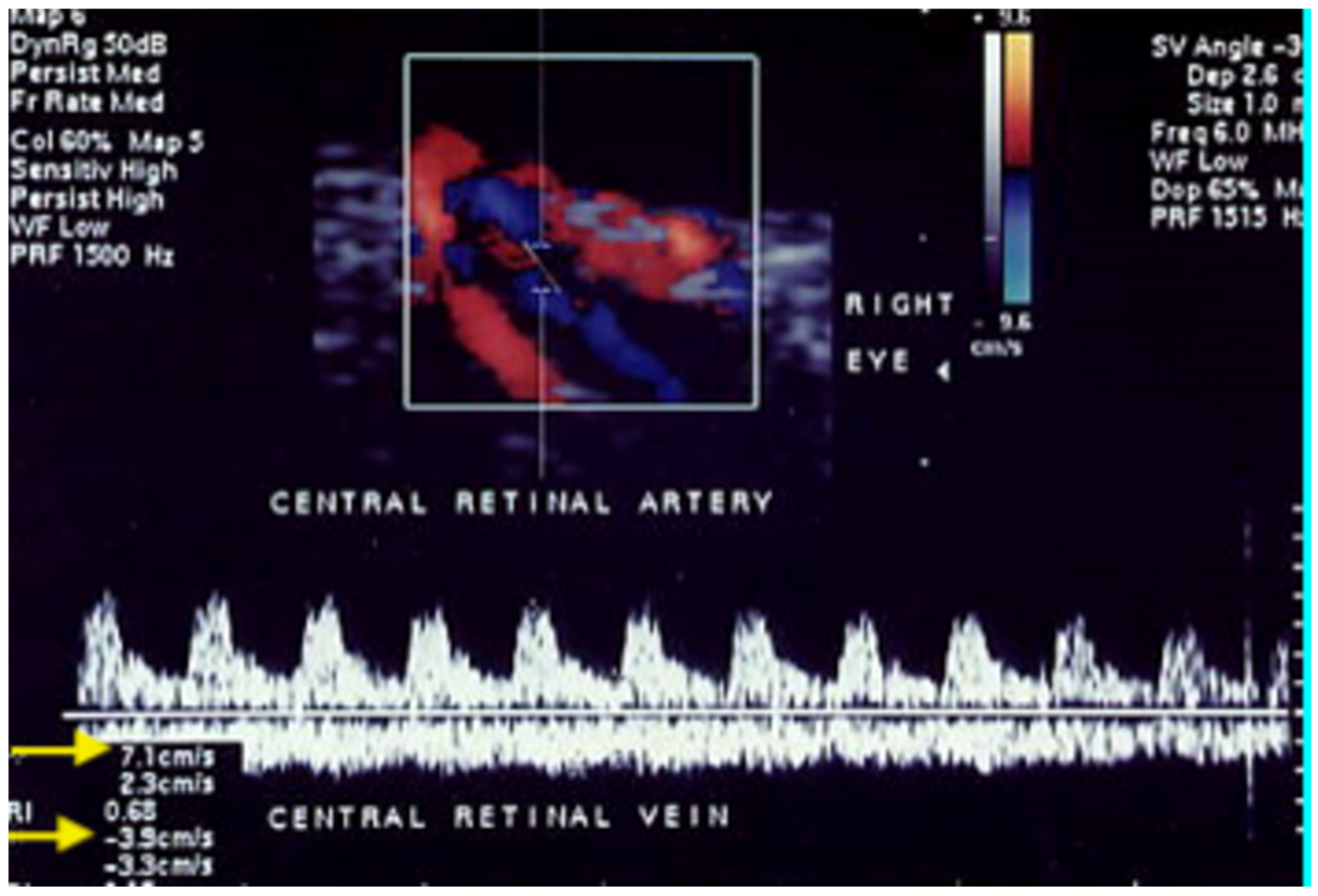
Ocular Color Doppler Ultrasound (OCDUS) in Diagnosis and Monitoring of Ophthalmological, Cerebrovascular and Systemic Diseases: A Narrative Review
 , , , and
Abstract
1. Introduction
2. Ocular Color Doppler Ultrasound (OCDUS)
2.1. Examination Technique
2.2. Orbital Vessels in Normal Subjects
3. Ophthalmological Diseases
3.1. Central Retinal Vein Occlusion (CRVO)
3.2. Central Retinal Artery Occlusion (CRAO)
3.3. Non-Arteritic Anterior Ischemic Optic Neuropathy (NAION)
3.4. Orbital Varix
3.5. Glaucoma
3.6. Age-Related Macular Degeneration (AMD)
3.7. Retinitis Pigmentosa
3.8. Myopia
3.9. Ocular Tumors
4. Cerebrovascular Diseases
4.1. Carotid Cavernous Sinus Fistulas (CCSFs)
4.2. Carotid Artery Stenosis
4.3. OCDUS as an Alternative to Transcranial Doppler to Monitor Cerebral Circulation
5. Systemic Diseases
5.1. Diabetes
5.2. Hypertension
5.3. Takayasu Arteritis
5.4. Graves’ Disease
5.5. Pseudoexfoliation Syndrome
6. Conclusions
Author Contributions
Funding
Institutional Review Board Statement
Informed Consent Statement
Data Availability Statement
Conflicts of Interest
Abbreviations
| AAION | Arteritic Anterior Ischemic Optic Neuropathy |
| AMD | Age-Related Macular Degeneration |
| CCSFs | Carotid Cavernous Sinus Fistulas |
| CEUS | Contrast-Enhanced UltraSound |
| CRA | Central Retinal Artery |
| CRAO | Central Retinal Artery Occlusion |
| CRV | Central Retinal Vein |
| CRVO | Central Retinal Vein Occlusion |
| CT | Computed Tomography |
| CTA | Computed Tomography Angiography |
| DR | Diabetic Retinopathy |
| EDV | End Diastolic Velocity |
| GO | Graves’ Orbitopathy |
| IOV | Inferior Ophthalmic Vein |
| LPCAs | Long Posterior Ciliary Arteries |
| MRA | Magnetic Resonance Angiography |
| maxV | Maximum Velocity |
| minV | Minimum Velocity |
| NAION | Non-arteritic Anterior Ischemic Optic Neuropathy |
| NPG | Normal Pressure Glaucoma |
| OA | Ophthalmic Artery |
| OCDUS | Ocular Color Doppler UltraSound |
| OCT | Optical Coherence Tomography |
| OCT-A | OCT-Angiography |
| PACG | Primary Angle-Closure Glaucoma |
| PCAs | Posterior Ciliary Arteries |
| POAG | Primary Open-Angle Glaucoma |
| PRF | Pulse Repetition Frequency |
| PSV | Peak Systolic Velocity |
| RI | Resistive Index |
| RVO | Retinal Vein Occlusion |
| SOV | Superior Ophthalmic Vein |
| SPCAs | Short Posterior Ciliary Arteries |
References
- Patz, A. Principles of Fluorescein Angiography. Int. Ophthalmol. Clin. 1977, 17, 1–19. [Google Scholar] [CrossRef]
- Tsang, S.H.; Sharma, T. Fluorescein Angiography. In Atlas of Inherited Retinal Diseases; Springer Nature: Berlin/Heidelberg, Germany, 2018; Volume 1085, pp. 7–10. [Google Scholar] [CrossRef]
- Rabb, M.F.; Burton, T.C.; Schatz, H.; Yannuzzi, L.A. Fluorescein Angiography of the Fundus: A Schematic Approach to Interpretation. Surv. Ophthalmol. 1978, 22, 387–403. [Google Scholar] [CrossRef]
- Maram, J.; Srinivas, S.; Sadda, S.R. Evaluating Ocular Blood Flow. Indian J. Ophthalmol. 2017, 65, 337–346. [Google Scholar] [CrossRef]
- Aumann, S.; Donner, S.; Fischer, J.; Müller, F. Optical Coherence Tomography (OCT): Principle and Technical Realization. In High Resolution Imaging in Microscopy and Ophthalmology: New Frontiers in Biomedical Optics; Bille, J.F., Ed.; Springer: Cham, Switzerland, 2019. [Google Scholar]
- Chalam, K.V.; Sambhav, K. Optical Coherence Tomography Angiography in Retinal Diseases. J. Ophthalmic Vis. Res. 2016, 11, 84–92. [Google Scholar] [CrossRef]
- Naseripour, M.; Ghasemi Falavarjani, K.; Mirshahi, R.; Sedaghat, A. Optical Coherence Tomography Angiography (OCTA) Applications in Ocular Oncology. Eye 2020, 34, 1535–1545. [Google Scholar] [CrossRef] [PubMed]
- Kim, J.D.; Hashemi, N.; Gelman, R.; Lee, A.G. Neuroimaging in Ophthalmology. Saudi J. Ophthalmol. 2012, 26, 401–407. [Google Scholar] [CrossRef] [PubMed]
- Masaryk, A.M.; Ross, J.S.; DiCello, M.C.; Modic, M.T.; Paranandi, L.; Masaryk, T.J. 3DFT MR Angiography of the Carotid Bifurcation: Potential and Limitations as a Screening Examination. Radiology 1991, 179, 797–804. [Google Scholar] [CrossRef]
- Modrzejewska, M. Guidelines for Ultrasound Examination in Ophthalmology. Part III: Color Doppler Ultrasonography. J. Ultrason. 2019, 19, 128–136. [Google Scholar] [CrossRef] [PubMed]
- Lizzi, F.; Mortimer, A.; Carstensen, E.; Kremkau, F.; Miller, D.; Miller, M.; Nyborg, W.; O’Brien, W.; Ziskin, M. Bioeffects Considerations for the Safety of Diagnostic Ultrasound. American Institute of Ultrasound in Medicine. Bioeffects Committee. J. Ultrasound Med. 1988, 7, S1–S38. [Google Scholar]
- Wright, J.E. Doyne Lecture: Current Concepts in Orbital Disease. Eye 1988, 2, 1–11. [Google Scholar] [CrossRef][Green Version]
- Fielding, J.A. Ocular Ultrasound. Clin. Radiol. 1996, 51, 533–544. [Google Scholar] [CrossRef]
- Natale, F.; Tedesco, M.A.; Mocerino, R.; Tassinario, G.; Morra, S.; Rinaldi, G.; Gregorio, G.; Calabrò, R. Evaluation of retinal abnormalities in essential hypertension: Qualitative fundoscopy versus central retinal artery resistance index as indicators of target organ damage. J. Clin. Ultrasound 2008, 36, 485–491. [Google Scholar] [CrossRef] [PubMed]
- Williamson, T.H.; Harris, A. Color Doppler Ultrasound Imaging of the Eye and Orbit. Surv. Ophthalmol. 1996, 40, 255–267. [Google Scholar] [CrossRef]
- Krejza, J.; Mariak, Z.; Babikian, V.L. Importance of angle correction in the measurement of blood flow velocity with transcranial Doppler sonography. AJNR Am. J. Neuroradiol. 2001, 22, 1743–1747. [Google Scholar] [PubMed] [PubMed Central]
- Gao, J.; Hentel, K.; Zhu, Q.; Ma, T.; Shih, G.; Mennitt, K.; Min, R. Doppler angle correction in the measurement of intrarenal parameters. Int. J. Nephrol. Renov. Dis. 2011, 4, 49–55. [Google Scholar] [CrossRef] [PubMed] [PubMed Central][Green Version]
- Ruma, M.S.; Swartz, A.E.; Kim, E.; Herring, A.H.; Menard, M.K.; Moise, K.J., Jr. Angle correction can be used to measure peak systolic velocity in the fetal middle cerebral artery. Am. J. Obstet. Gynecol. 2009, 200, 397.e1–397.e3. [Google Scholar] [CrossRef] [PubMed]
- Lui, E.Y.; Steinman, A.H.; Cobbold, R.S.; Johnston, K.W. Human factors as a source of error in peak Doppler velocity measurement. J. Vasc. Surg. 2005, 42, 972.e1–972.e10. [Google Scholar] [CrossRef] [PubMed]
- Venturini, M.; Zaganelli, E.; Angeli, E.; Castrucci, M.; Pierro, L.; Salvioni, M.; Brancato, R.; Del Maschio, A. Ocular color Doppler echography: The examination technic, identification and flowmetry of the orbital vessels. Radiol. Med. 1996, 91, 60–65. [Google Scholar]
- Venturini, M.; Fiorina, P.; Maffi, P.; Losio, C.; Vergani, A.; Secchi, A.; Del Maschio, A. Early Increase of Retinal Arterial and Venous Blood Flow Velocities at Color Doppler Imaging in Brittle Type 1 Diabetes after Islet Transplant Alone. Transplantation 2006, 81, 1274–1277. [Google Scholar] [CrossRef]
- Guthoff, R.F.; Berger, R.W.; Winkler, P.; Helmke, K.; Chumbley, L.C. Doppler ultrasonography of the ophthalmic and central retinal vessels. Arch. Ophthalmol. 1991, 109, 532–536. [Google Scholar] [CrossRef] [PubMed]
- Ciloglu, E.; Yıldırım Celikdemir, A. The short-term effects of intravitreal aflibercept injections and dexamethasone implant on ocular hemodynamics in retinal vein occlusions. Ther. Clin. Risk Manag. 2019, 15, 823–830. [Google Scholar] [CrossRef] [PubMed] [PubMed Central]
- Williamson, T.H. A “throttle” mechanism in the central retinal vein in the region of the lamina cribrosa. Br. J. Ophthalmol. 2007, 91, 1190–1193. [Google Scholar] [CrossRef] [PubMed] [PubMed Central]
- Adiyeke, S.K.; Kutlu, N.; Aytogan, H.; Aras, B.; Yoyler, G.; Ture, G.; Talay, E.; Talay Dayangac, G. Thicknesses of sclera and lamina cribrosa in patients with central retinal vein occlusion. Retina 2020, 40, 2050–2054. [Google Scholar] [CrossRef] [PubMed]
- Baxter, G.M.; Williamson, T.H. Color Doppler flow imaging in central retinal vein occlusion: A new diagnostic technique? Radiology 1993, 187, 847–850. [Google Scholar] [CrossRef] [PubMed]
- Tranquart, F.; Arsene, S.; Giraudeau, B.; Piquemal, R.; Eder, V.; Le Lez, M.L.; Rossazza, C.; Pourcelot, L. Initial Color Doppler Findings in Retinal Vein Occlusion. J. Clin. Ultrasound 2000, 28, 28–33. [Google Scholar] [CrossRef]
- Keyser, B.J.; Flaharty, P.M.; Sergott, R.C.; Brown, G.C.; Lieb, W.E.; Annesley, W.H. Color Doppler Imaging of Arterial Blood Flow in Central Retinal Vein Occlusion. Ophthalmology 1994, 101, 1357–1361. [Google Scholar] [CrossRef]
- Baxter, G.M.; Williamson, T.H. The Value of Serial Doppler Imaging in Central Retinal Vein Occlusion: Correlation with Visual Recovery. Clin. Radiol. 1996, 51, 411–414. [Google Scholar] [CrossRef]
- Suzuki, A.; Okamoto, N.; Ohnishi, M.; Tsubakimori, S.; Fukuda, M. Investigation of Blood Flow Velocity by Color Doppler Imaging in Nonischemic Central Retinal Vein Occlusion with Collateral Veins. Jpn. J. Ophthalmol. 2000, 44, 685–687. [Google Scholar] [CrossRef] [PubMed]
- Arséne, S.; Giraudeau, B.; Le Lez, M.-L.; Pisella, P.J.; Pourcelot, L.; Tranquart, F. Follow up by Colour Doppler Imaging of 102 Patients with Retinal Vein Occlusion over 1 Year. Br. J. Ophthalmol. 2002, 86, 1243–1247. [Google Scholar] [CrossRef] [PubMed]
- Williamson, T.H.; Baxter, G.M. Central Retinal Vein Occlusion, an Investigation by Color Doppler Imaging. Blood Velocity Characteristics and Prediction of Iris Neovascularization. Ophthalmology 1994, 101, 1362–1372. [Google Scholar] [CrossRef] [PubMed]
- Tranquart, F.; Bergès, O.; Koskas, P.; Arsene, S.; Rossazza, C.; Pisella, P.-J.; Pourcelot, L. Color Doppler Imaging of Orbital Vessels: Personal Experience and Literature Review. J. Clin. Ultrasound 2003, 31, 258–273. [Google Scholar] [CrossRef] [PubMed]
- Hayreh, S.S. Management of central retinal vein occlusion. Ophthalmologica 2003, 217, 167–188. [Google Scholar] [CrossRef] [PubMed]
- Gok, M.; Altas, H.; Kapti, H.B. The impact of intravitreal dexamethasone implant (Ozurdex®) on retrobulbar hemodynamics in patients with diabetic macular edema and retinal vein occlusions. Cutan. Ocul. Toxicol. 2019, 38, 240–248. [Google Scholar] [CrossRef] [PubMed]
- Anayol, M.A.; Toklu, Y.; Kamberoglu, E.A.; Raza, S.; Arifoglu, H.B.; Simavli, H.; Altintas, A.G.; Simsek, S. Short-term effects of intravitreal triamcinolone acetonide injection on ocular blood flow evaluated with color Doppler ultrasonography. Int. J. Ophthalmol. 2014, 7, 811–815. [Google Scholar] [CrossRef] [PubMed] [PubMed Central][Green Version]
- Skevas, C.; Wagenfeld, L.; Feucht, M.; Galambos, P.; Richard, G.; Zeitz, O. Radial optic neurotomy in central retinal vein occlusion does not influence ocular hemodynamics. Ophthalmologica 2011, 225, 41–46. [Google Scholar] [CrossRef] [PubMed]
- Avunduk, A.M.; Dinç, H.; Kapicioğlu, Z.; Uğurlu, S.; Dayanir, V.; Korkmaz, E. Arterial blood flow characteristics in central retinal vein occlusion and effects of panretinal photocoagulation treatment: An investigation by colour Doppler imaging. Br. J. Ophthalmol. 1999, 83, 50–53. [Google Scholar] [CrossRef] [PubMed] [PubMed Central]
- Hattenbach, L.O.; Friedrich Arndt, C.; Lerche, R.; Scharrer, I.; Baatz, H.; Margaron, F.; Richard, G.; Behrens-Baumann, W.; Ohrloff, C. Retinal vein occlusion and low-dose fibrinolytic therapy (R.O.L.F.): A prospective, randomized, controlled multicenter study of low-dose recombinant tissue plasminogen activator versus hemodilution in retinal vein occlusion. Retina 2009, 29, 932–940. [Google Scholar] [CrossRef] [PubMed]
- Chen, S.N.; Hwang, J.F.; Huang, J.; Wu, S.L. Retinal arterial occlusion with multiple retinal emboli and carotid artery occlusion disease. Haemodynamic changes and pathways of embolism. BMJ Open Ophthalmol. 2020, 5, e000467. [Google Scholar] [CrossRef] [PubMed] [PubMed Central]
- Kambara, A.; Kitada, Y.; Sawamura, S.; Kakita, H.; Shimizu, F.; Akiyama, Y.; Yoshida, T.; Sakai, N. Central retinal artery occlusion caused by external carotid artery plaque dispersal during carotid artery stenting: A case report. Surg. Neurol. Int. 2025, 16, 352. [Google Scholar] [CrossRef] [PubMed] [PubMed Central]
- Huang, P.; Liu, A.; Ren, H.; Xue, K. Color Doppler Flow Imaging of Retrobulbar Ocular Blood Flow Changes in Retinal Artery Occlusions Caused by Cosmetic Facial Filler Injections. Ophthalmic Plast. Reconstr. Surg. 2019, 35, 227–231. [Google Scholar] [CrossRef] [PubMed]
- Rumelt, S.; Dorenboim, Y.; Rehany, U. Aggressive Systematic Treatment for Central Retinal Artery Occlusion. Am. J. Ophthalmol. 1999, 128, 733–738. [Google Scholar] [CrossRef] [PubMed]
- Varma, D.D.; Cugati, S.; Lee, A.W.; Chen, C.S. A Review of Central Retinal Artery Occlusion: Clinical Presentation and Management. Eye 2013, 27, 688–697. [Google Scholar] [CrossRef]
- Hayreh, S.S.; Zimmerman, M.B. Central Retinal Artery Occlusion: Visual Outcome. Am. J. Ophthalmol. 2005, 140, 376–391. [Google Scholar] [CrossRef] [PubMed]
- Williamson, T.H.; Baxter, G.M.; Dutton, G.N. Color Doppler Velocimetry of the Optic Nerve Head in Arterial Occlusion. Ophthalmology 1993, 100, 312–317. [Google Scholar] [CrossRef]
- Ruiz-Ares, G.; Fuentes, B.; Rodríguez-Pardo de Donlebún, J.; Alonso de Leciñana, M.; Gutiérrez-Zúñiga, R.; Rigual, R.; Díez-Tejedor, E. Usefulness of orbital colour Doppler ultrasound in vascular-related monocular vision loss. Vasc. Med. 2021, 26, 302–309. [Google Scholar] [CrossRef] [PubMed]
- Hayreh, S.S. Ischemic optic neuropathies—Where are we now? Graefe’s Arch. Clin. Exp. Ophthalmol. 2013, 251, 1873–1884. [Google Scholar] [CrossRef] [PubMed]
- Kuerten, D.; Fuest, M.; Bienert, M.; Walter, P.; Plange, N. Ocular Hemodynamics in Acute Nonarteritic Anterior Ischemic Optic Neuropathy Compared with Normal Tension Glaucoma. Eur. J. Gastroenterol. Hepatol. 2019, 28, 334–340. [Google Scholar] [CrossRef] [PubMed]
- Boutigny, A.; Mohamedi, N.; Pardo, S.J.; Bonnin, S.; Ketfi, C.; Ghaouti, H.; Viana, V.; Frazier, A.; Roos, C.; Burlacu, R.; et al. Impairment of central retinal artery hemodynamics in affected and fellow eyes in giant cell arteritis patients with unilateral vision loss. Autoimmun. Rev. 2023, 22, 103272. [Google Scholar] [CrossRef] [PubMed]
- Conticini, E.; Falsetti, P.; Fabiani, C.; Baldi, C.; Grazzini, S.; Tosi, G.M.; Cantarini, L.; Frediani, B. Color Doppler Eye Ultrasonography in giant cell arteritis: Differential diagnosis between arteritic and non-arteritic sudden blindness. J. Ultrasound 2023, 26, 313–320. [Google Scholar] [CrossRef] [PubMed] [PubMed Central]
- Lin, L.; Zhu, W.; Ma, N.; Lin, X.; Yang, H. Evaluation of enhanced external counterpulsation therapy for nonarteritic anterior ischemic optic neuropathy. BMC Ophthalmol. 2020, 20, 236. [Google Scholar] [CrossRef] [PubMed] [PubMed Central]
- Flaharty, P.M.; Sergott, R.C.; Lieb, W.; Bosley, T.M.; Savino, P.J. Optic nerve sheath decompression may improve blood flow in anterior ischemic optic neuropathy. Ophthalmology 1993, 100, 297–305; discussion 303–305. [Google Scholar] [CrossRef] [PubMed]
- Kaup, M.; Plange, N.; Arend, K.O.; Remky, A. Retrobulbar haemodynamics in non-arteritic anterior ischaemic optic neuropathy. Br. J. Ophthalmol. 2006, 90, 1350–1353. [Google Scholar] [CrossRef] [PubMed] [PubMed Central]
- Secil, M.; Soylev, M.; Ada, E.; Saatci, A.O. Orbital varices: Imaging findings and the role of color Doppler sonography in the diagnosis. Comput. Med. Imaging Graph. 2001, 25, 243–247. [Google Scholar] [CrossRef] [PubMed]
- Kawaguchi, S.; Nakase, H.; Noguchi, H.; Yonezawa, T.; Morimoto, T.; Sakaki, T. Orbital varix diagnosed by color Doppler flow imaging–case report. Neurol. Med.-Chir. 1997, 37, 616–619. [Google Scholar] [CrossRef] [PubMed][Green Version]
- Gündüz, K.; Shields, C.L.; Shields, J.A. Varix of the vortex vein ampulla simulating choroidal melanoma: Report of four cases. Retina 1998, 18, 343–347. [Google Scholar] [CrossRef] [PubMed]
- Lieb, W.E.; Merton, D.A.; Shields, J.A.; Cohen, S.M.; Mitchell, D.D.; Goldberg, B.B. Colour Doppler imaging in the demonstration of an orbital varix. Br. J. Ophthalmol. 1990, 74, 305–308. [Google Scholar] [CrossRef] [PubMed] [PubMed Central]
- Erickson, S.J.; Hendrix, L.E.; Massaro, B.M.; Harris, G.J.; Lewandowski, M.F.; Foley, W.D.; Lawson, T.L. Color Doppler flow imaging of the normal and abnormal orbit. Radiology 1989, 173, 511–516. [Google Scholar] [CrossRef] [PubMed]
- Venturini, M.; Cristel, G.; Marzoli, S.B.; Simionato, F.; Agostini, G.; Barboni, P.; De Cobelli, F.; Falini, A.; Bandello, F.; Del Maschio, A. Orbital color Doppler ultrasound as noninvasive tool in the diagnosis of anterior-draining carotid-cavernous fistula. Radiol. Med. 2016, 121, 301–307. [Google Scholar] [CrossRef] [PubMed]
- Kimyon, S.; Mete, A.; Mete, A.; Mete, D. Doppler ultrasonographic measurement of short-term effects of valsalva maneuver on retrobulbar blood flow. J. Clin. Ultrasound 2017, 45, 551–555. [Google Scholar] [CrossRef] [PubMed]
- Weinreb, R.N.; Khaw, P.T. Primary Open-Angle Glaucoma. Lancet 2004, 363, 1711–1720. [Google Scholar] [CrossRef]
- Tham, Y.-C.; Li, X.; Wong, T.Y.; Quigley, H.A.; Aung, T.; Cheng, C.-Y. Global Prevalence of Glaucoma and Projections of Glaucoma Burden through 2040: A Systematic Review and Meta-Analysis. Ophthalmology 2014, 121, 2081–2090. [Google Scholar] [CrossRef] [PubMed]
- Agarwal, R.; Gupta, S.K.; Agarwal, P.; Saxena, R.; Agrawal, S.S. Current Concepts in the Pathophysiology of Glaucoma. Indian J. Ophthalmol. 2009, 57, 257–266. [Google Scholar] [CrossRef] [PubMed]
- Weinreb, R.N.; Aung, T.; Medeiros, F.A. The Pathophysiology and Treatment of Glaucoma: A Review. JAMA 2014, 311, 1901–1911. [Google Scholar] [CrossRef]
- Banou, L.; Dastiridou, A.; Giannoukas, A.; Kouvelos, G.; Baros, C.; Androudi, S. The Role of Color Doppler Imaging in the Diagnosis of Glaucoma: A Review of the Literature. Diagnostics 2023, 13, 588. [Google Scholar] [CrossRef] [PubMed]
- Meng, N.; Zhang, P.; Huang, H.; Ma, J.; Zhang, Y.; Li, H.; Qu, Y. Color Doppler Imaging Analysis of Retrobulbar Blood Flow Velocities in Primary Open-Angle Glaucomatous Eyes: A Meta-Analysis. PLoS ONE 2013, 8, e62723. [Google Scholar] [CrossRef]
- Xu, S.; Huang, S.; Lin, Z.; Liu, W.; Zhong, Y. Color Doppler Imaging Analysis of Ocular Blood Flow Velocities in Normal Tension Glaucoma Patients: A Meta-Analysis. J. Ophthalmol. 2015, 2015, 919610. [Google Scholar] [CrossRef]
- Abegão Pinto, L.; Willekens, K.; Van Keer, K.; Shibesh, A.; Molenberghs, G.; Vandewalle, E.; Stalmans, I. Ocular Blood Flow in Glaucoma—The Leuven Eye Study. Acta Ophthalmol. 2016, 94, 592–598. [Google Scholar] [CrossRef]
- Marjanovic, I.; Milic, N.; Martinez, A.; Benitez-del-Castillo, J. Retrobulbar Hemodynamic Parameters in Open-Angle and Angle-Closure Glaucoma Patients. Eye 2012, 26, 523–528. [Google Scholar] [CrossRef]
- Cheng, C.Y.; Liu, C.J.; Chiou, H.J.; Chou, J.C.; Hsu, W.M.; Liu, J.H. Color Doppler Imaging Study of Retrobulbar Hemodynamics in Chronic Angle-Closure Glaucoma. Ophthalmology 2001, 108, 1445–1451. [Google Scholar] [CrossRef]
- Kalayci, M.; Tahtabasi, M. Assessment of Doppler Flow Parameters of the Retrobulbar Arteries and Internal Carotid Artery in Patients with Glaucoma: The Significance of Ophthalmic Artery Peak Ratio and the Intima-Media Thickness of the Internal Carotid Artery. Int. Ophthalmol. 2020, 40, 3337–3348. [Google Scholar] [CrossRef]
- Martínez, A.; Sánchez, M. Predictive Value of Colour Doppler Imaging in a Prospective Study of Visual Field Progression in Primary Open-Angle Glaucoma. Acta Ophthalmol. Scand. 2005, 83, 716–722. [Google Scholar] [CrossRef]
- Calvo, P.; Ferreras, A.; Polo, V.; Güerri, N.; Seral, P.; Fuertes-Lazaro, I.; Pablo, L.E. Predictive Value of Retrobulbar Blood Flow Velocities in Glaucoma Suspects. Investig. Ophthalmol. Vis. Sci. 2012, 53, 3875–3884. [Google Scholar] [CrossRef] [PubMed]
- Harris, A.; Sergott, R.C.; Spaeth, G.L.; Katz, J.L.; Shoemaker, J.A.; Martin, B.J. Color Doppler analysis of ocular vessel blood velocity in normal-tension glaucoma. Arch. Ophthalmol. 1994, 118, 642–649. [Google Scholar] [CrossRef] [PubMed]
- Rankin, S.J.; Drance, S.M.; Buckley, A.R.; Walman, B.E. Visual field correlations with color Doppler studies in open angle glaucoma. J. Gastroenterol. 1996, 5, 15–21. [Google Scholar] [CrossRef] [PubMed]
- Yamazaki, Y.; Drance, S.M. The relationship between progression of visual field defects and retrobulbar circulation in patients with glaucoma. Am. J. Ophthalmol. 1997, 124, 287–295. [Google Scholar] [CrossRef] [PubMed]
- Wang, Z.; Zhong, X.; Lu, X.; Shi, J.; Shi, W.; Zhao, C.; Wang, T. Correlation of retrobulbar perfusion deficits with glaucomatous visual field defects. Graefe’s Arch. Clin. Exp. Ophthalmol. 2024, 262, 2961–2970. [Google Scholar] [CrossRef] [PubMed]
- Montanari, P.; Marangoni, P.; Oldani, A.; Ratiglia, R.; Raiteri, M.; Berardinelli, L. Color Doppler imaging study in patients with primary open-angle glaucoma treated with timolol 0.5% and carteolol 2%. Eur. J. Ophthalmol. 2001, 11, 240–244. [Google Scholar] [CrossRef] [PubMed]
- Altan-Yaycioglu, R.; Türker, G.; Akdöl, S.; Acunaş, G.; Izgi, B. The effects of beta-blockers on ocular blood flow in patients with primary open angle glaucoma: A color Doppler imaging study. Eur. J. Ophthalmol. 2001, 11, 37–46. [Google Scholar] [CrossRef] [PubMed]
- Bergstrand, I.C.; Heijl, A.; Harris, A. Dorzolamide and ocular blood flow in previously untreated glaucoma patients: A controlled double-masked study. Acta Ophthalmol. Scand. 2002, 80, 176–182. [Google Scholar] [CrossRef] [PubMed]
- Erkin, E.F.; Tarhan, S.; Kayikçioğlu, O.R.; Deveci, H.; Güler, C.; Göktan, C. Effects of betaxolol and latanoprost on ocular blood flow and visual fields in patients with primary open-angle glaucoma. Eur. J. Ophthalmol. 2004, 14, 211–219. [Google Scholar] [CrossRef] [PubMed]
- Zeitz, O.; Matthiessen, E.T.; Reuss, J.; Wiermann, A.; Wagenfeld, L.; Galambos, P.; Richard, G.; Klemm, M. Effects of glaucoma drugs on ocular hemodynamics in normal tension glaucoma: A randomized trial comparing bimatoprost and latanoprost with dorzolamide [ISRCTN18873428]. BMC Ophthalmol. 2005, 5, 6. [Google Scholar] [CrossRef] [PubMed] [PubMed Central]
- Uva, M.G.; Longo, A.; Reibaldi, M.; Reibaldi, A. The effect of timolol-dorzolamide and timolol-pilocarpine combinations on ocular blood flow in patients with glaucoma. Am. J. Ophthalmol. 2006, 141, 1158–1160. [Google Scholar] [CrossRef] [PubMed]
- Aydin, B.; Onol, M.; Hondur, A.; Kaya, M.G.; Ozdemir, H.; Cengel, A.; Hasanreisoglu, B. The effect of oral magnesium therapy on visual field and ocular blood flow in normotensive glaucoma. Eur. J. Ophthalmol. 2010, 20, 131–135. [Google Scholar] [CrossRef] [PubMed]
- Stankiewicz, A.; Misiuk-Hojło, M.; Grabska-Liberek, I.; Romanowska-Dixon, B.; Wierzbowska, J.; Wasyluk, J.; Mulak, M.; Szuścik, I.; Sierdziński, J.; Ehrlich, R.; et al. Intraocular pressure and ocular hemodynamics in patients with primary open-angle glaucoma treated with the combination of morning dosing of bimatoprost and dorzolamide hydrochloride. Acta Ophthalmol. 2011, 89, e57–e63. [Google Scholar] [CrossRef] [PubMed]
- Januleviciene, I.; Siaudvytyte, L.; Barsauskaite, R. Ophthalmic drug delivery in glaucoma-a review. Pharmaceutics 2012, 4, 243–251. [Google Scholar] [CrossRef] [PubMed] [PubMed Central]
- Siesky, B.; Harris, A.; Ehrlich, R.; Cantor, L.; Shoja, M.M.; Rusia, D.; Hollander, D.A.; Abrams, L.; Williams, J.M.; Shoshani, Y. Short-term effects of brimonidine/timolol and dorzolamide/timolol on ocular perfusion pressure and blood flow in glaucoma. Adv. Ther. 2012, 29, 53–63. [Google Scholar] [CrossRef] [PubMed]
- Hommer, A.; Sperl, P.; Resch, H.; Popa-Cherecheanu, A.; Qiao, C.; Schmetterer, L.; Garhöfer, G. A double-masked randomized crossover study comparing the effect of latanoprost/timolol and brimonidine/timolol fixed combination on intraocular pressure and ocular blood flow in patients with primary open-angle glaucoma or ocular hypertension. J. Ocul. Pharmacol. Ther. 2012, 28, 569–575. [Google Scholar] [CrossRef] [PubMed]
- Trible, J.R.; Sergott, R.C.; Spaeth, G.L.; Wilson, R.P.; Katz, L.J.; Moster, M.R.; Schmidt, C.M. Trabeculectomy is associated with retrobulbar hemodynamic changes. A color Doppler analysis. Ophthalmology 1994, 101, 340–351. [Google Scholar] [CrossRef] [PubMed]
- Yamazaki, Y.; Hayamizu, F. Effect of trabeculectomy on retrobulbar circulation and visual field progression in patients with primary open-angle glaucoma. Clin. Ophthalmol. 2012, 6, 1539–1545. [Google Scholar] [CrossRef] [PubMed] [PubMed Central]
- Kuerten, D.; Fuest, M.; Koch, E.C.; Remky, A.; Plange, N. Long term effect of trabeculectomy on retrobulbar haemodynamics in glaucoma. Ophthalmic Physiol. Opt. 2015, 35, 194–200. [Google Scholar] [CrossRef] [PubMed]
- Januleviciene, I.; Siaudvytyte, L.; Diliene, V.; Barsauskaite, R.; Siesky, B.; Harris, A. Effect of Trabeculectomy on Ocular Hemodynamic Parameters in Pseudoexfoliative and Primary Open-angle Glaucoma Patients. J. Gastroenterol. 2015, 24, e52–e56. [Google Scholar] [CrossRef] [PubMed]
- Galassi, F.; Giambene, B.; Corvi, A.; Falaschi, G.; Menchini, U. Retrobulbar hemodynamics and corneal surface temperature in glaucoma surgery. Int. Ophthalmol. 2008, 28, 399–405. [Google Scholar] [CrossRef] [PubMed]
- Liu, H.; Dai, Y.; Yu, M.; Zeng, J.; Wang, C.; Tan, S.; Xiong, M.; Zhang, R.; Yu, X.; Shi, M.; et al. Effects and Safety of Press-Needle Therapy for Improving Visual Function and Eye Blood Circulation in Patients with Glaucoma with Controlled Intraocular Pressure: Study Protocol for a Multicenter Randomized Controlled Trial. JMIR Res. Protoc. 2025, 14, e67737. [Google Scholar] [CrossRef] [PubMed] [PubMed Central]
- Venturini, M.; Piacentino, F.; Coppola, A.; Recaldini, C.; Pacetti, S.; Fontana, F.; Vannucci, V. Early Increase in Retinal Arterial and Venous Blood Flow Velocities on Color Doppler Ultrasound after Acupuncture. Ultraschall Med. 2022, 43, 621–622. [Google Scholar] [CrossRef] [PubMed]
- Klein, R.; Chou, C.-F.; Klein, B.E.K.; Zhang, X.; Meuer, S.M.; Saaddine, J.B. Prevalence of Age-Related Macular Degeneration in the US Population. Arch. Ophthalmol. 2011, 129, 75–80. [Google Scholar] [CrossRef]
- Pauleikhoff, D.; Chen, J.C.; Chisholm, I.H.; Bird, A.C. Choroidal Perfusion Abnormality with Age-Related Bruch’s Membrane Change. Am. J. Ophthalmol. 1990, 109, 211–217. [Google Scholar] [CrossRef]
- Böker, T.; Fang, T.; Steinmetz, R. Refractive Error and Choroidal Perfusion Characteristics in Patients with Choroidal Neovascularization and Age-Related Macular Degeneration. Ger. J. Ophthalmol. 1993, 2, 10–13. [Google Scholar]
- Ferris, F.L.; Fine, S.L.; Hyman, L. Age-Related Macular Degeneration and Blindness Due to Neovascular Maculopathy. Arch. Ophthalmol. 1984, 102, 1640–1642. [Google Scholar] [CrossRef]
- Friedman, E.; Krupsky, S.; Lane, A.M.; Oak, S.S.; Friedman, E.S.; Egan, K.; Gragoudas, E.S. Ocular Blood Flow Velocity in Age-Related Macular Degeneration. Ophthalmology 1995, 102, 640–646. [Google Scholar] [CrossRef]
- Rodrigo, F.; Ruiz-Moreno, J.M.; García, J.B.; Torregrosa, M.E.; Segura, J.V.; Piñero, D.P. Color Doppler Imaging of the Retrobulbar Circulation and Plasmatic Biomarkers of Vascular Risk in Age-Related Macular Degeneration: A Pilot Study. Indian J. Ophthalmol. 2018, 66, 89–93. [Google Scholar] [CrossRef]
- Finzi, A.; Ottoboni, S.; Cellini, M.; Corcioni, B.; Gaudiano, C.; Fontana, L. Color Doppler Imaging, Endothelin-1, Corneal Biomechanics and Scleral Rigidity in Asymmetric Age-Related Macular Degeneration. Clin. Ophthalmol. 2024, 18, 2583–2591. [Google Scholar] [CrossRef] [PubMed] [PubMed Central]
- Mete, A.; Saygili, O.; Mete, A.; Bayram, M.; Bekir, N. Effects of intravitreal bevacizumab (Avastin) therapy on retrobulbar blood flow parameters in patients with neovascular age-related macular degeneration. J. Clin. Ultrasound 2010, 38, 66–70. [Google Scholar] [CrossRef] [PubMed]
- Toklu, Y.; Cakmak, H.B.; Raza, S.; Anayol, A.; Asik, E.; Simşek, S. Short-term effects of intravitreal bevacizumab (Avastin(®)) on retrobulbar hemodynamics in patients with neovascular age-related macular degeneration. Acta Ophthalmol. 2011, 89, e41–e45. [Google Scholar] [CrossRef] [PubMed]
- Pemp, B.; Garhofer, G.; Lasta, M.; Schmidl, D.; Wolzt, M.; Schmetterer, L. The effects of moxaverine on ocular blood flow in patients with age-related macular degeneration or primary open angle glaucoma and in healthy control subjects. Acta Ophthalmol. 2012, 90, 139–145. [Google Scholar] [CrossRef] [PubMed]
- Sakalar, Y.B.; Senturk, S.; Yildirim, M.; Keklikci, U.; Alakus, M.F.; Unlu, K. Evaluation of retrobulbar blood flow by color doppler ultrasonography after intravitreal ranibizumab injection in patients with neovascular age-related macular degeneration. J. Clin. Ultrasound 2013, 41, 32–37. [Google Scholar] [CrossRef] [PubMed]
- Yuksel, K.; Altinkaynak, H.; Kina, A.; Kara, N.; Yazici, A.T.; Demirok, A. Can intravitreal ranibizumab alter retrobulbar circulation in eyes with age-related macular degeneration? J. Ocul. Pharmacol. Ther. 2013, 29, 723–727. [Google Scholar] [CrossRef] [PubMed]
- Bonnin, P.; Pournaras, J.A.; Makowiecka, K.; Krivosic, V.; Kedra, A.W.; Le Gargasson, J.F.; Gaudric, A.; Levy, B.I.; Cohen, Y.S.; Tadayoni, R.; et al. Ultrasound assessment of ocular vascular effects of repeated intravitreal injections of ranibizumab for wet age-related macular degeneration. Acta Ophthalmol. 2014, 92, e382–e387. [Google Scholar] [CrossRef] [PubMed]
- Raza, S.; Ergun, S.B.; Toklu, Y.; Cakmak, H.B.; Ipek, A.; Cagil, N. Evaluating the Effect of Intravitreal Ranibizumab on Retrobulbar Hemodynamics by Color Doppler Ultrasonography in Neovascular AMD. Ophthalmic Surg. Lasers Imaging Retin. 2019, 50, 437–443. [Google Scholar] [CrossRef] [PubMed]
- Chelazzi, P.; Prati, M.; Contini, F.; Sivelli, P.; Viganò, V.; Donati, S.; Azzolini, C. Orbital Color Doppler Imaging Before and After Pars Plana Vitrectomy. Investig. Ophthalmol. Vis. Sci. 2007, 48, 2256. [Google Scholar]
- Cellini, M.; Strobbe, E.; Gizzi, C.; Campos, E.C. ET-1 plasma levels and ocular blood flow in retinitis pigmentosa. Can. J. Physiol. Pharmacol. 2010, 88, 630–635. [Google Scholar] [CrossRef] [PubMed]
- Akyol, N.; Kükner, S.; Celiker, U.; Koyu, H.; Lüleci, C. Decreased retinal blood flow in retinitis pigmentosa. Can. J. Ophthalmol. 1995, 30, 28–32. [Google Scholar] [PubMed]
- Taner, P.; Kara, S.A.; Akarsu, C.; Ergin, A. The effects of darkness on retrobulbar hemodynamics in patients with early stages retinitis pigmentosa. Int. Ophthalmol. 2004, 25, 95–99. [Google Scholar] [CrossRef] [PubMed]
- Lang, M.; Harris, A.; Ciulla, T.A.; Siesky, B.; Patel, P.; Belamkar, A.; Mathew, S.; Verticchio Vercellin, A.C. Vascular dysfunction in retinitis pigmentosa. Acta Ophthalmol. 2019, 97, 660–664. [Google Scholar] [CrossRef] [PubMed]
- Wang, X.N.; Zhao, Q.; Li, D.J.; Wang, Z.Y.; Chen, W.; Li, Y.F.; Cui, R.; Shen, L.; Wang, R.K.; Peng, X.Y.; et al. Quantitative evaluation of primary retinitis pigmentosa patients using colour Doppler flow imaging and optical coherence tomography angiography. Acta Ophthalmol. 2019, 97, E993–E997. [Google Scholar] [CrossRef] [PubMed]
- Kayser, S.; Vargas, P.; Mendelsohn, D.; Han, J.; Bi, H.; Benavente, A.; Bittner, A.K. Reduced Central Retinal Artery Blood Flow Is Related to Impaired Central Visual Function in Retinitis Pigmentosa Patients. Curr. Eye Res. 2017, 42, 1503–1510. [Google Scholar] [CrossRef] [PubMed] [PubMed Central]
- Bittner, A.K.; Seger, K.; Salveson, R.; Kayser, S.; Morrison, N.; Vargas, P.; Mendelsohn, D.; Han, J.; Bi, H.; Dagnelie, G.; et al. Randomized controlled trial of electro-stimulation therapies to modulate retinal blood flow and visual function in retinitis pigmentosa. Acta Ophthalmol. 2018, 96, e366–e376. [Google Scholar] [CrossRef] [PubMed] [PubMed Central]
- Yu, C.; Xu, C.; Wang, Z.; Zhang, X.; Huang, X. Color doppler ultrasound analysis of pathological myopia induced changes in retrobulbar blood flow and its relationship with characteristic changes in myopia. Pak. J. Med Sci. 2023, 39, 853–857. [Google Scholar] [CrossRef] [PubMed] [PubMed Central]
- Zhao, M.; Lam, A.K.; Ying, M.T.; Cheong, A.M. Hemodynamic and morphological changes of the central retinal artery in myopic eyes. Sci. Rep. 2022, 12, 7104, Erratum in Sci. Rep. 2022, 12, 8680. https://doi.org/10.1038/s41598-022-12902-1. [Google Scholar] [CrossRef] [PubMed] [PubMed Central]
- Gulani, A.C.; Morparia, H.; Bhatti, S.S.; Jehangir, R.P. Colour Doppler sonography: A new investigative modality for intraocular space-occupying lesions. Eye 1994, 8, 307–310. [Google Scholar] [CrossRef] [PubMed][Green Version]
- Yang, W.; Hu, S.; Wang, J.; Wang, L.; Zheng, B. Color Doppler imaging diagnosis of intra-ocular tumor. Chin. Med. J. 1997, 110, 664–666. [Google Scholar] [PubMed]
- Ozdemir, H.; Yücel, C.; Aytekin, C.; Onal, B.; Ataman, A.; Ozsunar, Y.; Işik, S. Intraocular tumors. The value of spectral and color Doppler sonography. Clin. Imaging 1997, 21, 77–81. [Google Scholar] [CrossRef] [PubMed]
- Iveković, R.; Lovrencić-Huzjan, A.; Novak-Laus, K.; Demarin, V. Value of color-Doppler imaging in diagnosis of intrabulbar and intraorbital tumors. Coll. Antropol. 2000, 24, 205–210. [Google Scholar] [PubMed]
- Neudorfer, M.; Leibovitch, I.; Stolovitch, C.; Dray, J.P.; Hermush, V.; Nagar, H.; Kessler, A. Intraorbital and periorbital tumors in children–value of ultrasound and color Doppler imaging in the differential diagnosis. Am. J. Ophthalmol. 2004, 137, 1065–1072. [Google Scholar] [CrossRef] [PubMed]
- Xue, K.; Liu, A.; Hui, R.; Zhang, J.; Qian, J. Retrobulbar ocular blood flow changes measured by colour Doppler imaging after intra-arterial chemotherapy in retinoblastoma. Br. J. Ophthalmol. 2017, 101, 1419–1422. [Google Scholar] [CrossRef] [PubMed]
- McInnis-Smith, K.; Abruzzo, T.; Riemann, M.; Goncalves, L.F.; Ramasubramanian, A. Utility of color Doppler imaging in patients with retinoblastoma treated by intra-arterial chemotherapy. J. Am. Assoc. Pediatr. Ophthalmol. Strabismus 2025, 29, 104093. [Google Scholar] [CrossRef] [PubMed]
- Walter, U.; Niendorf, T.; Graessl, A.; Rieger, J.; Krüger, P.C.; Langner, S.; Guthoff, R.F.; Stachs, O. Ultrahigh field magnetic resonance and colour Doppler real-time fusion imaging of the orbit–a hybrid tool for assessment of choroidal melanoma. Eur. Radiol. 2014, 24, 1112–1117. [Google Scholar] [CrossRef] [PubMed]
- Yang, W.L.; Wei, W.B.; Li, D.J. Quantitative parameter character of choroidal melanoma in contrast-enhanced ultrasound. Chin. Med. J. 2012, 125, 4440–4444. [Google Scholar] [PubMed]
- Venturini, M.; Colantoni, C.; Modorati, G.; Di Nicola, M.; Colucci, A.; Agostini, G.; Picozzi, P.; De Cobelli, F.; Parmiani, G.; Mortini, P.; et al. Preliminary results of contrast-enhanced sonography in the evaluation of the response of uveal melanoma to gamma-knife radiosurgery. Clin. Ultrasound 2015, 43, 421–430. [Google Scholar] [CrossRef] [PubMed]
- Yan, L.; He, G.; Zhou, X.; Zheng, Y.; Zhu, Y.; Yang, J.; Zhang, M.; Zhou, Y. Contrast-enhanced ultrasound in the diagnosis of orbital space-occupying lesions. Clin. Radiol. 2017, 72, 798.e1–798.e6. [Google Scholar] [CrossRef] [PubMed]
- Li, D.; Wang, Z.; Chen, W.; Zhao, Q.; Li, Y.; Cui, R.; Shen, L.; Liu, Q.; Cao, K.; Liu, Y.; et al. Quantitative Analysis of Perfusion Characteristics Using Contrast-Enhanced Ultrasound in Patients with Choroidal Metastasis. Ophthalmic Res. 2022, 65, 86–93. [Google Scholar] [CrossRef] [PubMed] [PubMed Central]
- Wildner, D.; Heinzerling, L.; Scheulen, M.E.; Kaempgen, E.; Schuler, G.; Strobel, D.; Janka, R.; Neurath, M.F.; Sturm, J.; Knieling, F. Assessment of sorafenib induced changes in tumor perfusion of uveal melanoma metastases with dynamic contrast-enhanced ultrasound (DCE-US). J. Cancer Res. Clin. Oncol. 2022, 148, 955–965. [Google Scholar] [CrossRef] [PubMed] [PubMed Central]
- Gobin, Y.P.; Duckwiler, G.R.; Viñuela, F. Direct Arteriovenous Fistulas (Carotid-Cavernous and Vertebral-Venous). Diagnosis and Intervention. Neuroimaging Clin. N. Am. 1998, 8, 425–443. [Google Scholar] [CrossRef]
- Miller, N.R. Dural Carotid-Cavernous Fistulas: Epidemiology, Clinical Presentation, and Management. Neurosurg. Clin. N. Am. 2012, 23, 179–192. [Google Scholar] [CrossRef] [PubMed]
- Feiner, L.; Bennett, J.; Volpe, N.J. Cavernous Sinus Fistulas: Carotid Cavernous Fistulas and Dural Arteriovenous Malformations. Curr. Neurol. Neurosci. Rep. 2003, 3, 415–420. [Google Scholar] [CrossRef]
- Liu, H.M.; Wang, Y.H.; Chen, Y.F.; Cheng, J.S.; Yip, P.K.; Tu, Y.K. Long-Term Clinical Outcome of Spontaneous Carotid Cavernous Sinus Fistulae Supplied by Dural Branches of the Internal Carotid Artery. Neuroradiology 2001, 43, 1007–1014. [Google Scholar] [CrossRef] [PubMed]
- Wu, H.-C.; Ro, L.-S.; Chen, C.-J.; Chen, S.-T.; Lee, T.-H.; Chen, Y.-C.; Chen, C.-M. Isolated Ocular Motor Nerve Palsy in Dural Carotid-Cavernous Sinus Fistula. Eur. J. Neurol. 2006, 13, 1221–1225. [Google Scholar] [CrossRef]
- Kawaguchi, S.; Sakaki, T.; Uranishi, R. Color Doppler Flow Imaging of the Superior Ophthalmic Vein in Dural Arteriovenous Fistulas. Stroke 2002, 33, 2009–2013. [Google Scholar] [CrossRef][Green Version]
- Polat, P.; Suma, S.; Kantarcý, M.; Alper, F.; Levent, A. Color Doppler US in the Evaluation of Uterine Vascular Abnormalities. Radiographics 2002, 22, 47–53. [Google Scholar] [CrossRef] [PubMed]
- Chiou, H.J.; Chou, Y.H.; Guo, W.Y.; Teng, M.M.; Hsu, C.C.; Tiu, C.M.; Lirng, J.F.; Luo, C.B.; Shiau, C.Y.; Pan, D.H. Verifying complete obliteration of carotid artery-cavernous sinus fistula: Role of color Doppler ultrasonography. J. Ultrasound Med. 1998, 17, 289–295. [Google Scholar] [CrossRef] [PubMed][Green Version]
- Müller, M.; Wessel, K.; Mehdorn, E.; Kömpf, D.; Kessler, C.M. Carotid artery disease in vascular ocular syndromes. J. Clin. Neuroophthalmol. 1993, 13, 175–180. [Google Scholar] [PubMed]
- Souma, N.; Tasaka, Y.; Nakauchi, K.; Kubota, Y.; Amano, Y.; Sogabe, T. A Case of Reversed Ophthalmic Artery Flow without Occlusion of the Internal Carotid Artery. Jpn. J. Ophthalmol. 2000, 44, 572. [Google Scholar] [CrossRef] [PubMed]
- Riiheläinen, K.; Päivänsalo, M.; Suramo, I.; Laatikainen, L. The effect of carotid endarterectomy on ocular blood velocity. Ophthalmology 1997, 104, 672–675. [Google Scholar] [CrossRef] [PubMed]
- Kawaguchi, S.; Iida, J.; Uchiyama, Y. Ocular Circulation and Chronic Ocular Ischemic Syndrome before and after Carotid Artery Revascularization Surgery. J. Ophthalmol. 2012, 2012, 350475. [Google Scholar] [CrossRef] [PubMed] [PubMed Central]
- Ma, F.; Su, J.; Shang, Q.; Ma, J.; Zhang, T.; Wang, X.; Ma, H.; Yu, J. Changes in Ocular Hemodynamics after Carotid Artery Angioplasty and Stenting (CAAS) in Patients with Different Severity of Ocular Ischemic Syndrome. Curr. Eye Res. 2018, 43, 266–272. [Google Scholar] [CrossRef] [PubMed]
- Markus, H. Transcranial Doppler detection of circulating cerebral emboli. A review. Stroke 1993, 24, 1246–1250. [Google Scholar] [CrossRef] [PubMed]
- Arrowsmith, J.E.; Grocott, H.P.; Reves, J.G.; Newman, M.F. Central nervous system complications of cardiac surgery. Br. J. Anaesth. 2000, 84, 378–393. [Google Scholar] [CrossRef] [PubMed]
- van der Linden, J.; Casimir-Ahn, H. When do cerebral emboli appear during open heart operations? A transcranial Doppler study. Ann. Thorac. Surg. 1991, 51, 237–241. [Google Scholar] [CrossRef] [PubMed]
- Rodriguez, R.A.; Ruel, M.; Labrosse, M.; Mesana, T. Transcranial Doppler and acoustic pressure fluctuations for the assessment of cavitation and thromboembolism in patients with mechanical heart valves. Interact. Cardiovasc. Thorac. Surg. 2008, 7, 179–183. [Google Scholar] [CrossRef] [PubMed]
- Rodriguez, R.A.; Rubens, F.D.; Wozny, D.; Nathan, H.J. Cerebral emboli detected by transcranial Doppler during cardiopulmonary bypass are not correlated with postoperative cognitive deficits. Stroke 2010, 41, 2229–2235. [Google Scholar] [CrossRef] [PubMed][Green Version]
- Jarry, S.; Couture, E.J.; Beaubien-Souligny, W.; Fernandes, A.; Fortier, A.; Ben-Ali, W.; Desjardins, G.; Huard, K.; Mailhot, T.; Denault, A.Y. Clinical relevance of transcranial Doppler in a cardiac surgery setting: Embolic load predicts difficult separation from cardiopulmonary bypass. J. Cardiothorac. Surg. 2024, 19, 90. [Google Scholar] [CrossRef] [PubMed] [PubMed Central]
- Kochi, K.; Sueda, T.; Orihashi, K.; Matsuura, Y. Central retinal artery color Doppler monitoring versus transcranial Doppler monitoring in cardiac surgery. Heart Vessel. 2000, 15, 7–10. [Google Scholar] [CrossRef] [PubMed]
- Venturini, M.; Zambon, M.; Cristel, G.; Agostini, G.; Querques, G.; Colombo, M.; Benussi, S.; Landoni, G.; Zangrillo, A.; Del Maschio, A. Monitoring of central retinal artery and vein with color Doppler ultrasound during heart surgery as an alternative to transcranial Doppler ultrasonography: A case report. J. Clin. Ultrasound 2014, 42, 112–115. [Google Scholar] [CrossRef] [PubMed]
- Pauk-Domańska, M.; Walasik-Szemplińska, D. Color Doppler imaging of the retrobulbar vessels in diabetic retinopathy. J. Ultrason. 2014, 14, 28–35. [Google Scholar] [CrossRef] [PubMed] [PubMed Central]
- Noureldine, A.M.; Abdelmaksoud, A.M.S.; Mostafa, H.A.A.F.; Macky, T.; ElBohy, A.E. Orbital Blood Vessels Changes on Color Duplex Imaging in Diabetics with and without Diabetic Retinopathy. Sci. Rep. 2023, 13, 17115. [Google Scholar] [CrossRef] [PubMed]
- Pillai, G.S.; Kutumbaka, A.; Nazar, P.K.; Chandiri, A.; Ravindran, G.C. Association between Resistivity Index of Central Retinal Artery and Severity of Diabetic Retinopathy. Indian J. Ophthalmol. 2023, 71, 3539–3543. [Google Scholar] [CrossRef] [PubMed]
- Sirkeci, O.; Erkus Sirkeci, E.; Ergün Süzer, N.; Tatlıparmak, A.C.; Küçükçiloğlu, Y.; Ulaş, T. Can Orbital Artery Resistive Indexes Detect Complications of Diabetes in the Prediabetic Phase? Wien. Klin. Wochenschr. 2023, 135, 343–348. [Google Scholar] [CrossRef]
- Khatri, M.; Saxena, S.; Kumar, M.; Chabbra, A.K.; Bhasker, S.K.; Akduman, E.I.; Pham, H.; Akduman, L. Resistive index of central retinal artery is a bioimaging biomarker for severity of diabetic retinopathy. Int. J. Retin. Vitr. 2019, 5, 38. [Google Scholar] [CrossRef] [PubMed] [PubMed Central]
- Pauk-Domańska, M.; Wilczewska, A.; Jaguś, D.; Kaczyński, B.; Jakubowski, W. Doppler Ultrasound-Based Evaluation of Hemodynamic Changes in the Ophthalmic Artery and Central Retinal Artery in Patients with Type 1 Diabetes Mellitus without Retinopathy and with Mild Non-Proliferative Retinopathy. J. Ultrason. 2024, 24, 20240009. [Google Scholar] [CrossRef]
- Venturini, M.; Losio, C.; Del Maschio, A.; Maffi, P.; Fiorina, P.; Gremizzi, C.; Secchi, A. Kidney-pancreas transplantation does not improve retinal arterial flow velocities in type 1 diabetic uremic patients. Transplantation 2010, 89, 261–263. [Google Scholar] [CrossRef] [PubMed]
- Mendivil, A.; Cuartero, V. Ocular blood flow velocities in patients with proliferative diabetic retinopathy after scatter photocoagulation. Two years of follow-up. Retina 1996, 16, 222–227. [Google Scholar] [CrossRef] [PubMed]
- Sullu, Y.; Hamidova, R.; Beden, U.; Yakupov, K.; Canbaz, S.; Danaci, M. Effects of pars plana vitrectomy on retrobulbar haemodynamics in diabetic retinopathy. Clin. Exp. Ophthalmol. 2005, 33, 246–251. [Google Scholar] [CrossRef] [PubMed]
- Örnek, N.; Inal, M.; Erbahceci, I.E.; Oğurel, T.; Örnek, K. Effect of intravitreal bevacizumab on retrobulbar blood flow of patients with diabetic macular edema. Eur. J. Ophthalmol. 2015, 25, 539–545. [Google Scholar] [CrossRef] [PubMed]
- Ashraf, H.; Lotfi, M.; Akbari, M.; Jahanbani-Ardakani, H.; Ghaffari, M.; Khalili, M.R. Effect of calcium dobesilate on retrobulbar blood flow and choroidal thickness in patients with non-proliferative diabetic retinopathy. Int. Ophthalmol. 2021, 41, 3479–3486. [Google Scholar] [CrossRef] [PubMed]
- Bayraktar, S.; İpek, A.; Takmaz, T.; Yildiz Tasci, Y.; Gezer, M.C. Ocular blood flow and choroidal thickness in ocular hypertension. Int. Ophthalmol. 2022, 42, 1357–1368. [Google Scholar] [CrossRef] [PubMed]
- Akal, A.; Ulas, T.; Goncu, T.; Karakas, E.; Karakas, O.; Kurnaz, F.; Boyaci, F.N.; Yilmaz, O.F.; Bata, A.; Yildiz, S. Evaluation of resistive index using color Doppler imaging of orbital arteries in geriatric patients with hypertension. Indian J. Ophthalmol. 2014, 62, 671–674. [Google Scholar] [CrossRef] [PubMed] [PubMed Central]
- Hung, J.H.; Yen, M.Y.; Pan, Y.P.; Hsu, L.P. The effect of methyldopa on retinal artery circulation in pre-eclamptic gravidae. Ultrasound Obstet. Gynecol. 2000, 15, 513–519. [Google Scholar] [CrossRef] [PubMed]
- Ahmetoğlu, A.; Erdöl, H.; Simşek, A.; Gökçe, M.; Dinç, H.; Gümele, H.R. Effect of hypertension and candesartan on the blood flow velocity of the extraocular vessels in hypertensive patients. Eur. J. Ultrasound 2003, 16, 177–182. [Google Scholar] [CrossRef] [PubMed]
- Koz, O.G.; Ozsoy, A.; Yarangumeli, A.; Kose, S.K.; Kural, G. Comparison of the effects of travoprost, latanoprost and bimatoprost on ocular circulation: A 6-month clinical trial. Acta Ophthalmol. Scand. 2007, 85, 838–843. [Google Scholar] [CrossRef] [PubMed]
- Mruthyunjaya, P.; Misra, R. Update on Takayasu arteritis: Year in review 2024. Int. J. Rheum. Dis. 2024, 27, e15314. [Google Scholar] [CrossRef] [PubMed]
- Esatoglu, S.N.; Hatemi, G. Takayasu arteritis. Curr. Opin. Rheumatol. 2022, 34, 18–24. [Google Scholar] [CrossRef] [PubMed]
- Grayson, P.C.; Ponte, C.; Suppiah, R.; Robson, J.C.; Craven, A.; Judge, A.; Khalid, S.; Hutchings, A.; Luqmani, R.A.; Watts, R.A.; et al. 2022 American College of Rheumatology/European Alliance of Associations for Rheumatology Classification Criteria for Eosinophilic Granulomatosis with Polyangiitis. Ann. Rheum. Dis. 2022, 81, 309–314. [Google Scholar] [CrossRef] [PubMed]
- Shyu, T.C.; Wu, C.J.; Fu, Y.C.; Peng, Y.C.; Chuang, T.Y.; Kuo, H.C.; Hsieh, K.S.; Tai, I.H. Prevalence of antecedent Kawasaki disease in young adults with suspected acute coronary syndrome in high incidence cohort. Front. Cardiovasc. Med. 2023, 10, 1167771. [Google Scholar] [CrossRef] [PubMed] [PubMed Central]
- Angeli, E.; Vanzulli, A.; Venturini, M.; Zoccai, G.B.; Del Maschio, A. The role of radiology in the diagnosis and management of Takayasu’s arteritis. J. Nephrol. 2001, 14, 514–524. [Google Scholar] [PubMed]
- Sun, Y.; Huang, Q.; Jiang, L. Radiology and biomarkers in assessing disease activity in Takayasu arteritis. Int. J. Rheum. Dis. 2018, 22, 53–59. [Google Scholar] [CrossRef] [PubMed]
- Bajgai, P.; Singh, R. Retinal Vasculitis in Takayasu’s Arteritis. N. Engl. J. Med. 2018, 378, e28. [Google Scholar] [CrossRef] [PubMed]
- Poignet, B.; Bonnin, P.; Gaudric, J.; Chehaibou, I.; Vautier, M.; Tadayoni, R.; Gaudric, A.; Paques, M.; Bodaghi, B.; Saadoun, D.; et al. Correlation between Ultra-Wide-Field Retinal Imaging Findings and Vascular Supra-Aortic Changes in Takayasu Arteritis. J. Clin. Med. 2021, 10, 4916. [Google Scholar] [CrossRef] [PubMed] [PubMed Central]
- Szydełko-Paśko, U.; Przeździecka-Dołyk, J.; Dołyk, A.; Małyszczak, A.; Misiuk-Hojło, M. Evaluation of Choroidal and Retinal Features in Patients with Primary Vasculitis-An Original Optical Coherence Tomography and Optical Coherence Tomography Angiography Study. J. Clin. Med. 2023, 12, 6827. [Google Scholar] [CrossRef] [PubMed] [PubMed Central]
- Esen, F.; Ergelen, R.; Alibaz-Öner, F.; Çelik, G.; Direskeneli, H.; Kazokoğlu, H. Ocular findings and blood flow in patients with Takayasu arteritis: A cross-sectional study. Br. J. Ophthalmol. 2019, 103, 928–932. [Google Scholar] [CrossRef] [PubMed]
- Matos, K.T.; Arantes, T.; Souza, A.W.; Ramos, M.H.; Allemann, N.; Muccioli, C. Retinal angiography and colour Doppler of retrobulbar vessels in Takayasu arteritis. Can. J. Ophthalmol. 2014, 49, 80–86. [Google Scholar] [CrossRef] [PubMed]
- Guclu, H.; Gurlu, V.P.; Ozal, S.A.; Guclu, O. Central Retinal Artery Occlusion in Takayasu’s Arteritis as the First Presentation of the Disease. Case Rep. Ophthalmol. Med. 2016, 2016, 6492513. [Google Scholar] [CrossRef] [PubMed] [PubMed Central]
- Reche-Sainz, J.A.; Mulero-López, J.M.; Sanabria-Hernández, M. Central retinal artery occlusion as an onset form of Takayasu’s arteritis. Med. Clin. 2018, 152, e57–e58. (In Spanish) [Google Scholar] [CrossRef] [PubMed]
- Mumtaz, S.; Wilson, C.; Vibhute, P.; Eggenberger, E.R.; Berianu, F.; Abril, A. Central Retinal Artery Occlusion Associated with Takayasu Arteritis. Diagnostics 2024, 14, 1329. [Google Scholar] [CrossRef] [PubMed] [PubMed Central]
- Gupta, V.; Luthra, S.; Shrinkhal, N.; Sinha, S. Takayasu’s arteritis: A unique ophthalmic presentation with CRAO and BRVO. BMJ Case Rep. 2019, 12, e228909. [Google Scholar] [CrossRef] [PubMed] [PubMed Central]
- Bulut, O.; Degirmenci, C.; Keser, G.; Palamar, M. Multimodal intervention of acute central retinal vein occlusion and Takayasu retinopathy in long-standing Takayasu arteritis. Clin. Exp. Optom. 2025, 109, 131–133. [Google Scholar] [CrossRef] [PubMed]
- Taylor, P.N.; Albrecht, D.; Scholz, A.; Gutierrez-Buey, G.; Lazarus, J.H.; Dayan, C.M.; Okosieme, O.E. Global Epidemiology of Hyperthyroidism and Hypothyroidism. Nat. Rev. Endocrinol. 2018, 14, 301–316. [Google Scholar] [CrossRef] [PubMed]
- Ippolito, S.; Cusini, C.; Lasalvia, P.; Gianfagna, F.; Veronesi, G.; Gallo, D.; Masiello, E.; Premoli, P.; Sabatino, J.; Mercuriali, A.; et al. Change in Newly Diagnosed Graves’ Disease Phenotype between the Twentieth and the Twenty-First Centuries: Meta-Analysis and Meta-Regression. J. Endocrinol. Investig. 2021, 44, 1707–1718. [Google Scholar] [CrossRef]
- Monteiro, M.L.R.; Angotti-Neto, H.; Benabou, J.E.; Betinjane, A.J. Color Doppler imaging of the superior ophthalmic vein in different clinical forms of Graves’ orbitopathy. Jpn. J. Ophthalmol. 2008, 52, 483–488. [Google Scholar] [CrossRef] [PubMed]
- Konuk, O.; Onaran, Z.; Ozhan Oktar, S.; Yucel, C.; Unal, M. Intraocular pressure and superior ophthalmic vein blood flow velocity in Graves’ orbitopathy: Relation with the clinical features. Graefe’s Arch. Clin. Exp. Ophthalmol. 2009, 247, 1555–1559. [Google Scholar] [CrossRef] [PubMed]
- Monteiro, M.L.; Moritz, R.B.; Angotti Neto, H.; Benabou, J.E. Color Doppler imaging of the superior ophthalmic vein in patients with Graves’ orbitopathy before and after treatment of congestive disease. Clinics 2011, 66, 1329–1333. [Google Scholar] [CrossRef] [PubMed] [PubMed Central]
- Pérez-López, M.; Sales-Sanz, M.; Rebolleda, G.; Casas-Llera, P.; González-Gordaliza, C.; Jarrín, E.; Muñoz-Negrete, F.J. Retrobulbar ocular blood flow changes after orbital decompression in Graves’ ophthalmopathy measured by color Doppler imaging. Investig. Ophthalmol. Vis. Sci. 2011, 52, 5612–5617. [Google Scholar] [CrossRef] [PubMed]
- Jamshidian-Tehrani, M.; Nekoozadeh, S.; Alami, E.; Ghadimi, H.; Nabavi, A.; Ameli, K.; Sharif Kashani, S.; Kashkouli, M.B.; Sadeghi-Tari, A. Color Doppler imaging of orbital vasculature before and after orbital decompression in thyroid eye disease. Orbit 2019, 38, 173–179. [Google Scholar] [CrossRef] [PubMed]
- Plateroti, P.; Plateroti, A.M.; Abdolrahimzadeh, S.; Scuderi, G. Pseudoexfoliation Syndrome and Pseudoexfoliation Glaucoma: A Review of the Literature with Updates on Surgical Management. J. Ophthalmol. 2015, 2015, 370371. [Google Scholar] [CrossRef] [PubMed] [PubMed Central]
- Aydın, N.; Tufek, M.; Kaya, A.T.; Akman, B.; Nalcacıoglu, P. The effect of phacoemulsification surgery on early stage retrobulbar blood flow in patients with pseudoexfoliation syndrome. Int. Ophthalmol. 2025, 45, 100. [Google Scholar] [CrossRef] [PubMed]
- Yüksel, N.; Karabaş, V.L.; Arslan, A.; Demirci, A.; Cağlar, Y. Ocular hemodynamics in pseudoexfoliation syndrome and pseudoexfoliation glaucoma. Ophthalmology 2001, 108, 1043–1049. [Google Scholar] [CrossRef] [PubMed]
- Galassi, F.; Giambene, B.; Menchini, U. Ocular perfusion pressure and retrobulbar haemodynamics in pseudoexfoliative glaucoma. Graefe’s Arch. Clin. Exp. Ophthalmol. 2008, 246, 411–416. [Google Scholar] [CrossRef] [PubMed]
- Martinez, A.; Sanchez, M. Ocular haemodynamics in pseudoexfoliative and primary open-angle glaucoma. Eye 2008, 22, 515–520. [Google Scholar] [CrossRef] [PubMed]
- Cetin Doğan, N.; Ozdemir, N.; Aikimbaev, K.; Ciloglu, E. Hemodynamics of the temporal and nasal short posterior ciliary arteries in pseudoexfoliation syndrome. Int. Ophthalmol. 2020, 41, 335–340. [Google Scholar] [CrossRef] [PubMed]

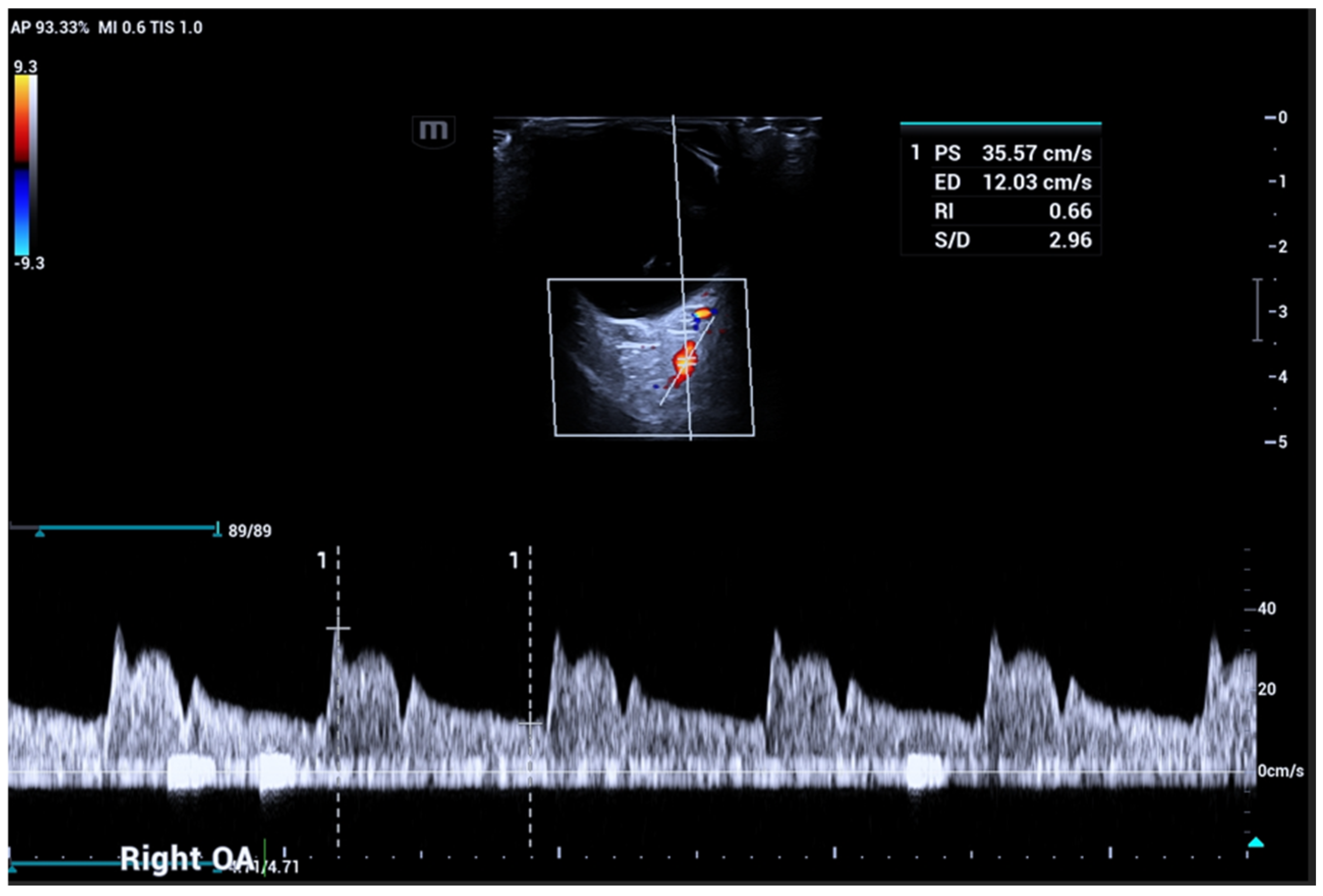

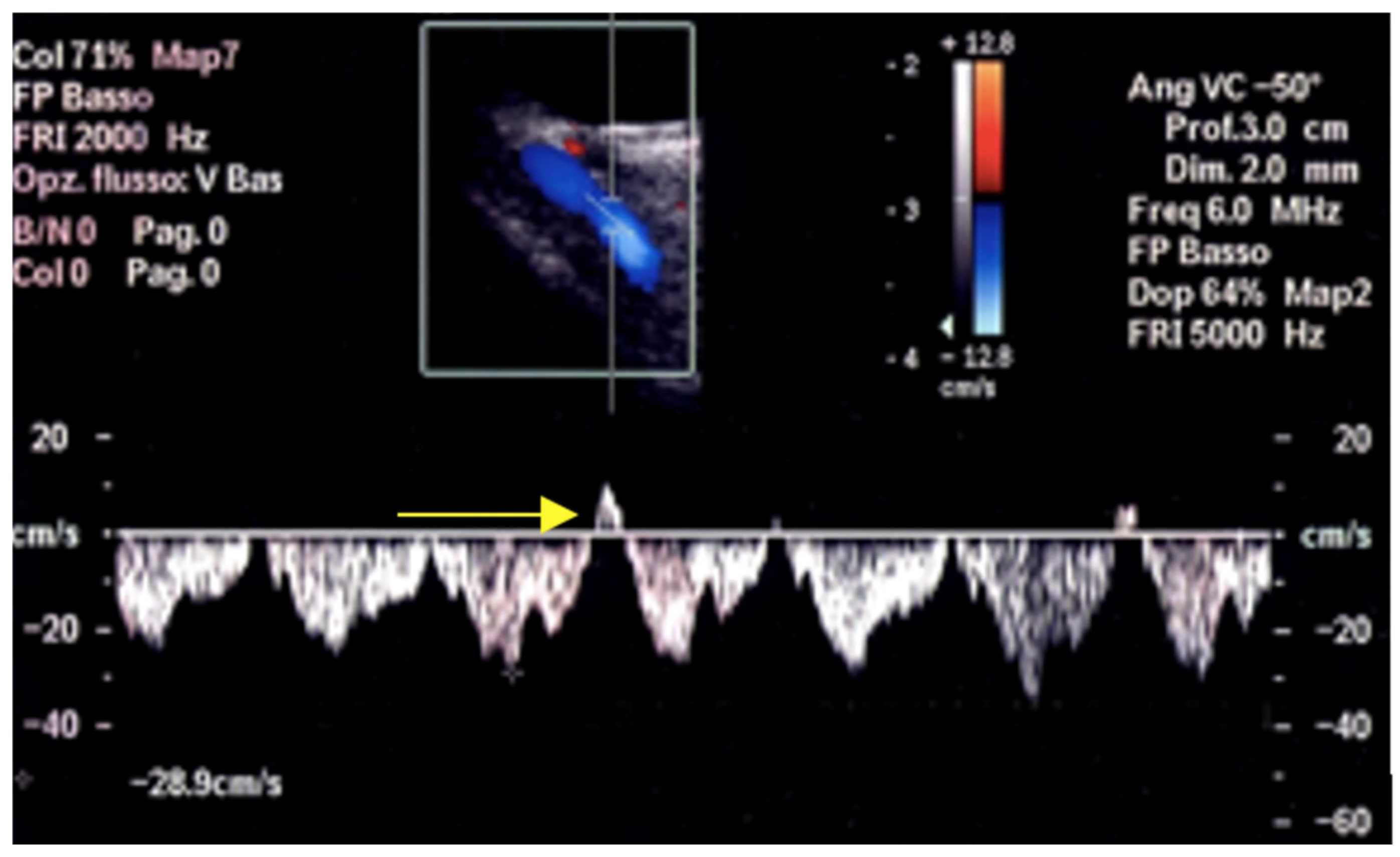

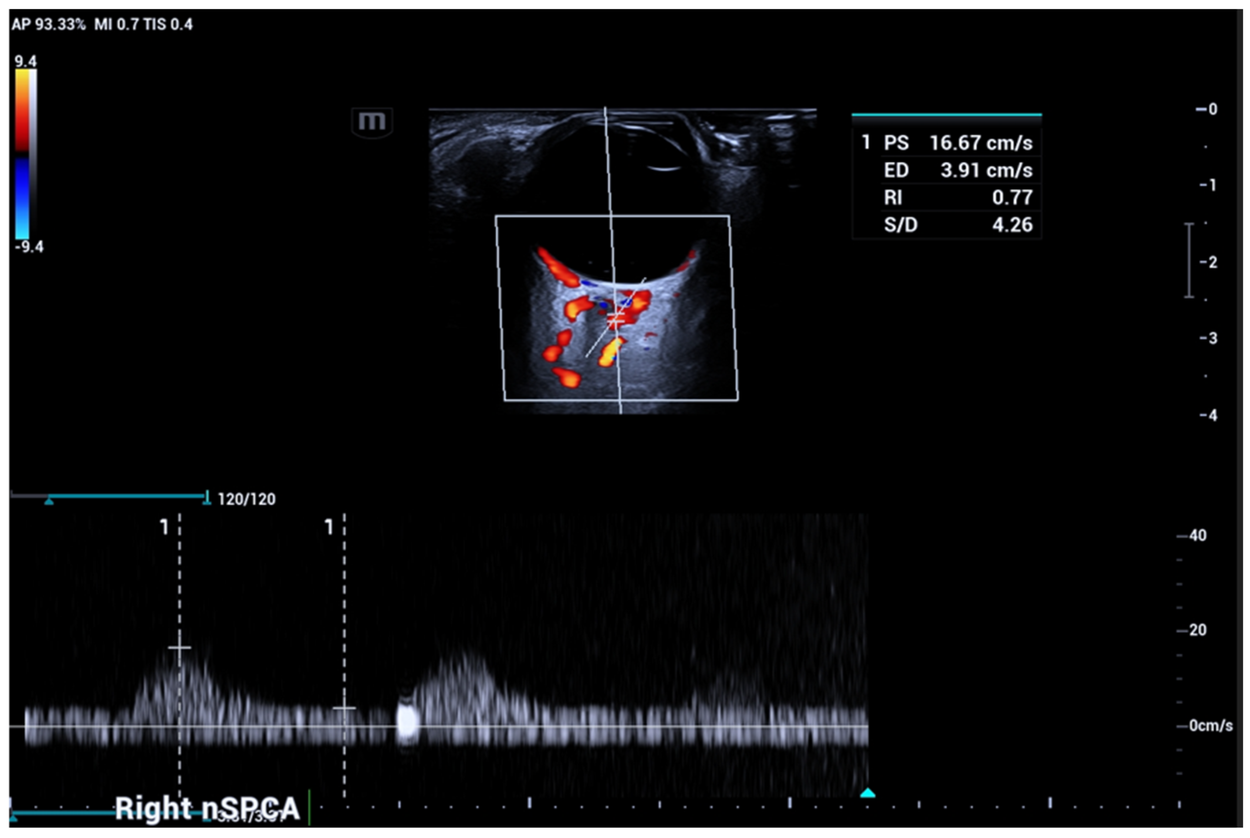


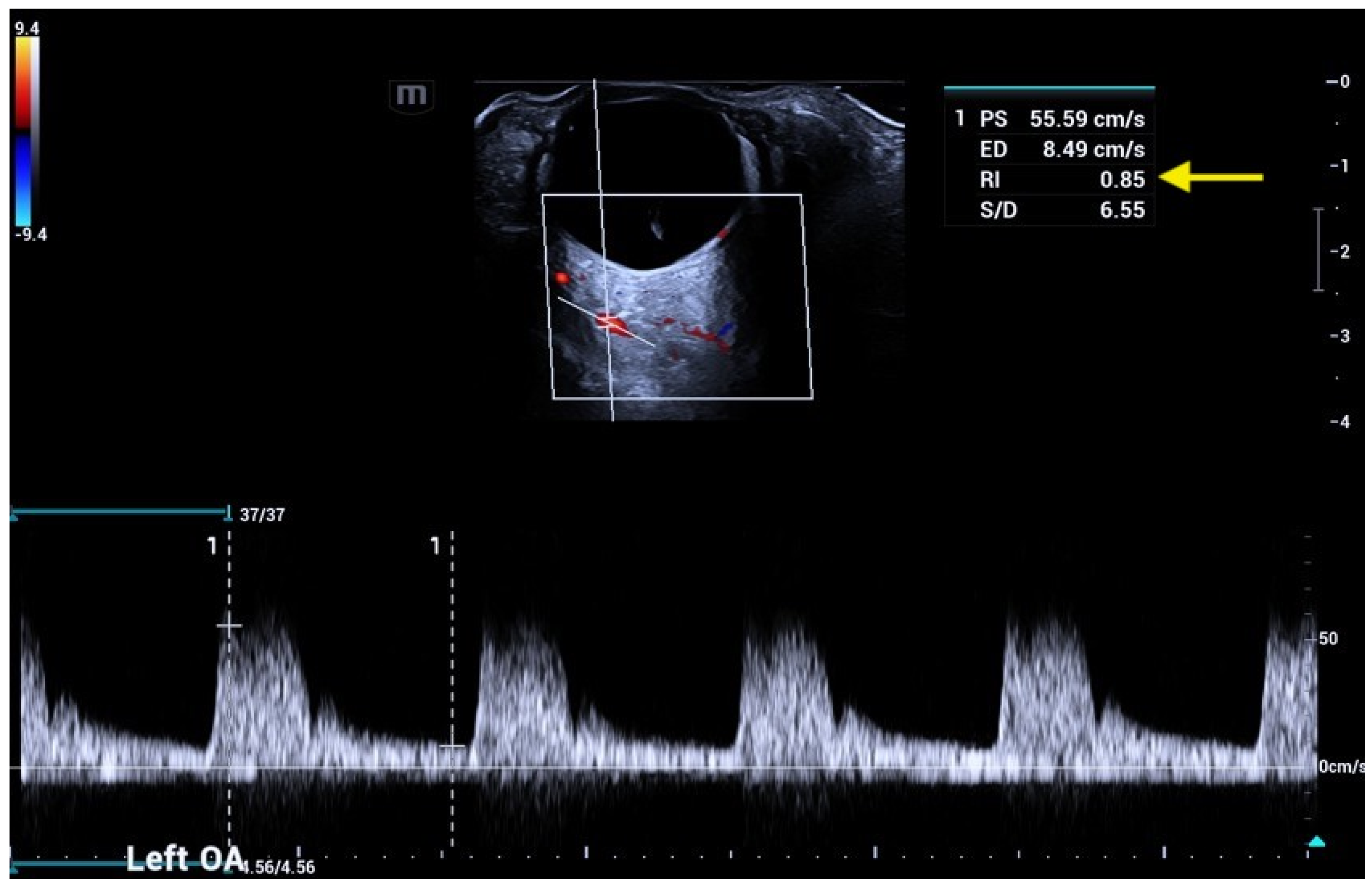
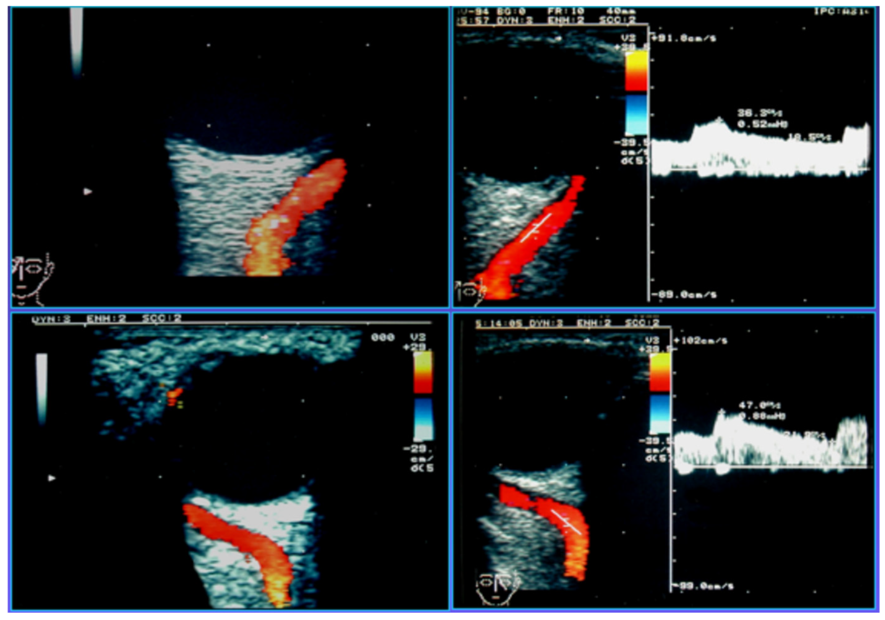
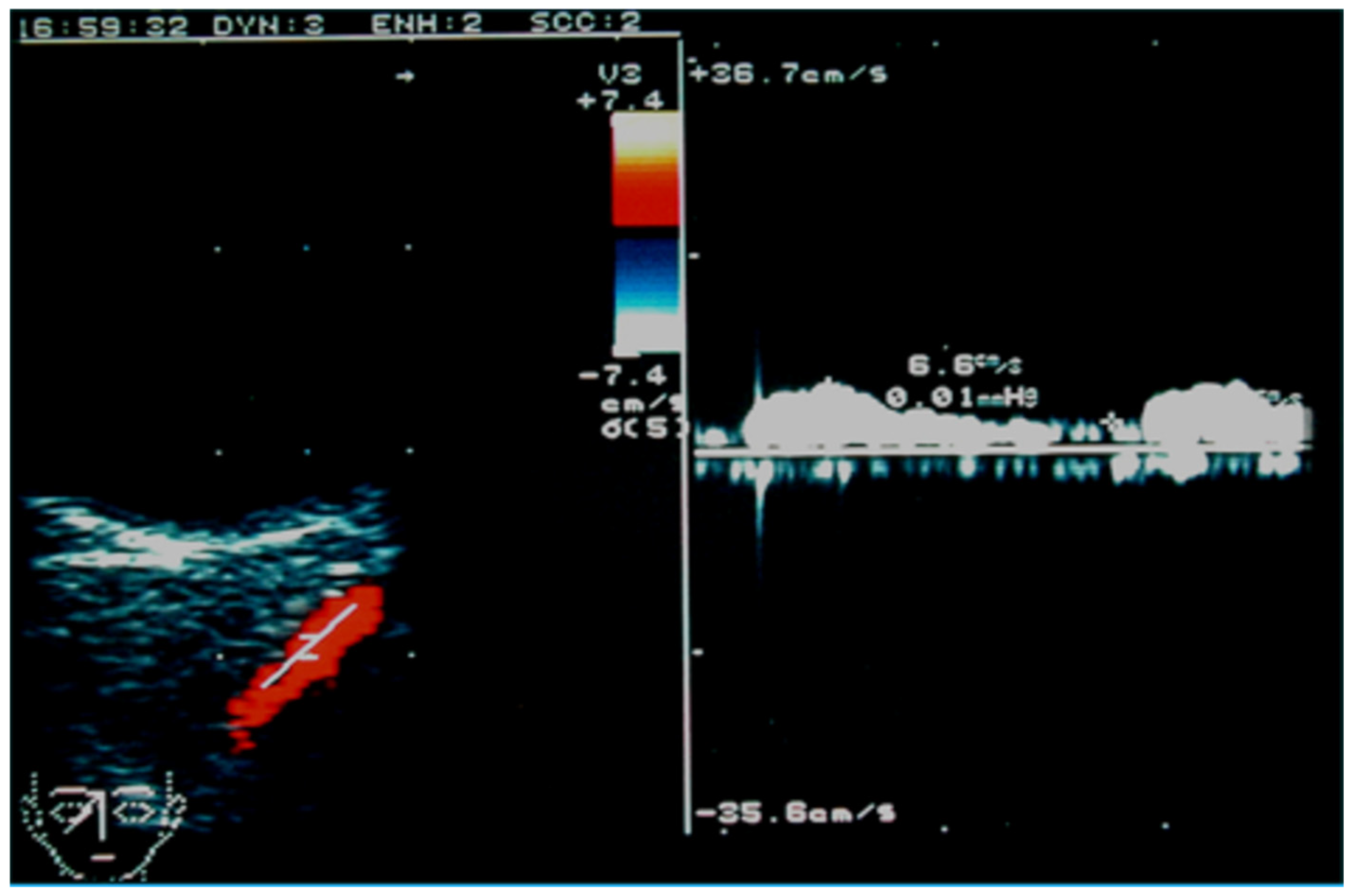
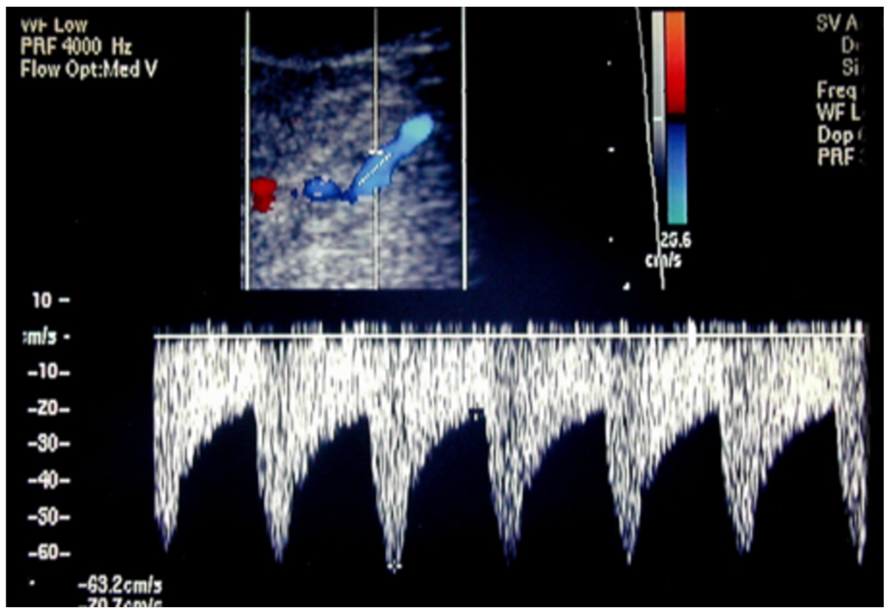

| Ophthalmological Diseases | ||
|---|---|---|
| Disease | Vessel Involved | Main OCDUS Findings |
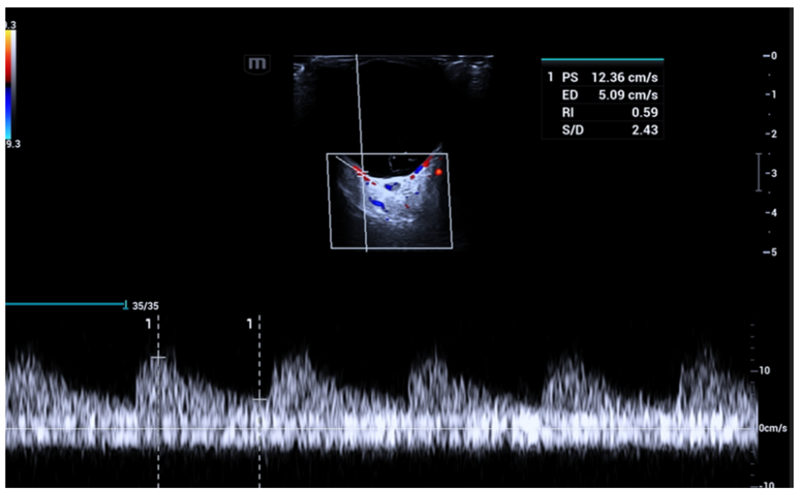
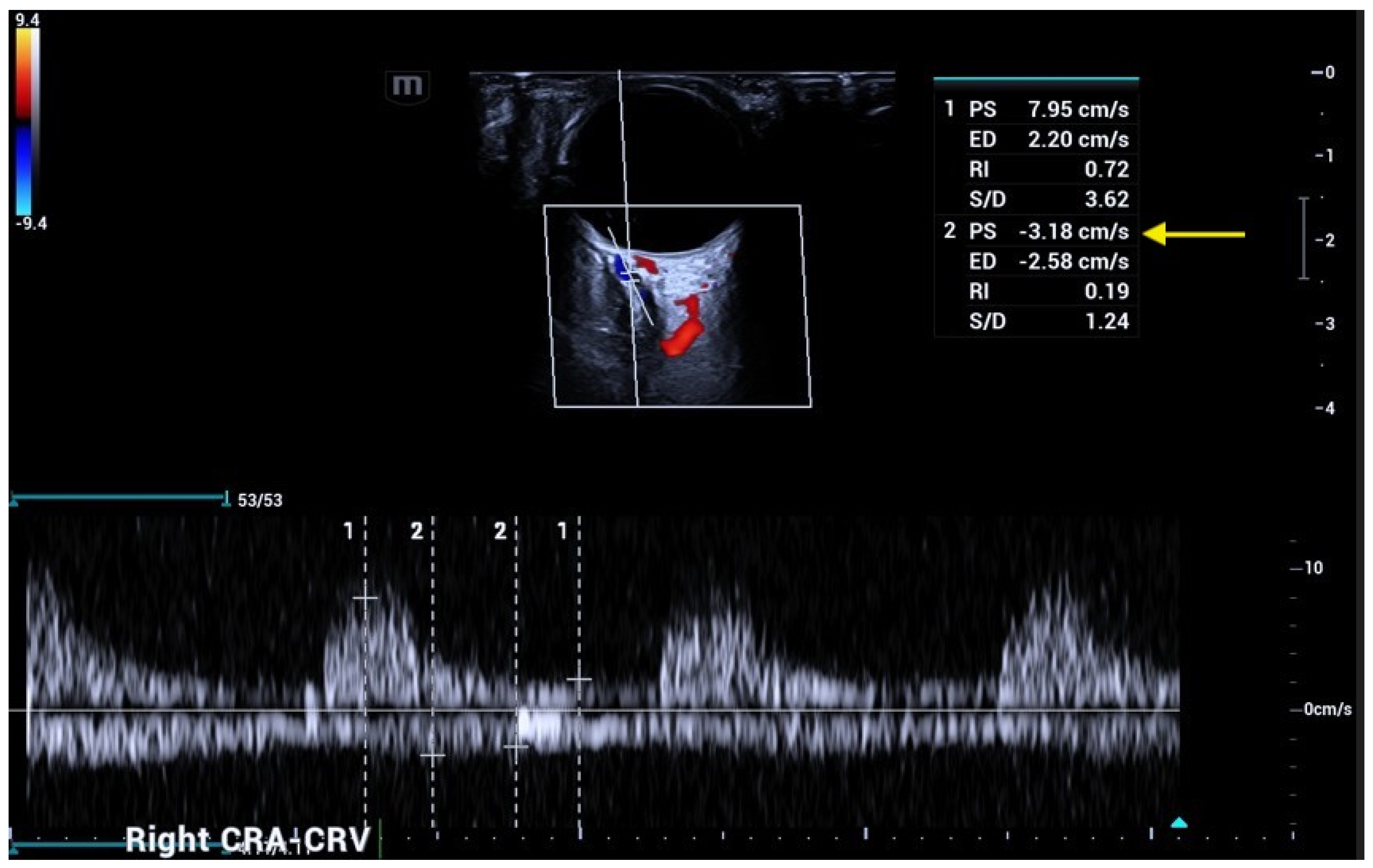
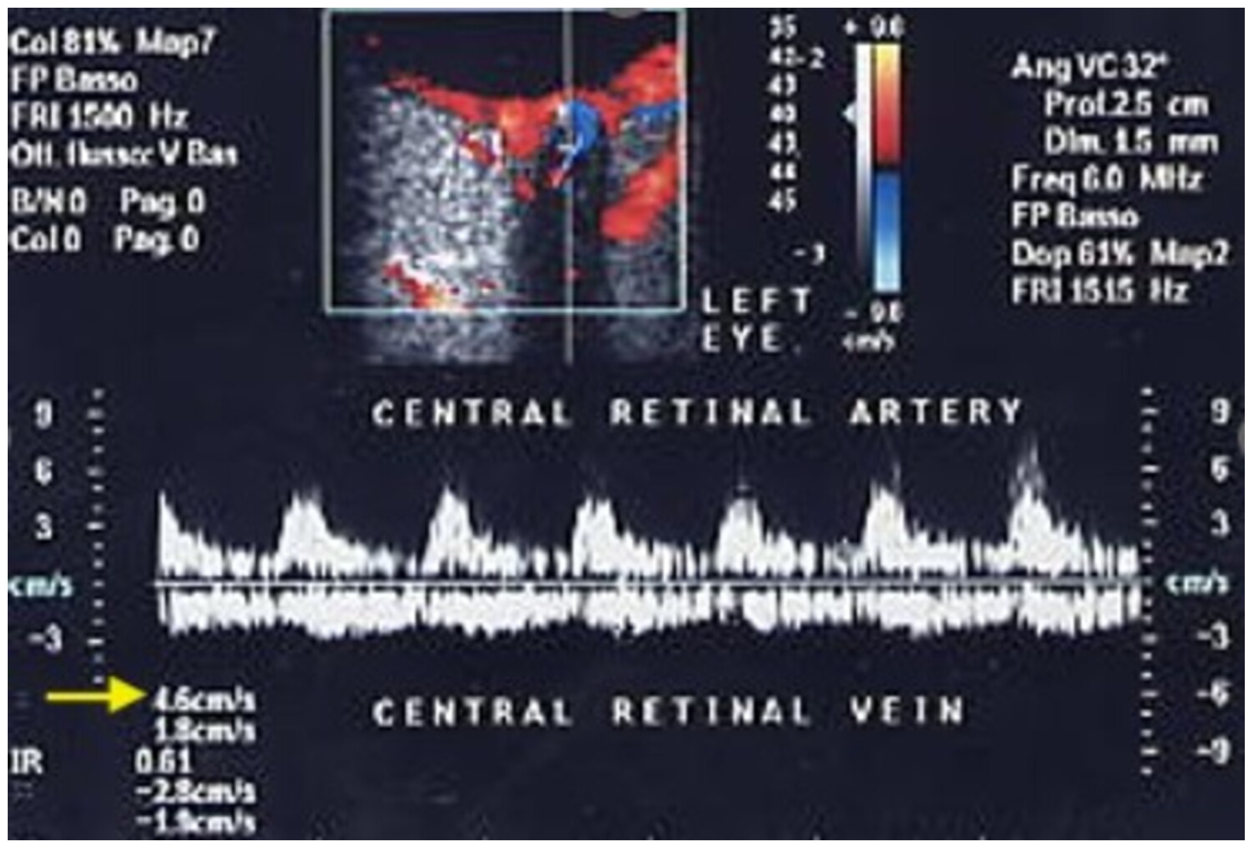
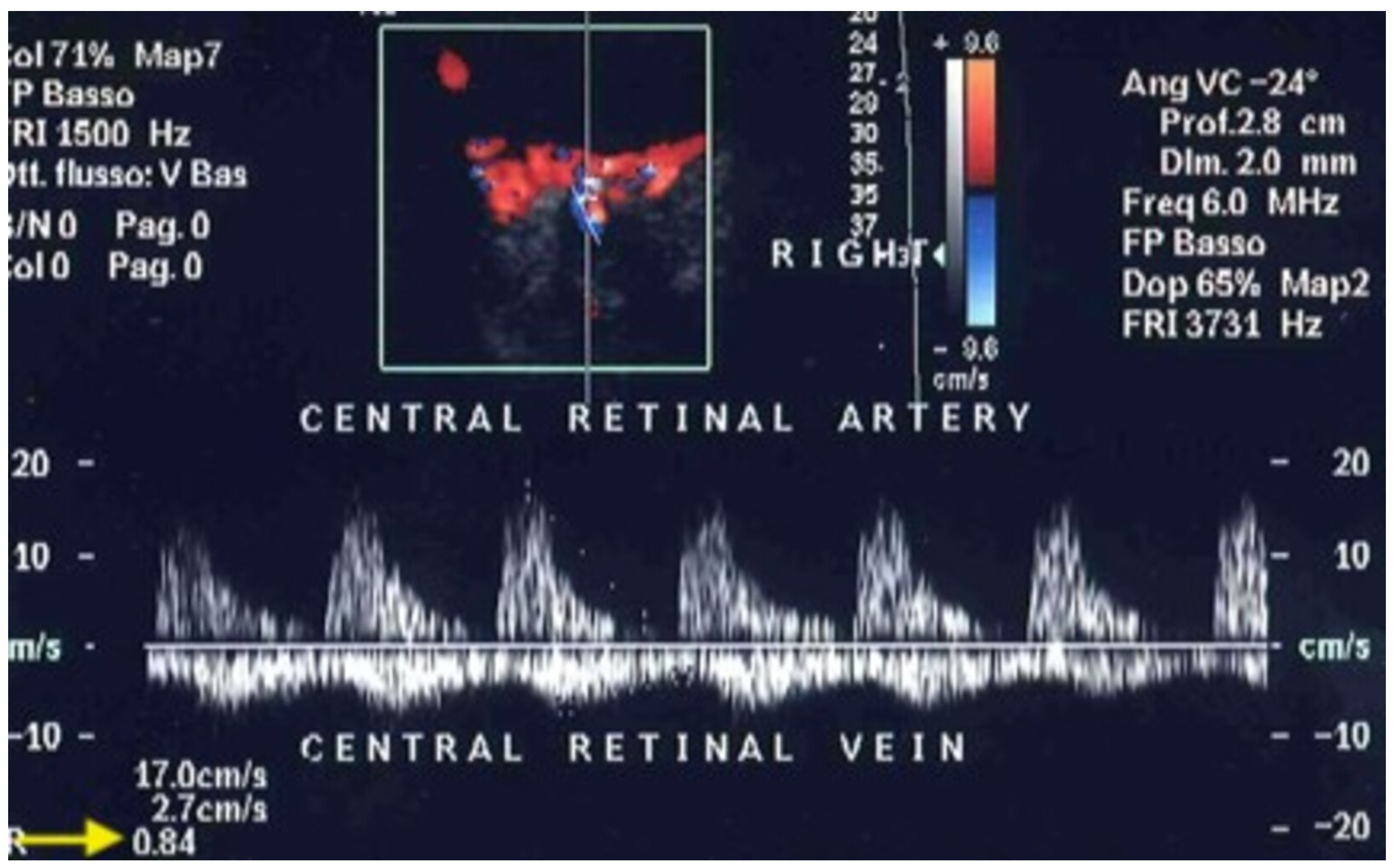
| Central retinal vein occlusion (CRVO) | CRV | Reduced maxV and minV |
| Central retinal artery occlusion (CRAO) | CRA | Reduced PSV and EDV |
| Non-arteritic anterior ischemic optic neuropathy (NAION) | CRA, CRV | Reduced PSV and EDV |
| Orbital varix | Orbital veins | Venous dilation |
| Glaucoma | CRA, SPCAs | Increased RI and reduced EDV |
| Age-related macular degeneration (AMD) | OA, CRA, SPCAs | Increased RI and reduced EDV |
| Retinitis pigmentosa | CRA | Reduced PSV and EDV |
| Myopia | OA, CRA, SPCAs | Reduced EDV and increased RI |
| Orbital tumors | Tumor-feeding vessels | Tumor vascularization |
| Cerebrovascular Diseases | ||
| Disease | Vessel Involved | Main OCDUS Findings |
| Carotid cavernous sinus fistulas (CCSFs) | SOV | Dilated, reversed, arterialized low RI blood flow |
| Carotid artery stenosis | OA, CRA, SPCAs | Reduced PSV and EDV |
| Cerebral circulation monitoring | CRA | CRA flow changes reflect intracranial perfusion |
| Systemic Diseases | ||
| Disease | Vessel Involved | Main OCDUS Findings |
| Diabetes | OA, CRA, PCAs | Reduced EDV and increased RI |
| Hypertension | CRA, SPCAs | Reduced EDV and increased RI |
| Takayasu Arteritis | OA, CRA | Reduced EDV and increased RI |
| Graves’ disease | SOV, OA, CRA | Reduced venous flow, reduced EDV and increased RI |
| Pseudoexfoliation syndrome | CRA, SPCAs | Reduced EDV and increased RI |
Disclaimer/Publisher’s Note: The statements, opinions and data contained in all publications are solely those of the individual author(s) and contributor(s) and not of MDPI and/or the editor(s). MDPI and/or the editor(s) disclaim responsibility for any injury to people or property resulting from any ideas, methods, instructions or products referred to in the content. |
© 2026 by the authors. Licensee MDPI, Basel, Switzerland. This article is an open access article distributed under the terms and conditions of the Creative Commons Attribution (CC BY) license.
Share and Cite
Venturini, M.; Malnati, S.; Catania, N.T.; Coppola, A.; Recaldini, C.; Gnesutta, A.; Ciani, M.; Tamietti, S.; Simonini, E.; Cappelli, A.; et al. Ocular Color Doppler Ultrasound (OCDUS) in Diagnosis and Monitoring of Ophthalmological, Cerebrovascular and Systemic Diseases: A Narrative Review. J. Clin. Med. 2026, 15, 2458. https://doi.org/10.3390/jcm15062458
Venturini M, Malnati S, Catania NT, Coppola A, Recaldini C, Gnesutta A, Ciani M, Tamietti S, Simonini E, Cappelli A, et al. Ocular Color Doppler Ultrasound (OCDUS) in Diagnosis and Monitoring of Ophthalmological, Cerebrovascular and Systemic Diseases: A Narrative Review. Journal of Clinical Medicine. 2026; 15(6):2458. https://doi.org/10.3390/jcm15062458
Chicago/Turabian StyleVenturini, Massimo, Silvia Malnati, Noemi Teresa Catania, Andrea Coppola, Chiara Recaldini, Aroa Gnesutta, Marianna Ciani, Silvia Tamietti, Emilio Simonini, Alberta Cappelli, and et al. 2026. "Ocular Color Doppler Ultrasound (OCDUS) in Diagnosis and Monitoring of Ophthalmological, Cerebrovascular and Systemic Diseases: A Narrative Review" Journal of Clinical Medicine 15, no. 6: 2458. https://doi.org/10.3390/jcm15062458
APA StyleVenturini, M., Malnati, S., Catania, N. T., Coppola, A., Recaldini, C., Gnesutta, A., Ciani, M., Tamietti, S., Simonini, E., Cappelli, A., Donati, S., Piacentino, F., & Fontana, F. (2026). Ocular Color Doppler Ultrasound (OCDUS) in Diagnosis and Monitoring of Ophthalmological, Cerebrovascular and Systemic Diseases: A Narrative Review. Journal of Clinical Medicine, 15(6), 2458. https://doi.org/10.3390/jcm15062458