Article

Pioglitazone Attenuates Sepsis-Associated Acute Kidney Injury by Modulating TLR-4/NF-κB Signaling and Improving Survival and Renal Function

1
Department of General Surgery, Güneşli Erdem Hospital, 34212 İstanbul, Turkey
2
Department of General Surgery, Faculty of Medicine, Karadeniz Technical University, 61080 Trabzon, Turkey
3
Department of Obstetrics and Gynecology, Faculty of Medicine, Karadeniz Technical University, 61080 Trabzon, Turkey
4
Department of General Surgery, Sultan 2. Abdulhamid Han Educational and Research Hospital, University of Health Sciences, 34668 Istanbul, Turkey
5
Department of Internal Medicine, Faculty of Medicine, Karadeniz Technical University, 61080 Trabzon, Turkey
6
Neuroscience Laboratory, BAMER, Biruni University, 34010 Istanbul, Turkey
7
Faculty of Medicine, BAMER, Biruni University, 34010 Istanbul, Turkey
*
Author to whom correspondence should be addressed.
J. Clin. Med. 2026, 15(6), 2270; https://doi.org/10.3390/jcm15062270
Submission received: 30 January 2026 / Revised: 8 March 2026 / Accepted: 12 March 2026 / Published: 17 March 2026
(This article belongs to the Section Nephrology & Urology)

Abstract

Aim: Sepsis-associated acute kidney injury (SA-AKI) remains a major cause of mortality, driven by inflammation and oxidative stress. Pioglitazone, a PPAR-γ agonist, has demonstrated anti-inflammatory and antioxidant effects beyond glycemic control. This study evaluated its renoprotective efficacy in a rat model of sepsis induced by cecal ligation and puncture (CLP). Methods: Thirty-six female Wistar rats were divided into Control, CLP + Saline, and CLP + Pioglitazone (10 mg/kg/day) groups. Survival was analyzed for 5 days. Renal function (BUN, creatinine, NGAL), oxidative stress (MDA), antioxidant signaling (NRF2), and inflammatory mediators (TNF-α, IL-6, HMGB1, TLR-4, NF-κB) were quantified by ELISA. Tubular epithelial necrosis, luminal debris, dilatation, hemorrhage, and inflammation were semi-quantitatively scored. Results: CLP caused marked renal dysfunction with elevated BUN, creatinine, and NGAL (p all <0.001 vs. Control). Pioglitazone significantly reduced these markers (p < 0.001 vs. CLP + Saline) and improved survival. Plasma MDA levels increased and renal Nrf2 levels decreased following CLP induction (both p < 0.001 vs. Control), whereas pioglitazone treatment significantly reduced MDA levels and increased NRF2 expression (p = 0.002 and p < 0.001 vs. CLP + Saline, respectively). Inflammatory mediators were markedly increased in sepsis (TNF-α, IL-6, HMGB1, TLR-4, and NF-κB; all p < 0.001 vs. Control) and significantly downregulated by pioglitazone (p < 0.01, p < 0.001, p < 0.001, p < 0.01, p < 0.01 vs. CLP + Saline, respectively). Histopathological injury was pronounced in septic rats (all p < 0.01 vs. Control) but was markedly ameliorated by pioglitazone p < 0.05, indicating substantial structural recovery. Conclusions: Pioglitazone markedly ameliorates CLP-induced SA-AKI by suppressing TLR-4/NF-κB/TNF-α signaling and oxidative stress, improving renal structure, function, and survival. These findings support its potential repurposing as a therapeutic adjunct in sepsis management.

1. Introduction

Sepsis represents a critical clinical condition characterized by a maladaptive immune response to infection that precipitates systemic inflammation, circulatory collapse, and multiorgan failure. Despite advances in intensive care, it remains a major global cause of mortality, contributing to approximately one-fifth of all deaths worldwide [1,2]. Global burden analyses estimated approximately 48.9 million sepsis cases and nearly 11 million sepsis-related deaths worldwide in 2017, underscoring the substantial global impact of the disease [2]. Among affected organs, the kidney is particularly susceptible to septic injury, and the development of sepsis-associated acute kidney injury (SA-AKI) markedly worsens patient outcomes and increases morbidity and mortality [3,4,5]. Current international guidelines also emphasize that early recognition and prompt management during the first hours of sepsis are critical for improving clinical outcomes [6].
The cecal ligation and puncture (CLP) procedure is widely regarded as the most reliable experimental model for reproducing polymicrobial sepsis in animals, as it closely mimics the hemodynamic, metabolic, and immune disturbances observed in human sepsis [7,8,9,10]. In this model, fecal leakage into the peritoneal cavity triggers generalized inflammation and leads to systemic organ dysfunction similar to clinical sepsis. Recent studies have further demonstrated that the CLP model reproduces a clinically relevant spectrum of sepsis severity, including hypotension, lactate elevation, cytokine surges, and multi-organ injury patterns [11].
SA-AKI is characterized by an abrupt decline in renal function, reflected by elevated serum urea and creatinine concentrations and decreased glomerular filtration rate. The underlying pathophysiology involves intertwined mechanisms, including cytokine-driven inflammation, oxidative stress, mitochondrial impairment, and endothelial injury [12,13]. Recent consensus reports further emphasize that microcirculatory dysfunction, dysregulated immune responses, and metabolic reprogramming play key roles in SA-AKI development, even when global renal blood flow is preserved [14]. Among these, activation of the Toll-like receptor-4 (TLR-4)/nuclear factor-κB (NF-κB) signaling pathway is central, as it stimulates the release of tumor necrosis factor-α (TNF-α) and interleukin-6, promoting tubular apoptosis and microcirculatory dysfunction [15,16]. Concomitantly, reactive oxygen species-mediated lipid peroxidation exacerbates epithelial and endothelial injury [17,18]. Recent reviews also highlight emerging serum and urinary biomarkers (e.g., NGAL and HMGB1) for SA-AKI risk stratification; however, current therapeutic strategies remain largely supportive [19]. Current therapeutic approaches remain largely supportive because no pharmacological agent has yet been proven to reverse these processes effectively [20].
Pioglitazone, a peroxisome proliferator-activated receptor-γ (PPAR-γ) agonist primarily used for type 2 diabetes, exerts pleiotropic biological effects that extend beyond glycemic regulation. Its activation of PPAR-γ modulates transcriptional control of inflammation and oxidative balance, suppressing NF-κB-dependent cytokine expression while enhancing Nrf2-mediated antioxidant responses [21,22,23]. Recent mechanistic studies further highlight the crosstalk between NF-κB-driven inflammatory signaling and Nrf2-regulated antioxidant pathways, supporting therapeutic strategies that simultaneously suppress cytokine amplification and restore redox balance in sepsis-related organ injury [24]. Evidence from preclinical studies indicates that pioglitazone can attenuate TLR-4 signaling and reduce oxidative and inflammatory injury in models of hepatic and cardiac sepsis [25]. However, its efficacy as a post-treatment strategy for renal protection in polymicrobial sepsis has not yet been comprehensively examined.
Based on these considerations, the present study aimed to determine whether pioglitazone could mitigate oxidative stress and inflammatory damage in the kidneys of rats subjected to CLP-induced sepsis. To this end, renal functional markers (BUN, creatinine, NGAL), oxidative parameters (MDA), inflammatory mediators (TNF-α, TLR-4, NF-κB), and histopathological alterations were systematically evaluated.

2. Materials and Methods

2.1. Animals and Ethical Approval

A total of 36 adult female Wistar albino rats (weighing 200–250 g) were included. All procedures were conducted in compliance with the ethical guidelines of the Guide for the Care and Use of Laboratory Animals (National Institutes of Health [NIH], USA) and approved by the Institutional Animal Research Ethics Committee (Approval No: 2125095711, 28 May 2023). The animals were housed individually in polycarbonate cages under standard conditions (temperature 22 ± 2 °C, 12 h light/dark cycle) with unrestricted access to food and water. All efforts were made to minimize animal suffering, following the principles of Replacement, Reduction, and Refinement (3Rs). Statistical analyses were performed using GraphPad Prism (version 9.0, GraphPad Software, San Diego, CA, USA) and IBM SPSS Statistics (version 25.0/IBM Corp./Armonk, NY, USA).

2.2. Experimental Design

Rats were randomly assigned into three experimental groups (n = 12 per group):
Control: sham-operated rats without cecal manipulation, maintained on standard diet and water.
CLP + Saline: sepsis induced by cecal ligation and puncture (CLP), followed by intraperitoneal injection of physiological saline (0.9% NaCl).
CLP + Pioglitazone: CLP-induced sepsis treated with pioglitazone (10 mg/kg/day, intraperitoneally).
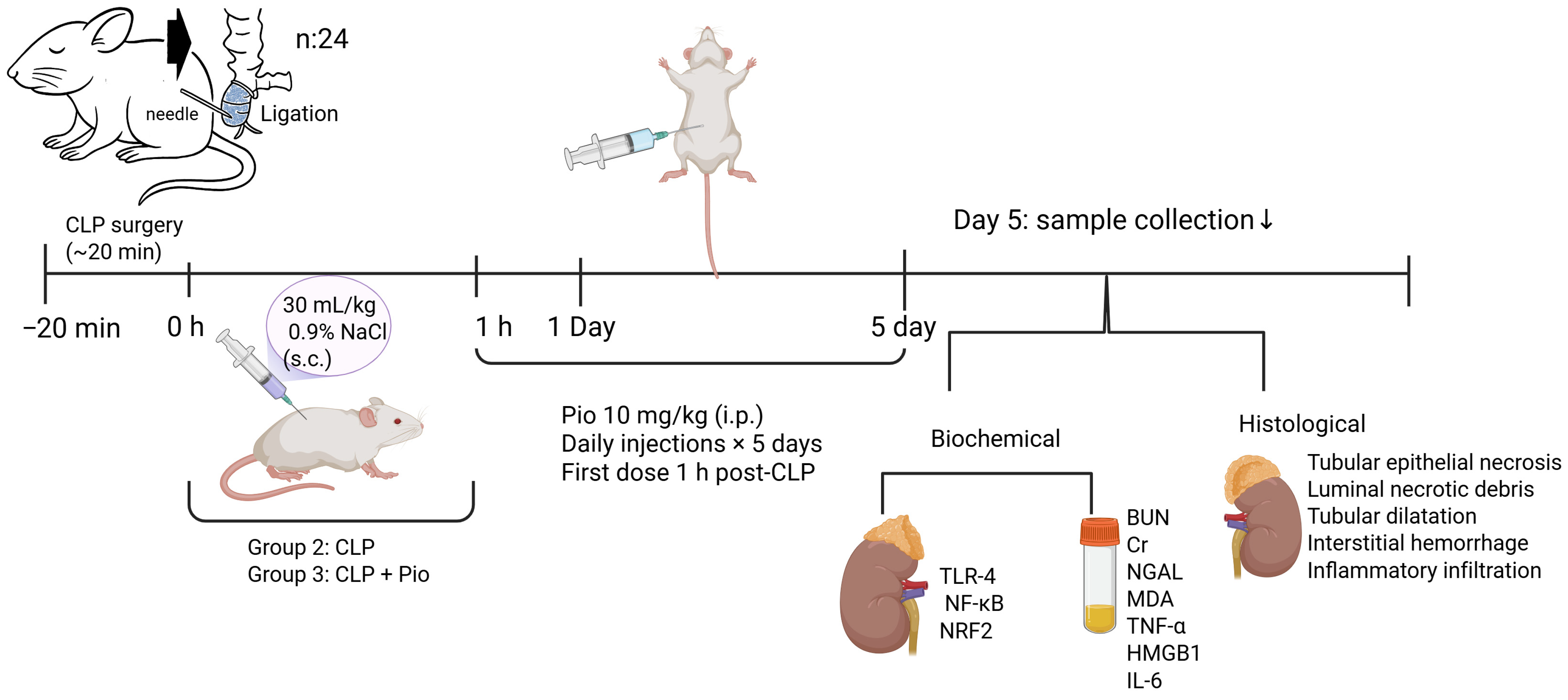
In the CLP + Pioglitazone group, sepsis was induced by cecal ligation and puncture, and pioglitazone was administered intraperitoneally at a dose of 10 mg/kg. The first injection was performed 1 h after the CLP surgery, followed by subsequent injections once every 24 h during the 5-day experimental period. This post-treatment regimen was designed to evaluate the therapeutic efficacy of pioglitazone rather than its prophylactic potential in sepsis-associated acute kidney injury.
Fluid resuscitation was administered equally in septic groups (30 mL/kg 0.9% NaCl within the first postoperative hour, followed by 10 mL/kg/day in divided doses). The experiment continued for five days; during this period, six rats from the CLP + Saline and two from the CLP + Pioglitazone group did not survive.
At the study endpoint, surviving animals were anesthetized with ketamine/xylazine, and blood samples were obtained by cardiac puncture. Both kidneys were excised for biochemical and histopathological analyses (Figure 1).

2.3. Cecal Ligation and Puncture (CLP) Procedure

Under aseptic conditions and general anesthesia, a midline laparotomy (~3 cm) was performed to expose the cecum. The distal third of the cecum was ligated using 3–0 silk suture approximately 1 cm from the ileocecal junction to prevent intestinal obstruction, and the cecal wall was punctured once with a 22-gauge sterile needle. A small amount of fecal material was gently extruded to confirm perforation before the cecum was repositioned and the abdominal wall was closed in two layers with 4–0 absorbable sutures.
Sham-operated animals underwent laparotomy without ligation or puncture. In accordance with previously described CLP protocols, systemic sepsis was induced following the procedure [26].

2.4. Histopathological Examination of the Kidney

At the endpoint, both kidneys were carefully removed, rinsed with cold saline, and immersed in 10% neutral buffered formalin (pH 7.4) for 72 h. Following fixation, tissues were processed using standard paraffin-embedding techniques and sectioned at 5 µm thickness. The sections were stained with hematoxylin and eosin (H&E) for morphological evaluation under a light microscope (Olympus BX51, Tokyo, Japan).
Digital images were captured from ten randomly selected non-overlapping cortical fields (×20 magnification) using an integrated imaging system (Image-Pro Express, Media Cybernetics, Rockville, MD, USA). A pathologist blinded to experimental grouping performed semi-quantitative scoring of histological changes, including tubular epithelial necrosis, luminal debris accumulation, tubular dilatation, interstitial hemorrhage, and inflammatory cell infiltration.
Each parameter was graded according to the percentage of affected cortical area as follows:
0 = 0–5%, 1 = 6–20%, 2 = 21–40%, 3 = 41–60%, 4 = 61–80%, and 5 = 81–100% of tissue involvement, modified from Erbas et al. [26].

2.5. Biochemical Measurements

2.5.1. Plasma Biomarkers

After euthanasia, blood was collected into heparinized tubes via cardiac puncture. Samples were centrifuged at 3000 rpm for 10 min, and plasma was separated and stored at −20 °C until analysis. Blood urea nitrogen (BUN) and creatinine concentrations were determined using an automated chemistry analyzer (Beckman Coulter AU640, Brea, CA, USA) and standard commercial assay kits.
Plasma levels of tumor necrosis factor-α (TNF-α), neutrophil gelatinase-associated lipocalin (NGAL) and IL-6 were quantified using rat-specific enzyme-linked immunosorbent assay (ELISA) kits (Abcam, UK), performed in duplicate according to the manufacturers’ protocols. HMGB1 levels were measured using a commercially available ELISA kit (IBL International, Hamburg, Germany, Cat. No. ST51011). Optical densities were read at 450 nm using a microplate spectrophotometer.

2.5.2. Renal Tissue Assays

Kidney samples were homogenized in ice-cold phosphate-buffered saline (pH 7.4) at a ratio of 1:5 (w/v). Homogenates were centrifuged at 5000× g for 15 min at 4 °C, and supernatants were collected for biochemical analyses. Total protein content was determined by the Bradford method using bovine serum albumin as the calibration standard.
Levels of toll-like receptor 4 (TLR-4) and nuclear factor-κB (NF-κB) in renal tissue were quantified using commercial rat-specific ELISA kits (Abcam, Waltham, MA, USA). Nuclear factor erythroid 2–related factor 2 (Nrf2) levels in tissue homogenates were measured using a rat-specific quantitative sandwich ELISA kit (MyBioSource, MBS1602926, San Diego, CA, USA) according to the manufacturer’s instructions. Lipid peroxidation was evaluated by measuring malondialdehyde (MDA) using the thiobarbituric acid reactive substances (TBARS) method. Briefly, samples were incubated with trichloroacetic acid and thiobarbituric acid at 100 °C for 1 h, cooled on ice, centrifuged, and the absorbance of the supernatant was measured at 535 nm. MDA concentrations were expressed as nmol/mL using 1,1,3,3-tetraethoxypropane as the calibration standard.

2.6. Statistical Analysis

Data distribution was assessed using the Shapiro–Wilk test for normality and Levene’s test for homogeneity of variances. Normally distributed data were analyzed using one-way ANOVA, followed by Tukey’s or Tamhane’s T2 post hoc tests where appropriate. Non-parametric data were expressed as median [interquartile range, IQR] and compared using the Kruskal–Wallis test, followed by Mann–Whitney U for pairwise analyses. Kaplan–Meier survival analysis was conducted with the log-rank (Mantel–Cox) test. All statistical analyses were performed using SPSS version 25.0 (IBM Corp., Armonk, NY, USA), and a p value < 0.05 was considered statistically significant.

3. Results

3.1. Survival Analysis

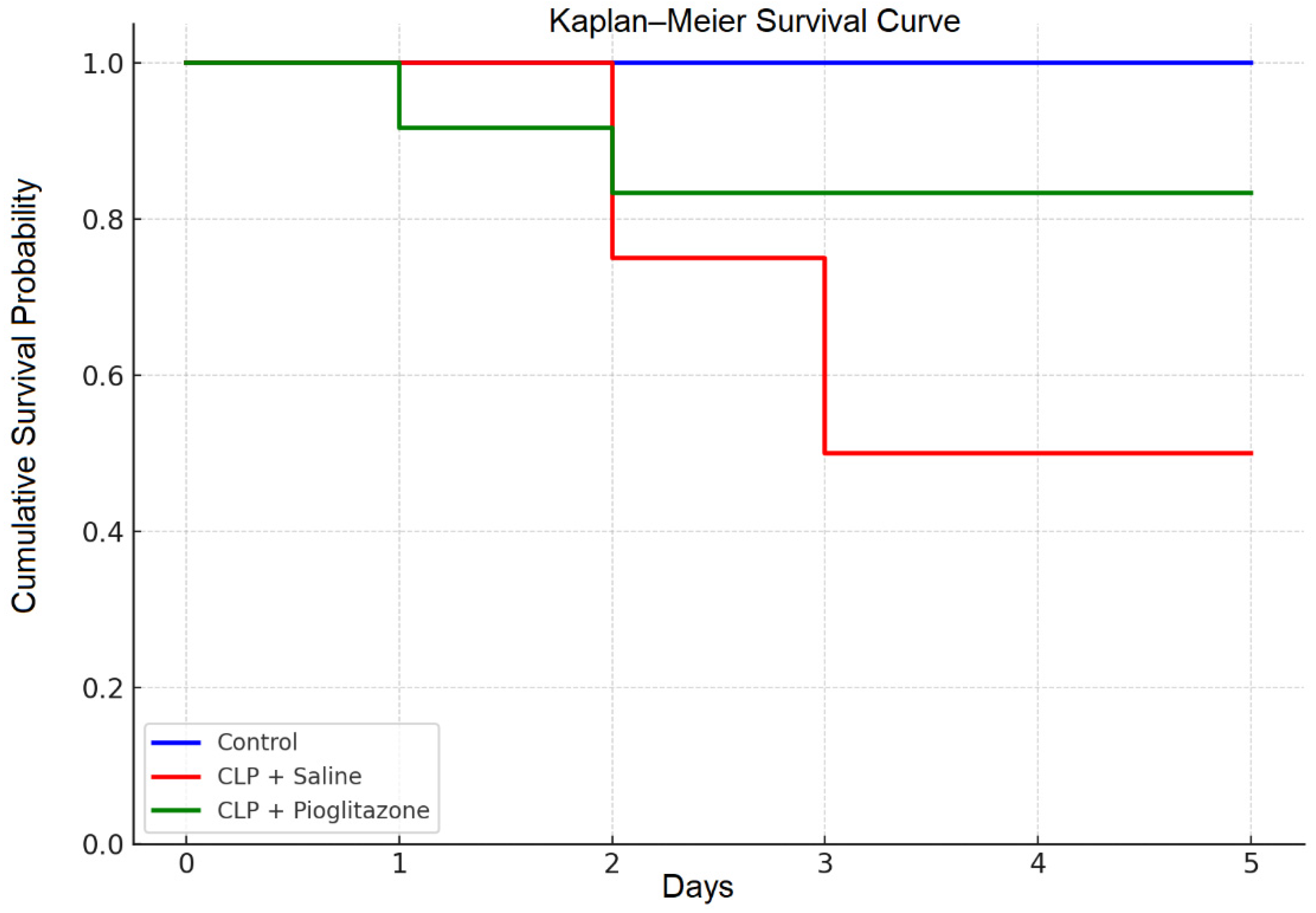
Kaplan–Meier survival analysis demonstrated statistically significant differences among the experimental groups (Log-rank test: χ2 = 8.97, df = 2, p = 0.011). The CLP + saline group exhibited a substantial reduction in 5-day survival (50%), while pioglitazone treatment markedly improved survival to 83.3%, closely approaching the 100% survival observed in the non-septic control group. These results suggest that pioglitazone provides a significant protective effect in this rat sepsis model (Figure 2, Table 1).
The table summarizes the number of surviving rats on each experimental day. Pioglitazone treatment improved overall survival compared with the CLP + saline group.

3.2. Renal Function and Histopathology

3.2.1. Quantitative Histopathological Scores

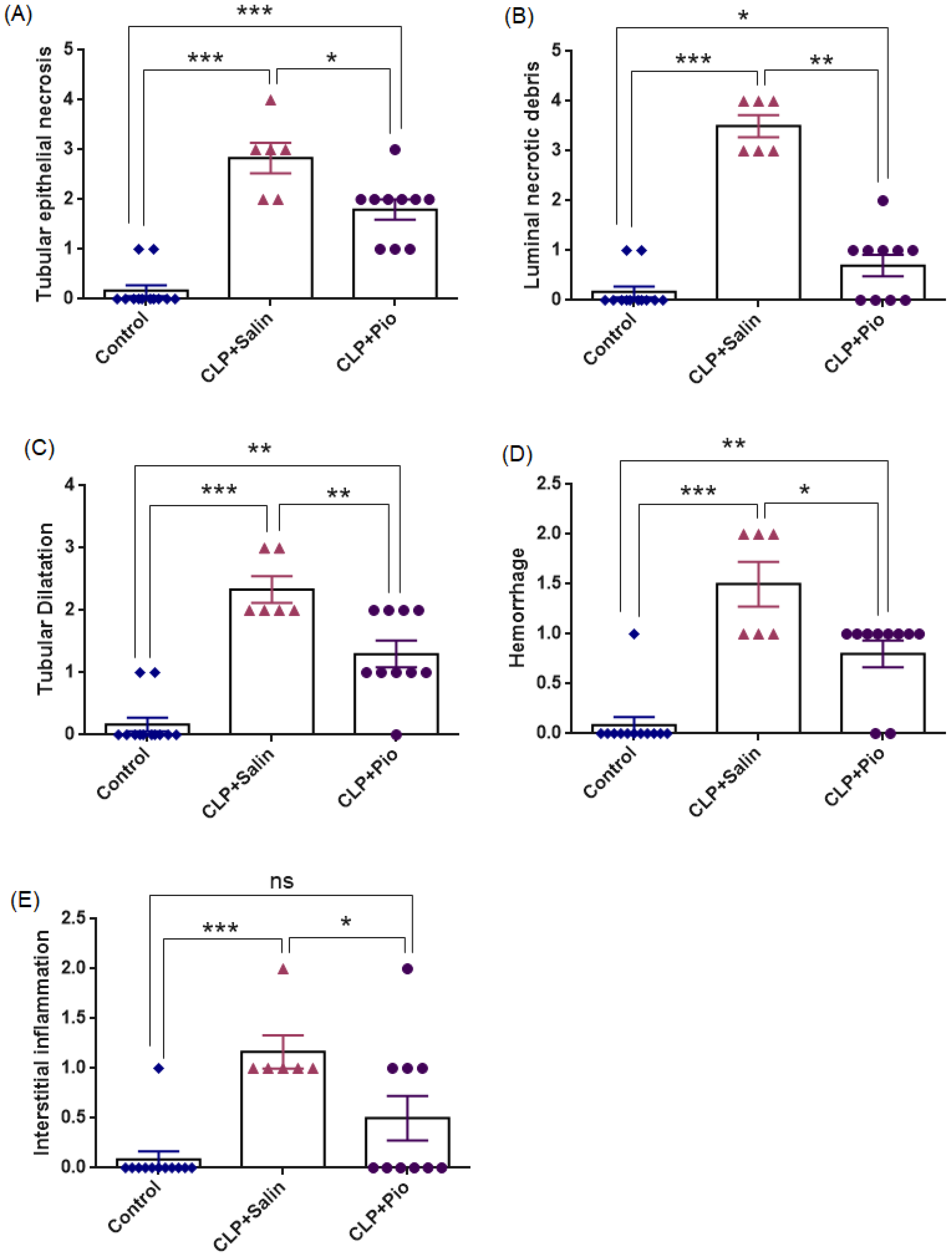
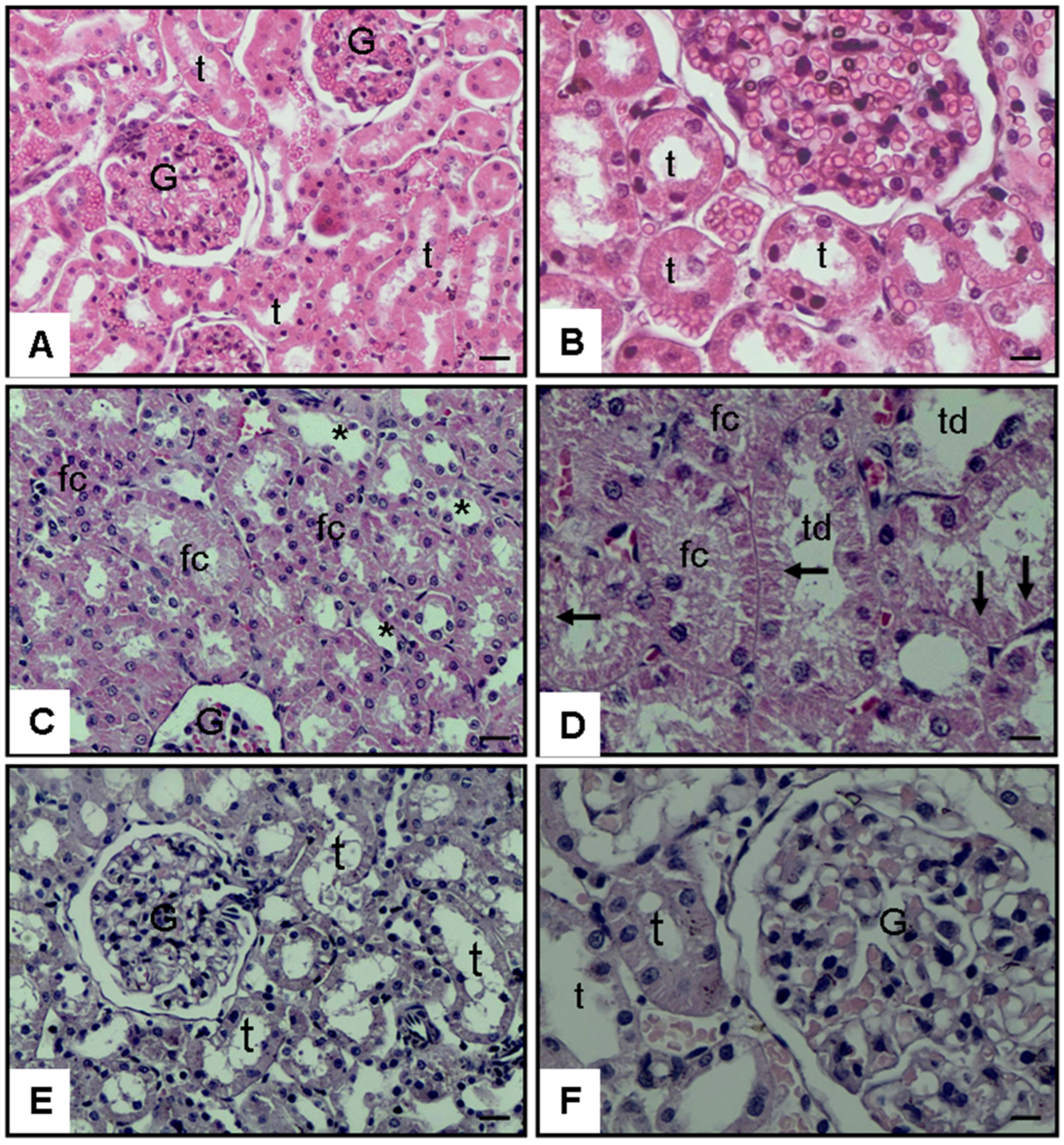
Representative histopathological findings are presented in Figure 3 and Figure 4 and Table 2, demonstrating that CLP-induced sepsis caused extensive renal structural injury, whereas pioglitazone treatment markedly alleviated these alterations.
Histological examination revealed intact corticomedullary architecture in the control group. In contrast, the CLP + Saline group exhibited prominent tubular epithelial necrosis, luminal necrotic debris accumulation, tubular dilatation, hemorrhage, and interstitial inflammation (overall Kruskal–Wallis p < 0.001; pairwise comparisons p < 0.001), indicating marked tubular injury accompanied by microvascular bleeding, leukocyte infiltration, and tissue inflammation after sepsis induction. Pioglitazone treatment significantly ameliorated these lesions (p = 0.016; 0.001; 0.009; 0.017; and 0.042 vs. CLP + Saline, respectively), although complete normalization was not achieved. Tubular epithelial necrosis (p < 0.001), luminal necrotic debris (p = 0.035), tubular dilatation (p = 0.001), and hemorrhage (p = 0.017) scores remained higher than in controls, suggesting partial yet meaningful structural recovery rather than full restoration. Pioglitazone group did not differ significantly from Control (p = 0.078), suggesting that inflammation levels were nearly normalized after treatment, indicating a strong trend toward complete restoration of tissue inflammatory status to baseline levels.

3.2.2. Representative Micrographs of Renal Injury

Histopathological evaluation of kidney sections revealed marked differences among the experimental groups. The control group exhibited normal renal morphology with intact glomeruli and tubular structures (Figure 3A,B). In contrast, the CLP + Saline group demonstrated extensive tubular epithelial necrosis, luminal necrotic debris, tubular dilatation, and interstitial inflammatory cell infiltration (Figure 3C,D). Tubular epithelial flattening and focal vascular congestion were also evident, indicating severe sepsis-induced renal injury. Treatment with pioglitazone notably alleviated these pathological alterations, as reflected by preserved tubular integrity, reduced inflammatory infiltration, and nearly normal glomerular appearance (Figure 3E,F). These findings corroborate the quantitative histopathological scores, confirming the renoprotective effect of pioglitazone against CLP-induced septic damage.

3.2.3. Renal Function Biomarkers

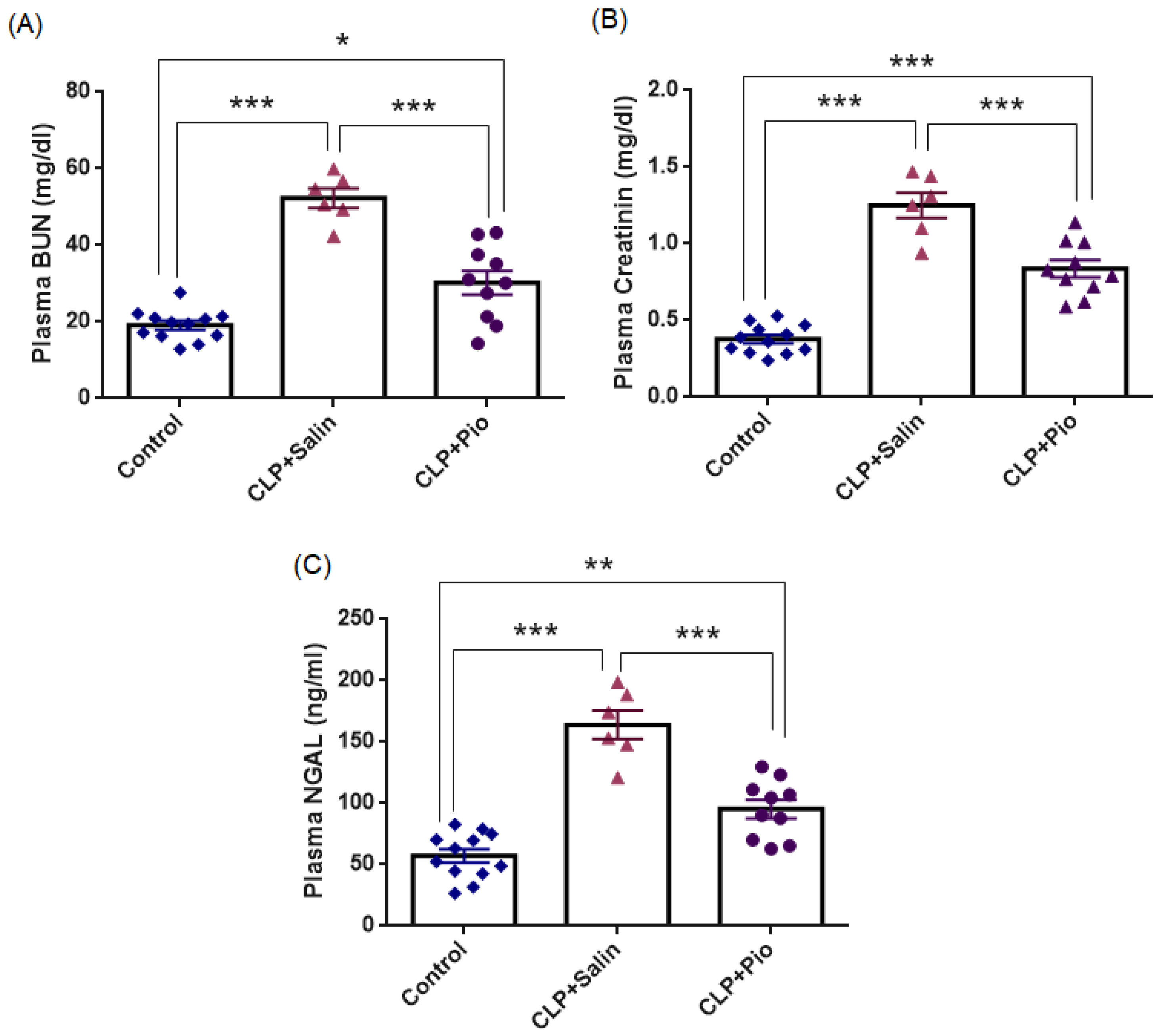
To evaluate the effect of CLP-induced sepsis on renal injury across groups, plasma BUN, creatinine, and NGAL levels were quantified. As shown in Figure 5 and Table 3, the CLP + Saline group exhibited significantly higher BUN, creatinine, and NGAL values than the control group (all p < 0.001). When the mean control value was taken as the baseline, the CLP + Saline group showed 2.73-fold, 3.38-fold, and 2.87-fold increases in BUN, creatinine, and NGAL, respectively. Pioglitazone treatment markedly attenuated these elevations (all p < 0.001 vs. CLP + Saline), corresponding to reductions of 42.1%, 33.6%, and 41.9%, respectively. Nevertheless, values remained modestly above baseline (BUN p = 0.018; creatinine p < 0.001; NGAL p = 0.002), indicating partial but meaningful recovery of renal function. Collectively, the biochemical improvements in renal function biomarkers (BUN, creatinine, and NGAL) were consistent with the histopathological recovery, indicating that pioglitazone substantially mitigated sepsis-induced acute tubular injury.

3.3. Oxidative Stress and Inflammatory Biomarkers

3.3.1. Oxidative Stress Biomarker

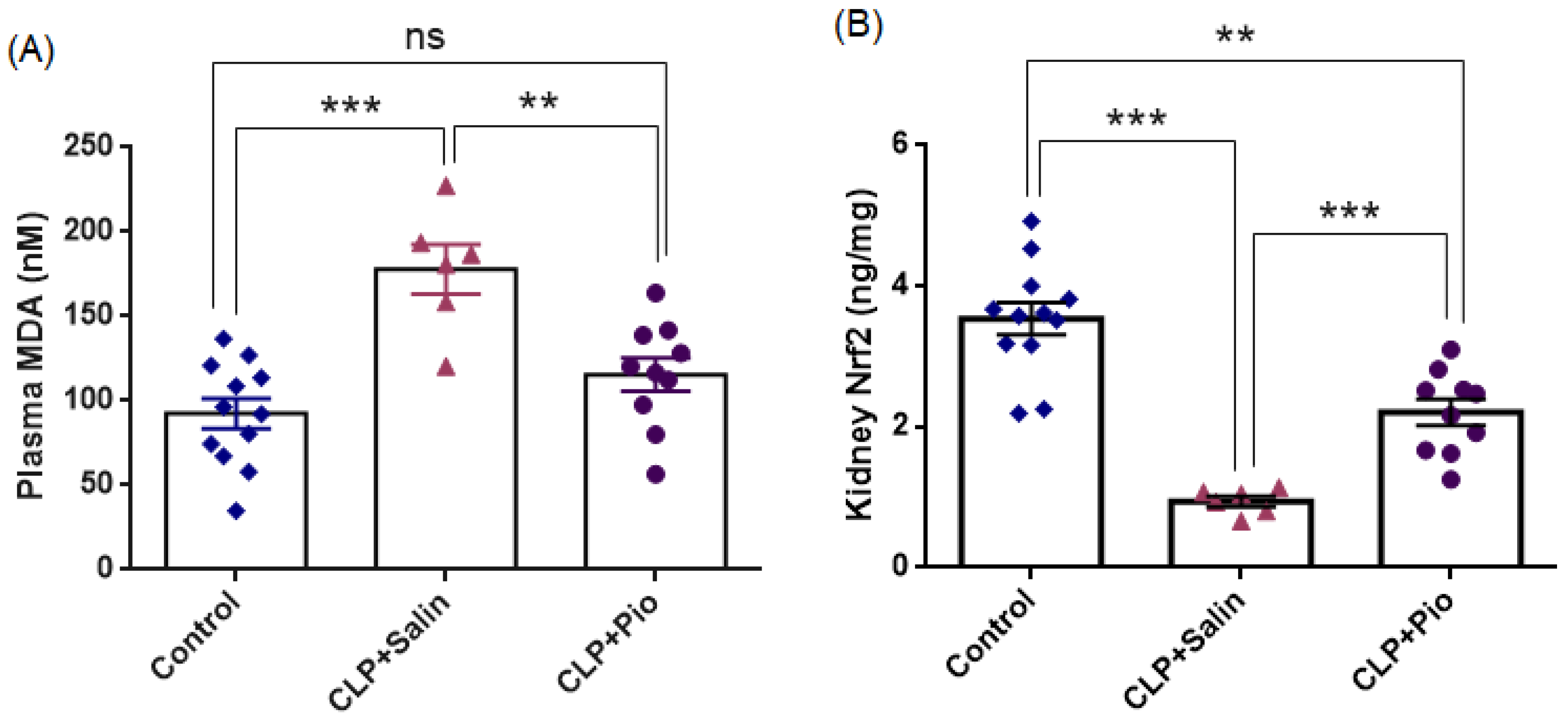
An overview of the oxidative stress findings is presented in Figure 6 and Table 4, showing that CLP-induced sepsis markedly increased systemic lipid peroxidation and suppressed renal antioxidant signaling, whereas pioglitazone treatment substantially attenuated lipid peroxidation and partially improved antioxidant responses.
Plasma MDA levels were significantly higher in the CLP + Saline group (177.9 ± 14.6 nmol/mL) than in the Control group (92.5 ± 8.8 nmol/mL; p < 0.001), representing an approximately 1.9-fold increase in lipid peroxidation following sepsis induction. Pioglitazone treatment markedly reduced MDA concentrations (p < 0.01 vs. CLP + Saline), corresponding to an estimated 35% decrease relative to untreated sepsis. Importantly, MDA values in the pioglitazone group were statistically comparable to controls (p = 0.231), indicating near-complete restoration of oxidative balance.
Collectively, these findings suggest that pioglitazone mitigated sepsis-induced oxidative damage, likely by limiting lipid peroxidation and enhancing redox balance.
Renal Nrf2 levels were significantly lower in the CLP + Saline group (0.942 ± 0.074 ng/mL) than in the Control group (3.537 ± 0.229 ng/mL; p < 0.001), representing an approximately 3.8-fold decrease in antioxidant signaling following sepsis induction. Pioglitazone treatment markedly increased NRF2 concentrations (p < 0.01 vs. CLP + Saline), corresponding to an approximately 2.3-fold increase relative to untreated sepsis. Importantly, NRF2 values in the pioglitazone group remained significantly lower than those observed in controls (p < 0.01), indicating partial improvement of antioxidant defense.
Collectively, these findings suggest that pioglitazone mitigated sepsis-induced renal oxidative damage, likely by modulating NRF2-mediated antioxidant signaling and improving renal redox balance.

3.3.2. Inflammatory Biomarkers

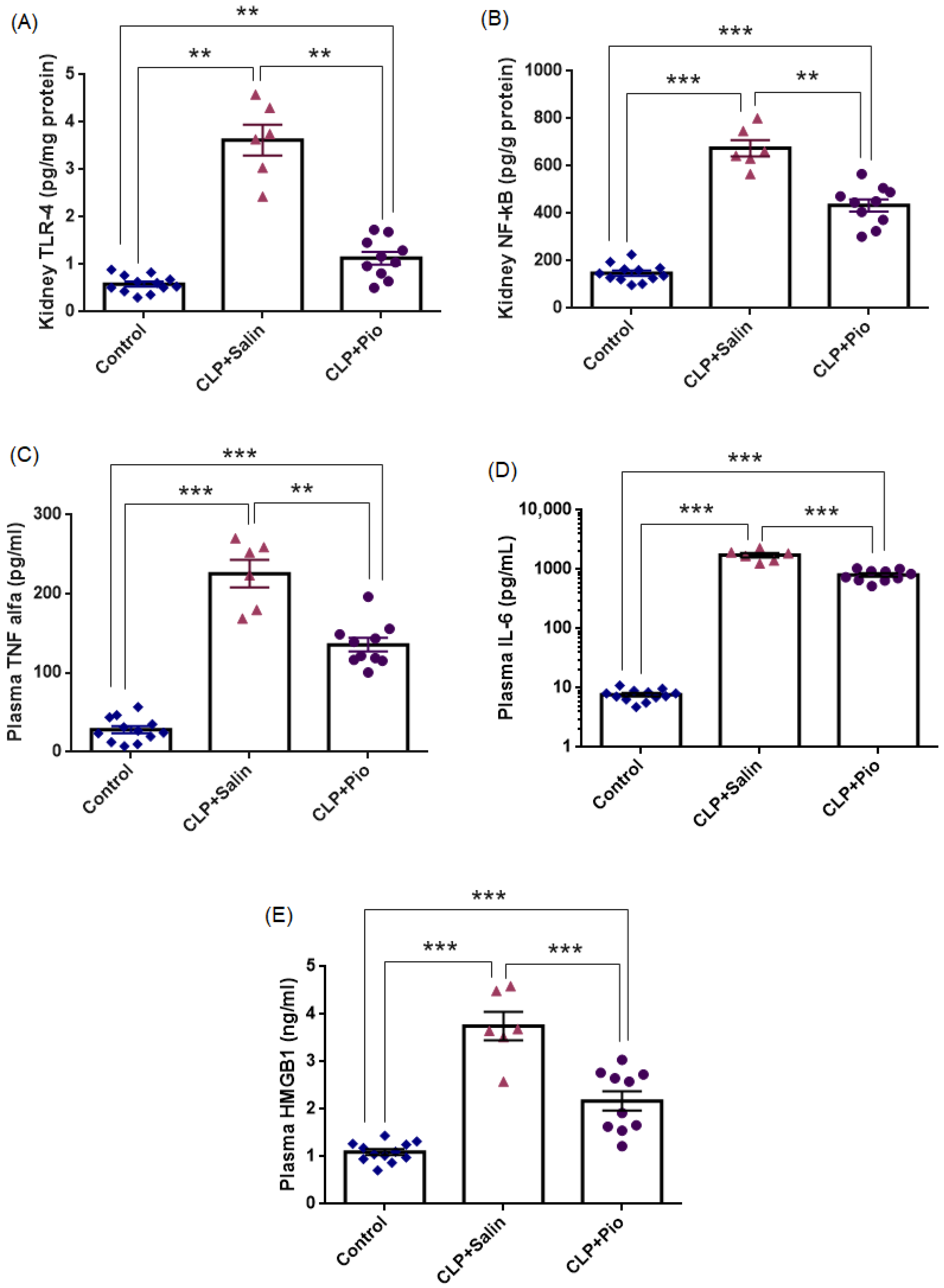
Figure 7 and Table 4 summarize the principal inflammatory findings, demonstrating marked activation of innate inflammatory responses following CLP-induced sepsis, which were substantially attenuated by pioglitazone treatment. As shown in Figure 7, the CLP + Saline group exhibited significant up-regulation of TLR-4, NF-κB, and TNF-α and a marked elevation of plasma IL-6 compared with the Control group (p < 0.01; p < 0.001; p < 0.001; and p < 0.001, respectively). Relative to baseline, TLR-4, NF-κB, and TNF-α levels increased approximately 6.2-fold, 4.6-fold, and 8.0-fold, respectively, while plasma IL-6 increased approximately 226-fold, indicating a robust activation of the TLR-4/NF-κB signaling axis and a pronounced systemic inflammatory response characteristic of sepsis.
Pioglitazone treatment markedly attenuated these elevations (all p < 0.001 vs. CLP + Saline), corresponding to reductions of 69%, 36%, and 40%, respectively. Similarly, plasma IL-6 levels were substantially reduced in the CLP + Pio group compared with the CLP + Saline group, representing an approximate 54% decrease (p < 0.001).
Despite this marked attenuation, values in the pioglitazone group remained significantly higher than those observed in the Control group (p < 0.01; p < 0.001; p < 0.001; and p < 0.001, respectively), indicating partial yet biologically meaningful suppression of inflammation. Collectively, these findings demonstrate that pioglitazone downregulated renal TLR-4 and NF-κB expression and reduced circulating TNF-α and IL-6 levels, thereby mitigating the excessive inflammatory and cytokine responses characteristic of sepsis.
As shown in Figure 7, the CLP + Saline group exhibited significantly higher HMGB1 levels than the control group (p < 0.001). When the mean control value was taken as the baseline, the CLP + Saline group showed an approximately 3.43-fold increase in plasma HMGB1 concentrations. Pioglitazone treatment markedly attenuated this elevation (p < 0.001 vs. CLP + Saline), corresponding to a 42.0% reduction relative to untreated sepsis. Nevertheless, HMGB1 levels remained significantly higher than the control values (p = 0.002), indicating partial but meaningful suppression of systemic inflammatory activation. Collectively, these findings suggest that pioglitazone substantially mitigated sepsis-associated inflammatory signaling reflected by circulating HMGB1 levels.

4. Discussion

In the present study, we demonstrated that pioglitazone, a PPAR-γ agonist, significantly alleviates sepsis-induced acute kidney injury (SA-AKI) by suppressing TLR-4/NF-κB–mediated inflammation and oxidative stress, thereby improving renal structure, function, and survival. Within this framework, activation of PPARγ may represent a promising therapeutic strategy for mitigating sepsis-associated renal injury.
Sepsis was induced via cecal ligation and puncture (CLP), a well-established model that replicates the hemodynamic, immunological, and microbial complexity of human polymicrobial sepsis [7]. Recent methodological analyses note that CLP severity and organ injury are highly sensitive to surgical parameters and supportive care conditions, highlighting the importance of standardized protocols for reproducibility [27]. Moreover, graded CLP paradigms with longitudinal phenotyping have been proposed to better capture clinically relevant heterogeneity ranging from moderate illness to fulminant septic shock [11]. In this model, the leakage of fecal material into the peritoneal cavity produces polymicrobial peritonitis, which precipitates a systemic inflammatory response and subsequent multiorgan dysfunction. Model validity was confirmed by marked increases in plasma BUN, creatinine, and NGAL levels, accompanied by the classic histopathological features of acute tubular necrosis—tubular epithelial necrosis, luminal debris, tubular dilatation, interstitial hemorrhage, and inflammatory infiltration—consistent with prior CLP-based sepsis studies [28,29,30,31].
In the present study, pioglitazone treatment markedly attenuated renal dysfunction, as evidenced by normalization of BUN and creatinine levels and a significant reduction in NGAL, a sensitive biomarker that increases earlier than creatinine during early tubular injury [32,33,34]. Contemporary SA-AKI frameworks emphasize that creatinine-based functional criteria may lag behind early tubular stress, and therefore recommend the combined interpretation of functional parameters with damage biomarkers such as NGAL [14]. However, NGAL levels may also rise in sepsis in the absence of overt AKI due to systemic inflammatory activation, highlighting the importance of interpreting NGAL in conjunction with creatinine dynamics and the broader inflammatory context [32,35]. Accordingly, the recent literature increasingly supports multi-biomarker approaches rather than reliance on a single marker for a more accurate characterization of kidney injury [36]. In this context, the reduction in NGAL observed in our study, together with the improvement in BUN and creatinine levels and the marked attenuation of histopathological features such as tubular injury and inflammatory infiltration, suggests that pioglitazone exerts both functional and structural protective effects against sepsis-associated renal injury.
This biochemical recovery was paralleled by a survival benefit: mortality decreased from 50% in untreated septic rats to 16.7% with pioglitazone administration. These findings align with previous reports showing that PPAR-γ activation improves survival and reduces systemic inflammation during sepsis. For instance, pioglitazone pretreatment was shown to lower IL-6 and MCP-1 expression while increasing 7-day survival [37], whereas prolonged administration decreased TNF-α and IL-1β levels and improved outcomes in severe sepsis [38]. Together with our results, these studies reinforce the notion that pioglitazone mitigates the systemic inflammatory injury that drives SA-AKI-related mortality.
CLP-induced sepsis markedly increased renal TLR-4 and NF-κB expression together with elevated TNF-α and IL-6, indicating activation of innate immune signaling in the kidney. Consistent with CLP-based sepsis models, robust activation of the TLR-4/NF-κB inflammatory axis during sepsis-associated AKI has been widely reported [39,40,41,42]. In addition to classical cytokines, the damage-associated molecular pattern HMGB1 has been implicated in the regulation of inflammatory signaling during sepsis. HMGB1 released during tissue injury activates TLRs and RAGE receptors, amplifying NF-κB–driven inflammatory signaling and contributing to renal injury in SA-AKI [43,44]. Consistent with this mechanism, circulating IL-6, TNF-α and HMGB1 levels were elevated in the CLP group, reflecting systemic inflammatory activation.
Pioglitazone significantly reduced renal TLR-4 and NF-κB expression together with decreased TNF-α and IL-6 levels, indicating suppression of this pro-inflammatory cascade. These findings agree with previous studies showing that pioglitazone downregulates TLR-4/MyD88 signaling, inhibits NF-κB nuclear translocation, and stabilizes IκB-α, ultimately reducing systemic TNF-α and IL-6 release [21,37]. In parallel, pioglitazone also reduced circulating HMGB1 levels, suggesting attenuation of DAMP-mediated inflammatory amplification. Upregulation of adiponectin, a downstream effector of PPAR-γ, may further suppress NF-κB activity and reinforce this anti-inflammatory effect [21]. Although our measurements reflect protein levels rather than direct pathway activation, the findings are consistent with attenuation of the HMGB1/TLR4/NF-κB inflammatory axis [45].
IL-6 is an early biomarker of sepsis and correlates with disease severity in ICU cohorts [46]. In the present study, the concurrent reduction in IL-6 and HMGB1 following pioglitazone treatment further supports its systemic immunomodulatory effect. Future studies should evaluate anti-inflammatory cytokines such as IL-10 to better characterize sepsis-associated immune dysregulation and its potential links to tumor immune escape [47].
In addition to inflammation, oxidative stress plays a key role in the pathogenesis of sepsis. Excessive production of reactive oxygen species (ROS) during systemic inflammation promotes lipid peroxidation, resulting in malondialdehyde (MDA) accumulation and cellular injury [48]. Clinical observations also suggest that oxidative stress biomarkers such as MDA dynamically change during early sepsis and may reflect developing organ dysfunction [49]. Consistent with these reports, MDA levels were markedly elevated in the CLP + Saline group in the present study, indicating pronounced oxidative stress in sepsis-associated AKI. Similar increases in plasma and renal MDA have been reported in experimental CLP models [50,51,52,53].
Parallel to its anti-inflammatory effects, pioglitazone exerted a significant antioxidant action, as evidenced by reduced plasma MDA levels and partial restoration of renal Nrf2 protein suppressed by CLP. Nrf2 is a central regulator of antioxidant defense and plays a protective role in sepsis-related organ injury, including septic AKI [54]. Consistent with previous studies showing that pioglitazone reduces oxidative stress markers such as MDA and myeloperoxidase (MPO), these findings suggest attenuation of neutrophil-mediated oxidative injury [25,55].
Mechanistically, our results support a PPARγ-linked redox regulatory pathway. CLP-induced sepsis decreased renal Nrf2 protein, whereas pioglitazone partially restored Nrf2 toward control values. Nrf2 signaling interacts with inflammatory pathways through reciprocal antagonism with NF-κB, potentially explaining the concurrent reduction in inflammatory cytokines and oxidative damage observed in the present study [24]. In addition, experimental evidence indicates that PPARγ activation can enhance Nrf2 signaling in kidney injury models, supporting a mechanistic link between pioglitazone treatment and restoration of antioxidant defense [56].
Histopathological examination confirmed the biochemical findings, showing that pioglitazone markedly reduced tubular necrosis, debris accumulation, interstitial inflammation, and hemorrhage, restoring near-normal renal architecture. The parallel structural and functional recovery underscores its renoprotective efficacy. Comparable effects observed with other TLR-4/NF-κB inhibitors, such as paeonol in endotoxin-induced AKI [57], validate this pathway as a common therapeutic target in septic renal injury. By simultaneously suppressing TLR-4/NF-κB-driven inflammation and oxidative stress, pioglitazone disrupts the cytokine–ROS cycle underlying multiorgan dysfunction, preserving renal function and improving survival.
The translational relevance lies in the potential repurposing of pioglitazone for sepsis—an area that still urgently requires effective adjunctive therapies. As an U.S. Food and Drug Administration approved antidiabetic drug with a well-characterized clinical safety profile, pioglitazone represents a practical repurposing candidate. Epidemiological data indicate lower sepsis-related mortality among chronic users (OR = 0.80; ref. [58]), and phase I studies have confirmed feasible administration in critically ill patients [59]. Recent immunotherapy frameworks further emphasize that successful sepsis interventions will likely require phenotype-guided and carefully timed immunomodulation rather than uniform anti-inflammatory strategies [60]. Importantly, sepsis is a major complication in immunocompromised oncology populations—particularly after cytotoxic chemotherapy where febrile neutropenia/neutropenic sepsis remains associated with substantial ICU mortality and distinct risk profiles across cancer subtypes [61]. In addition, septic AKI is clinically relevant in cancer patients with sepsis and is linked to shock severity and organ-failure burden, strengthening the need for renoprotective adjuncts that can be deployed without compromising antimicrobial priorities [62]. Beyond infection-driven organ failure, chemotherapy-induced AKI remains a key limitation to treatment intensity in cancer care [63], and pioglitazone’s anti-inflammatory/antioxidant mechanisms (TLR4/NF κB suppression and restoration of cytoprotective redox programs) may therefore be mechanistically relevant to kidney vulnerability in oncology. At the same time, immunometabolic targeting of PPARγ is actively explored in cancer biology and therapy [64]; notably, pioglitazone has been reported to reduce tumor PD L1 protein and to enhance anti PD 1 efficacy in preclinical colorectal cancer models [65], whereas tumor-cell PPARγ signaling has also been implicated in immune evasion and resistance to immune-checkpoint blockade in other contexts, highlighting that the direction and timing of PPARγ modulation may be tumor- and setting-dependent [66]. Finally, potential oncologic safety signals should be acknowledged: evidence regarding pioglitazone and bladder cancer remains mixed and appears most relevant to long-term exposure, so short-course use as an acute sepsis adjunct would be expected to entail a different risk profile but warrants caution in patients with prior urothelial malignancy or unexplained hematuria [67,68].
Long-standing male bias in preclinical research has resulted in substantial sex underrepresentation, with male-only designs remaining common across biomedical disciplines [69,70]. Quantitative analyses further show that inclusion of females does not inherently increase experimental variability, even without strict estrous-cycle control [71,72]. Sex differences in sepsis and acute kidney injury (AKI) appear model-dependent. While sex effects are evident in some inflammatory injury settings, CLP-based polymicrobial sepsis models often show limited differences in early survival or organ injury [73], and clinical studies similarly report comparable SA-AKI incidence between men and women after adjustment [74,75]. Accordingly, the use of female rats in this study served as an initial proof-of-concept evaluation in an underrepresented sex, although confirmation in both sexes is warranted. Estrous-cycle stage was not monitored. Because estrogen can influence inflammatory signaling, including NF-κB-related pathways, hormonal variation may affect responses and should be addressed in future studies [76]. In addition, sex-related differences in immune responses and drug metabolism—including variation in CYP450 activity relevant to anticancer therapies—may influence translational outcomes in oncology populations [77,78,79].
To ensure adequate PPARγ target engagement in polymicrobial sepsis, we selected pioglitazone at 10 mg/kg/day based on prior CLP studies showing that post-treatment PPARγ activation attenuates NF-κB signaling and systemic inflammation [21], as well as oncology-relevant models demonstrating reduced NF-κB activation and oxidative stress at the same dose [80]. The first dose was administered 1 h after CLP induction to model a clinically relevant post-treatment scenario reflecting early therapeutic intervention after the onset of sepsis. Early bacteremia and endotoxemia have been reported within the first hours following CLP, supporting early post-treatment designs in experimental sepsis studies [81,82].
We administered pioglitazone for a short course (5 days), consistent with a phase-1 sepsis pharmacokinetic and safety study [59], which may limit duration-dependent adverse effects such as fluid retention. Because PPARγ agonists can promote fluid retention and hemodynamic stress at higher exposures, careful dose optimization will be important for future translational applications, particularly in oncology populations. Importantly, epidemiological concerns regarding pioglitazone and bladder cancer have primarily been reported in the context of long-term cumulative exposure [83,84]. In contrast, short-course administration in acute inflammatory conditions such as sepsis is unlikely to involve comparable exposure profiles.

Limitation

In this study, a single dose derived from prior efficacy studies was employed rather than a full dose–response design. Although this approach demonstrated consistent effects on survival, renal biomarkers, and inflammatory and oxidative endpoints in our CLP-SA-AKI post-treatment model, it does not allow identification of the minimal effective dose. Therefore, future studies should evaluate lower doses, characterize pharmacokinetic profiles in septic versus non-septic hosts, and monitor potential adverse effects such as fluid retention, which may be clinically relevant for oncology populations [85,86,87].
We did not assess emerging renal stress biomarkers such as urinary [TIMP-2]·[IGFBP7] or circulating cell-free DNA, which have been highlighted in recent AKI and sepsis-AKI biomarker literature; incorporating these panels may strengthen oncology-relevant phenotyping in future studies [88,89,90]. In addition, downstream metabolic targets of PPAR-γ activation, such as CD36 and GLUT4, were not evaluated in the present study, and their assessment may further clarify on-target pharmacological effects of pioglitazone in septic AKI.

5. Conclusions

In this study, findings establish that pioglitazone confers potent renoprotective, anti-inflammatory, and antioxidant effects in sepsis-associated AKI. By downregulating TLR-4/NF-κB signaling and mitigating oxidative stress, the compound interrupts the cytokine–redox cycle that drives multiorgan failure, leading to improved renal structure, function, and survival.

Author Contributions

Concept—N.A.H.; Design—N.A.H. and O.E.; Data Collection or Processing—B.A., A.A., H.A., G.E. and O.E.; Analysis or Interpretation—B.A., H.A., A.A., G.E., A.S., N.A.H. and O.E.; Literature Search—B.A., H.A., G.E., N.A.H., A.S., A.A. and O.E.; Written by N.A.H. and H.A., Final approval—B.A., A.S., A.A., N.A.H., G.E., H.A. and O.E. All authors have read and agreed to the published version of the manuscript.

Funding

The authors declared that this study has received no financial support.

Institutional Review Board Statement

All experimental procedures conformed to the ethical standards outlined in the Guide for the Care and Use of Laboratory Animals (National Institutes of Health [NIH], USA). The study protocol received prior approval from the Animal Ethics Committee of Science University (Number: 2125095711, 28 May 2023).

Data Availability Statement

The data that support the findings of this study are not publicly available due to ethical reasons but are available from the corresponding author upon request.

Conflicts of Interest

The authors declare that there are no conflicts of interest.

References

  1. Fleischmann, C.; Scherag, A.; Adhikari, N.K.J.; Hartog, C.S.; Tsaganos, T.; Schlattmann, P.; Angus, D.C.; Reinhart, K.; International Forum of Acute Care Trialists. Assessment of global incidence and mortality of hospital-treated sepsis: Current estimates and limitations. Am. J. Respir. Crit. Care Med. 2016, 193, 259–272. [Google Scholar] [CrossRef] [PubMed]
  2. Rudd, K.E.; Johnson, S.C.; Agesa, K.M.; Shackelford, K.A.; Tsoi, D.; Kievlan, D.R.; Colombara, D.V.; Ikuta, K.S.; Kissoon, N.; Finfer, S.; et al. Global, regional, and national sepsis incidence and mortality, 1990–2017: Analysis for the Global Burden of Disease Study. Lancet 2020, 395, 200–211. [Google Scholar] [CrossRef] [PubMed]
  3. Uchino, S.; Kellum, J.A.; Bellomo, R.; Doig, G.S.; Morimatsu, H.; Morgera, S.; Schetz, M.; Tan, I.; Bouman, C.; Macedo, E.; et al. Acute renal failure in critically ill patients: A multinational, multicenter study. JAMA 2005, 294, 813–818. [Google Scholar] [CrossRef]
  4. The Cooperative Antimicrobial Therapy of Septic Shock (CATSS) Database Research Group; Bagshaw, S.M.; Lapinsky, S.; Dial, S.; Arabi, Y.; Dodek, P.; Wood, G.; Ellis, P.; Guzman, J.; Marshall, J.; et al. Acute kidney injury in septic shock: Clinical outcomes and impact of duration of hypotension prior to initiation of antimicrobial therapy. Intensive Care Med. 2009, 35, 871–881. [Google Scholar] [CrossRef]
  5. Zarjou, A.; Agarwal, A. Sepsis and acute kidney injury. J. Am. Soc. Nephrol. 2011, 22, 999–1006. [Google Scholar] [CrossRef]
  6. Evans, L.; Rhodes, A.; Alhazzani, W.; Antonelli, M.; Coopersmith, C.M.; French, C.; Machado, F.R.; Mcintyre, L.; Ostermann, M.; Prescott, H.C.; et al. Surviving sepsis campaign: International guidelines for management of sepsis and septic shock 2021. Crit. Care Med. 2021, 49, e1063–e1143. [Google Scholar] [PubMed]
  7. Dejager, L.; Pinheiro, I.; Dejonckheere, E.; Libert, C. Cecal ligation and puncture: The gold standard model for polymicrobial sepsis? Trends Microbiol. 2011, 19, 198–208. [Google Scholar] [CrossRef]
  8. Li, J.L.; Li, G.; Jing, X.Z.; Li, Y.F.; Ye, Q.Y.; Jia, H.H.; Liu, S.H.; Li, X.J.; Li, H.; Huang, R.; et al. Assessment of clinical sepsis-associated biomarkers in a septic mouse model. J. Int. Med. Res. 2018, 46, 2410–2422. [Google Scholar]
  9. Cai, L.; Rodgers, E.; Schoenmann, N.; Raju, R.P. Advances in rodent experimental models of sepsis. Int. J. Mol. Sci. 2023, 24, 9578. [Google Scholar] [CrossRef]
  10. Kannan, S.K.; Kim, C.Y.; Heidarian, M.; Berton, R.R.; Jensen, I.J.; Griffith, T.S.; Badovinac, V.P. Mouse Models of Sepsis. Curr. Protoc. 2024, 4, e997. [Google Scholar]
  11. Ramsay, S.D.; Kilgariff, D.E.; Young, B.J.; Plummer, M.P.; Nenke, M.A.; Meyer, E.J.; Torpy, D.J.; Young, R.L. Precision Profiling of Disease Progression in Murine Models of Sepsis and Septic Shock. Int. J. Mol. Sci. 2025, 26, 9954. [Google Scholar] [CrossRef]
  12. Fani, F.; Regolisti, G.; Delsante, M.; Cantaluppi, V.; Castellano, G.; Gesualdo, L.; Villa, G.; Fiaccadori, E. Recent advances in the pathogenetic mechanisms of sepsis-associated acute kidney injury. J. Nephrol. 2018, 31, 351–359. [Google Scholar] [CrossRef] [PubMed]
  13. Gómez, H.; Kellum, J.A.; Ronco, C. Metabolic reprogramming and tolerance during sepsis-induced AKI. Nat. Rev. Nephrol. 2017, 13, 143–151. [Google Scholar] [CrossRef] [PubMed]
  14. Zarbock, A.; Nadim, M.K.; Pickkers, P.; Gomez, H.; Bell, S.; Joannidis, M.; Kashani, K.; Koyner, J.L.; Pannu, N.; Meersch, M.; et al. Sepsis-associated acute kidney injury: Consensus report of the 28th Acute Disease Quality Initiative workgroup. Nat. Rev. Nephrol. 2023, 19, 401–417. [Google Scholar] [CrossRef] [PubMed]
  15. Gong, S.; Xiong, H.; Lei, Y.; Huang, S.; Ouyang, Y.; Cao, C.; Wang, Y. Usp9x contributes to the development of sepsis-induced acute kidney injury by promoting inflammation and apoptosis in renal tubular epithelial cells via activation of the TLR4/nf-κb pathway. Ren. Fail. 2024, 46, 2361089. [Google Scholar]
  16. Yue, L.; Liu, X.; Wu, C.; Lai, J.; Wang, J.; Zhong, H.; Chen, F. Toll-like receptor 4 promotes the inflammatory response in septic acute kidney injury by promoting p38 mitogen-activated protein kinase phosphorylation. J. Bioenerg. Biomembr. 2023, 55, 353–363. [Google Scholar] [CrossRef]
  17. Zhang, Y.; Liu, T.; Zuo, Z. Fortunellin attenuates sepsis-induced acute kidney injury by inhibiting inflammation and ferroptosis via the TLR4/NF-κB pathway. Histol. Histopathol. 2024, 40, 1083–1093. [Google Scholar] [CrossRef]
  18. Wang, H.-W.; Wu, M.-M.; Zhu, M.-M.; Qin, Y.-Y.; Wang, K.-Q.; Wu, C.-Y.; Zhang, R.-R.; Wang, Y.; Zhou, C.; Luo, S.; et al. Growth differentiation factor 11 attenuates sepsis-associated acute kidney injury by reducing inflammation and coagulation via PGC-1α/Nrf2 activation. Cell. Mol. Biol. Lett. 2025, 30, 102. [Google Scholar] [CrossRef]
  19. Kounatidis, D.; Tzivaki, I.; Daskalopoulou, S.; Daskou, A.; Adamou, A.; Rigatou, A.; Sdogkos, E.; Karampela, I.; Dalamaga, M.; Vallianou, N.G. Sepsis-Associated Acute Kidney Injury: What’s New Regarding Its Diagnostics and Therapeutics? Diagnostics 2024, 14, 2845. [Google Scholar] [CrossRef] [PubMed] [PubMed Central]
  20. He, F.F.; Wang, Y.M.; Chen, Y.Y.; Huang, W.; Li, Z.Q.; Zhang, C. Sepsis-induced AKI: From pathogenesis to therapeutic approaches. Front. Pharmacol. 2022, 13, 981578. [Google Scholar] [CrossRef]
  21. Kaplan, J.; Nowell, M.; Chima, R.; Zingarelli, B. Pioglitazone reduces inflammation through inhibition of NF-kappaB in polymicrobial sepsis. Innate Immun. 2014, 20, 519–528. [Google Scholar] [CrossRef] [PubMed]
  22. Cai, W.; Yang, T.; Liu, H.; Han, L.; Zhang, K.; Hu, X.; Zhang, X.; Yin, K.-J.; Gao, Y.; Bennett, M.V.; et al. Peroxisome proliferator-activated receptor γ (PPARγ): A master gatekeeper in CNS injury and repair. Prog. Neurobiol. 2018, 163, 27–58. [Google Scholar] [CrossRef] [PubMed]
  23. Dana, N.; Vaseghi, G.; Haghjooy Javanmard, S. Activation of PPARgamma Inhibits TLR4 Signal Transduction Pathway in Melanoma Cancer in Vitro. Adv. Pharm. Bull. 2020, 10, 458–463. [Google Scholar] [CrossRef] [PubMed]
  24. Gao, W.; Guo, L.; Yang, Y.; Wang, Y.; Xia, S.; Gong, H.; Zhang, B.K.; Yan, M. Dissecting the Crosstalk Between Nrf2 and NF-κB Response Pathways in Drug-Induced Toxicity. Front. Cell Dev. Biol. 2022, 9, 809952. [Google Scholar] [CrossRef] [PubMed] [PubMed Central]
  25. Jafari-Khataylou, Y.; Kazemi-Darabadi, S.; Afshar, S.A. Effects of adenosine N1-Oxide and pioglitazone on in-flammatory and antioxidant state in sepsis caused by experimental cecal puncture in rat. Vet. Res. Forum. 2023, 14, 381–387. [Google Scholar] [CrossRef]
  26. Erbas, O.; Taskiran, D. Sepsis-induced changes in behavioral stereotypy in rats; involvement of tumor necrosis factor-alpha, oxidative stress, and dopamine turnover. J. Surg. Res. 2014, 186, 262–268. [Google Scholar] [CrossRef]
  27. Dong, L.; Xie, Y.L.; Zhang, R.T.; Hu, Q.Y. Models of sepsis-induced acute kidney injury. Life Sci. 2024, 352, 122873. [Google Scholar] [CrossRef]
  28. Sun, S.; Wang, J.; Wang, J.; Wang, F.; Yao, S.; Xia, H. Maresin 1 mitigates sepsis-associated acute kidney injury in mice via inhibition of the NF-κB/STAT3/MAPK pathways. Front. Pharmacol. 2019, 10, 1323. [Google Scholar] [CrossRef]
  29. Yubolphan, R.; Kobroob, A.; Kongkaew, A.; Chiranthanut, N.; Jinadang, N.; Wongmekiat, O. Berberine mitigates Sepsis-Associated acute kidney injury in aged rats by preserving mitochondrial integrity and inhibiting TLR4/NF-κB and NLRP3 inflammasome activations. Antioxidants 2024, 13, 1398. [Google Scholar] [CrossRef]
  30. Nakamura, I.; Umehara, M.; Yagi-Tomita, A.; Yamamoto, S.; Sawai, S.; Nakamura, M.; Minamida, A.; Yamauchi-Sawada, H.; Sunahara, Y.; Matoba, Y.; et al. Toll like receptor 2 mediated exacerbation of sepsis associated acute kidney injury by renal congestion in mice. Sci. Rep. 2025, 15, 22081. [Google Scholar] [CrossRef]
  31. Çakır, M.; Tarakçı, B.; Aydın, A.; Bircan, B.; Fırat, S.; Şekerci, G. JAK1/JAK2 inhibitor baricitinib ameliorates sepsis-induced acute kidney injury in rats. Eur. J. Pharmacol. 2025, 1002, 177770. [Google Scholar] [PubMed]
  32. Ferreira, G.S.; Frota, M.L.; Gonzaga, M.J.D.; Vattimo, M.D.F.F.; Lima, C. The role of biomarkers in diagnosis of sepsis and acute kidney injury. Biomedicines 2024, 12, 931. [Google Scholar] [CrossRef] [PubMed]
  33. Zhou, H.; Hu, X.; Yu, S.; Chen, Y.; He, W. Diagnostic value of urinary neutrophil gelatinase-associated lipocalin for acute kidney injury in patients with suspected sepsis: A meta-analysis. BMC Nephrol. 2021, 22, 19. [Google Scholar]
  34. Albert, C.; Haase, M.; Albert, A.; Zapf, A.; Braun-Dullaeus, R.; Haase-Fielitz, A. Biomarker-guided risk assessment for acute kidney injury: Time for clinical implementation? Ann. Lab. Med. 2020, 40, 453–464. [Google Scholar] [CrossRef]
  35. Paul, A.; Newbigging, N.S.; Lenin, A.; Gowri, M.; Varghese, J.S.; Nell, A.J.; Abhilash, K.P.P.; Binu, A.J.; Chandiraseharan, V.K.; Iyyadurai, R.; et al. Role of Neutrophil Gelatinase-associated Lipocalin (NGAL) and Other Clinical Parameters as Predictors of Bacterial Sepsis in Patients Presenting to the Emergency Department with Fever. Indian J. Crit. Care Med. 2023, 27, 176–182. [Google Scholar] [CrossRef] [PubMed] [PubMed Central]
  36. Baeseman, L.; Gunning, S.; Koyner, J.L. Biomarker Enrichment in Sepsis-Associated Acute Kidney Injury: Finding High-Risk Patients in the Intensive Care Unit. Am. J. Nephrol. 2024, 55, 72–85. [Google Scholar] [CrossRef] [PubMed] [PubMed Central]
  37. Tsujimura, Y.; Matsutani, T.; Matsuda, A.; Kutsukake, M.; Uchida, E.; Sasajima, K.; Tamura, K. Effects of pioglitazone on survival and omental adipocyte function in mice with sepsis induced by cecal ligation and puncture. J. Surg. Res. 2011, 171, e215–e221. [Google Scholar] [CrossRef]
  38. Shafaroodi, H.; Hassanipour, M.; Mousavi, Z.; Rahimi, N.; Dehpour, A.R. The effects of sub-chronic treatment with pioglitazone on the septic mice mortality in the model of cecal ligation and puncture: Involvement of nitric oxide pathway. Acta Medica Iran. 2015, 53, 608–616. [Google Scholar]
  39. Xia, Y.M.; Guan, Y.Q.; Liang, J.F.; Wu, W.D. TAK-242 improves sepsis-associated acute kidney injury in rats by inhibiting the TLR4/NF-κB signaling pathway. Ren. Fail. 2024, 46, 2313176. [Google Scholar]
  40. Abdelnaser, M.; Attya, M.E.; El-Rehany, M.A.; Fathy, M. Clemastine mitigates sepsis-induced acute kidney injury in rats; the role of α-Klotho/TLR-4/MYD-88/NF-κB/Caspase-3/p-P38 MAPK signaling pathways. Arch. Biochem. Biophys. 2025, 763, 110229. [Google Scholar]
  41. Shen, Y.; Shi, Q.; Dai, T.; Cai, Y.; Sheng, M.; Qiu, Y.; Yang, M.; Hu, S.; Cheng, B. Spermidine alleviates sepsis-induced acute kidney injury by suppressing apoptosis and inflammation through modulation of TLR4-mediated signaling. J. Toxicol. Sci. 2025, 50, 555–567. [Google Scholar] [PubMed]
  42. Liu, B.; Wang, J.; Duan, Y.; Hussain, S.A.; Tripathi, A.S.; Li, K. Ononin promotes survival rate in cecal ligation puncture-induced sepsis rat model by regulating inflammatory pathway. Acta Cir. Bras. 2025, 40, e407825. [Google Scholar] [CrossRef] [PubMed] [PubMed Central]
  43. Li, X.; Jing, G.; Guo, Z.; Guo, Z. High-mobility group box 1 in acute kidney injury. Front. Pharmacol. 2025, 16, 1618971. [Google Scholar] [CrossRef] [PubMed]
  44. Wei, S.; Dai, Z.; Wu, L.; Xiang, Z.; Yang, X.; Jiang, L.; Du, Z. Lactate-induced macrophage HMGB1 lactylation promotes neutrophil extracellular trap formation in sepsis-associated acute kidney injury. Cell Biol. Toxicol. 2025, 41, 78. [Google Scholar] [CrossRef] [PubMed] [PubMed Central]
  45. Zhang, J.; Chen, Q.; Dai, Z.; Pan, H. miR-22 alleviates sepsis-induced acute kidney injury via targeting the HMGB1/TLR4/NF-κB signaling pathway. Int. Urol. Nephrol. 2023, 55, 409–421. [Google Scholar] [CrossRef]
  46. Gamarra-Morales, Y.; Molina-López, J.; Santiago-Ruiz, F.C.; Herrera-Quintana, L.; Vázquez-Lorente, H.; Gascón-Luna, F.; Planells, E. Efficiency of IL-6 in early prognosis and follow-up in critically ill patients with septic shock. Diseases 2024, 12, 298. [Google Scholar] [CrossRef]
  47. Gao, X.; Cai, S.; Li, X.; Wu, G. Sepsis-induced immunosuppression: Mechanisms, biomarkers and immunotherapy. Front. Immunol. 2025, 16, 1577105. [Google Scholar] [CrossRef]
  48. Murphy, M.P. How mitochondria produce reactive oxygen species. Biochem. J. 2009, 417, 1–13. [Google Scholar] [CrossRef]
  49. Helan, M.; Malaska, J.; Tomandl, J.; Jarkovsky, J.; Helanova, K.; Benesova, K.; Sitina, M.; Dastych, M.; Ondrus, T.; Goldbergova, M.P.; et al. Kinetics of Biomarkers of Oxidative Stress in Septic Shock: A Pilot Study. Antioxidants 2022, 11, 640. [Google Scholar] [CrossRef]
  50. Rodrigues, F.A.D.P.; Santos, A.D.D.C.; de Medeiros, P.H.Q.S.; Prata, M.D.M.G.; Santos, T.C.D.S.; da Silva, J.A.; Brito, G.A.D.C.; Dos Santos, A.A.; Silveira, E.R.; Lima, A.Â.M. Gingerol suppresses sepsis-induced acute kidney injury by modulating methylsulfonylmethane and dimethylamine production. Sci. Rep. 2018, 8, 12154. [Google Scholar] [CrossRef]
  51. Anter, A.; Ahmed, A.S.F.; Hammad, A.S.; Almalki, W.H.; Abdel Hafez, S.M.N.; Kasem, A.W.; El-Moselhy, M.A.; Alrabia, M.W.; Ibrahim, A.R.N.; El-Daly, M. The severity of acute kidney and lung injuries induced by cecal ligation and puncture is attenuated by menthol: Role of proliferating cell nuclear antigen and apoptotic markers. Front. Med. 2022, 9, 904286. [Google Scholar] [PubMed]
  52. Qiongyue, Z.; Xin, Y.; Meng, P.; Sulin, M.; Yanlin, W.; Xinyi, L.; Xuemin, S. Post-treatment with irisin attenuates acute kidney injury in sepsis mice through anti-ferroptosis via the SIRT1/Nrf2 pathway. Front. Pharmacol. 2022, 13, 857067. [Google Scholar]
  53. Li, X.; Zhou, W.; Chen, J.; Zhou, L.; Li, Y.; Wu, X.; Peng, X. Circ_001653 alleviates sepsis associated-acute kidney injury by recruiting BUD13 to regulate KEAP1/NRF2/HO-1 signaling pathway. J. Inflamm. 2024, 21, 37. [Google Scholar]
  54. Liu, H.; Wang, L.; Zhou, J. Nrf2 and its signaling pathways in sepsis and its complications: A comprehensive review of research progress. Medicine 2025, 104, e42132. [Google Scholar] [CrossRef] [PubMed] [PubMed Central]
  55. Gao, M.; Jiang, Y.; Xiao, X.; Peng, Y.; Xiao, X.; Yang, M. Protective effect of pioglitazone on sepsis-induced intestinal injury in a rodent model. J. Surg. Res. 2015, 195, 550–558. [Google Scholar] [CrossRef]
  56. Alharbi, M.; Mahmoud, M.A.; Alshammari, A.; Almutairi, M.M.; Al-Ghamdi, J.M.; Alsabhan, J.F.; Shabanah, O.A.; Alshalawi, N.A.; Alzarea, S.I.; Alasmari, A.F. The ameliorative effect of pioglitazone against colistin-induced nephrotoxicity is mediated by inhibition of NF-κB and restoration of Nrf2 signaling: An integrative bioinformatics prediction-guided in vitro study. PLoS ONE 2024, 19, e0314092. [Google Scholar] [CrossRef] [PubMed] [PubMed Central]
  57. Fan, H.Y.; Qi, D.; Yu, C.; Zhao, F.; Liu, T.; Zhang, Z.K.; Yang, M.-Y.; Zhang, L.-M.; Chen, D.-Q.; Du, Y. Paeonol protects endotoxin-induced acute kidney injury: Potential mechanism of inhibiting TLR4-NF-κB signal pathway. Oncotarget 2016, 7, 39497. [Google Scholar]
  58. Hsieh, M.S.; Hu, S.Y.; Liao, S.H.; Chang, C.M.; Hsieh, V.C.R.; How, C.K. Type 2 Diabetic Sepsis Patients Have a Lower Mortality Rate in Pioglitazone Use: A Nationwide 15-Year Propensity Score Matching Observational Study in Taiwan. Emerg. Med. Int. 2021, 2021, 4916777. [Google Scholar] [CrossRef]
  59. Kaplan, J.M.; Zingarelli, B.; Krallman, K.; Girdwood, S.T.; Lagory, D.; Mizuno, T.; Fei, L.; Wong, H.R.; Vinks, A.A. Phase 1 safety and pharmacokinetic study on the use of pioglitazone in critically ill patients with sepsis. Intensive Care Med. 2018, 44, 2006–2008. [Google Scholar] [CrossRef]
  60. Slim, M.A.; van Mourik, N.; Bakkerus, L.; Fuller, K.; Acharya, L.; Giannidis, T.; Dionne, J.C.; Oczkowski, S.J.W.; Netea, M.G.; Pickkers, P.; et al. Towards personalized medicine: A scoping review of immunotherapy in sepsis. Crit. Care 2024, 28, 183. [Google Scholar] [CrossRef]
  61. MacPhail, A.; Dendle, C.; Slavin, M.; Weinkove, R.; Bailey, M.; Pilcher, D.; McQuilten, Z. Neutropenic sepsis in the intensive care unit: Differences in clinical profile and outcomes according to the cause of neutropenia. In Open Forum Infectious Diseases; Oxford University Press: New York, NY, USA, 2024; Volume 11, p. ofae289. [Google Scholar]
  62. Yang, Y.; Dong, J.; Chen, X.; Chen, R.; Wang, H. Incidence, risk factors and clinical outcomes of septic acute renal injury in cancer patients with sepsis admitted to the ICU: A retrospective study. Front. Med. 2022, 9, 1015735. [Google Scholar] [CrossRef] [PubMed]
  63. Lyrio, R.M.D.C.; Rocha, B.R.A.; Corrêa, A.L.R.M.; Mascarenhas, M.G.S.; Santos, F.L.; Maia, R.D.H.; Segundo, L.B.; de Almeida, P.A.A.; Moreira, C.M.O.; Sassi, R.H. Chemotherapy-induced acute kidney injury: Epidemiology, pathophysiology, and therapeutic approaches. Front. Nephrol. 2024, 4, 1436896. [Google Scholar] [CrossRef] [PubMed]
  64. Vasileiou, M.; Diamantoudis, S.C.; Tsianava, C.; Nguyen, N.P. Therapeutic potential and challenges of pioglitazone in cancer treatment. Appl. Sci. 2025, 15, 1925. [Google Scholar] [CrossRef]
  65. Jia, X.; Qian, J.; Chen, H.; Liu, Q.; Hussain, S.; Jin, J.; Shi, J.; Hou, Y. PPARγ agonist pioglitazone enhances colorectal cancer immunotherapy by inducing PD-L1 autophagic degradation. Eur. J. Pharmacol. 2023, 950, 175749. [Google Scholar] [CrossRef]
  66. Xiong, Z.; Chan, S.L.; Zhou, J.; Vong, J.S.; Kwong, T.T.; Zeng, X.; Wu, H.; Cao, J.; Tu, Y.; Feng, Y.; et al. Targeting PPAR-gamma counteracts tumour adaptation to immune-checkpoint blockade in hepatocellular carcinoma. Gut 2023, 72, 1758–1773. [Google Scholar] [CrossRef]
  67. Baddam, S.; Banka, A.V.; Divity, S.; Sandesara, M.; Vityala, Y. Association between pioglitazone use and bladder cancer: A systematic review. Bladder 2024, 11, e21200023. [Google Scholar] [CrossRef]
  68. Malhotra, B.; Hiteshi, P.; Khalkho, P.; Malik, R.; Bhadada, S.K.; Bhansali, A.; Shafiq, N.; Malhotra, S.; Kumar, N.; Rajput, R.; et al. Bladder cancer with pioglitazone: A case-control study. Diabetes Metab. Syndr. 2022, 16, 102637. [Google Scholar] [CrossRef]
  69. Beery, A.K.; Zucker, I. Sex bias in neuroscience and biomedical research. Neurosci. Biobehav. Rev. 2011, 35, 565–572. [Google Scholar] [CrossRef]
  70. Justice, M.J. Sex matters in preclinical research. Dis. Models Mech. 2024, 17, dmm050759. [Google Scholar]
  71. Zucker, I.; Prendergast, B.J.; Beery, A.K. Pervasive neglect of sex differences in biomedical research. Cold Spring Harb. Perspect. Biol. 2022, 14, a039156. [Google Scholar] [CrossRef]
  72. Smarr, B.; Kriegsfeld, L.J. Female mice exhibit less overall variance, with a higher proportion of structured variance, than males at multiple timescales of continuous body temperature and locomotive activity records. Biol. Sex Differ. 2022, 13, 41. [Google Scholar] [CrossRef] [PubMed]
  73. Bojalil, R.; Ruíz-Hernández, A.; Villanueva-Arias, A.; Amezcua-Guerra, L.M.; Cásarez-Alvarado, S.; Hernández-Dueñas, A.M.; Rodríguez-Galicia, V.; Pavón, L.; Marquina, B.; Becerril-Villanueva, E.; et al. Two murine models of sepsis: Immunopathological differences between the sexes—Possible role of TGFβ1 in female resistance to endotoxemia. Biol. Res. 2023, 56, 54. [Google Scholar] [CrossRef] [PubMed]
  74. Peng, J.; Tang, R.; Yu, Q.; Wang, D.; Qi, D. No sex differences in the incidence, risk factors and clinical impact of acute kidney injury in critically ill patients with sepsis. Front. Immunol. 2022, 13, 895018. [Google Scholar] [CrossRef] [PubMed]
  75. Min, S.Y.; Yong, H.J.; Kim, D. Sex or gender differences in treatment outcomes of sepsis and septic shock. Acute Crit. Care 2024, 39, 207–213. [Google Scholar] [CrossRef] [PubMed] [PubMed Central]
  76. Harding, A.T.; Heaton, N.S. The Impact of Estrogens and Their Receptors on Immunity and Inflammation during Infection. Cancers 2022, 14, 909. [Google Scholar] [CrossRef] [PubMed] [PubMed Central]
  77. Guo, Y.; Lin, Z.; Zhang, W.; Chen, H.; Liu, Y.; Liu, J.; Zhang, J.; Zhang, A.; Mao, S.; Yao, X. Gender-Stratified analysis of sepsis mortality in cancer: A 45-Year Population-Based cohort study. JNCI Cancer Spectr. 2025, 10, pkaf109. [Google Scholar] [CrossRef]
  78. Wang, F.; Zhang, X.; Wang, Y.; Chen, Y.; Lu, H.; Meng, X.; Ye, X.; Chen, W. Activation/inactivation of anticancer drugs by CYP3A4: Influencing factors for personalized cancer therapy. Drug Metab. Dispos. 2023, 51, 543–559. [Google Scholar]
  79. Behal, M.; McCalla, Z.; Wang, X. Sex differences in hepatic enzymes and transporters involved in pharmacokinetics. Liver Res. 2025, 9, 253–272. [Google Scholar] [CrossRef] [PubMed] [PubMed Central]
  80. Kamel, G.A.M.; Elariny, H.A. Pioglitazone attenuates tamoxifen-induced liver damage in rats via modulating Keap1/Nrf2/HO-1 and SIRT1/Notch1 signaling pathways: In-vivo investigations, and molecular docking analysis. Mol. Biol. Rep. 2023, 50, 10219–10233. [Google Scholar] [CrossRef] [PubMed] [PubMed Central]
  81. Otero-Anton, E.; Gonzalez-Quintela, A.; Lopez-Soto, A.; Lopez-Ben, S.; Llovo, J.; Perez, L.F. Cecal ligation and puncture as a model of sepsis in the rat: Influence of the puncture size on mortality, bacteremia, endotoxemia and tumor necrosis factor alpha levels. Eur. Surg. Res. 2001, 33, 77–79. [Google Scholar] [CrossRef]
  82. Fang, H.; Gong, C.; Fu, J.; Liu, X.; Bi, H.; Cheng, Y.; Liu, Y.; Tang, Y.; Wang, D. Evaluation of 2 Rat Models for Sepsis Developed by Improved Cecal Ligation/Puncture or Feces Intraperitoneal-Injection. Med. Sci. Monit. Int. Med. J. Exp. Clin. Res. 2020, 26, e919054. [Google Scholar] [CrossRef]
  83. Tuccori, M.; Filion, K.B.; Yin, H.; Yu, O.H.; Platt, R.W.; Azoulay, L. Pioglitazone use and risk of bladder cancer: Population based cohort study. BMJ 2016, 352, i1541. [Google Scholar] [CrossRef] [PubMed] [PubMed Central]
  84. Tang, H.; Shi, W.; Fu, S.; Wang, T.; Zhai, S.; Song, Y.; Han, J. Pioglitazone and bladder cancer risk: A systematic review and meta-analysis. Cancer Med. 2018, 7, 1070–1080. [Google Scholar] [CrossRef] [PubMed] [PubMed Central]
  85. Percie du Sert, N.; Hurst, V.; Ahluwalia, A.; Alam, S.; Avey, M.T.; Baker, M.; Browne, W.J.; Clark, A.; Cuthill, I.C.; Dirnagl, U.; et al. The ARRIVE guidelines 2.0: Updated guidelines for reporting animal research. J. Cereb. Blood Flow Metab. 2020, 40, 1769–1777. [Google Scholar] [PubMed]
  86. Ewoldt, T.M.; Abdulla, A.; Hunfeld, N.; Li, L.; Smeets, T.J.; Gommers, D.; Koch, B.C.; Endeman, H. The impact of sepsis on hepatic drug metabolism in critically ill patients: A narrative review. Expert Opin. Drug Metab. Toxicol. 2022, 18, 413–421. [Google Scholar] [CrossRef] [PubMed]
  87. Morales Castro, D.; Dresser, L.; Granton, J.; Fan, E. Pharmacokinetic Alterations Associated with Critical Illness. Clin. Pharmacokinet. 2023, 62, 209–220. [Google Scholar] [CrossRef] [PubMed] [PubMed Central]
  88. Nusshag, C.; Rupp, C.; Schmitt, F.; Krautkraemer, E.; Speer, C.; Kaelble, F.; Tamulyte, S.; Bruckner, T.; Zeier, M.; Reiser, J.; et al. Cell cycle biomarkers and soluble urokinase-type plasminogen activator receptor for the prediction of sepsis-induced acute kidney injury requiring renal replacement therapy: A prospective, exploratory study. Crit. Care Med. 2019, 47, e999–e1007. [Google Scholar]
  89. Palmowski, L.; Lindau, S.; Henk, L.C.; Marko, B.; Witowski, A.; Nowak, H.; Stoll, S.E.; Zacharowski, K.; Böttiger, B.W.; Peters, J.; et al. Predictive enrichment for the need of renal replacement in sepsis-associated acute kidney injury: Combination of furosemide stress test and urinary biomarkers TIMP-2 and IGFBP-7. Ann. Intensive Care 2024, 14, 111. [Google Scholar]
  90. Licheng; Jia, H.; Jiang, Y.; Li, W. Predictive performance of two types of urinary biomarkers for renal non-recovery in sepsis-associated acute kidney injury: A prospective observational study. BMC Nephrol. 2024, 25, 153. [Google Scholar] [CrossRef]
Figure 1. Schematic representation of the experimental design and procedures. Polymicrobial sepsis was induced by cecal ligation and puncture (CLP) under anesthesia. Rats received fluid resuscitation (30 mL/kg, 0.9% NaCl, s.c.) immediately after CLP. Pioglitazone (10 mg/kg, i.p.) was administered 1 h after surgery and repeated once daily for five days. On Day 5, blood and kidney samples were collected for biochemical (BUN, creatinine, NGAL, MDA, TNF-α, TLR-4, NF-Κb, HMGB1, IL-6, NRF2) and histopathological (tubular epithelial necrosis, luminal necrotic debris, tubular dilatation, interstitial hemorrhage, inflammatory infiltration) analyses.
Jcm 15 02270 g001
Figure 2. Kaplan–Meier survival curves of experimental groups. Pioglitazone (10 mg/kg, i.p.) markedly improved 5-day survival (83.3%) compared with the CLP + saline group (50%), approaching control levels (100%).
Jcm 15 02270 g002
Figure 3. Kidney histopathology ×20 and ×40 magnification, hematoxylin and eosin (H&E) stain. (A,B): Normal group kidney, renal tubules (t); Glomerulus (G). (C,D): CLP and saline groups showed severe histopathologic alterations with tubular dilatation (td), tubular epithelial necrosis (arrow), and luminal fibrin cast (fc). (E,F): CLP and 10 mg/kg pioglitazone groups showed decreased injury with nearly normal tubules and tubular epithelial cells (t).
Jcm 15 02270 g003
Figure 4. Histopathological scoring of renal injury parameters in CLP-induced sepsis and the protective effect of pioglitazone. Data show (A) tubular epithelial necrosis, (B) luminal necrotic debris, (C) tubular dilatation, (D) hemorrhage, and (E) interstitial inflammation across groups (Control, CLP + Saline, CLP + Pioglitazone). (n = 6–10). * p < 0.05, ** p < 0.01, *** p < 0.001; ns = not significant.
Jcm 15 02270 g004
Figure 5. Plasma renal function markers in experimental groups. (A) Plasma blood urea nitrogen (BUN), (B) plasma creatinine, and (C) plasma neutrophil gelatinase-associated lipocalin (NGAL) level across groups. CLP + Saline induced marked elevations, while pioglitazone significantly reduced these increases. * p < 0.05, ** p < 0.01, *** p < 0.001.
Jcm 15 02270 g005
Figure 6. Pioglitazone attenuates sepsis-induced oxidative stress and restores antioxidant signaling. (A) Plasma MDA and (B) kidney Nrf2 levels across experimental groups (Control, CLP + Saline, CLP + Pio). Bars represent mean ± SEM with individual animal values (Control n = 12; CLP + Saline n = 6; CLP + Pio n = 10). Statistical significance was determined by one-way ANOVA followed by Tukey’s multiple comparisons test. ** p < 0.01, *** p < 0.001; ns, not significant.
Jcm 15 02270 g006
Figure 7. Pioglitazone attenuates sepsis-induced inflammatory responses. (A) Kidney TLR-4, (B) kidney NF-κB, (C) plasma TNF-α, (D) plasma IL-6, and (E) plasma HMGB1 levels across experimental groups (Control, CLP + Saline, CLP + Pio). Bars represent mean ± SEM with individual animal values (Control n = 12; CLP + Saline n = 6; CLP + Pio n = 10). Statistical significance was determined using one-way ANOVA followed by Tukey’s multiple comparisons test. For IL-6 (panel D), the y-axis is presented on a logarithmic scale to facilitate visualization of the wide range of cytokine concentrations. ** p < 0.01, *** p < 0.001.
Jcm 15 02270 g007
Table 1. Daily survival counts of experimental groups after CLP.
Baseline (n)Day 1Day 2Day 3Day 4Day 5
Control121212121212
CLP + Salin12129666
CLP + Pio121110101010
Table 2. Histopathological scoring parameters in CLP-induced sepsis and the effect of pioglitazone treatment.
ControlCLP + SalineCLP + Pio
Tubular epithelial necrosis0.0 [0.0–0.0]3.0 [2.0–3.25] a2.0 [1.0–2] b
Luminal necrotic debris0.0 [0.0–0.0]3.5 [3.0–4.0] a1.0 [0.0–1.0] c
Tubular Dilatation0.0 [0.0–0.0]2.0 [2.0–3.0] a1.0 [1.0–2.0] c
Hemorrhage0.0 [0.0–0.0]1.5 [1.0–2.0] a1.0 [0.75–1.0] b
Interstitial inflammation0.0 [0.0–0.0]1.0 [1.0–1.25] a0.5 [0.0–1.0] b
Superscript letters indicate statistical significance: a p < 0.001 vs. Control; b p < 0.05, c p < 0.01 vs. CLP + Saline.
Table 3. Renal function biomarkers across experimental groups.
Control
(n = 12)
CLP + Saline
(n = 6)
CLP + Pio
(n = 10)
Plasma BUN (mg/dL)19.1 ± 1.152.2 ± 2.5 a30.2 ± 3.1 b
Plasma Creatinine (mg/dL)0.37 ± 0.021.25 ± 0.08 a0.83 ± 0.05 b
Plasma NGAL (ng/mL)57.2 ± 5.4163.9 ± 11.7 a95.2 ± 7.5 b
Superscript letters indicate significance levels: a p < 0.001 vs. Control group; b p < 0.001 vs. CLP + Saline group.
Table 4. Oxidative stress and inflammatory markers across experimental groups.
Control
(n = 12)
CLP + Saline
(n = 6)
CLP + Pio
(n = 10)
Plasma MDA (nM)92.5 ± 8.8177.9 ± 14.6 b115.6 ± 9.8 c
NRF2 (ng/mg)3.537 ± 0.2290.942 ± 0.074 b2.206 ± 0.185 a,d
Kidney TLR-4 (pg/mg protein)0.58 ± 0.053.62 ± 0.32 a1.13 ± 0.14 a,c
Kidney NF-κB (pg/g protein) 148.2 ± 10.8675.2 ± 34.79 b433.8 ± 26.16 b,c
Plasma TNF-α (pg/mL)28.17 ± 4.4225.7 ± 17.4 b135.7 ± 8.6 b,c
Plasma IL-6 (pg/mL)7.72 ± 0.501744 ± 152.9 b804.6 ± 55.25 b,d
HMGB1 (ng/mL)1.09 ± 0.063.75 ± 0.29 b2.17 ± 0.20 b,d
Superscript letters indicate statistical significance: a p < 0.01, b p < 0.001 vs. Control; c p < 0.01, d p < 0.001 vs. CLP + Saline.
Disclaimer/Publisher’s Note: The statements, opinions and data contained in all publications are solely those of the individual author(s) and contributor(s) and not of MDPI and/or the editor(s). MDPI and/or the editor(s) disclaim responsibility for any injury to people or property resulting from any ideas, methods, instructions or products referred to in the content.