



Laser-Based Therapies in Rosacea: A Comprehensive Review of Mechanisms, Clinical Efficacy, and Future Directions
 , ,
, ,  ,
,  and
and
Abstract
1. Introduction
2. Methods
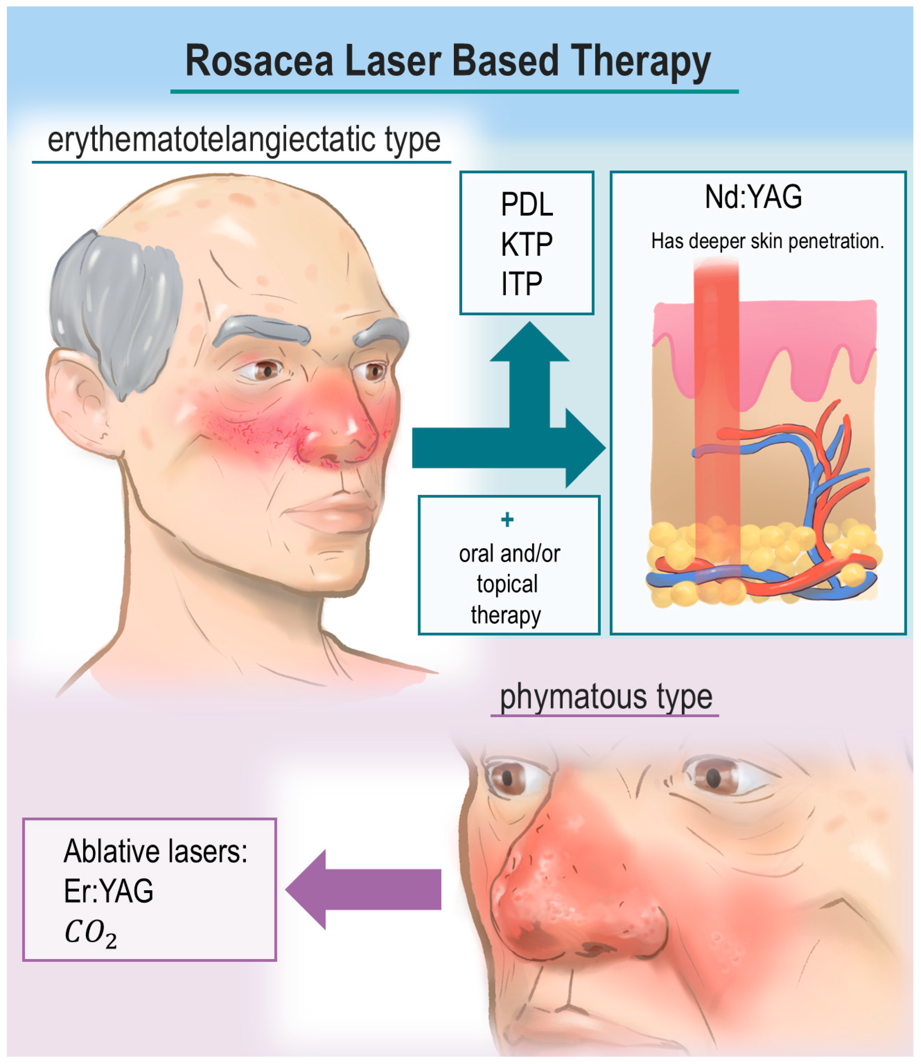
3. Lasers in the Treatment of Rosacea
3.1. PDL
3.2. KTP Laser
3.3. IPL Laser
3.4. Nd:YAG Laser
3.5. CO2 Laser
3.6. Er:YAG Laser
4. Practical Treatment Considerations
5. Combination Therapy
6. Ongoing Clinical Trials
7. Conclusions
Author Contributions
Funding
Institutional Review Board Statement
Informed Consent Statement
Data Availability Statement
Conflicts of Interest
References
- Grzech-Leśniak, Z.; Szwach, J.; Lelonkiewicz, M.; Migas, K.; Pyrkosz, J.; Szwajkowski, M.; Kosidło, P.; Pajączkowska, M.; Wiench, R.; Matys, J.; et al. Effect of Nd:YAG Laser Irradiation on the Growth of Oral Biofilm. Microorganisms 2024, 12, 2231. [Google Scholar] [CrossRef]
- Grzech-Leśniak, Z.; Pyrkosz, J.; Szwach, J.; Kosidło, P.; Matys, J.; Wiench, R.; Pajączkowska, M.; Nowicka, J.; Dominiak, M.; Grzech-Leśniak, K. Antibacterial Effects of Er:YAG Laser Irradiation on Candida–Streptococcal Biofilms. Life 2025, 15, 474. [Google Scholar] [CrossRef]
- Nammour, S.; El Mobadder, M.; Namour, A.; Namour, M.; Romeo, U.; España-Tost, A.-J.; Arnabat-Dominguez, J.; Grzech-Lesniak, K.; Zeinoun, T.; Vescovi, P. Success Rate of Benign Oral Squamous Papilloma Treatments After Different Surgical Protocols (Conventional, Nd:YAG, CO2 and Diode 980 nm Lasers): A 34-Year Retrospective Study. Photobiomodulation Photomed. Laser Surg. 2021, 39, 123–130. [Google Scholar] [CrossRef] [PubMed]
- El Mobadder, M.; Nammour, S.; Matys, J.; Grzech-Leśniak, K. Sodium Hypochlorite and Diode Laser in Non-Surgical Treatment of Periodontitis: Clinical and Bacteriological Study with Real Time Polymerase Chain Reaction (PCR). Life 2022, 12, 1637. [Google Scholar] [CrossRef] [PubMed]
- Zeaiter, N.; Grzech-Leśniak, K.; Grzech-Leśniak, Z.; Ghandour, M.; El Mobadder, M. Facial Aesthetic Laser-Assisted Protocol for the Management of Acne and Pigmentation: A Case Report. Cureus 2022, 14, e28871. [Google Scholar] [CrossRef]
- Geng, R.S.Q.; Bourkas, A.N.; Mufti, A.; Sibbald, R.G. Rosacea: Pathogenesis and Therapeutic Correlates. J. Cutan. Med. Surg. 2024, 28, 178–189. [Google Scholar] [CrossRef]
- Andrén, J.; Najeeb, M.; Ludwig, R.J.; Curman, P. Increased long-term risks of depression, anxiety, stress, and suicidality in rosacea and perioral dermatitis. J. Affect. Disord. 2025, 394, 120683. [Google Scholar] [CrossRef] [PubMed]
- Gether, L.; Overgaard, L.K.; Egeberg, A.; Thyssen, J.P. Incidence and prevalence of rosacea: A systematic review and meta-analysis. Br. J. Dermatol. 2018, 179, 282–289. [Google Scholar] [CrossRef]
- Tan, J.; Almeida, L.; Bewley, A.; Cribier, B.; Dlova, N.; Gallo, R.; Kautz, G.; Mannis, M.; Oon, H.; Rajagopalan, M.; et al. Updating the diagnosis, classification and assessment of rosacea: Recommendations from the global ROS acea CO nsensus (ROSCO) panel. Br. J. Dermatol. 2016, 176, 431–438. [Google Scholar] [CrossRef]
- Tan, J.; Schöfer, H.; Araviiskaia, E.; Audibert, F.; Kerrouche, N.; Berg, M. The RISE Study Group. Prevalence of rosacea in the general population of Germany and Russia—The RISE study. J. Eur. Acad. Dermatol. Venereol. 2016, 30, 428–434. [Google Scholar] [CrossRef]
- Wang, H.; Zhou, C. Advances in the pathogenesis of rosacea. Front. Immunol. 2026, 16, 1705588. [Google Scholar] [CrossRef]
- Maden, S. Rosacea: An Overview of Its Etiological Factors, Pathogenesis, Classification and Therapy Options. Dermato 2023, 3, 241–262. [Google Scholar] [CrossRef]
- Forton, F.M.N. The Pathogenic Role of Demodex Mites in Rosacea: A Potential Therapeutic Target Already in Erythematotelangiectatic Rosacea? Dermatol. Ther. 2020, 10, 1229–1253. [Google Scholar] [CrossRef]
- Lacey, N.; Russell-Hallinan, A.; Zouboulis, C.; Powell, F. Demodex mites modulate sebocyte immune reaction: Possible role in the pathogenesis of rosacea. Br. J. Dermatol. 2018, 179, 420–430. [Google Scholar] [CrossRef] [PubMed]
- Chang, A.L.S.; Raber, I.; Xu, J.; Li, R.; Spitale, R.; Chen, J.; Kiefer, A.K.; Tian, C.; Eriksson, N.K.; Hinds, D.A.; et al. Assessment of the Genetic Basis of Rosacea by Genome-Wide Association Study. J. Investig. Dermatol. 2015, 135, 1548–1555. [Google Scholar] [CrossRef]
- Aldrich, N.; Gerstenblith, M.; Fu, P.; Tuttle, M.S.; Varma, P.; Gotow, E.; Cooper, K.D.; Mann, M.; Popkin, D.L. Genetic vs environmental factors that correlate with rosacea: A cohort-based survey of twins. JAMA Dermatol. 2015, 151, 1213–1219. [Google Scholar] [CrossRef] [PubMed]
- Schaller, M.; Almeida, L.; Bewley, A.; Cribier, B.; Del Rosso, J.; Dlova, N.; Gallo, R.; Granstein, R.; Kautz, G.; Mannis, M.; et al. Recommendations for rosacea diagnosis, classification and management: Update from the global ROS acea CO nsensus 2019 panel. Br. J. Dermatol. 2019, 182, 1269–1276. [Google Scholar] [CrossRef]
- van Zuuren, E.J.; Arents, B.W.M.; van der Linden, M.M.D.; Vermeulen, S.; Fedorowicz, Z.; Tan, J. Rosacea: New Concepts in Classification and Treatment. Am. J. Clin. Dermatol. 2021, 22, 457–465. [Google Scholar] [CrossRef] [PubMed]
- Paiva-Santos, A.C.; Gonçalves, T.; Peixoto, D.; Pires, P.C.; Velsankar, K.; Jha, N.K.; Chavda, V.P.; Mohammad, I.S.; Cefali, L.C.; Mazzola, P.G.; et al. Rosacea Topical Treatment and Care: From Traditional to New Drug Delivery Systems. Mol. Pharm. 2023, 20, 3804–3828. [Google Scholar] [CrossRef]
- Sharma, A.; Kroumpouzos, G.; Kassir, M.; Galadari, H.; Goren, A.; Grabbe, S.; Goldust, M. Rosacea management: A comprehensive review. J. Cosmet. Dermatol. 2022, 21, 1895–1904. [Google Scholar] [CrossRef]
- Chen, A.; Choi, J.; Balazic, E.; Zhu, T.R.; Kobets, K. Review of laser and energy-based devices to treat rosacea in skin of color. J. Cosmet. Laser Ther. 2024, 26, 43–53. [Google Scholar] [CrossRef]
- Goldman, M.P.; Weiss, R.A.; Brody, H.J.; Coleman, W.P.; Fitzpatrick, R.E. Treatment of Facial Telangiectasia with Sclero-therapy, Laser Surgery, and/or Electrodesiccation: A Review. J. Dermatol. Surg. Oncol. 1993, 19, 899–906. [Google Scholar] [CrossRef]
- Alster, T.S.; Lupton, J.R. Lasers in Dermatology An Overview of Types and Indications. Am. J. Clin. Dermatol. 2001, 2, 291–303. [Google Scholar] [CrossRef]
- Nguyen, C.; Kuceki, G.; Birdsall, M.; Sahni, D.R.; Sahni, V.N.; Hull, C.M. Rosacea: Practical Guidance and Challenges for Clinical Management. Clin. Cosmet. Investig. Dermatol. 2024, 17, 175–190. [Google Scholar] [CrossRef]
- Clementi, A.; Cannarozzo, G.; Amato, S.; Zappia, E.; Bennardo, L.; Michelini, S.; Morini, C.; Sannino, M.; Longo, C.; Nistico, S.P. Dye Laser Applications in Cosmetic Dermatology: Efficacy and Safety in Treating Vascular Lesions and Scars. Cosmetics 2024, 11, 227. [Google Scholar] [CrossRef]
- Liu, A.; Moy, R.L.; Ross, E.V.; Hamzavi, I.; Ozog, D.M. Pulsed dye laser and pulsed dye laser–mediated photodynamic therapy in the treatment of dermatologic disorders. Dermatol. Surg. 2012, 38, 351–366. [Google Scholar] [CrossRef]
- Zhai, Q.; Cheng, S.; Liu, R.; Xie, J.; Han, X.; Yu, Z. Meta-Analysis of the Efficacy of Intense Pulsed Light and Pulsed-Dye Laser Therapy in the Management of Rosacea. J. Cosmet. Dermatol. 2024, 23, 3821–3827. [Google Scholar] [CrossRef]
- Yepuri, V.; Patil, A.D.; Fritz, K.; Salavastru, C.; Kroumpouzos, G.; Nisticò, S.P.; Piccolo, D.; Sadek, A.; Badawi, A.; Kassir, M.; et al. Light-Based Devices for the Treatment of Facial Erythema and Telangiectasia. Dermatol. Ther. 2021, 11, 1879–1887. [Google Scholar] [CrossRef] [PubMed]
- Clark, S.; Lanigan, S.; Marks, R. Laser Treatment of Erythema and Telangiectasia Associated with Rosacea. Lasers Med. Sci. 2002, 17, 26–33. [Google Scholar] [CrossRef] [PubMed]
- Kim, B.Y.; Moon, H.-R.; Ryu, H.J. Comparative efficacy of short-pulsed intense pulsed light and pulsed dye laser to treat rosacea. J. Cosmet. Laser Ther. 2018, 21, 291–296. [Google Scholar] [CrossRef] [PubMed]
- Baskan, E.B.; Belli, A.A. Evaluation of long-term efficacy, safety, and effect on life quality of pulsed dye laser in rosacea patients. J. Cosmet. Laser Ther. 2018, 21, 185–189. [Google Scholar] [CrossRef] [PubMed]
- Tan, S.R.; Tope, W.D. Pulsed dye laser treatment of rosacea improves erythema, symptomatology, and quality of life. J. Am. Acad. Dermatol. 2004, 51, 592–599. [Google Scholar] [CrossRef]
- Tan, S.; Bialostocki, A.; Armstrong, J. Pulsed dye laser therapy for rosacea. Br. J. Plast. Surg. 2004, 57, 303–310. [Google Scholar] [CrossRef]
- Kim, S.-J.; Lee, Y.; Seo, Y.-J.; Lee, J.-H.; Im, M. Comparative efficacy of radiofrequency and pulsed dye laser in the treatment of rosacea. Dermatol. Surg. 2017, 43, 204–209. [Google Scholar] [CrossRef] [PubMed]
- Haedersdal, M.; Gniadecka, M.; Efsen, J.; Bech-Thomsen, N.; Keiding, J.; Wulf, H.C. Side effects from the Pulsed Dye Laser: The Importance of Skin Pigmentation and Skin Redness. Acta Derm. Venereol. 1998, 78, 445–450. [Google Scholar] [CrossRef] [PubMed]
- Wall, T.L. Current Concepts: Laser Treatment of Adult Vascular Lesions. Semin. Plast. Surg. 2007, 21, 147–158. [Google Scholar] [CrossRef]
- Cassuto, D.A.; Ancona, D.M.; Emanuelli, G. Treatment of facial telangiectasias with a diode-pumped Nd:YAG laser at 532 nm. J. Cutan. Laser Ther. 2000, 2, 141–146. [Google Scholar] [CrossRef]
- Butler, E.G.; McClellan, S.D.; Ross, E.V. Split treatment of photodamaged skin with KTP 532 nm laser with 10 mm handpiece versus IPL: A cheek-to-cheek comparison. Lasers Surg. Med. 2005, 38, 124–128. [Google Scholar] [CrossRef]
- Uebelhoer, N.S.; Bogle, M.A.; Stewart, B.; Arndt, K.A.; Dover, J.S. A split-face comparison study of pulsed 532-nm KTP laser and 595-nm pulsed dye laser in the treatment of facial telangiectasias and diffuse telangiectatic facial erythema. Dermatol. Surg. 2007, 33, 441–448. [Google Scholar]
- Nguyen, L.; Dierckxsens, C.; Kerscher, M.; Hartjen, A.; Schneider, S.W.; Herberger, K. Rosacea treatment with 532 nm KTP versus 595 nm pulsed dye laser—A prospective, controlled study. J. Cosmet. Dermatol. 2024, 23, 2443–2449. [Google Scholar] [CrossRef]
- Becher, G.L.; Cameron, H.; Moseley, H. Treatment of superficial vascular lesions with the KTP 532-nm laser: Experience with 647 patients. Lasers Med. Sci. 2014, 29, 267–271. [Google Scholar] [CrossRef]
- Clark, C.; Cameron, H.; Moseley, H.; Ferguson, J.; Ibbotson, S.H. Treatment of superficial cutaneous vascular lesions: Experience with the KTP 532 nm laser. Lasers Med. Sci. 2004, 19, 1–5. [Google Scholar] [CrossRef]
- Bernstein, E.F. A new 532 nm, variable-pulse-structure, dual-wavelength, KTP laser incorporating cryogen spray cooling, effectively treats rosacea. Lasers Surg. Med. 2023, 55, 734–740. [Google Scholar] [CrossRef] [PubMed]
- Piccolo, D.; Di Marcantonio, D.; Crisman, G.; Cannarozzo, G.; Sannino, M.; Chiricozzi, A.; Chimenti, S. Unconventional Use of Intense Pulsed Light. BioMed Res. Int. 2014, 2014, 618206. [Google Scholar] [CrossRef] [PubMed]
- Ruan, J.; Zheng, Y.; Cai, S. Efficacy and safety comparison between pulsed dye laser and intense pulsed light configured with different wavelength bands in treating erythematotelangiectatic rosacea. Lasers Med. Sci. 2024, 39, 146. [Google Scholar] [CrossRef] [PubMed]
- Raulin, C.; Greve, B.; Grema, H. IPL technology: A review. Lasers Surg. Med. 2003, 32, 78–87. [Google Scholar] [CrossRef]
- Schroeter, C.A.; Haaf-Von Below, S.; Neumann, H.A.M. Effective Treatment of Rosacea Using Intense Pulsed Light Systems. Dermatol. Surg. 2005, 31, 1285–1289. [Google Scholar] [CrossRef]
- González-Rodríguez, A.; Lorente-Gual, R. Indicaciones actuales y nuevas aplicaciones de los sistemas de luz pulsada intensa. Actas Dermo-Sifiliogr. 2015, 106, 350–364. [Google Scholar] [CrossRef]
- Myers, P.; Bowler, P.; Hills, S. A retrospective study of the efficacy of intense pulsed light for the treatment of dermatologic disorders presenting to a cosmetic skin clinic. J. Cosmet. Dermatol. 2005, 4, 262–266. [Google Scholar] [CrossRef]
- Luo, Y.; Luan, X.; Zhang, J.; Wu, L.; Zhou, N. Improved telangiectasia and reduced recurrence rate of rosacea after treatment with 540 nm-wavelength intense pulsed light: A prospective randomized controlled trial with a 2-year follow-up. Exp. Ther. Med. 2020, 19, 3543–3550. [Google Scholar] [CrossRef]
- Arminda, A. Intense Pulsed Light (IPL) for the treatment of vascular and pigmented lesions. J. Cosmet. Dermatol. 2024, 23, 1–6. [Google Scholar] [CrossRef]
- Neuhaus, I.M.; Zane, L.T.; Tope, W.D. Comparative efficacy of nonpurpuragenic pulsed dye laser and intense pulsed light for Erythematotelangiectatic Rosacea. Dermatol. Surg. 2009, 35, 920–928. [Google Scholar] [CrossRef] [PubMed]
- Nestor, M.; Andriessen, A.; Berman, B.; Katz, B.E.; Gilbert, D.; Goldberg, D.J.; Gold, M.H.; Kirsner, R.S.; Lorenc, P.Z. Photobiomodulation with non-thermal lasers: Mechanisms of action and therapeutic uses in dermatology and aesthetic medicine. J. Cosmet. Laser Ther. 2017, 19, 190–198. [Google Scholar] [CrossRef]
- Piccolo, D.; Fusco, I.; Crisman, G.; Zingoni, T.; Conforti, C. Efficacy and Safety of Q-Switched 1064/532 nm Nd:YAG Lasers on Benign Hypermelanosis in Dark-Skinned Individuals—A Preliminary Study. J. Clin. Med. 2024, 13, 1615. [Google Scholar] [CrossRef]
- Adamič, M.; Troilius, A.; Adatto, M.; Drosner, M.; Dahmane, R. Vascular lasers and IPLS: Guidelines for care from the European Society for Laser Dermatology (ESLD). J. Cosmet. Laser Ther. 2007, 9, 113–124. [Google Scholar] [CrossRef]
- Rudert, H.; Werner, J.A. Therapy of Vascular Lesions in the Head and Neck Area by Means of Argon, Nd:YAG, CO2 and Flashlamp-Pumped Pulsed Dye Lasers. In Lasers in Otorhinolaryngology, and in Head and Neck Surgery: 4th International Symposium, Kiel, January 1994; Karger Medical and Scientific Publishers: Basel, Switzerland, 1995; Volume 49, pp. 81–86. [Google Scholar] [CrossRef]
- Kumaresan, M.; Srinivas, C. Lasers for vascular lesions: Standard guidelines of care. Indian. J. Dermatol. Venereol. Leprol. 2011, 77, 349–368. [Google Scholar] [CrossRef] [PubMed]
- Say, E.M.; Okan, G.; Gökdemir, G. Treatment Outcomes of Long-Pulsed Nd: YAG Laser for Two Different Subtypes of Rosacea. J. Clin. Aesthet. Dermatol. 2015, 8, 16–20. [Google Scholar] [PubMed]
- Lee, J.H.; Kim, M.; Bae, J.M.; Cho, B.K.; Park, H.J. Efficacy of the long-pulsed 1064-nm neodymium:yttrium-aluminum-garnet laser (LPND) (rejuvena-tion mode) in the treatment of papulopustular rosacea (PPR): A pilot study of clinical outcomes and patient satisfaction in 30 cases. J. Am. Acad. Dermatol. 2015, 73, 331–333.e1. [Google Scholar] [CrossRef]
- Salem, S.A.M.; Fattah, N.S.A.; Tantawy, S.M.; El-Badawy, N.M.; El-Aziz, Y.A.A. Neodymium-yttrium aluminum garnet laser versus pulsed dye laser in erythemato-telangiectatic rosacea: Comparison of clinical efficacy and effect on cutaneous substance (P) expression. J. Cosmet. Dermatol. 2013, 12, 187–194. [Google Scholar] [CrossRef]
- Alam, M.; Voravutinon, N.; Warycha, M.; Whiting, D.; Nodzenski, M.; Yoo, S.; West, D.P.; Veledar, E.; Poon, E. Comparative effectiveness of nonpurpuragenic 595-nm pulsed dye laser and microsecond 1064-nm neodymium:yttrium-aluminum-garnet laser for treatment of diffuse facial erythema: A double-blind randomized controlled trial. J. Am. Acad. Dermatol. 2013, 69, 438–443. [Google Scholar] [CrossRef]
- Campos, M.A.; Sousa, A.C.; Varela, P.; Baptista, A.; Menezes, N. Comparative effectiveness of purpuragenic 595 nm pulsed dye laser versus sequential emission of 595 nm pulsed dye laser and 1064 nm Nd:YAG laser: A double-blind randomized controlled study. Acta Dermatovenerol. Alp. Pannonica Adriat. 2019, 28, 1–5. [Google Scholar] [CrossRef]
- Kwon, H.H.; Jung, J.Y.; Lee, W.; Bae, Y.; Park, G. Combined treatment of recalcitrant papulopustular rosacea involving pulsed dye laser and fractional microneedling radiofrequency with low-dose isotretinoin. J. Cosmet. Dermatol. 2019, 19, 105–111. [Google Scholar] [CrossRef] [PubMed]
- Xanar, Inc. CO2 Surgical Laser System Xml-10 [Internet]; FDA Browser: Silver Spring, MD, USA, 1984. [Google Scholar]
- Omi, T.; Numano, K. The Role of the CO2 Laser and Fractional CO2 Laser in Dermatology. Laser Ther. 2014, 23, 49–60. [Google Scholar] [CrossRef] [PubMed]
- Campolmi, P.; Bonan, P.; Cannarozzo, G.; Bassi, A.; Bruscino, N.; Arunachalam, M.; Troiano, M.; Lotti, T.; Moretti, S. Highlights of Thirty-Year Experience of Laser Use at the Florence (Italy) Department of Dermatology. Sci. World J. 2012, 2012, 546528. [Google Scholar] [CrossRef]
- Olafsdottir, B.; Jebril, W.; Curman, P.; Bachar-Wikström, E.; Wikström, J.D. Long-term Satisfaction with CO2 Laser Treatment for Moderate to Major Rhinophyma: A Single-centre Study. Acta Dermato-Venereologica 2025, 105, adv41335. [Google Scholar] [CrossRef]
- Amaral, M.T.S.S.D.; Haddad, A.; Nahas, F.X.; Juliano, Y.; Ferreira, L.M. Impact of Fractional Ablative Carbon Dioxide Laser on the Treatment of Rhinophyma. Aesthet. Surg. J. 2019, 39, NP68–NP75. [Google Scholar] [CrossRef] [PubMed]
- Sakhiya, J.; Sakhiya, D.; Sakhiya, M.; Daruwala, F. Rhinophyma successfully treated with ultra plus CO2 laser: Report of a case and literature review. Indian. Dermatol. Online J. 2020, 11, 619–622. [Google Scholar] [CrossRef]
- Madan, V.; Ferguson, J.; August, P. Carbon dioxide laser treatment of rhinophyma: A review of 124 patients. Br. J. Dermatol. 2009, 161, 814–818. [Google Scholar] [CrossRef]
- Noyman, Y.; Friedman, H.O.; Lapidoth, M.; Rimon, G.H.; Solomon-Cohen, E.; Snast, I.; Rozenblat, M.; Mimouni, D.; Levi, A. Efficacy and safety of non-fractional ablative carbon dioxide laser resurfacing for the treatment of rhinophyma—A retrospective cohort and questionnaires-based study. Lasers Med. Sci. 2025, 40, 261. [Google Scholar] [CrossRef]
- Dugourd, P.-M.; Guillot, P.; Beylot-Barry, M.; Cogrel, O. Surgical treatment of rhinophyma: Retrospective monocentric study and literature review. Ann. Dermatol. Venereol. 2021, 148, 172–176. [Google Scholar] [CrossRef]
- Amiri, S.V.; Laustsen-Kiel, C.M.; Carlsen, B.; Taudorf, E.; Hesselfeldt, J.; Jemec, G.B.; Kaae, J. Rhinophyma treatment: An observational study comparing the results of the cold blade technique and ablative fractional CO2-laser. J. Plast. Reconstr. Aesthetic Surg. 2024, 101, 90–96. [Google Scholar] [CrossRef]
- Moreira, A.; Leite, I.; Guedes, R.; Baptista, A.; Mota, G. Surgical treatment of rhinophyma using carbon dioxide (CO2) laser and pulsed dye laser (PDL). J. Cosmet. Laser Ther. 2010, 12, 73–76. [Google Scholar] [CrossRef] [PubMed]
- Ismail, D.; Asfour, L.; Madan, V. Rhinophyma in women: A case series. Lasers Med. Sci. 2021, 36, 1283–1287. [Google Scholar] [CrossRef]
- Tortorella, R.; Mori, E.; Rovesti, M.; Casanova, D.; D’Astolto, R.; Scrivani, S.; Bertolani, M.; Zucchi, A.; Feliciani, C. Huge rhinophyma in a complicated patient successfully treated with CO2laser. Dermatol. Ther. 2019, 32, e12850. [Google Scholar] [CrossRef] [PubMed]
- Sara Yumeen, M.H.H.T.K. Laser Erbium-Yag Resurfacing; StatPearls: Treasure Island, FL, USA, 2023. [Google Scholar]
- Orenstein, A.; Haik, J.; Tamir, J.; Winkler, E.; Frand, J.; Zilinsky, I.; Kaplan, H. Treatment of rhinophyma with Er:YAG Laser. Lasers Surg. Med. 2001, 29, 230–235. [Google Scholar] [CrossRef]
- Fincher, E.F.; Gladstone, H.B. Use of a Dual-Mode Erbium:YAG Laser for the Surgical Correction of Rhinophyma. Arch. Facial Plast. Surg. 2004, 6, 267–271. [Google Scholar] [CrossRef]
- Mathis, J.; Ibrahim, S.F. Erbium-doped Yttrium Aluminium Garnet (Er:YAG) Laser Resurfacing Restores Normal Function and Cosmesis in Patients with Severe Rhinophyma. J. Clin. Aesthet. Dermatol. 2019, 12, 28–33. [Google Scholar] [PubMed]
- Goon, P.K.; Dalal, M.; Peart, F.C. The Gold Standard for Decortication of Rhinophyma: Combined Erbium-YAG/CO2 Laser. Aesthetic Plast. Surg. 2004, 28, 456–460. [Google Scholar] [CrossRef]
- Thiboutot, D.; Anderson, R.; Cook-Bolden, F.; Draelos, Z.; Gallo, R.L.; Granstein, R.D.; Kang, S.; Macsai, M.; Gold, L.S.; Tan, J. Standard management options for rosacea: The 2019 update by the National Rosacea Society Expert Committee. J. Am. Acad. Dermatol. 2020, 82, 1501–1510. [Google Scholar] [CrossRef]
- Lev-Tov, H.; Rill, J.S.; Liu, G.; Kirby, J.S. Trends in utilization of topical medications for treatment of rosacea in the United States (2005–2014): A cohort analysis. J. Am. Acad. Dermatol. 2019, 80, 1135–1137. [Google Scholar] [CrossRef]
- Clinical Trials [Internet]. Split-Face Comparison of 532 nm Potassium Titanyl Phosphate (KTP) Laser Treatment Versus Radiofrequency Microneedling in Combination with 532 KTP Laser for Erythematotelangiectatic or Pap-ulopustular Rosacea. 2025. Available online: https://clinicaltrials.gov/study/NCT06801717?cond=Rosacea&term=laser&aggFilters=status:rec&rank=1&fbclid=IwY2xjawOAPV1leHRuA2FlbQIxMABicmlkETA5Sm9EenlnemJCMWhxaERXc3J0YwZhcHBfaWQQMjIyMDM5MTc4ODIwMDg5MgABHgXaQc-coo3evjCfddZlSUAPlxFnrcmsdi3cb_3SAQ5DuuhbmS44unsCGHVA7_aem_4dzPvJmXS3sVTUTmzOt1-w#more-information (accessed on 11 November 2025).
- Alster, T.S.; Husain, Z. The role of lasers and intense pulsed light technology in dermatology. Clin. Cosmet. Investig. Dermatol. 2016, 9, 29–40. [Google Scholar] [CrossRef] [PubMed]
- Hernandez, L.; Mohsin, N.; Frech, F.S.; Dreyfuss, I.; Does, A.V.; Nouri, K. Laser tattoo removal: Laser principles and an updated guide for clinicians. Lasers Med. Sci. 2022, 37, 2581–2587. [Google Scholar] [CrossRef] [PubMed]
- Nistico, S.P.; Bennardo, L.; Bennardo, S.; Marigliano, M.; Zappia, E.; Silvestri, M.; Cannarozzo, G. Comparing Traditional and in Motion Nd:YAG Laser in Hair Removal: A Prospective Study. Medicina 2022, 58, 1205. [Google Scholar] [CrossRef] [PubMed]

| Laser Type | Wavelength(s) | Pulse Duration | Primary Chromophore | Penetration Depth | Common Clinical Indications |
|---|---|---|---|---|---|
| PDL (Pulsed Dye Laser) | 585–595 nm | 0.45–40 ms | Oxyhemoglobin | Superficial–medium | Port-wine stains, rosacea, telangiectasia, spider nevi, inflammatory acne, pediatric vascular lesions |
| KTP (Potassium Titanyl Phosphate) | 532 nm (frequency-doubled Nd:YAG) | 1–20 ms | Oxyhemoglobin, Melanin | Superficial | Telangiectasia, rosacea, cherry angiomas, pigmented lesions, lentigines |
| IPL (Intense Pulsed Light) | 500–1200 nm broadband) | 2–30 ms (varies) | Oxyhemoglobin, Melanin, Water | Superficial–medium (adjustable) | Vascular and pigmented lesions, rosacea, photorejuvenation, hair removal, acne |
| Nd:YAG (Neodymium:Yttrium-Aluminum-Garnet) | 1064 nm (long-pulsed), 532 nm (Q-switched) | ns (Q-switched), ms (long-pulsed) | Hemoglobin (1064 nm) Melanin (532 nm) | Deep (especially 1064 nm) | Leg veins, vascular lesions, tattoo removal, melasma, hair removal in darker skin types |
| Er:YAG (Erbium:Yttrium-Aluminum-Garnet) | 2940 nm | 300 μs–1 ms (ablative fractional) | Water | Very superficial | Acne scars, photodamage, melasma, epidermal lesions, skin resurfacing |
| CO2 (Carbon Dioxide Laser) | 10,600 nm | 0.1–1 ms (ablative fractional) | Water | Superficial–medium | Scar revision, resurfacing, warts, photodamage, rhinophyma |
| Author (Year) | Study Population | Laser Settings and Sessions | Rosacea Subtypes | Time of Follow Up | Outcome | Adverse Events |
|---|---|---|---|---|---|---|
| Clark, Lanigan, Marks (2002) [29] | 12 patients | PDL 585 nm, 450 µs pulse duration, fluences 5.5–7.5 J/cm2, 5–7 mm spot; mean 3 sessions | Not specified | After end of treatment | 50% reduction in erythema, 55% in flushing, 75% in telangiectasia overall severity decreased | Bruising hyperpigmentation mild crusting, small atrophic scars |
| Kim et al. (2018) [30] | 30 patients | PDL 595 nm, 7 mm spot, 8–9 J/cm2, 6 ms pulse; 3 sessions at 4-week intervals; RF on other side | erythematotelangiectatic rosacea, papulopustular rosacea | 4 weeks after last treatment | Significant improvement in rosacea severity and erythema index; RF superior to PDL for PPR lesion count | Transient erythema and edema mild pain; no scarring or pigmentation |
| Bubul Baskan & Belli (2018) [31] | 14 patients | PDL 595 nm, spot size 7–10 mm, fluence 8–12 J/cm2, 10–20 ms pulse; 1–4 sessions mode 2 | erythematotelangiectatic rosacea papulopustular rosacea | Mean 21.64 months | Significant improvement in erythema telangiectasia, and quality of life; benefit maintained long-term | Transient erythema pain (VAS mode 1), purpura, edema crusting; no serious side effects |
| Tan et al. (2004) [32] | 40 patients | PDL 585 nm, 0.45 ms pulse width, fluences 5.4–7.5 J/cm2, spot sizes 3, 5, 7 mm; average 2.4 treatments | Not specified | Mean 23.3 months | Clinical improvement in erythema, telangiectasia, papules; high patient satisfaction | Purpura lasting 7–10 days; post-inflammatory hyperpigmentation in 6 patients; one exacerbation requiring antibiotics |
| Author (Year) | Study Population | Laser Settings and Sessions | Rosacea Subtypes | Time of Follow Up | Outcome | Adverse Events |
|---|---|---|---|---|---|---|
| Nguyen et al. (2024) [40] | 45 patients (30 KTP, 15 PDL), Fitzpatrick I–IV | KTP: 6–10 J/cm2, spot 8-12 mm, 10 ms, cryogen cooling; PDL: 5–8 J/cm2, spot 10 mm, 0.45–2 ms | Not specified | 6 weeks after last treatment | Significant decrease in erythema with both lasers; high improvement | KTP: mild-moderate redness, edema, purpura 20% (mean 1.3 days); PDL: swelling, purpura 100% (mean 6.9 days) crusting 35%, no serious AE |
| Becher et al. (2014) [41] | 647 patients, Fitzpatrick I–IV, | KTP 532 nm: spot 1–4 mm, fluence ~11 J/cm2, pulse ~10 ms, ~2.8 sessions | Telangiectasia, angioma, rosacea | 6 weeks after last treatment | 94% clinician, 91% patient marked improvement or clearance | 5.8% AE: mainly swelling crusts, blisters; 1 atrophic scar (many treatments) hypopigmentation (n = 2), purpura (n = 1) |
| Clark et al. (2004) [42] | 204 patients, Fitzpatrick I–III | KTP 532nm: spot 1–4mm, fluence 10–14 J/cm2, pulse 10–14 ms, ~4 sessions for telangiectasia | Telangiectasia spider angioma | up to 2 years | Spider angioma 98%, telangiectasia 90% marked improvement | Mainly transient erythema mild hyperpigmentation (n = 2) 1 atrophic scar, 1 purpura, 1 blister after steroids |
| Author (Year) | Study Population | Laser Settings and Sessions | Rosacea Subtypes | Time of Follow Up | Outcome | Adverse Events |
|---|---|---|---|---|---|---|
| Luo et al. (2020) [50] | 227 patients (107 IPL, 120 control); Fitzpatrick III–IV | IPL: 540 nm wavelength, spot size 1.5 × 4 cm2, fluence 10–16 J/cm2, pulse width 12 ms, pulse interval 10–15 ms; 3 sessions at 4-week intervals | Late-stage rosacea with persistent telangiectasia after anti-mite therapy | 2 years | IPL: Effective treatment (≥90% clearance) in 66.36%; total efficacy 95.33%; improvement rate and recurrence rate significantly better than control | Mild burning (n = 11 cases) swelling (n = 8), hyperpigmentation (n = 3), facial blisters (n = 2); temporary redness/blisters subsided within 1 week, hyperpigmentation within 3 months |
| Neuhaus et al. (2009) [52] | 29 patients (20 F, 9 M), Fitzpatrick I–III | IPL: 560 nm filter, pulse train (2.4 + 6.0 ms, 15 ms delay), starting fluence 25 J/cm2, 3 sessions (1 per month), split-face, fluence increased per session if tolerated. | ET rosacea | 1 month after 3rd treatment (visit 4) | Significant reduction in erythema, telangiectasia, and symptoms; no significant difference between PDL and IPL; | 1 patient (IPL group) excessive swelling and reaction (did not attend follow-up). No other adverse events reported. |
| Arminda (2024) [51] | 100 patients (50 vascular, 50 pigmented), adults | Narrow-band AFT-IPL, wavelength 450–600 nm, spot 3 cm2, pulse 13 s, 1–4 sessions (every 3 weeks), 3-month follow-up | Vascular and pigmented lesions of face & body | 3 months after last session | Mean GAIS score for vascular: 8.02/10; pigmented: 8.14/10; satisfaction: 8/10. High, comparable efficacy | 2 mild, transient body burns (resolved in 8–10 weeks); no pain; no other relevant adverse events |
| Author (Year) | Study Population | Laser Settings And Sessions | Rosacea Subtypes | Time of Follow Up | Outcome | Adverse Events |
|---|---|---|---|---|---|---|
| Ekin Mese Say et al. (2015) [58] | 66 patients (39 ETR, 27 PPR); Fitzpatrick II–III | Nd:YAG 1064 nm, spot size 2–3 mm, fluence 100–160 J/cm2, pulse duration 15–20 ms, average number of sessions: 3.95 (ETR 2–8; PPR 1–10), interval 3–4 weeks | ETR, PPR | 4 weeks after completing treatment | ETR: 79.49% global improvement; PPR: 62.96% improvement; in both groups, most patients reported significant, or excellent improvement. | 2 patients hypopigmentation atrophic scars; no other serious adverse events occurred |
| J.H. Lee et al. (2015) [59] | 30 patients with PPR; group A: 22 patients mild moderate PPR, laser only), group B: 8 patients (severe PPR, laser + doxycycline) Fitzpatrick IV–V | Nd:YAG (GentleMax Candela), 1064 nm, rejuvenation mode, spot size 10 mm, fluence 40–50 J/cm2, pulse duration 50 ms, 3 sessions every 4 weeks | PPR | 4 weeks after completing treatment | 77.3% (17 out of 22) in group A and 87.5% (7 out of 8) in group B | Temporary redness and mild pain during the procedure; no scars, discoloration, swelling, or purpura |
| Samar A.M. Salem et al. (2013) [60] | 15 women, Fitzpatrick III | Nd:YAG 1064 nm (Candela GentleYAG), fluence 22 J/cm2, spot size 18 mm, pulse 10 ms, 3 sessions every 4 weeks | ETR | 4 weeks after completing treatment | one patient (6.7%) had mild improvement, 3 patients (20.0%) had moderate improvement, and eleven patients (73.3%) | Transient erythema; no purpura, discoloration or scarring |
| M. Alam (2013) [61] | 14 patients Fitzpatrick I–III | Nd:YAG (1064 nm, Genesis module): 6 J/cm2; 8 mm spot; 0.3 ms pulse. 4 sessions, 3–4 week intervals | ETR | 4 weeks after completing treatment | mean 2.5% (not significant) subjectively 34% improvement | Nd:YAG associated with less pain, no bruising |
| Campos et al. (2019) [62] | 29 patients enrolled, 27 completed | Multiplexed PDL/Nd:YAG (595 + 1064 nm): PDL 7.0 J/cm2 (10 ms), Nd:YAG 35 J/cm2 (15 ms, long delay), 7 mm spot, DCD level 3/5, 1 pass minimal overlap 3 sessions at 3–4 week intervals | ETR | 4 weeks after completing treatment | Multiplexed PDL/Nd:YAG: fewer side effects, higher satisfaction (96.3% would recommend) | 2 patients dropped out (purpura interfering with excessive pain) |
| Kwon et al. (2018) [63] | 20 patients Fitzpatrick III–IV | LPNY (1064 nm, AILEEN plus): 2 mm spot, 10–25 ms pulse, 150–250 J. 3 sessions at 4-week intervals | ETR, nasal telangiectasia | 4 weeks after completing treatment | Nd:YAG: better for thick dilated vessels High patient satisfaction | Nd:YAG erythema (n = 17), purpura (n = 3), pain (n = 11), blister (n = 1), linear furrow (n = 1) |
| Author (Year) | Study Population | Laser Settings and Sessions | Rosacea Subtypes | Time of Follow Up | Outcome | Adverse Events |
|---|---|---|---|---|---|---|
| Madan et al. (2009) [70] | 124 patients mostly Caucasian | Sharplan 40C CO2 laser; continuous mode 10–20 W (1–3 mm beam) resurfacing mode Silk Touch 20–40 W (4–7 mm spot); mostly single session (115/124), some multiple up to 4) | Rhinophyma (end-stage rosacea) classified as minor, moderate, major | 3 months post-treatment questionnaire follow-up 2–12 years | Good–excellent in 118/124, poor in 6, high long-term satisfaction improved confidence and well-being | Pain from local anaesthesia scarring (n = 4), hypopigmentation (n = 4), open pores (n = 2), infection with scarring (n = 1), notching nasal ala (n = 2), hypertrophic scarring in 1 Asian patient |
| Noyman et al. (2025) [71] | 16 patients | Sharplan 40C CO2 laser, non-fractional continuous wave, 15–20 W, 1.2 mm focal spot, 300 mm handpiece; single-session under local anesthesia | Severe rhinophyma some with erythematotelangiectatic papulopustular rosacea (2 patients) | Follow-up at 1, 2, 4, 12 weeks main evaluation at 3 months long-term questionnaire | Good or excellent aesthetic outcome in 81%. 85% would recommend this treatment | Most none mild scarring (n = 1), temporary swelling (n = 1), pain mild moderate in most, no long-term PIH, hypertrophic or keloid scars |
| Dugourd et al. (2021) [72] | 25 patients Fitzpatrick II–III | CO2 laser (CO2RE, Syneron-Candela) continuous mode 9 W for debulking pulsed mode 2–3 mJ fractional mode (fusion 40%, 90/50 mJ) for mild cases and “risk areas | Rhinophyma graded by NRS: mild, moderate, severe | Photographic evaluation at 3 months patient questionnaires | Good–excellent improvement in 80% (20/25); moderate in 16% (4/25); poor in 4% (1/25). Patient satisfaction 88% (satisfied or very satisfied | Only 1 hypertrophic scar |
| Amiri et al. (2025) [73] | 15 patients, Fitzpatrick II–III | Lumenis Ultrapulse Active FX fractional CO2 laser: 125 mJ/cm2, 150–450 Hz, spot size 2–7, density 9; single session after medical downstaging local anesthesia (lidocaine + epinephrine) | Rhinophyma (severity scored with RHISI) | The changes in RHISI score were assessed at 23 and 17 months after | 71% rated cosmetic outcome as good excellent, 86% would recommend | no major scarring pigmentary changes, or severe complications reported |
| Author (Year) | Study Population | Laser Settings and Sessions | Rosacea Subtypes | Time of Follow Up | Outcome | Adverse Events |
|---|---|---|---|---|---|---|
| Orenstein et al. (2001) [78] | 6 patients | Er:YAG (Derma TM20), 5 mm beam, 1.2 J/pulse, single session | Rhinophyma | 1 month, 1–2 years | 4 excellent normal nasal contour, no scarring); 2 good (acceptable contour minimal scarring); re-epithelization 7–14 days, erythema resolved in 1 month | None reported |
| Fincher et al. (2004) [79] | 6 patients Fitzpatrick II–IV | Dual-mode Er:YAG laser (Contour Sciton Inc.); 3 mm spot, scanner handpiece; 100 µm ablation (25 J/cm2) + 50 coagulation, single session | Rhinophyma | 3–6 months | All patients achieved very good or excellent outcomes; re-epithelialization complete by 4 weeks; high satisfaction | None reported |
| Mathis et al. (2019) [80] | 11 patients Fitzpatrick I–II | Dual-mode Er:YAG Joule Sciton Inc.); 2940 nm; 4 mm spot; 100 µm ablation + 50 µm coagulation per pulse; 7 Hz, single session | Rhinophyma | 1 month | 10/11 patients had excellent results normal contour, no scarring); 1 patient very good (mild scarring after debulking) all reported satisfaction | None reported |
| Goon et al. (2004) [81] | 6 patients | Combined Er:YAG/CO2 laser; Er:YAG: 1.2 J/pulse, 5 mm spot; CO2: 10 W, single session | Rhinophyma | 1 year | Re-epithelialization within 1–2 weeks all patients achieved good contour color alar symmetry minimal scarring all satisfied with cosmetic results | None reported |
Disclaimer/Publisher’s Note: The statements, opinions and data contained in all publications are solely those of the individual author(s) and contributor(s) and not of MDPI and/or the editor(s). MDPI and/or the editor(s) disclaim responsibility for any injury to people or property resulting from any ideas, methods, instructions or products referred to in the content. |
© 2026 by the authors. Licensee MDPI, Basel, Switzerland. This article is an open access article distributed under the terms and conditions of the Creative Commons Attribution (CC BY) license.
Share and Cite
Szwach, J.; Szwajkowski, M.; Makówka, J.; Pyrkosz, J.; Łyko, M.; Grzech-Leśniak, K.; Jankowska-Konsur, A. Laser-Based Therapies in Rosacea: A Comprehensive Review of Mechanisms, Clinical Efficacy, and Future Directions. J. Clin. Med. 2026, 15, 1771. https://doi.org/10.3390/jcm15051771
Szwach J, Szwajkowski M, Makówka J, Pyrkosz J, Łyko M, Grzech-Leśniak K, Jankowska-Konsur A. Laser-Based Therapies in Rosacea: A Comprehensive Review of Mechanisms, Clinical Efficacy, and Future Directions. Journal of Clinical Medicine. 2026; 15(5):1771. https://doi.org/10.3390/jcm15051771
Chicago/Turabian StyleSzwach, Jagoda, Maciej Szwajkowski, Julia Makówka, Jakub Pyrkosz, Magdalena Łyko, Kinga Grzech-Leśniak, and Alina Jankowska-Konsur. 2026. "Laser-Based Therapies in Rosacea: A Comprehensive Review of Mechanisms, Clinical Efficacy, and Future Directions" Journal of Clinical Medicine 15, no. 5: 1771. https://doi.org/10.3390/jcm15051771
APA StyleSzwach, J., Szwajkowski, M., Makówka, J., Pyrkosz, J., Łyko, M., Grzech-Leśniak, K., & Jankowska-Konsur, A. (2026). Laser-Based Therapies in Rosacea: A Comprehensive Review of Mechanisms, Clinical Efficacy, and Future Directions. Journal of Clinical Medicine, 15(5), 1771. https://doi.org/10.3390/jcm15051771

