Cardiovascular CT in Bicuspid Aortic Valve Disease: A State-of-the-Art Narrative Review of Advances, Clinical Integration, and Future Directions

 , , , , , and
Abstract
1. Introduction
1.1. Literature Search and Review Methodology
- PubMed/MEDLINE: Specific Boolean strings were created using: (“Bicuspid Aortic Valve” [MeSH] OR “BAV”) AND (“Multidetector Computed Tomography” [MeSH] OR “Cardiac Imaging Techniques” [MeSH]).
- Scopus and Google Scholar: Additional broader keyword combinations were developed for Scopus and Google Scholar to identify emerging technologies and procedural applications; examples include: (“ECG-gated computed tomography” AND “BAV”), (“MSCT” OR “MDCT” AND “BAV”), and (“Transcatheter aortic valve implantation” AND (“artificial intelligence” OR “machine learning” OR “deep learning”)).
- Refine: To ensure the included studies link pathophysiology to intervention, additional specific terms were added to the refinement; examples include “Aortic Stenosis,” “Aortic Regurgitation,” and procedural acronyms like “TAVI/TAVR”.
1.2. Study Selection and Eligibility Criteria
| Category | Inclusion Criteria | Role in Review |
| Primary Research | Original studies utilizing cardiovascular CT for diagnosis, morphological characterization, or procedural planning in confirmed BAV patients. | Formed the core evidence base for diagnostic accuracy and outcomes. |
| Secondary Literature | Major society guidelines, consensus statements, and high-quality systematic reviews/meta-analyses. | Used to establish current standards of care and clinical context. |
| Outcomes Data | Studies reporting diagnostic accuracy, prognostic value, or therapeutic planning outcomes (e.g., TAVR sizing). | Used for comparative analysis of sizing methodologies. |
| Language | English language literature only. | N/A |
1.3. Data Synthesis and Organization
- Physiological and Pathological Characterization: Anatomical evaluation of BAV morphology and calcium burden.
- Diagnostic Accuracy: Effectiveness of CT compared to other modalities.
- Interventional Planning: Use of imaging for sizing and simulation for surgical (SAVR) or transcatheter (TAVR) procedures.
- Emerging Technologies: Incorporation of AI, radiomics, and photon-counting CT.
1.4. Contemporary Landscape of BAV Disease and Imaging Needs
1.5. Principles of Cardiovascular CT in BAV
1.6. Aortic Valve Planimetry
1.7. Cusp Calcification
1.8. Aortic Regurgitation
1.9. CT-Based Prediction of Aortic Valve Repair in BAV
1.10. Aortopathy in BAV
1.11. Morphological Classification and Emerging Challenges
2. Integration of CTCA and CT-FFR in Pre-Procedural CT in BAV
2.1. Established Evidence in Standard Aortic Stenosis
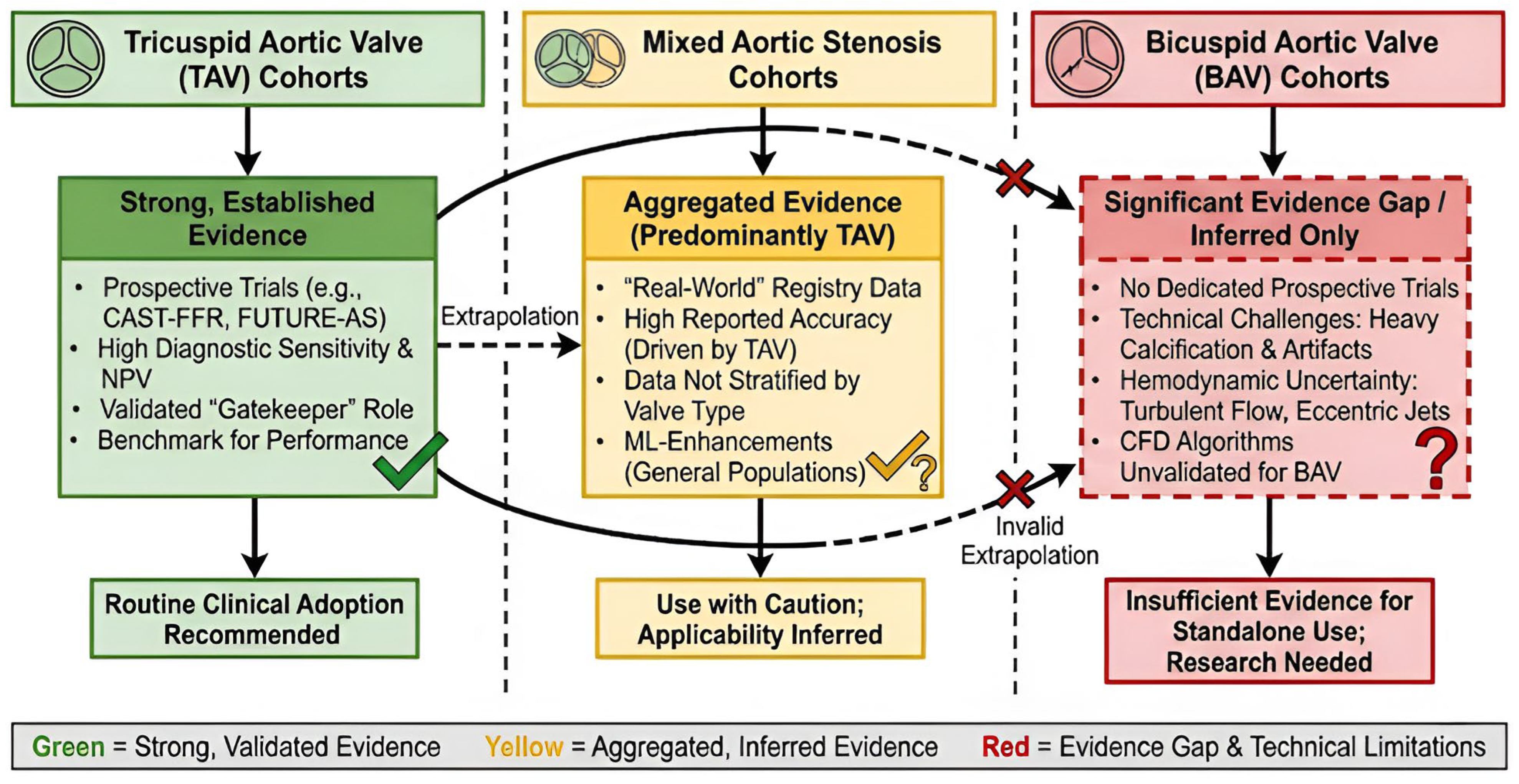
2.2. Exploratory Status and Challenges in BAV Anatomy
- Imaging Calcifications: BAV has greater and more asymmetric leaflet calcification than tricuspid valves [21], and this calcification burden results in increased artifact from both beam hardening and blooming that can hide the coronary ostia and result in decreased sensitivity (false positives) for CTCA-based assessments of coronary stenosis [33,35].
- Functional Hemodynamic: The function assessments by CT-FFR rely on computational fluid dynamics (CFD), assuming normal laminar flow conditions. Due to the presence of a raphe and annulus ellipticity in BAV, eccentric turbulent flow jets occur [35], and it is currently unknown if these flow disturbances would decrease the accuracy of current CT-FFR algorithms that have not been validated in the complex hemodynamic environment of bicuspid aortopathy [37].
3. CT for Interventional Procedural Planning in BAV
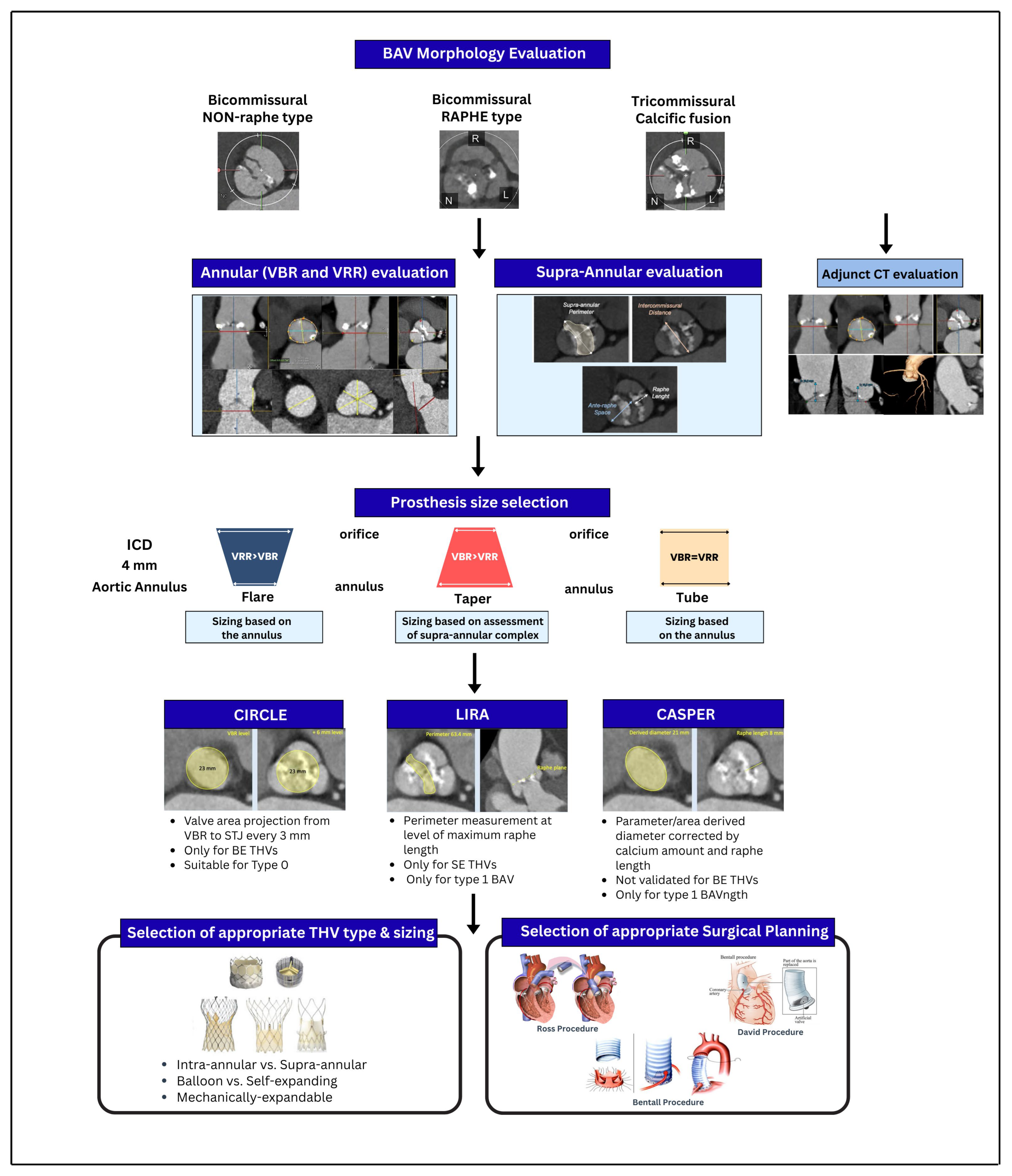
3.1. The Foundation: Annular vs. Supra-Annular Sizing
3.1.1. Annular Sizing (Standard of Care)
3.1.2. Supra-Annular Sizing (Exploratory/Complex Anatomy)
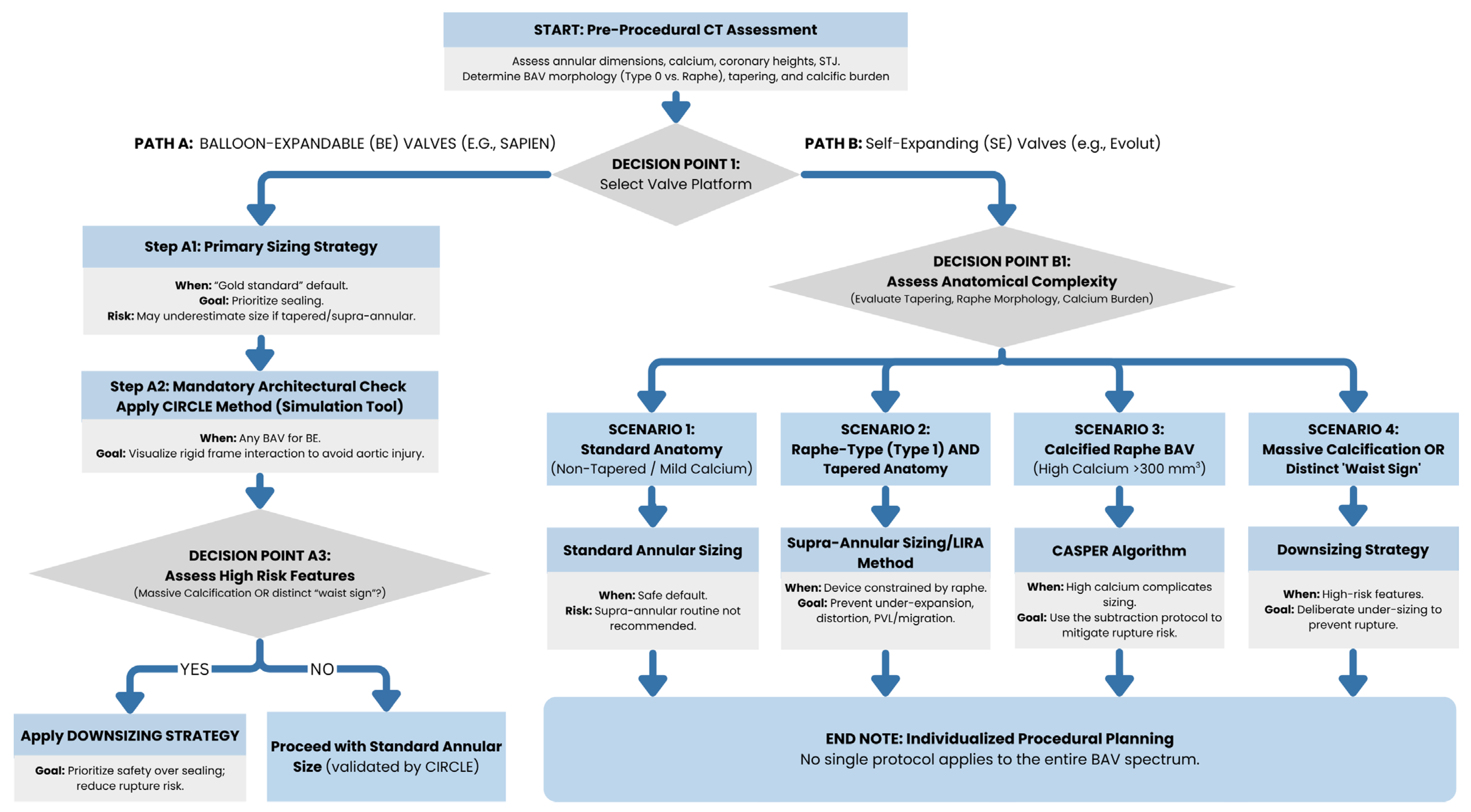
3.2. Advanced Sizing Algorithms: When and Why to Use
3.2.1. LIRA (Level of Implantation at the Raphe)
3.2.2. CASPER (Calcium Algorithm Sizing for Bicuspid Evaluation with Raphe)
3.2.3. CIRCLE Method
3.3. The Role of Downsizing
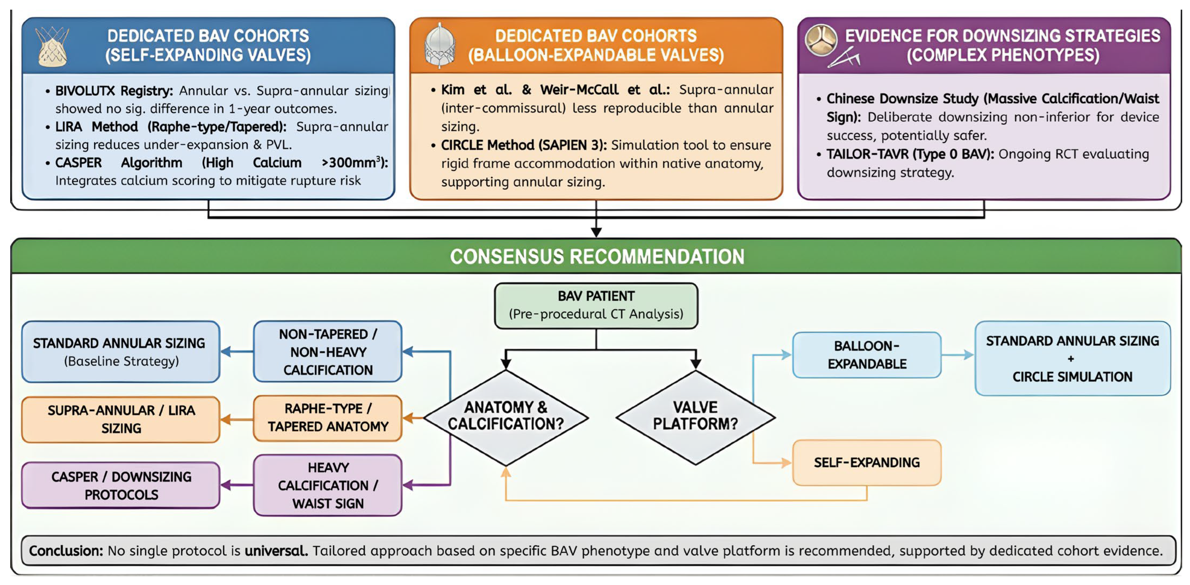
3.4. Evidence Summary and Clinical Consensus
4. Innovation Spotlight: Downsizing, Trials, and Artificial Intelligence
4.1. Procedural Innovation: Sizing Trials and the Downsizing Strategy
4.2. Artificial Intelligence: Differentiating Statistical Learning from Deep Learning
4.2.1. Logistic Regression (LR) vs. Advanced ML
4.2.2. Deep Learning (DL) and the “Black Box” Challenge
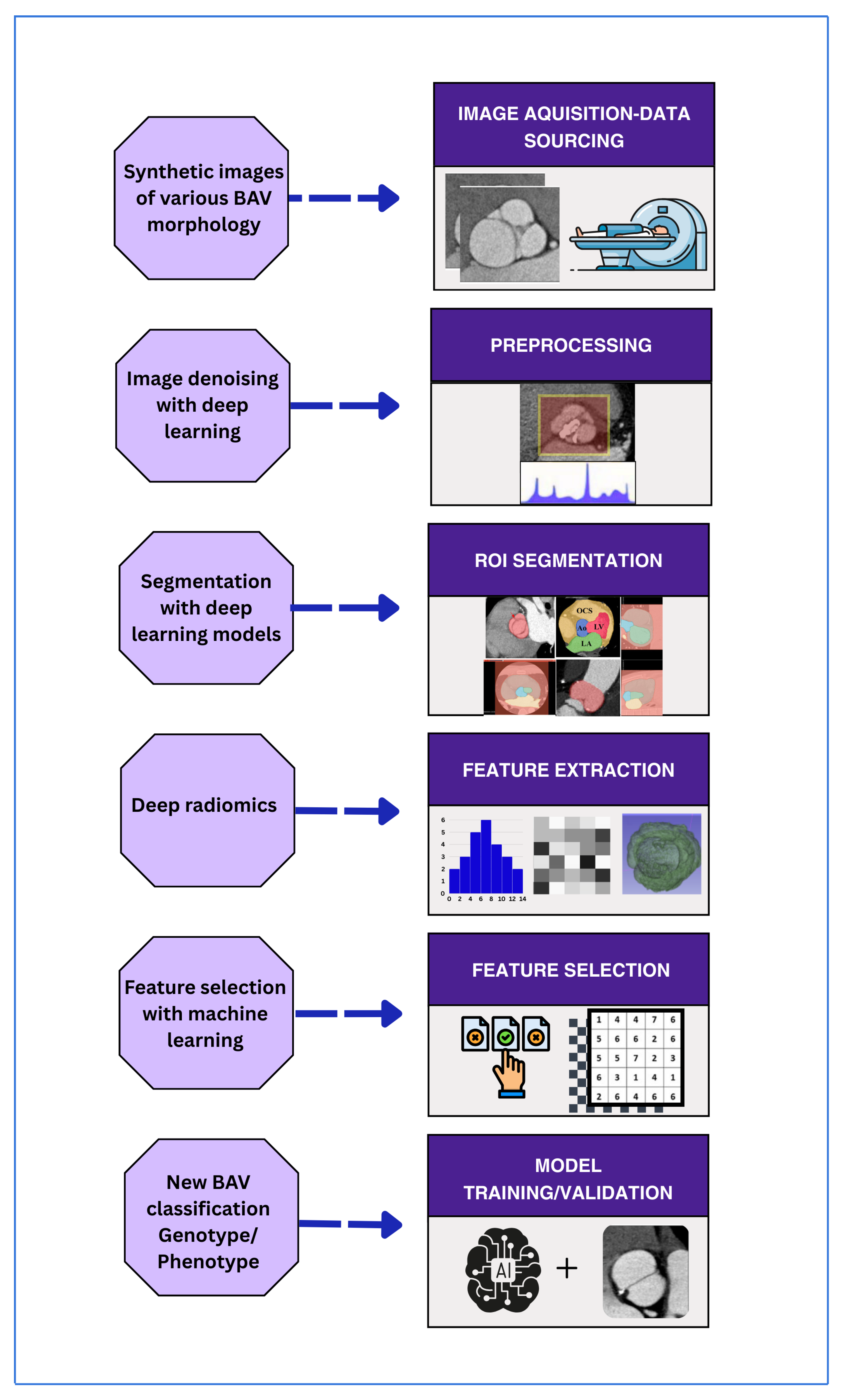
4.3. Radiomics: Technical Capabilities and Reproducibility Crises
4.3.1. Segmentation Variability
4.3.2. Lack of Standardization
4.3.3. Validation Gaps
4.3.4. Post-Procedural CT Surveillance
4.3.5. Limitations, Unmet Needs, and Controversies
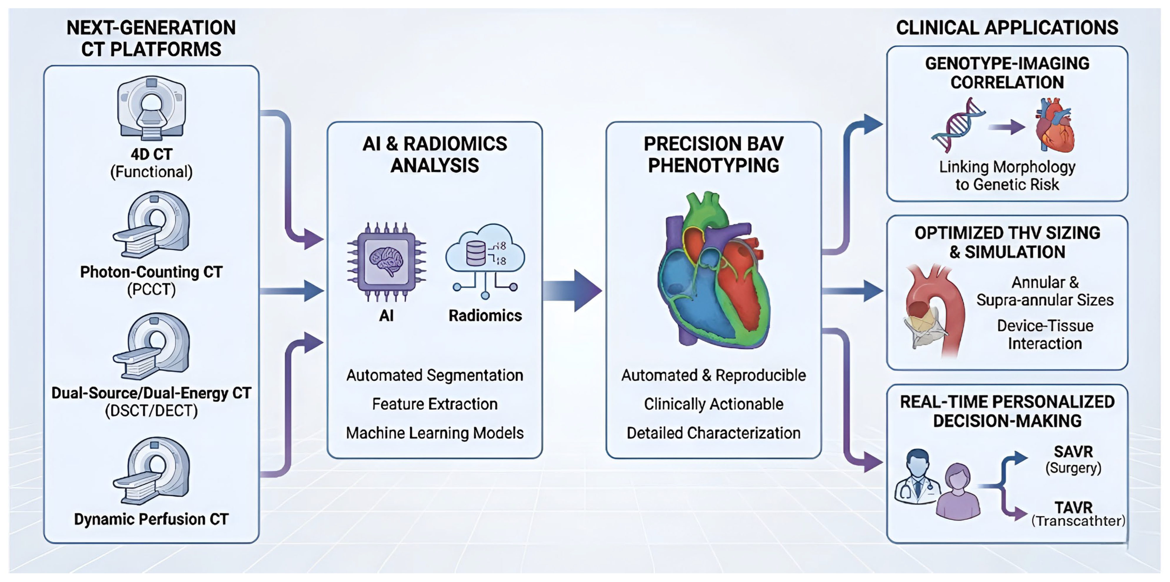
5. Future Directions: Imaging in the Precision Era
5.1. D CT Imaging
5.2. Photon-Counting CT
5.3. Dynamic Perfusion CT
6. Conclusions
Author Contributions
Funding
Data Availability Statement
Conflicts of Interest
References
- Schneider, U.; Karliova, I.; Giebels, C.; Ehrlich, T.; Schäfers, H.-J. Concepts and techniques of bicuspid aortic valve repair. J. Vis. Surg. 2020, 6, 3. [Google Scholar] [CrossRef]
- Jahanyar, J.; El Khoury, G.; de Kerchove, L. Commissural geometry and cusp fusion insights to guide bicuspid aortic valve repair. JTCVS Tech. 2021, 7, 83–92. [Google Scholar] [CrossRef] [PubMed]
- Sundström, E.; Tretter, J.T. Impact of Variation in Commissural Angle Between Fused Leaflets in the Functionally Bicuspid Aortic Valve on Hemodynamics and Tissue Biomechanics. Bioengineering 2023, 10, 1219. [Google Scholar] [CrossRef] [PubMed]
- Sievers, H.-H.; Schmidtke, C. A classification system for the bicuspid aortic valve from 304 surgical specimens. J. Thorac. Cardiovasc. Surg. 2007, 133, 1226–1233. [Google Scholar] [CrossRef]
- Schaefer, B.M.; Lewin, M.B.; Stout, K.K.; Gill, E.; Prueitt, A.; Byers, P.H.; Otto, C.M. The bicuspid aortic valve: An integrated phenotypic classification of leaflet morphology and aortic root shape. Heart 2008, 94, 1634–1638. [Google Scholar] [CrossRef]
- Braverman, A.C.; Güven, H.; Beardslee, M.A.; Makan, M.; Kates, A.M.; Moon, M.R. The bicuspid aortic valve. Curr. Probl. Cardiol. 2005, 30, 470–522. [Google Scholar] [CrossRef]
- de Kerchove, L.; Mastrobuoni, S.; Froede, L.; Tamer, S.; Boodhwani, M.; Van Dyck, M.; el Khoury, G.; Schäfers, H.-J. Variability of repairable bicuspid aortic valve phenotypes: Towards an anatomical and repair-oriented classification. Eur. J. Cardio-Thorac. Surg. 2019, 56, 351–359. [Google Scholar] [CrossRef]
- Michelena, H.I.; Della Corte, A.; Evangelista, A.; Maleszewski, J.J.; Edwards, W.D.; Roman, M.J.; Devereux, R.B.; Fernández, B.; Asch, F.M.; Barker, A.J.; et al. International consensus statement on nomenclature and classification of the congenital bicuspid aortic valve and its aortopathy, for clinical, surgical, interventional and research purposes. Eur. J. Cardio-Thorac. Surg. 2021, 60, 448–476. [Google Scholar] [CrossRef]
- Amoretti, F.; Cerillo, A.G.; Mariani, M.; Stefano, P. A simple method to visualize the bicuspid aortic valve pathology by cardiac computed tomography. J. Cardiovasc. Comput. Tomogr. 2020, 14, 195–198. [Google Scholar] [CrossRef]
- Devos, D.; Van Langenhove, C.; Campens, L. The Hammock Sign in Computed Tomography as a Detection Aid for Bicuspid Aortic Valves. J. Belg. Soc. Radiol. 2023, 107, 5. [Google Scholar] [CrossRef]
- Kim, J.; Kim, S.M.; Ahn, J.; Kim, J.; Choe, Y.H. Diagnostic performance of cardiac CT and transthoracic echocardiography for detection of surgically confirmed bicuspid aortic valve: Effect of calcium extent and valve subtypes. J. Korean Soc. Radiol. 2023, 84, 1324. [Google Scholar] [CrossRef]
- Williams, M.C.; Abbas, A.; Tirr, E.; Alam, S.; Nicol, E.; Shambrook, J.; Schmitt, M.; Hughes, G.M.; Stirrup, J.; Holloway, B.; et al. Reporting incidental coronary, aortic valve and cardiac calcification on non-gated thoracic computed tomography, a consensus statement from the BSCI/BSCCT and BSTI. Br. J. Radiol. 2021, 94, 20200894. [Google Scholar] [CrossRef] [PubMed]
- Forrest, J.K.; Ramlawi, B.; Deeb, G.M.; Zahr, F.; Song, H.K.; Kleiman, N.S.; Chetcuti, S.J.; Michelena, H.I.; Mangi, A.A.; Skiles, J.A.; et al. Transcatheter aortic valve replacement in low-risk patients with bicuspid aortic valve stenosis. JAMA Cardiol. 2021, 6, 50–57. [Google Scholar] [CrossRef] [PubMed]
- Xiong, T.-Y.; Li, Y.-J.; Feng, Y.; Liao, Y.-B.; Zhao, Z.-G.; Mylotte, D.; Wei, X.; Xu, Y.-N.; Peng, Y.; Wei, J.-F.; et al. Understanding the interaction between transcatheter aortic valve prostheses and supra-annular structures from post-implant stent geometry. JACC Cardiovasc. Interv. 2019, 12, 1164–1171. [Google Scholar] [CrossRef]
- Ahmad, Y.; Agarwal, V.; Williams, M.L.; Wang, D.D.; Reardon, M.J.; Cavalcante, J.L.; Makkar, R.; Forrest, J.K. Imaging, treatment options, patient selection, and outcome considerations for patients with bicuspid aortic valve disease. J. Soc. Cardiovasc. Angiogr. Interv. 2022, 1, 100506. [Google Scholar] [CrossRef]
- Alkadhi, H.; Wildermuth, S.; Plass, A.; Bettex, D.; Baumert, B.; Leschka, S.; Desbiolles, L.M.; Marincek, B.; Boehm, T. Aortic stenosis: Comparative evaluation of 16–detector row CT and echocardiography. Radiology 2006, 240, 47–55. [Google Scholar] [CrossRef]
- Blanke, P.; Weir-McCall, J.R.; Achenbach, S.; Delgado, V.; Hausleiter, J.; Jilaihawi, H.; Marwan, M.; Nørgaard, B.L.; Piazza, N.; Schoenhagen, P.; et al. Computed tomography imaging in the context of transcatheter aortic valve implantation (TAVI)/transcatheter aortic valve replacement (TAVR) an expert consensus document of the Society of Cardiovascular Computed Tomography. JACC Cardiovasc. Imaging 2019, 12, 1–24. [Google Scholar] [CrossRef]
- Halpern, E.J.; Mallya, R.; Sewell, M.; Shulman, M.; Zwas, D.R. Differences in aortic valve area measured with CT planimetry and echocardiography (continuity equation) are related to divergent estimates of left ventricular outflow tract area. Am. J. Roentgenol. 2009, 192, 1668–1673. [Google Scholar] [CrossRef]
- Clavel, M.-A.; Malouf, J.; Messika-Zeitoun, D.; Araoz, P.A.; Michelena, H.I.; Enriquez-Sarano, M. Aortic valve area calculation in aortic stenosis by CT and Doppler echocardiography. Cardiovasc. Imaging 2015, 8, 248–257. [Google Scholar] [CrossRef]
- Pawade, T.; Sheth, T.; Guzzetti, E.; Dweck, M.R.; Clavel, M.-A. Why and how to measure aortic valve calcification in patients with aortic stenosis. JACC Cardiovasc. Imaging 2019, 12, 1835–1848. [Google Scholar] [CrossRef]
- Gollmann-Tepeköylü, C.; Nägele, F.; Engler, C.; Stoessel, L.; Zellmer, B.; Graber, M.; Hirsch, J.; Pölzl, L.; Ruttmann, E.; Tancevski, I.; et al. Different calcification patterns of tricuspid and bicuspid aortic valves and their clinical impact. Interact. Cardiovasc. Thorac. Surg. 2022, 35, ivac274. [Google Scholar] [CrossRef] [PubMed]
- Kim, W.-K.; Renker, M.; Rolf, A.; Fischer-Rasokat, U.; Wiedemeyer, J.; Doss, M.; Möllmann, H.; Walther, T.; Nef, H.; Hamm, C.W.; et al. Annular versus supra-annular sizing for TAVI in bicuspid aortic valve stenosis. EuroIntervention 2019, 15, e231–e238. [Google Scholar] [CrossRef] [PubMed]
- Evangelista Masip, A.; Galian-Gay, L.; Guala, A.; Lopez-Sainz, A.; Teixido-Tura, G.; Ruiz Munoz, A.; Valente, F.; Gutierrez, L.; Fernandez-Galera, R.; Casas, G.; et al. Unraveling Bicuspid Aortic Valve Enigmas by Multimodality Imaging: Clinical Implications. J. Clin. Med. 2022, 11, 456. [Google Scholar] [CrossRef]
- Irace, F.G.; Chirichilli, I.; de Paulis, R. CT scan as a tool to evaluate root reconstruction and aortic valve repair. Vessel Plus 2025, 9, 4. [Google Scholar] [CrossRef]
- Kong, W.K.; Delgado, V.; Poh, K.K.; Regeer, M.V.; Ng, A.C.; McCormack, L.; Yeo, T.C.; Shanks, M.; Parent, S.; Enache, R.; et al. Prognostic Implications of Raphe in Bicuspid Aortic Valve Anatomy. JAMA Cardiol. 2017, 2, 285–292. [Google Scholar] [CrossRef]
- Kong, W.K.F.; Delgado, V.; Bax, J.J. Bicuspid Aortic Valve: What to Image in Patients Considered for Transcatheter Aortic Valve Replacement? Circ. Cardiovasc. Imaging 2017, 10, e005987. [Google Scholar] [CrossRef]
- Michelena, H.I.; Khanna, A.D.; Mahoney, D.; Margaryan, E.; Topilsky, Y.; Suri, R.M.; Eidem, B.; Edwards, W.D.; Sundt, T.M.; Enriquez-Sarano, M. Incidence of aortic complications in patients with bicuspid aortic valves. JAMA 2011, 306, 1104–1112. [Google Scholar] [CrossRef]
- Wojnarski, C.M.; Svensson, L.G.; Roselli, E.E.; Idrees, J.J.; Lowry, A.M.; Ehrlinger, J.; Pettersson, G.B.; Gillinov, A.M.; Johnston, D.R.; Soltesz, E.G.; et al. Aortic dissection in patients with bicuspid aortic valve–associated aneurysms. Ann. Thorac. Surg. 2015, 100, 1666–1674. [Google Scholar] [CrossRef]
- Yoon, S.-H.; Kim, W.-K.; Dhoble, A.; Milhorini Pio, S.; Babaliaros, V.; Jilaihawi, H.; Pilgrim, T.; De Backer, O.; Bleiziffer, S.; Vincent, F.; et al. Bicuspid aortic valve morphology and outcomes after transcatheter aortic valve replacement. J. Am. Coll. Cardiol. 2020, 76, 1018–1030. [Google Scholar] [CrossRef]
- Chirichilli, I.; Irace, F.; Weltert, L.; Tsuda, K.; Scaffa, R.; Salica, A.; Galea, N.; De Paulis, R. Morphological modification of the aortic annulus in tricuspid and bicuspid valves after aortic valve reimplantation: An electrocardiography-gated computed tomography study. Eur. J. Cardio-Thorac. Surg. 2019, 56, 778–784. [Google Scholar] [CrossRef]
- Chirichilli, I.; Irace, F.G.; Weltert, L.P.; Salica, A.; Guerrieri Wolf, L.; Fusca, S.; Ricci, A.; De Paulis, R. A direct correlation between commissural orientation and annular shape in bicuspid aortic valves: A new anatomical and computed tomography classification. Interact. Cardiovasc. Thorac. Surg. 2020, 30, 666–670. [Google Scholar] [CrossRef]
- Corcione, N.; Morello, A.; Ferraro, P.; Cimmino, M.; Albanese, M.; Pepe, M.; Nestola, P.L.; Giordano, S.; Bardi, L.; Biondi-Zoccai, G.; et al. TAVI-CT score to evaluate the anatomic risk in patients undergoing transcatheter aortic valve implantation. Sci. Rep. 2022, 12, 7612. [Google Scholar] [CrossRef] [PubMed]
- Michail, M.; Ihdayhid, A.-R.; Comella, A.; Thakur, U.; Cameron, J.D.; McCormick, L.M.; Gooley, R.P.; Nicholls, S.J.; Mathur, A.; Hughes, A.D.; et al. Feasibility and Validity of Computed Tomography-Derived Fractional Flow Reserve in Patients With Severe Aortic Stenosis. Circ. Cardiovasc. Interv. 2021, 14, 20–28. [Google Scholar] [CrossRef] [PubMed]
- Khoo, J.K.; Sellers, S.; Fairbairn, T.; Polsani, V.; Liu, S.; Yong, G.; Shetty, S.; Corrigan, F.; Ko, B.; Vucic, E.; et al. Feasibility and Utility of Anatomical and Physiological Evaluation of Coronary Disease With Cardiac CT in Severe Aortic Stenosis (FUTURE-AS Registry): Rationale and Design. J. Soc. Cardiovasc. Angiogr. Interv. 2024, 3, 101293. [Google Scholar] [CrossRef]
- Gohmann, R.F.; Pawelka, K.; Seitz, P.; Majunke, N.; Heiser, L.; Renatus, K.; Desch, S.; Lauten, P.; Holzhey, D.; Noack, T.; et al. Combined cCTA and TAVR Planning for Ruling Out Significant CAD. JACC Cardiovasc. Imaging 2022, 15, 476–486. [Google Scholar] [CrossRef]
- Hussain, K.; Lee, K.; Minga, I.; Wathen, L.; Balasubramanian, S.S.; Vyas, N.; Singh, L.; Shetty, M.; Rosenberg, J.R.; Levisay, J.P.; et al. Real-world application of CCTA with CT-FFR for coronary assessment pre-TAVI: The CT2TAVI study. Int. J. Cardiovasc. Imaging 2025, 41, 523–535. [Google Scholar] [CrossRef]
- Wang, R.; Pan, D.; Sun, X.; Yang, G.; Yao, J.; Shen, X.; Xiao, W. Two birds with one stone: Pre-TAVI coronary CT angiography combined with FFR helps screen for coronary stenosis. BMC Med. Imaging 2025, 25, 1–7. [Google Scholar] [CrossRef]
- Tchetche, D.; De Biase, C.; van Gils, L.; Parma, R.; Ochala, A.; Lefevre, T.; Hovasse, T.; De Backer, O.; Sondergaard, L.; Bleiziffer, S.; et al. Bicuspid aortic valve anatomy and relationship with devices: The Bavard multicenter Registry: A European picture of contemporary multidetector computed tomography sizing for bicuspid valves. Circ. Cardiovasc. Interv. 2019, 12, e007107. [Google Scholar] [CrossRef]
- Iannopollo, G.; Romano, V.; Esposito, A.; Guazzoni, G.; Ancona, M.; Ferri, L.; Russo, F.; Bellini, B.; Montorfano, M. Update on supra-annular sizing of transcatheter aortic valve prostheses in raphe-type bicuspid aortic valve disease according to the LIRA method. Eur. Heart J. Suppl. 2022, 24, C233–C242. [Google Scholar] [CrossRef]
- Iannopollo, G.; Romano, V.; Buzzatti, N.; Ancona, M.; Ferri, L.; Russo, F.; Bellini, B.; Granada, J.F.; Chieffo, A.; Montorfano, M. Supra-annular sizing of transcatheter aortic valve prostheses in raphe-type bicuspid aortic valve disease: The LIRA method. Int. J. Cardiol. 2020, 317, 144–151. [Google Scholar] [CrossRef]
- Petronio, A.S.; Angelillis, M.; De Backer, O.; Giannini, C.; Costa, G.; Fiorina, C.; Castriota, F.; Bedogni, F.; Laborde, J.C.; Søndergaard, L. Bicuspid aortic valve sizing for transcatheter aortic valve implantation: Development and validation of an algorithm based on multi-slice computed tomography. J. Cardiovasc. Comput. Tomogr. 2020, 14, 452–461. [Google Scholar] [CrossRef]
- Blackman, D.; Gabbieri, D.; Del Blanco, B.G.; Kempfert, J.; Laine, M.; Mascherbauer, J.; Parma, R.; Tchétché, D. Expert Consensus on Sizing and Positioning of SAPIEN 3/Ultra in Bicuspid Aortic Valves. Cardiol. Ther. 2021, 10, 277–288. [Google Scholar] [CrossRef] [PubMed]
- Guo, Y.; Liu, X.; Li, R.; Ng, S.; Liu, Q.; Wang, L.; Hu, P.; Ren, K.; Jiang, J.; Fan, J.; et al. Comparison of downsizing strategy (HANGZHOU Solution) and standard annulus sizing strategy in type 0 bicuspid aortic stenosis patients undergoing transcatheter aortic valve replacement: Rationale and design of a randomized clinical trial. Am. Heart J. 2024, 274, 65–74. [Google Scholar] [CrossRef] [PubMed]
- Niu, G.; Ali, W.B.; Wang, M.; Jilaihawi, H.; Zhang, H.; Zhang, Q.; Ye, Y.; Liu, X.; Yao, J.; Zhao, Q.; et al. Anatomical morphology of the aortic valve in Chinese aortic stenosis patients and clinical results after downsize strategy of transcatheter aortic valve replacement. Chin. Med. J. 2022, 135, 2968–2975. [Google Scholar] [CrossRef] [PubMed]
- Tchétché, D.; Ziviello, F.; De Biase, C.; De Backer, O.; Hovasse, T.; Leroux, L.; Petronio, A.-S.; Saint-Etienne, C.; Teles, R.C.; Modine, T.; et al. Transcatheter aortic valve implantation with the Evolut platform for bicuspid aortic valve stenosis: The international, multicentre, prospective BIVOLUTX registry. EuroIntervention 2023, 19, 502–511. [Google Scholar] [CrossRef]
- Bleiziffer, S. Bicuspid is different. EuroIntervention 2023, 19, 455–456. [Google Scholar] [CrossRef]
- Barker, C.M.; Reardon, M.J. TAVR Bicuspid Aortic Valves: Past Is Prologue Or Tempest A Teapot? Cardiovasc. Revascularization Med. 2019, 20, 5. [Google Scholar] [CrossRef]
- Bellini, B.; Romano, V.; Gorla, R.; Moccetti, F.; De Felice, F.; Buono, A.; Massussi, M.; Resta, F.; Iannopollo, G.; Zanda, G.; et al. Size up Bicuspid With the Lira Method: The SUBLIME Multicenter Study. Struct. Heart 2025, 9, 100527. [Google Scholar] [CrossRef]
- Costa, G.; Angelillis, M.; Petronio, A.S. Bicuspid valve sizing for transcatheter aortic valve implantation: The missing link. Front. Cardiovasc. Med. 2022, 8, 770924. [Google Scholar] [CrossRef]
- Weir-McCall, J.R.; Attinger-Toller, A.; Blanke, P.; Perlman, G.Y.; Sellers, S.L.; Wood, D.; Webb, J.G.; Leipsic, J. Annular versus supra-annular sizing for transcatheter aortic valve replacement in bicuspid aortic valve disease. J. Cardiovasc. Comput. Tomogr. 2020, 14, 407–413. [Google Scholar] [CrossRef]
- Zaka, A.; Mustafiz, C.; Mutahar, D.; Sinhal, S.; Gorcilov, J.; Muston, B.; Evans, S.; Gupta, A.; Stretton, B.; Kovoor, J.; et al. Machine-learning versus traditional methods for prediction of all-cause mortality after transcatheter aortic valve implantation: A systematic review and meta-analysis. Open Heart 2025, 12, e002779. [Google Scholar] [CrossRef] [PubMed]
- Benjamin, M.M.; Rabbat, M.G. Artificial Intelligence in Transcatheter Aortic Valve Replacement: Its Current Role and Ongoing Challenges. Diagnostics 2024, 14, 261. [Google Scholar] [CrossRef] [PubMed]
- Shojaei, S.; Mousavi, A.; Kazemian, S.; Armani, S.; Maleki, S.; Fallahtafti, P.; Arashlow, F.T.; Daryabari, Y.; Naderian, M.; Alkhouli, M.; et al. Artificial Intelligence in Risk Stratification and Outcome Prediction for Transcatheter Aortic Valve Replacement: A Systematic Review and Meta-Analysis. J. Pers. Med. 2025, 15, 302. [Google Scholar] [CrossRef] [PubMed]
- Williams, M.C.; Weir-McCall, J.R.; Baldassarre, L.A.; De Cecco, C.N.; Choi, A.D.; Dey, D.; Dweck, M.R.; Isgum, I.; Kolossvary, M.; Leipsic, J.; et al. Artificial Intelligence and Machine Learning for Cardiovascular Computed Tomography (CCT): A White Paper of the Society of Cardiovascular Computed Tomography (SCCT). J. Cardiovasc. Comput. Tomogr. 2024, 18, 519–532. [Google Scholar] [CrossRef]
- Maniaci, A.; Lavalle, S.; Gagliano, C.; Lentini, M.; Masiello, E.; Parisi, F.; Iannella, G.; Cilia, N.D.; Salerno, V.; Cusumano, G.; et al. The Integration of Radiomics and Artificial Intelligence in Modern Medicine. Life 2024, 14, 1248. [Google Scholar] [CrossRef]
- Guo, Z.; Dong, N.J.; Litt, H.; Yushkevich, N.; Freas, M.; Nunez, J.; Ferrari, V.; Hao, J.; Goldfinger, S.; Jolley, M.A.; et al. Towards Patient-Specific Surgical Planning for Bicuspid Aortic Valve Repair: Fully Automated Segmentation of the Aortic Valve in 4D. In Proceedings of the 2025 IEEE 22nd International Symposium on Biomedical Imaging (ISBI), Houston, TX, USA, 14–17 April 2025; IEEE: New York, NY, USA, 2025; pp. 1–5. [Google Scholar]
- Jamhour-Chelh, K.; Arzamendi, D.; Asmarats, L. Artificial intelligence for risk prediction in transcatheter heart valve interventions. Expert Rev. Cardiovasc. Ther. 2025, 23, 329–331. [Google Scholar] [CrossRef]
- Baessler, B.; Gotz, M.; Antoniades, C.; Heidenreich, J.F.; Leiner, T.; Beer, M. Artificial intelligence in coronary computed tomography angiography: Demands and solutions from a clinical perspective. Front. Cardiovasc. Med. 2023, 10, 1120361. [Google Scholar] [CrossRef]
- Kapoor, A.; Mahajan, G.; Kapoor, A. Onsite Computed Tomography Fractional Flow Reserve in Patients with Suspected Stable Coronary Artery Disease Initial Experience. Indian J. Radiol. Imaging 2021, 31, 291–296. [Google Scholar] [CrossRef]
- Shabbir, A.; Attia, A.; Weber, N.M.; Alhassan, M.; Radike, M.; Lane, C.M.; Challa, A.B.; Wamil, M. The Emerging Role of Computed Tomography Coronary Angiography in the Left Main Stem Percutaneous Coronary Intervention. Rev. Cardiovasc. Med. 2025, 26, 37379. [Google Scholar] [CrossRef]
- Alperi, A.; del Valle, R.; Pascual, I.; Antuna, P.; Almendárez, M.; Álvarez, R.; Hernández-Vaquero, D.; Avanzas, P. Transcatheter aortic valve implantation in bicuspid aortic valves: Present and future. Rev. Española Cardiol. Engl. Ed. 2025, 78, 724–735. [Google Scholar] [CrossRef]
- He, A.; Wilkins, B.; Lan, N.S.R.; Othman, F.; Sehly, A.; Bhat, V.; Jaltotage, B.; Dwivedi, G.; Leipsic, J.; Ihdayhid, A.R. Cardiac computed tomography post-transcatheter aortic valve replacement. J. Cardiovasc. Comput. Tomogr. 2024, 18, 319–326. [Google Scholar] [CrossRef] [PubMed]
- Tzimas, G.; Meier, D.; Beneki, E.; Antiochos, P.; Auberson, D.; Leboub, S.; Lu, H.; Liabot, Q.; Zimmerli, A.; Salihu, A.; et al. Current Trends and Future Challenges in Transcatheter Aortic Valve Replacement: Utility of Cardiac Computed Tomography Angiography. J. Clin. Med. 2025, 14, 2474. [Google Scholar] [CrossRef] [PubMed]
- Jia, Y.; Khokhar, A.A.; Pilgrim, T.; Costa, G.; Mylotte, D.; Sammartino, S.; Tomii, D.; Fosbøl, E.; Tamburino, C.; Kofoed, K.F.; et al. Incidence and predictors of continued ascending aortic dilatation after TAVI in patients with bicuspid aortic stenosis. Clin. Res. Cardiol. 2024, 114, 375–384. [Google Scholar] [CrossRef] [PubMed]
- Das, R.; Puri, R. Transcatheter Treatment of Bicuspid Aortic Valve Disease: Imaging and Interventional Considerations. Front. Cardiovasc. Med. 2018, 5, 91. [Google Scholar] [CrossRef]
- Flohr, T.; Petersilka, M.; Henning, A.; Ulzheimer, S.; Ferda, J.; Schmidt, B. Photon-counting CT review. Phys. Medica 2020, 79, 126–136. [Google Scholar] [CrossRef]
- Leng, S.; Bruesewitz, M.; Tao, S.; Rajendran, K.; Halaweish, A.F.; Campeau, N.G.; Fletcher, J.G.; McCollough, C.H. Photon-counting detector CT: System design and clinical applications of an emerging technology. Radiographics 2019, 39, 729–743. [Google Scholar] [CrossRef]
- Zanon, C.; Cademartiri, F.; Toniolo, A.; Bini, C.; Clemente, A.; Colacchio, E.C.; Cabrelle, G.; Mastro, F.; Antonello, M.; Quaia, E.; et al. Advantages of Photon-Counting Detector CT in Aortic Imaging. Tomography 2023, 10, 1–13. [Google Scholar] [CrossRef]
- Sliwicka, O.; Sechopoulos, I.; Baggiano, A.; Pontone, G.; Nijveldt, R.; Habets, J. Dynamic myocardial CT perfusion imaging—State of the art. Eur. Radiol. 2023, 33, 5509–5525. [Google Scholar] [CrossRef]
- Fikani, A.; Craiem, D.; Mousseaux, E.; Soulat, G.; Rouchaud, A.; Boulogne, C.; Martins, E.; Jouan, J. Morphological and dynamic analysis of the normal aortic valve with 4D computed tomography. Eur. J. Cardio-Thorac. Surg. 2024, 65, ezae113. [Google Scholar] [CrossRef]
- Bruns, S.; Wolterink, J.M.; Van Den Boogert, T.P.; Henriques, J.P.; Baan, J.; Planken, R.N.; Išgum, I. (Eds.) Automatic whole-heart segmentation in 4D TAVI treatment planning CT. In Medical Imaging 2021: Image Processing; SPIE: Bellingham, WA, USA, 2021. [Google Scholar]
- Piatti, F.; Sturla, F.; Bissell, M.M.; Pirola, S.; Lombardi, M.; Nesteruk, I.; Della Corte, A.; Redaelli, A.C.L.; Votta, E. 4D flow analysis of BAV-related fluid-dynamic alterations: Evidences of wall shear stress alterations in absence of clinically-relevant aortic anatomical remodeling. Front. Physiol. 2017, 8, 441. [Google Scholar] [CrossRef]
- Fikani, A.; Craiem, D.; Boulogne, C.; Soulat, G.; Mousseaux, E.; Jouan, J. Four-dimensional computed tomography analysis of bicuspid aortic valves. JTCVS Tech. 2024, 27, 60–67. [Google Scholar] [CrossRef]
- Zdanowicz, A.; Guzinski, M.; Pula, M.; Witkowska, A.; Reczuch, K. Dynamic CT Myocardial Perfusion: The Role of Functional Evaluation in the Diagnosis of Coronary Artery Disease. J. Clin. Med. 2023, 12, 7062. [Google Scholar] [CrossRef]





| Strategy | Primary Device Applicability | Key Anatomical Indication (When/Why) | Key Evidence/Outcome |
|---|---|---|---|
| Standard Annular Sizing | BE & SE | Default strategy. Best for Type 0 BAV or mild calcification, where anatomy is non-tapered. | BIVOLUTX [45,46,47]: Comparison showed similar safety/efficacy to supra-annular sizing. Remains the most reproducible standard. |
| Supra-Annular (ICD)/LIRA | SE | Raphe-Type BAV (Type 1). Used when the “functional” annulus is higher due to fused leaflets, or to avoid device constraint in tapered aortas. | LIRA Studies [39,40,48]: 100% device success in initial cohorts; reduced risk of PVL and migration in raphe-type valves. |
| CASPER Algorithm | SE | Calcified Raphe BAV. Used when a high calcium burden (>300 mm3) complicates sizing. Adjust size to prevent rupture. | Petronio et al. [41]: Validated algorithm integrating calcium score and raphe length to customize downsizing. |
| CIRCLE Method | BE | Any BAV (esp. Type 0/1). Simulation tool to check if the rigid BE valve frame fits the anatomy from annulus to STJ without injury. | Blackman et al. [42]: Consensus method for SAPIEN 3/Ultra to visualize device-tissue interaction at multiple levels. |
| Downsizing (e.g., Hangzhou) | BE & SE | High Calcium/Waist Sign. Deliberate under-sizing to prevent annular rupture in rigid, heavily calcified anatomy. | TAILOR-TAVR [43], Chinese Downsize [44]: Non-inferior device success; favourable for reducing rupture and pacemaker rates. |
| Trial/Study | Sizing Strategy | Methodology | Key Outcomes | Conclusion |
|---|---|---|---|---|
| TAILOR-TAVR (NCT05511792) | Downsizing (Hangzhou Solution) vs. Standard Annular | RCT in Type 0 BAV. Self-expanding valve. Valve one size smaller if “waist sign” and mild regurgitation after pre-dilatation. | Results awaited. Preliminary data suggests higher device success and fewer pacemakers in the downsizing arm. | Downsizing may improve clinical outcomes in challenging anatomies [43]. |
| BIVOLUTX Registry (NCT03495050) | Annular vs. Combined (Annular + Supra-annular) | 149 patients (Evolut). Compared outcomes by sizing strategy using CT-based measurements | No significant difference in valve performance, mortality, PVL, or PPM at 1 year. | Clinical outcomes were similar regardless of methodology; annular sizing remains safe [45,46,47]. |
| LIRA Method Studies (incl. SUBLIME) | Supra-annular (Level of Implantation at Raphe) | Cohorts (Single centres/registries). Sizing based on the minimum CT perimeter at the raphe versus the annulus. | 100% device success in initial series; reduced valve migration and moderate/severe PVL. | Supra-annular sizing appears safe and effective in select raphe-type BAV [39,40,48]. |
| Chinese Downsize Study | Annularvs.Downsize | Cohort of 293 (95 BAV). Outcomes at 1 year comparing device success, gradient, and PVL. | Device success: 82% (downsizing) vs. 83.3% (annular); similar gradients and PVL rates. | The downsizing strategy is non-inferior and safe, particularly in high-calcium anatomy [44]. |
| Weir-McCall et al. | Annular vs. Supra-annular (ICD-based) | 44 BAV patients (SAPIEN 3). Compared to CT-measured annulus vs. Intercommissural Diameter (ICD). | Supra-annular (ICD) sizing was less reproducible; annular sizing agreed best with implanted size. | Annular sizing is more consistent; routine supra-annular sizing is not recommended [22,49,50]. |
Disclaimer/Publisher’s Note: The statements, opinions and data contained in all publications are solely those of the individual author(s) and contributor(s) and not of MDPI and/or the editor(s). MDPI and/or the editor(s) disclaim responsibility for any injury to people or property resulting from any ideas, methods, instructions or products referred to in the content. |
© 2026 by the authors. Licensee MDPI, Basel, Switzerland. This article is an open access article distributed under the terms and conditions of the Creative Commons Attribution (CC BY) license.
Share and Cite
Jawed, M.A.; Ayhan, C.; Byrne, R.; Hothi, S.S.; Sultan, S.; Spence, M.; Soliman, O. Cardiovascular CT in Bicuspid Aortic Valve Disease: A State-of-the-Art Narrative Review of Advances, Clinical Integration, and Future Directions. J. Clin. Med. 2026, 15, 1268. https://doi.org/10.3390/jcm15031268
Jawed MA, Ayhan C, Byrne R, Hothi SS, Sultan S, Spence M, Soliman O. Cardiovascular CT in Bicuspid Aortic Valve Disease: A State-of-the-Art Narrative Review of Advances, Clinical Integration, and Future Directions. Journal of Clinical Medicine. 2026; 15(3):1268. https://doi.org/10.3390/jcm15031268
Chicago/Turabian StyleJawed, Muhammad Ali, Cagri Ayhan, Robert Byrne, Sandeep Singh Hothi, Sherif Sultan, Mark Spence, and Osama Soliman. 2026. "Cardiovascular CT in Bicuspid Aortic Valve Disease: A State-of-the-Art Narrative Review of Advances, Clinical Integration, and Future Directions" Journal of Clinical Medicine 15, no. 3: 1268. https://doi.org/10.3390/jcm15031268
APA StyleJawed, M. A., Ayhan, C., Byrne, R., Hothi, S. S., Sultan, S., Spence, M., & Soliman, O. (2026). Cardiovascular CT in Bicuspid Aortic Valve Disease: A State-of-the-Art Narrative Review of Advances, Clinical Integration, and Future Directions. Journal of Clinical Medicine, 15(3), 1268. https://doi.org/10.3390/jcm15031268