Using Patient Feedback to Improve Treatment Outcomes for Patients with Congenital Dyserythropoietic Anaemia Type I Receiving Interferon Therapy
 , , , and
Abstract
1. Introduction
- Would a national formal network of clinicians with expertise and/or a national MDT (multidisciplinary team meeting) improve care for patients with rare inherited anaemias?
- Would a register of all rare inherited anaemia patients in the UK (including data and samples) improve care?
2. Methods
3. Results
- Are my symptoms (e.g., fatigue) improving with interferon?
- Can I stop other treatments (e.g., iron chelation, blood transfusions) because of interferon?
- Will interferon reduce my risk of CDA-I-related problems in the future?
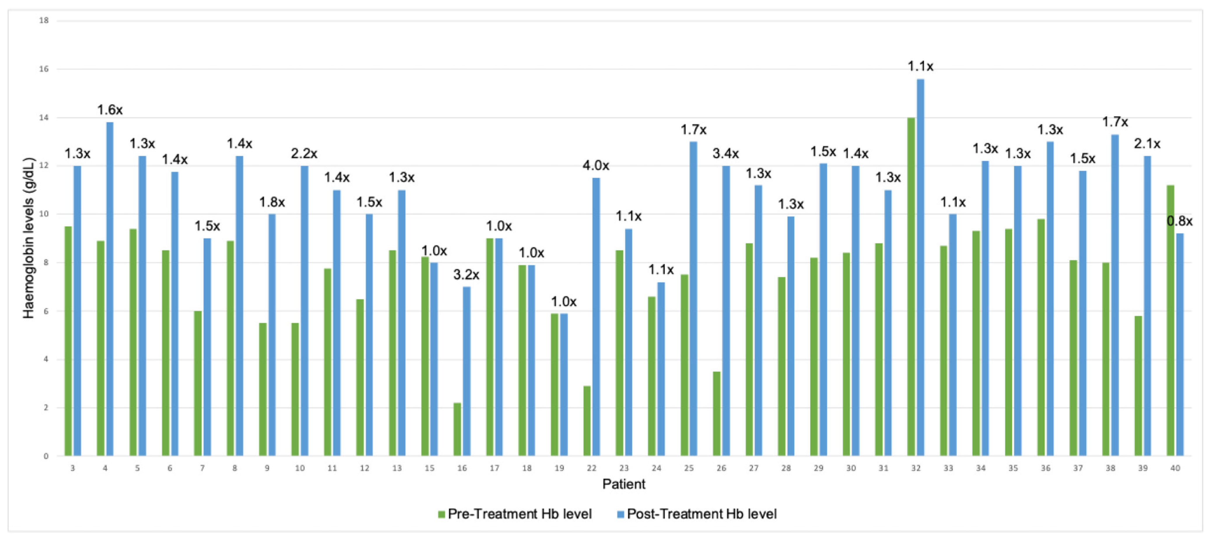
- Are my Hb levels better after interferon?
4. Discussion
- Quantifiable criteria
- 2.
- PROMs
- 3.
- Side Effects
Author Contributions
Funding
Data Availability Statement
Acknowledgments
Conflicts of Interest
References
- Roy, N.B.A.; Babbs, C. The pathogenesis, diagnosis and management of congenital dyserythropoietic anaemia type I. Br. J. Haematol. 2019, 185, 436–449. [Google Scholar] [CrossRef] [PubMed]
- Niss, O.; Lorsbach, R.B.; Berger, M.; Chonat, S.; McLemore, M.; Buchbinder, D.; McCavit, T.; Shaffer, L.G.; Simpson, J.; Schwartz, J.H.; et al. Congenital dyserythropoietic anemia type I: First report from the Congenital Dyserythropoietic Anemia Registry of North America (CDAR). Blood Cells Mol. Dis. 2021, 87, 102534. [Google Scholar] [CrossRef] [PubMed]
- Heimpel, H.; Schwarz, K.; Ebnöther, M.; Goede, J.S.; Heydrich, D.; Kamp, T.; Plaumann, L.; Rath, B.; Roessler, J.; Schildknecht, O.; et al. Congenital dyserythropoietic anemia type I (CDA I): Molecular genetics, clinical appearance, and prognosis based on long-term observation. Blood 2006, 107, 334–340. [Google Scholar] [CrossRef] [PubMed]
- Al-Fawaz, I.M.; Al-Mashhadani, S.A. Congenital Dyserythropoietic Anaemia Type I. Acta Haematol. 1995, 93, 50–53. [Google Scholar] [CrossRef]
- Shalev, H.; Kapelushnik, J.; Moser, A.; Dgany, O.; Krasnov, T.; Tamary, H. A Comprehensive Study of the Neonatal Manifestations of Congenital Dyserythropoietic Anemia Type I. J. Pediatr. Hematol. Oncol. 2004, 26, 746–748. [Google Scholar] [CrossRef]
- Olijnik, A.-A.; A Roy, N.B.; Scott, C.; A Marsh, J.; Brown, J.; Lauschke, K.; Ask, K.; Roberts, N.; Downes, D.J.; Brolih, S.; et al. Genetic and functional insights into CDA-I prevalence and pathogenesis. J. Med. Genet. 2021, 58, 185–195. [Google Scholar] [CrossRef]
- Iolascon, A.; Esposito, M.R.; Russo, R. Clinical aspects and pathogenesis of congenital dyserythropoietic anemias: From morphology to molecular approach. Haematologica 2012, 97, 1786–1794. [Google Scholar] [CrossRef]
- Iolascon, A.; Andolfo, I.; Barcellini, W.; Corcione, F.; Garçon, L.; De Franceschi, L.; Pignata, C.; Graziadei, G.; Pospisilova, D.; Rees, D.C.; et al. Recommendations regarding splenectomy in hereditary hemolytic anemias. Haematologica 2017, 102, 1304–1313. [Google Scholar] [CrossRef]
- Cazzola, M.; Barosi, G.; Bergamaschi, G.; Dezza, L.; Palestra, P.; Polino, G.; Ramella, S.; Spriano, P.; Ascari, E. Iron loading in congenital dyserythropoietic anaemias and congenital sideroblastic anaemias. Br. J. Haematol. 1983, 54, 649–654. [Google Scholar] [CrossRef]
- Scott, C.; Bartolovic, K.; Clark, S.; Waithe, D.; Hill, Q.A.; Okoli, S.; Renella, R.; Ryan, K.; Cahill, M.R.; Higgs, D.R.; et al. Functional impairment of erythropoiesis in Congenital Dyserythropoietic Anaemia type I arises at the progenitor level. Br. J. Haematol. 2022, 198, e10–e14. [Google Scholar] [CrossRef]
- Demerdash, Y.; Kain, B.; Essers, M.A.G.; King, K.Y. Yin and Yang: The dual effects of interferons on hematopoiesis. Exp. Hematol. 2021, 96, 1–12. [Google Scholar] [CrossRef]
- The James Lind Alliance. Top 10 Priorities for Research—Rare Inherited Anaemias—The James Lind Alliance. Available online: https://www.jla.nihr.ac.uk/priority-setting-partnerships/rare-inherited-anaemias#tab-28171 (accessed on 1 November 2025).
- NHR. National Haemoglobinopathy Register. 2025. Available online: https://nhr.mdsas.com/ (accessed on 1 November 2025).
- Abu-Quider, A.; Asleh, M.; Shalev, H.; Fruchtman, Y.; Ben-Harosh, M.; Beck, G.; Kapelushnik, J. Treatment of transfusion-dependent congenital dyserythropoietic anemia Type I patients with pegylated interferon alpha-2a. Eur. J. Haematol. 2020, 105, 216–222. [Google Scholar] [CrossRef] [PubMed]
- Rathe, M.; Møller, M.B.; Greisen, P.W.; Fisker, N. Successful management of transfusion-dependent congenital dyserythropoietic anemia type 1b with interferon alfa-2a. Pediatr. Blood Cancer 2018, 65, e26866. [Google Scholar] [CrossRef] [PubMed]
- Scott, C.; Downes, D.J.; Brown, J.M.; Beagrie, R.; Olijnik, A.-A.; Gosden, M.; Schwessinger, R.; Fisher, C.A.; Rose, A.; Ferguson, D.J.; et al. Recapitulation of erythropoiesis in congenital dyserythropoietic anemia type I (CDA-I) identifies defects in differentiation and nucleolar abnormalities. Haematologica 2020, 106, 2960–2970. [Google Scholar] [CrossRef] [PubMed]
- Roda, L.; Pasche, J.; Fournier, A.; Terorotua, V.; Wickramasinghe, S.N.; Tamary, H.; Schischmanoff, P.O.; Tchernia, G.; Delaunay, J. Congenital Dyserythropoietic Anemia, Type 1, in a Polynesian Patient: Response to Interferon α2b. J. Pediatr. Hematol. Oncol. 2002, 24, 503–506. [Google Scholar] [CrossRef]
- Agrigento, V.; Barone, R.; Sclafani, S.; Di Maggio, R.; Sacco, M.; Maggio, A.; D’aLcamo, E. Response to Alpha-Interferon Treatment of the Congenital Dyserythropoietic Anemia type I in Two Sicilian Beta Thalassemia Carriers. Indian J. Hematol. Blood Transfus. 2017, 33, 621–623. [Google Scholar] [CrossRef]
- Lavabre-Bertrand, T.; Blanc, P.; Navarro, R.; Saghroun, M.; Vannereau, H.; Braun, M.; Wagner, A.; Taiub, J.; Lavabre-Bertrand, C.; Navarro, M. Alpha-interferon therapy for congenital dyserythropoiesis type I. Br. J. Haematol. 1995, 89, 929–932. [Google Scholar] [CrossRef]
- Parez, N.; Dommergues, M.; Zupan, V.; Chambost, H.; Fieschi, J.B.; Delaunay, J.; Miélot, F.; Cramer, E.M.; Dommergues, J.; Wickramasinghe, S.N.; et al. Severe congenital dyserythropoietic anaemia type I: Prenatal management, transfusion support and alpha-interferon therapy: SHORT REPORT. Br. J. Haematol. 2000, 110, 420–423. [Google Scholar] [CrossRef]
- Yaralı, N.; Fışgın, T.; Duru, F.; Atilla, P.; Müftüoğlu, S.F.; Kaymaz, S.F. Successful management of congenital dyserythropoietic anemia type I with interferon alpha in a child. Pediatr. Hematol. Oncol. 2005, 22, 265–270. [Google Scholar] [CrossRef]
- Wickramasinghe, S.N. Response of CDA type I to alpha-interferon. Eur. J. Haematol. 1997, 58, 121–123. [Google Scholar] [CrossRef]
- Babbs, C.; Roberts, N.A.; Sanchez-Pulido, L.; McGowan, S.J.; Ahmed, M.R.; Brown, J.M.; Sabry, M.A.; WGS500 Consortium; Bentley, D.R.; McVean, G.A.; et al. Homozygous mutations in a predicted endonuclease are a novel cause of congenital dyserythropoietic anemia type I. Haematologica 2013, 98, 1383–1387. [Google Scholar] [CrossRef]
- Shamseddine, A.; Taher, A.; Jaafar, H.; Haidar, J.H.; Nasr, R.; Arzoumanian, V.; Salem, Z.; Bazarbachi, A. Interferon alpha is an effective therapy for congenital dyserythropoietic anaemia type I. Eur. J. Haematol. 2000, 65, 207–209. [Google Scholar] [CrossRef]
- Pease, S.; Mangrum, R.; Schantz, K.; Slota, C.; Rubin, L.; Martin, S.; Shu, C.; Scippa, K. Content validity and meaningful change for the FACIT-Fatigue scale in warm autoimmune hemolytic anemia: Results from qualitative interview studies with patients. J. Patient-Rep. Outcomes 2025, 9, 97. [Google Scholar] [CrossRef]
- FACIT.org. Functional Assessment of Chronic Illness Therapy—Fatigue Scale. 2025. Available online: https://www.facit.org/measures/facit-fatigue (accessed on 1 December 2025).
- Dickerhoof, R.; DeBusk, K.; Bernard, K.; Strauss, W.; Allen, L.; Acaster, S. Measuring fatigue in iron deficiency anemia patients: A psychometric validation study. Value Health 2013, 16, A36. [Google Scholar] [CrossRef][Green Version]
- CTCAE. Common Terminology Criteria for Adverse Events (CTCAE), Version 5.0; National Cancer Institute (NCI): Bethesda, MD, USA, 2017.


| Patient | Dem | Genomic Region of Mut | Mutation | Hom/Het | Pre-Treatment | Treatment Response | Response | Treatment Regime | Side Effects | Other Clinical Features | Reference |
|---|---|---|---|---|---|---|---|---|---|---|---|
| 1 | 20 F | CDAN1 Exon 2 CDAN1 Exon 14 | F52L P672L | Com. Het | Transfusion dependent for 19 years | Hb: 101–124 g/L MCV: 91–93 fL Transfusion independent | B1 | IFNα 2a 3 × 3 MU/wk | NS | Pulmonary hypertension, digital clubbing, osteoarthritis | (Niss et al. 2021) [2] |
| 2 | 1 M | CDAN1 Exon 2 CDAN1 Exon 23 | F52L E1009GfsTer10 | Com. Het | Transfusion dependent for 12 months | Hb: 78–101 g/L MCV: 89–91 fL Transfusion-independent for 4 years since discontinuing IFN treatment | B1 | IFNα 2a 3 × 3 MU/wk Discontinued after 2 yrs | NS | Hepatomegaly, pulmonary hypertension | (Niss et al. 2021) [2] |
| 3 | 48 F | CDAN1 Exon 12 CDAN1 Exon 25 | N599S A1086PfsTer11 | Com. Het | Hb: 90–100 g/L MCV: 100–120 fL Treated with iron chelation | Hb: 120 g/L Reduced need for chelation | A1 C2 | IFNα 2a 3 × 3 MU/wk Reduced to 2 MU/wk | Symptomatic neutropenia | Splenomegaly, syndactyly, liver siderosis, and cirrhosis | (Heimpel et al. 2006) [3] |
| 4 | F | CDAN1 Exon 14 CDAN1 Exon 20 | R682X L915-L922 del | Com. Het | Hb: 89 g/L MCV: 103 fL | Hb: 138 g/L MCV: 104 fL | A1 | IFNα 2 | NS | NS | (Olijnik et al. 2021) [6] |
| 5 | 51 F | CDAN1 Exon 14 CDAN1 Exon 23 | R725W 3133 TT insertion | Com. Het | Hb: 94 g/L MCV: 106 fL | Hb: 119–129 g/L | A1 | IFNα 2b 3 × 3 MU/wk | Reduced quality of life | NS | (Heimpel et al. 2006) [3] |
| 6 | 42 F | CDAN1 Exon 25 CDAN1 Exon 26 | A1086P fsTer11 P1130L | Com. Het | Hb: 80–90 g/L MCV: 110–120 fL Treated with iron chelation | Hb: 105–130 g/L Reduced need for chelation | A2 C2 | IFNα 2b 3 × 3 MU/wk | Asymptomatic neutropenia | Syndactyly | (Heimpel et al. 2006) [3] |
| 7 | 6 wksF | CDAN1 intron 12 | IVS −12 + 5G > A | Het | Hb: 60 g/L Transfusion dependent | Hb: 80–100 g/L Transfusion independent | A2 B1 | IFNα | NS | Nail hypoplasia, acral dysostosis | (Shalev et al. 2004) [5] |
| 8 | 54 F | CDAN1 Exon 28 | (−10 + 31bp) del | Het | Hb: 89 g/L MCV: 106 fL | Hb: 114–134 g/L | A1 | IFNα 2b 3 × 3 MU/wk | Reduced quality of life | NS | (Heimpel et al. 2006) [3] |
| 9 | 16 F | CDAN1 Exon 24 | R1042W | Hom | Hb: 50–60 g/L MCV: 94 fL | Hb: 100 g/L MCV: 96 fL | A2 | Pegylated IFNα 2a 180 µg/wk | Flu-like symptoms, myalgia | Thalassaemia minor trait | (Abu-Quider et al. 2020) [14] |
| 10 | 16 M | CDAN1 Exon 24 | R1042W | Hom | Hb: 50–60 g/L MCV: 91 fL | Hb: 120 g/L, MCV: 84 fL | A2 | Pegylated IFNα 2a 180 µg/wk | Flu-like symptoms, myalgia | Delayed puberty, growth hormone deficiency | (Abu-Quider et al. 2020) [14] |
| 11 | 4 F | CDAN1 Exon 24 | R1042W | Hom | Hb: 70–85 g/L MCV: 86 fL | Hb: 110 g/L, MCV: 84 fL | A2 | Pegylated IFNα 2a 90 µg/wk | Flu-like symptoms, myalgia | NS | (Abu-Quider et al. 2020) [14] |
| 12 | 5 F | CDAN1 Exon 24 | R1042W | Hom | Hb: 65 g/L MCV: 89 fL | Hb: 100 g/L MCV: 88 fL | A2 | Pegylated IFNα 2a 90 µg/wk | Flu-like symptoms, myalgia | NS | (Abu-Quider et al. 2020) [14] |
| 13 | 11 F | CDAN1 Exon 24 | R1042W | Hom | Hb: 75–95 g/L MCV: 88 fL | Hb: 100–120 g/L. MCV: 86 fL Returned to original baseline after discontinuation of IFN | A2 | Pegylated IFNα 2a 90 µg/wk | Flu-like symptoms, moon-face, abdominal distention | NS | (Abu-Quider et al. 2020) [14] |
| 14 | 12 F | CDAN1 Exon 24 | R1042W | Hom | Hb: 65–90 g/L MCV: 86 fL | No Hb response, MCV: 87 fL * | A0 | Pegylated IFNα 2a 180 µg/wk | Flu-like symptoms, myalgia | Bone manifestations | (Abu-Quider et al. 2020) [14] |
| 15 | 5 F | CDAN1 Exon 24 | R1042W | Hom | Hb: 55–110 g/L MCV: 89 fL | Hb: 80 g/L MCV: 97 fL | A0 | Pegylated IFNα 2a 90 µg/wk | Flu-like symptoms, myalgia | NS | (Abu-Quider et al. 2020) [14] |
| 16 | 4 M | CDIN1 Exon 8 | L178Q | Hom | Hb: 22 g/L at birth | Hb: 70 g/L | A2 | IFNα 2a 67.5 µg/wk reduced to 45 µg/wk | Neutropenia | Syndactyly, ventricular septal defect, growth delays, learning disability, jaundice | (Rathe et al. 2018) [15] |
| 17 | 29 F | CDIN1 Exon 8 | L178Q | Hom | Hb: 90 g/L MCV: 98 fL Transfusion dependent | No Response | A0 B0 | IFNα | NS | Jaundice, syndactyly, hepatosplenomegaly, growth delays, hand and foot hypoplasia | (Babbs et al. 2013) [23] |
| 18 | 20 M | CDIN1 Exon 8 | L178Q | Hom | Hb: 79 g/L MCV: 84 fL Transfusion dependent | No Response | A0 B0 | IFNα | NS | Jaundice, congenital ptosis, hepatomegaly, growth delays, hand and foot hypoplasia | (Babbs et al. 2013) [23] |
| 19 | 17 M | CDIN1 Exon 8 | L178Q | Hom | Hb: 59 g/L MCV: 79 fL Transfusion dependent | No Response | A0 | IFNα | NS | Jaundice, hepatosplenomegaly, growth delays, hand and foot hypoplasia | (Babbs et al. 2013) [23] |
| 20 † | 45 F | CDIN1 Exon 5 | Y94C | Hom | Transfusion dependent | Reduced need for transfusions | B2 | IFNα | Thrombocytopenia | NS | (Babbs et al. 2013) [23] (Scott et al. 2020) [16] |
| 21 | 15 M | CDA-1 confirmed by EM (no CDAN1 or CDIN1 mutations) | n/a | Hb: 90 g/L MCV: 87 fL | No response | A0 | IFNα 2 | NS | Splenomegaly | (Olijnik et al. 2021) [6] | |
| 22 | 32 F | NS | n/a | Hb: 29 g/L MCV: 91 fL Transfusion dependent Treated with iron chelation | Hb: 115 g/L Transfusion independent | A2 B1 | IFNα 3 × 3 MU/wk | Well tolerated | Jaundice, splenomegaly | (Roda et al., 2002) [17] | |
| 23 | 15 F | NS | n/a | Hb: 85 g/L MCV < 80 fL | Hb: 94 g/L | A3 | IFNα 2b 4.5 MU/wk | Flu-like symptoms | Beta thalassaemia trait | (Agrigento et al. 2017) [18] | |
| 24 | 50 F | NS | n/a | Hb: 66 g/L MCV < 80 fL Transfusion dependent (20 days) | Hb: 72 g/L Transfusion dependent (49 days) | A3 B2 | IFNα 2b 3 × 3 MU/wk | Well tolerated | NS | (Agrigento et al. 2017) [18] | |
| 25 | 28 F | NS | n/a | Hb: 70–80 g/L Transfusion dependent (monthly) | Hb: 130 g/L Normalised EM Transfusion independent | A1 B1 | IFNα 2a 3 × 3 MU/wk Then pegylated IFNα 2b 2 × 2 MU/wk | Well tolerated | NS | (Lavabre-Bertrand et al. 1995) [19] | |
| 26 | 14 mosF | NS | n/a | Neonatal Hb: 35 g/L Transfusion dependent (monthly) EM: 62% shc | Hb: 120 g/L EM (18 mos): 30% shc EM (23 mos): 24% shc Transfusion independent | A1 B1 | IFNα 3 × 1 MU/wk, then 2 MU/wk | Well tolerated | NS | (Parez et al. 2000) [20] | |
| 27 | 14 F | NS | n/a | Hb: 88 g/L MCV: 100 fL Transfusion dependent | Hb: 112 g/L MCV: 92 fL Transfusion independent | A2 B1 | IFNα 2a 3 × 3 MU/wk reduced to 2 × 3 MU/wk | Well tolerated | Syndactyly, failure to thrive, splenomegaly, gallstones | (Roda et al. 2002) [17] | |
| 28 | 3 mosM | NS | n/a | Hb: 74 g/L Transfusion dependent | Hb: 99 g/L Transfusion independent EM: reduced % shc | A2 B1 | IFNα 3 × 1 MU/wk | Well tolerated | Consanguine parents | (Yarali et al. 2005) [21] | |
| 29 | 30 F | NS | n/a | Hb: 82 g/L EM: 58% shc Treated with iron chelation | Hb: 121 g/dL EM: 16% shc Splenomegaly reversed (8cm to 5cm) | A1 | IFNα 2a 3 × 3 MU/wk | Well tolerated | Splenomegaly | (Wickramasinghe 1997) [22] | |
| 30 | 31 M | NS | n/a | Hb: 84 g/L MCV: 77 fL | Hb: 120 g/L Splenomegaly reversed (12cm to 3cm) | A2 | IFNα 3 × 3 MU/wk | Well tolerated | Splenomegaly | (Shamseddine 2000) [24] | |
| 31 | 64 F | NS | n/a | Hb: 88 g/L MCV: 85 fL | Hb: 110 g/L | A2 | IFNα 3 × 3 MU/wk | Nausea, Myalgia | Splenectomy at age 22 | (Shamseddine 2000) [24] | |
| 32 | 34 F | CDAN1 Exon14 CDAN1 Exon 20 | P672L E894VfsTer109 | Com. Het | Hb: 140 g/L Ferritin 2928, 4-weekly venesection for iron overload | Hb: 113–200 g/L reduced venesection requirement | A1 C2 | Pegylated IFNα 2a 135 µg/wk | Rash, joint pain, diarrhoea, polycythemia | Splenectomy, cholecystectomy, iron overload, IBS, abdominal pain, vitamin D deficiency, low BMI, skeletal abnormalities | (Olijnik et al. 2021) [6] |
| 33 | 11 M | CDAN1 Exon 6 CDAN1 Exon 15 | D365N Q754R | Com. Het | Hb: 87 g/L MCV: 75.2 fL Ferritin 2145.8 Transfusion dependent | Hb stable at 100 g/L, Transfusion independent | A2 B1 | Pegasys IFNα 105 mcg/wk | ?Growth retardation | Short stature, global developmental delay, beta thalassaemia trait, hypospadias, undescended testes, choanal atresia, abdominal pain | (Olijnik et al. 2021) [6] |
| 34 | 28 F | CDAN1 Exon 13 | R623W | Hom | Hb: 93 g/L MCV: 93 fL | Hb: 122 g/L | A1 | IFNα 2a 180mcg weekly | Severe muscle pain, unable to get out of bed | Thalassaemia minor trait, asthma, congenital hypothyroidism, joint pain | |
| 35 | F | CDAN1 Exon 14 CDAN1 Intron 22 | P672L Ivs22 + 5G to C | Com. Het | Hb: 94 g/L MCV: 96 f | Hb: 120 g/L | A1 | IFNα | NS | On/off treatment | |
| 36 | 32 F | CDAN1 Exon 14 CDAN1 Exon 2 | P672L F52L | Com. Het | Hb: 98 g/L. MCV: 104.2 fL Ferritin 501.4 Transfusion independent | Hb 130 g/L, MCV 85.1, Transfusion independent Good symptomatic response | A1 B1 | Pegylated IFNα 2a 65 µg/wk | Well tolerated | Neonatal Jaundice, syndactyly, leg length discrepancy, osteoporosis | |
| 37 | 4 M | CDAN1 Exon 14 CDAN1 Exon 2 | R725W P51L | Com. Het | Hb: 70–92 g/L Transfusion dependent (4 weekly) Ferritin 928 | Hb: 118 g/L Transfusion independent Ferritin 1223 | A2 B1 | Pegylated IFNα 2a 90 µg/wk | Well tolerated | Hydrops fetalis in 2nd trimester, 4 intra-uterine transfusions | |
| 38 | 41 F | CDAN1 Exon 13 CDAN1 Exon 13 | P632L A644S | Com. Het | Hb: 80 g/L Transfusion requirement only during pregnancy. Post-splenectomy: Hb: 102 g/L MCV: 110 fL | Hb: 133 g/L | A1 | Pegylated IFNα 2a 180 µg/wk | Deranged liver function tests—LFT 25 to 120. Led to dose reduction. Asymptomatic | Transfusion requirement at birth, developmental delay in childhood, congenital heart disease | |
| 39 | 14 mosM | CDAN1 Exon 12 CDAN1 Exon 14 | N599S P694L | Com. Het | Hb 58 g/L at birth, requiring occasional transfusions | Hb: 124 g/L, transfusion independent for 2 months | A2 | IFNα | Well Tolerated | Pulmonary HTN | |
| 40 | 29 F | CDAN1 | Q904 * c.1466T > C (M489T) or c.2833G > C (V945L) | Com. Het | Hb: 112g/L MCV: 104 fL Ferritin 445 | Hb: 92 g/L MCV 109.5 fL ferritin 555 | A2 | Pegylated IFNα 2a 180 µg/wk | Well Tolerated—great symptomatic response, improvement in nausea, abdominal pain, and diarrhoea. Best patient has felt in 5 years. | Abdominal Pain, Recurrent Syncope, Asthma, Gall Stones, Gilbert Syndrome, Postnatal Depression, Has Haemochromatosis gene heterozygous mutation. |
| Criteria | |
|---|---|
| Haemoglobin Response (HbA) | Requires durable response > 6 months |
| A1 | Hb increase to non-anaemic levels 120–160 g/L (females) or 130–170 g/L (males) |
| A2 | Hb increase > 20 g/L |
| A3 | Hb increase < 20 g/L |
| A0 | No significant response |
| Transfusion Requirement (Blood) | Requires durable response > 6 months |
| B1 | Transfusion independence |
| B2 | Reduced transfusion requirement/frequency |
| B0 | No significant response |
| Iron Chelation Status (Chelation) | Requires durable response > 6 months |
| C1 | Independent of iron chelation AND venesection |
| C2 | Reduced chelation requirements OR reduced venesection requirements |
| C0 | No significant response |
| Patient Reported Outcomes | Requires durable response > 6 months |
| Meaningful symptomatic response [25] | FACIT-Fatigue scale improvement of ≥ 3 |
| Side effects | Graded as per the Common Terminology Criteria for Adverse Events (CTCAE) |
| Grades 1–5 | Or a specific patient-feedback-led IFN therapy scale |
Disclaimer/Publisher’s Note: The statements, opinions and data contained in all publications are solely those of the individual author(s) and contributor(s) and not of MDPI and/or the editor(s). MDPI and/or the editor(s) disclaim responsibility for any injury to people or property resulting from any ideas, methods, instructions or products referred to in the content. |
© 2026 by the authors. Licensee MDPI, Basel, Switzerland. This article is an open access article distributed under the terms and conditions of the Creative Commons Attribution (CC BY) license.
Share and Cite
Frey, K.; Brolih, S.; Scott, C.; Fordham, N.; Burrows, S.; Cole, N.; Deem, K.; Jenkins, C.; Proven, M.; Babbs, C.; et al. Using Patient Feedback to Improve Treatment Outcomes for Patients with Congenital Dyserythropoietic Anaemia Type I Receiving Interferon Therapy. J. Clin. Med. 2026, 15, 901. https://doi.org/10.3390/jcm15020901
Frey K, Brolih S, Scott C, Fordham N, Burrows S, Cole N, Deem K, Jenkins C, Proven M, Babbs C, et al. Using Patient Feedback to Improve Treatment Outcomes for Patients with Congenital Dyserythropoietic Anaemia Type I Receiving Interferon Therapy. Journal of Clinical Medicine. 2026; 15(2):901. https://doi.org/10.3390/jcm15020901
Chicago/Turabian StyleFrey, Karl, Sanja Brolih, Caroline Scott, Nicholas Fordham, Sam Burrows, Nyree Cole, Karen Deem, Christopher Jenkins, Melanie Proven, Christian Babbs, and et al. 2026. "Using Patient Feedback to Improve Treatment Outcomes for Patients with Congenital Dyserythropoietic Anaemia Type I Receiving Interferon Therapy" Journal of Clinical Medicine 15, no. 2: 901. https://doi.org/10.3390/jcm15020901
APA StyleFrey, K., Brolih, S., Scott, C., Fordham, N., Burrows, S., Cole, N., Deem, K., Jenkins, C., Proven, M., Babbs, C., & Roy, N. B. A. (2026). Using Patient Feedback to Improve Treatment Outcomes for Patients with Congenital Dyserythropoietic Anaemia Type I Receiving Interferon Therapy. Journal of Clinical Medicine, 15(2), 901. https://doi.org/10.3390/jcm15020901