In Vivo Comparison of Laser Fluorescence, Near-Infrared Transillumination, and Conventional Methods for Approximal Caries Detection


Abstract
1. Introduction
- There is no difference among the tested diagnostic methods in detecting enamel and dentin approximal caries.
- There is no significant difference between the diagnostic methods evaluated and the reference standard.
- There is no difference between examiners in detecting enamel and dentin caries for each diagnostic method.
2. Materials and Methods
2.1. Study Design and Ethical Approval
2.2. Inclusion Criteria
- Systemically healthy individuals classified as ASA I according to the American Society of Anesthesiologists;
- Age between 15 and 60 years;
- Absence of fixed prosthetic restorations on the evaluated tooth or adjacent teeth;
- Not undergoing fixed orthodontic treatment;
- Absence of crowding that could cause anatomical variation;
- No systemic disease that could affect oral flora;
- No missing teeth or restorative treatment requirement in the mesial or distal approximal contact areas;
- Periodontally healthy condition;
- Absence of cavitation detected during visual examination;
- The evaluated approximal surfaces were restricted to posterior-to-posterior contacts and included the distal surfaces of permanent first premolars (14, 24, 34, and 44), the mesial and distal surfaces of permanent second premolars (15, 25, 35, and 45), the mesial and distal surfaces of permanent first molars (16, 26, 36, and 46), and the mesial surfaces of permanent second molars (17, 27, 37, and 47). Tooth numbers are reported according to the FDI tooth numbering system. This selection ensured that all evaluated approximal surfaces represented contacts between posterior teeth and reduced anatomical variability associated with anterior-to-posterior proximal contacts.
2.3. Evaluation by Visual Examination
2.4. Evaluation by Digital Bite-Wing Radiography
2.5. Evaluation by DIAGNOcam
2.6. Evaluation by VistaCam iX-Proxi Head
2.7. Evaluation by DIAGNOdent Pen
2.8. Clinical Evaluation
2.9. Statistical Analysis
3. Results
3.1. Comparison of the Effectiveness of the Caries Detection Methods in Identifying Enamel and Dentin Carious Lesions
3.2. Evaluation of the Agreement Between Caries Detection Methods and the Reference Standard
3.3. Evaluation of Inter-Examiner Agreement
4. Discussion
5. Conclusions
Author Contributions
Funding
Institutional Review Board Statement
Informed Consent Statement
Data Availability Statement
Conflicts of Interest
Abbreviations
| ALARA | As Low As Reasonably Achievable |
| ASA | American Society of Anesthesiologists |
| AUC | Area Under the Receiver Operating Characteristic Curve |
| DEJ | Dentinoenamel Junction |
| ICDAS II | International Caries Detection and Assessment System II |
| NIR | Near-Infrared |
| NIRT | Near-Infrared Transillumination |
| NPV | Negative Predictive Value |
| PPV | Positive Predictive Value |
| ROC | Receiver Operating Characteristic |
| SPSS | Statistical Package for the Social Sciences |
References
- Wenzel, A. Digital radiography and caries diagnosis. Dentomaxillofacial Radiol. 1998, 27, 3–11. [Google Scholar] [CrossRef] [Scilit] [PubMed]
- Marsh, P.D. Are dental diseases examples of ecological catastrophes? Microbiology 2003, 149, 279–294. [Google Scholar] [CrossRef] [Scilit] [PubMed]
- Daniels, A.; Owais, A.; Kanellis, M.; Blanchette, D.; Dawson, D.; Warren, J.; Geneser, M.; Gasparoni, A.; Weber-Gasparoni, K. Clinical versus radiographic caries diagnosis in primary tooth approximal surfaces. Pediatr. Dent. 2020, 42, 193–196. [Google Scholar] [PubMed]
- Hoskin, E.R.; Keenan, A.V. Can we trust visual methods alone for detecting caries in teeth? Evid. Based Dent. 2016, 17, 41–42. [Google Scholar] [CrossRef] [Scilit] [PubMed]
- Macey, R.; Walsh, T.; Riley, P.; Glenny, A.M.; Worthington, H.V.; O’Malley, L.; Clarkson, J.E.; Ricketts, D. Visual or visual–tactile examination to detect and inform the diagnosis of enamel caries. Cochrane Database Syst. Rev. 2021, 6, CD014546. [Google Scholar] [CrossRef] [Scilit] [PubMed]
- Wenzel, A. Radiographic display of carious lesions and cavitation in approximal surfaces: Advantages and drawbacks of conventional and advanced modalities. Acta Odontol. Scand. 2014, 72, 251–264. [Google Scholar] [CrossRef] [Scilit] [PubMed]
- Kühnisch, J.; Aps, J.K.M.; Splieth, C.; Lussi, A.; Jablonski-Momeni, A.; Mendes, F.M.; Schmalz, G.; Fontana, M.; Banerjee, A.; Ricketts, D.; et al. ORCA-EFCD consensus report on clinical recommendation for caries diagnosis. Paper I: Caries lesion detection and depth assessment. Clin. Oral Investig. 2024, 28, 227. [Google Scholar] [CrossRef] [Scilit] [PubMed]
- Abogazalah, N.; Ando, M. Alternative methods to visual and radiographic examinations for approximal caries detection. J. Oral Sci. 2017, 59, 315–322. [Google Scholar] [CrossRef] [Scilit] [PubMed]
- Al-Sehaibany, F.; White, G.; Rainey, J.T. The use of caries detector dye in diagnosis of occlusal carious lesions. J. Clin. Pediatr. Dent. 1996, 20, 293–298. [Google Scholar] [PubMed]
- Jablonski-Momeni, A.; Jablonski, B.; Lippe, N. Clinical performance of the near-infrared imaging system VistaCam iX Proxi for detection of approximal enamel lesions. BDJ Open 2017, 3, 17012. [Google Scholar] [CrossRef] [Scilit] [PubMed]
- Burin, C.; Burin, C.; Loguercio, A.D.; Grande, R.H.M.; Reis, A. Occlusal caries detection: A comparison of a laser fluorescence system and conventional methods. Pediatr. Dent. 2005, 27, 307–312. [Google Scholar] [PubMed]
- Marczuk-Kolada, G.; Luczaj-Cepowicz, E.; Obidzinska, M.; Rozycki, J. Performance of ICDAS II and fluorescence methods on detection of occlusal caries—An ex vivo study. Photodiagnosis Photodyn. Ther. 2020, 29, 101609. [Google Scholar] [CrossRef] [Scilit] [PubMed]
- Prudel-Babiuch, M.; Postek-Stefańska, L.; Babiuch, J. Diagnostic efficacy of light-induced fluorescence method and digital fiber-optic transillumination in the assessment of early caries in children. J. Stomatol. 2021, 74, 101–109. [Google Scholar] [CrossRef] [Scilit]
- Astvaldsdóttir, A.; Ahlund, K.; Holbrook, W.P.; de Verdier, B.; Tranæus, S. Approximal caries detection by DIFOTI: In vitro comparison of diagnostic accuracy/efficacy with film and digital radiography. Int. J. Dent. 2012, 2012, 326401. [Google Scholar] [CrossRef] [Scilit] [PubMed]
- Matalon, S.; Feuerstein, O.; Calderon, S.; Mittleman, A.; Kaffe, I. Detection of cavitated carious lesions in approximal tooth surfaces by ultrasonic caries detector. Oral Surg. Oral Med. Oral Pathol. Oral Radiol. Endodontology 2007, 103, 109–113. [Google Scholar] [CrossRef] [Scilit] [PubMed]
- Ozkan, G.; Ulu Guzel, K.G. Clinical evaluation of near-infrared light transillumination in approximal dentin caries detection. Lasers Med. Sci. 2017, 32, 1417–1422. [Google Scholar] [CrossRef] [Scilit] [PubMed]
- Dikmen, B. ICDAS II criteria (International Caries Detection and Assessment System). J. Istanb. Univ. Fac. Dent. 2015, 49, 63–72. [Google Scholar] [CrossRef] [Scilit] [PubMed]
- Lussi, A.; Hack, A.; Hug, I.; Heckenberger, H.; Megert, B.; Stich, H. Detection of approximal caries with a new laser fluorescence device. Caries Res. 2006, 40, 97–103. [Google Scholar] [CrossRef] [Scilit] [PubMed]
- Pitts, N.B. Systems for grading approximal carious lesions and overlaps diagnosed from bitewing radiographs. Proposals for future standardization. Community Dent. Oral Epidemiol. 1984, 12, 114–122. [Google Scholar] [CrossRef] [Scilit] [PubMed]
- Pitts, N.B. Score system for behaviour of radiologically diagnosed approximal carious lesions. Community Dent. Oral Epidemiol. 1985, 13, 268–272. [Google Scholar] [CrossRef] [Scilit] [PubMed]
- Söchtig, F.; Hickel, R.; Kühnisch, J. Caries detection and diagnostics with near-infrared light transillumination: Clinical experiences. Quintessence Int. 2014, 45, 531–538. [Google Scholar] [CrossRef] [Scilit] [PubMed]
- Valizadeh, S.; Safi, Y.; Beigvand, A.; Farahnaki, A. Comparative diagnostic accuracy of VistaCam IX Proxi and bitewing radiography for detection of interproximal caries. J. Dent. 2023, 24, 395–403. [Google Scholar] [CrossRef] [PubMed]
- Kühnisch, J.; Söchtig, F.; Pitchika, V.; Laubender, R.; Neuhaus, K.W.; Lussi, A.; Hickel, R. In vivo validation of near-infrared light transillumination for interproximal dentin caries detection. Clin. Oral Investig. 2016, 20, 821–829. [Google Scholar] [CrossRef] [Scilit] [PubMed]
- Kühnisch, J.; Ekstrand, K.R.; Pretty, I.; Twetman, S.; van Loveren, C.; Gizani, S.; Spyridonos Loizidou, M. Best clinical practice guidance for management of early caries lesions in children and young adults: An EAPD policy document. Eur. Arch. Paediatr. Dent. 2016, 17, 3–12. [Google Scholar] [CrossRef] [Scilit] [PubMed]
- Mortensen, D.; Hessing-Olsen, I.; Ekstrand, K.R.; Twetman, S. In-vivo performance of impedance spectroscopy, laser fluorescence, and bitewing radiographs for occlusal caries detection. Quintessence Int. 2018, 49, 293–299. [Google Scholar] [CrossRef] [Scilit] [PubMed]
- Ekstrand, K.R.; Zero, D.T.; Martignon, S.; Pitts, N.B. Lesion activity assessment. In Detection, Assessment, Diagnosis and Monitoring of Caries; Pitts, N.B., Ed.; Karger: Basel, Switzerland, 2009; Volume 21, pp. 63–90. [Google Scholar] [CrossRef] [Scilit] [PubMed]
- Ismail, A.I.; Sohn, W.; Tellez, M.; Amaya, A.; Sen, A.; Hasson, H.; Pitts, N.B. The International Caries Detection and Assessment System (ICDAS): An integrated system for measuring dental caries. Community Dent. Oral Epidemiol. 2007, 35, 170–178. [Google Scholar] [CrossRef] [Scilit] [PubMed]
- Bader, J.D.; Shugars, D.A.; Bonito, A.J. A systematic review of selected caries prevention and management methods. Community Dent. Oral Epidemiol. 2001, 29, 399–411. [Google Scholar] [CrossRef] [Scilit] [PubMed]
- Ekstrand, K.R.; Gimenez, T.; Ferreira, F.R.; Mendes, F.M.; Braga, M.M. The International Caries Detection and Assessment System—ICDAS: A systematic review. Caries Res. 2018, 52, 406–419. [Google Scholar] [CrossRef] [Scilit] [PubMed]
- Zandoná, A.F.; Zero, D.T. Diagnostic tools for early caries detection. J. Am. Dent. Assoc. 2006, 137, 1675–1684. [Google Scholar] [CrossRef] [Scilit] [PubMed]
- Dehghani, M.; Barzegari, R.; Tabatabai, H.; Ghanea, S. Diagnostic value of conventional and digital radiography for detection of cavitated and non-cavitated proximal caries. J. Dent. 2017, 14, 21–30. [Google Scholar]
- Khator, A.M.; Motwani, M.B.; Choudhary, A.B. A study for determination of various positioning errors in digital panoramic radiography for evaluation of diagnostic image quality. Indian J. Dent. Res. 2017, 28, 666–670. [Google Scholar] [CrossRef] [Scilit] [PubMed]
- Tadinada, A. Dental radiography. In Evidence-Based Oral Surgery: A Clinical Guide for the General Dental Practitioner; Ferneini, E.M., Goupil, M.T., Eds.; Springer International Publishing: Cham, Switzerland, 2019; pp. 67–90. [Google Scholar] [CrossRef] [Scilit]
- Elsawaf, A.E.; Al Deri, A.Y.; Armanious, P.S.; Khasawneh, A.M.; AlKhaja, A.M.; Yasin, A.R.; Al-Rawi, N.H.; Al Kawas, S.; Shetty, S.R. Efficiency of near-infrared technology in the clinical detection of carious lesions: A systematic review. Eur. J. Dent. 2024, 18, 14–25. [Google Scholar] [CrossRef] [Scilit] [PubMed]
- Abdelaziz, M.; Krejci, I. DIAGNOcam—A near-infrared digital imaging transillumination (NIDIT) technology. Int. J. Esthet. Dent. 2015, 10, 158–165. [Google Scholar] [PubMed]
- Abdelaziz, M.; Krejci, I.; Perneger, T.; Feilzer, A.; Vazquez, L. Near infrared transillumination compared with radiography to detect and monitor proximal caries: A clinical retrospective study. J. Dent. 2018, 70, 40–45. [Google Scholar] [CrossRef] [Scilit] [PubMed]
- Wang, F.; Su, C.; Yang, C.; von den Hoff, J.W.; Bian, Z.; Meng, L. Validity of near-infrared light transillumination for the assessment of proximal caries in permanent teeth. Aust. Dent. J. 2022, 67, 46–54. [Google Scholar] [CrossRef] [Scilit] [PubMed]
- Rodrigues, J.A.; Hug, I.; Diniz, M.B.; Lussi, A. Performance of fluorescence methods, radiographic examination and ICDAS II on occlusal surfaces in vitro. Caries Res. 2008, 42, 297–304. [Google Scholar] [CrossRef] [Scilit] [PubMed]
- Lederer, A.; Kunzelmann, K.H.; Heck, K.; Hickel, R.; Litzenburger, F. In vitro validation of near-infrared transillumination at 780 nm for the detection of caries on proximal surfaces. Clin. Oral Investig. 2019, 23, 3933–3940. [Google Scholar] [CrossRef] [Scilit] [PubMed]
- Tonkaboni, A.; Saffarpour, A.; Aghapourzangeneh, F.; Kharazi Fard, M.J. Comparison of diagnostic effects of infrared imaging and bitewing radiography in proximal caries of permanent teeth. Lasers Med. Sci. 2019, 34, 873–879. [Google Scholar] [CrossRef] [Scilit] [PubMed]
- Edrees, A.; Hassanein, O.; Shaalan, O.; Yassen, A. Accuracy of high definition near infrared transillumination camera in detection of hidden proximal caries. J. Clin. Exp. Dent. 2023, 15, e1–e8. [Google Scholar] [CrossRef] [Scilit] [PubMed]
- Shi, X.Q.; Welander, U.; Angmar-Månsson, B. Occlusal caries detection with KaVo DIAGNOdent and radiography: An in vitro comparison. Caries Res. 2000, 34, 151–158. [Google Scholar] [CrossRef] [Scilit] [PubMed]
- Lussi, A.; Hibst, R.; Paulus, R. DIAGNOdent: An optical method for caries detection. J. Dent. Res. 2004, 83, C80–C83. [Google Scholar] [CrossRef] [Scilit] [PubMed]
- Lussi, A.; Imwinkelried, S.; Pitts, N.; Longbottom, C.; Reich, E. Performance and reproducibility of a laser fluorescence system for detection of occlusal caries in vitro. Caries Res. 1999, 33, 261–266. [Google Scholar] [CrossRef] [Scilit] [PubMed]
- Ranković, M.J.; Kapor, S.; Khazaei, Y.; Crispin, A.; Schüler, I.; Krause, F.; Ekstrand, K.; Michou, S.; Eggmann, F.; Lussi, A.; et al. Systematic review and meta-analysis of diagnostic studies of proximal surface caries. Clin. Oral Investig. 2021, 25, 6069–6079. [Google Scholar] [CrossRef] [Scilit] [PubMed]
- Serban, C.; Lungeanu, D.; Bota, S.-D.; Cotca, C.C.; Negrutiu, M.L.; Duma, V.-F.; Sinescu, C.; Craciunescu, E.L. Emerging technologies for dentin caries detection—A systematic review and meta-analysis. J. Clin. Med. 2022, 11, 674. [Google Scholar] [CrossRef] [Scilit] [PubMed]
- Espelid, I.; Tveit, A.B. Radiographic diagnosis of mineral loss in approximal enamel. Caries Res. 1984, 18, 141–148. [Google Scholar] [CrossRef] [Scilit] [PubMed]
- Önem, E.; Baksı, B.G.; Şen, B.H.; Söğüt, Ö.; Mert, A. Diagnostic accuracy of proximal enamel subsurface demineralization and its relationship with calcium loss and lesion depth. Dentomaxillofacial Radiol. 2012, 41, 285–293. [Google Scholar] [CrossRef] [Scilit] [PubMed]



| ICDAS II Criteria | |||
| Threshold values | Scores | Diagnostic Criteria | |
| D0 | 0 | Sound | |
| D1 (Enamel lesion) | 1 | First visual change in enamel | |
| 2 | Distinct visual change in enamel | ||
| 3 | Localized enamel breakdown (without clinically visible signs of dentin involvement) | ||
| D2 (Dentin lesion) | 4 | Underlying dark shadow from dentin | |
| 5 | Distinct cavity with visible dentin involvement | ||
| 6 | Extensive distinct cavity with visible dentin involvement | ||
| Evaluation Criteria for Digital Bite-Wing Radiography | |||
| Threshold values | Scores | Category | Diagnostic Criteria |
| D0 | 0 | Sound | No radiolucency or restoration |
| D1 (Enamel lesion) | 1 | Enamel lesion located in the outer 1/2 of the enamel | Increased radiolucency limited to the outer half of the enamel |
| 2 | Enamel lesion located in the inner 1/2 of the enamel | Increased radiolucency involving both inner and outer halves of enamel without crossing the enamel-dentin junction | |
| D2 (Dentin lesion) | 3 | Dentin lesion located in the outer 1/2 of the dentin | Radiolucency crossing the enamel-dentin junction but limited to the outer half of dentin |
| 4 | Dentin lesion located in the inner 1/2 of the dentin | Radiolucency extending into the inner half of dentin; the pulp may or may not be involved | |
| Evaluation Criteria for DIAGNOcam | |||
| Threshold values | Scores | Definition | Diagnostic and Therapeutic Recommendations |
| D0 | 0 | Sound surface | Caries monitoring; no active treatment recommended |
| D1 (Enamel lesion) | 1 | First visible signs of enamel caries | Caries monitoring; preventive care recommended |
| 2 | Established enamel caries lesion | Caries monitoring; preventive care recommended | |
| 3 | Enamel caries reaching the DEJ | Caries monitoring; preventive care recommended | |
| D2 (Dentin lesion) | 4 | Dentin caries; lesion linearly crossing the DEJ | Bite-wing radiography; minimally invasive operative treatment recommended |
| 5 | Deep dentin caries | Bite-wing radiography; operative treatment recommended | |
| Evaluation Criteria for Images Obtained with VistaCam iX-Proxi Head | |||
| Scores | Diagnostic criteria | ||
| D0 | 0 | No change in enamel | |
| D1 (Enamel lesion) | 1 | Wide, bright band with wedge-shaped structures in dark translucent enamel. The lesion may extend to the enamel-dentin junction | |
| D2 (Dentin lesion) | 2 | Wide, bright band with wedge-shaped structures crossing the enamel-dentin junction | |
| Evaluation Criteria for Scores Obtained with DIAGNOdent Pen | |||
| Threshold values | Scores | Diagnostic Criteria | |
| D0 | 0–6 | Sound | |
| D1 (Enamel lesion) | 6.1–9 | Caries lesion in the outer half of the enamel | |
| 9.1–15 | Caries lesion in the inner half of the enamel | ||
| D1 (Dentin lesion) | >15 | Caries lesion in dentin | |
| Caries Diagnostic Methods | Sensitivity(%) | Specificity (%) | PPV (%) | NPV (%) | Accuracy (%) | AUC |
|---|---|---|---|---|---|---|
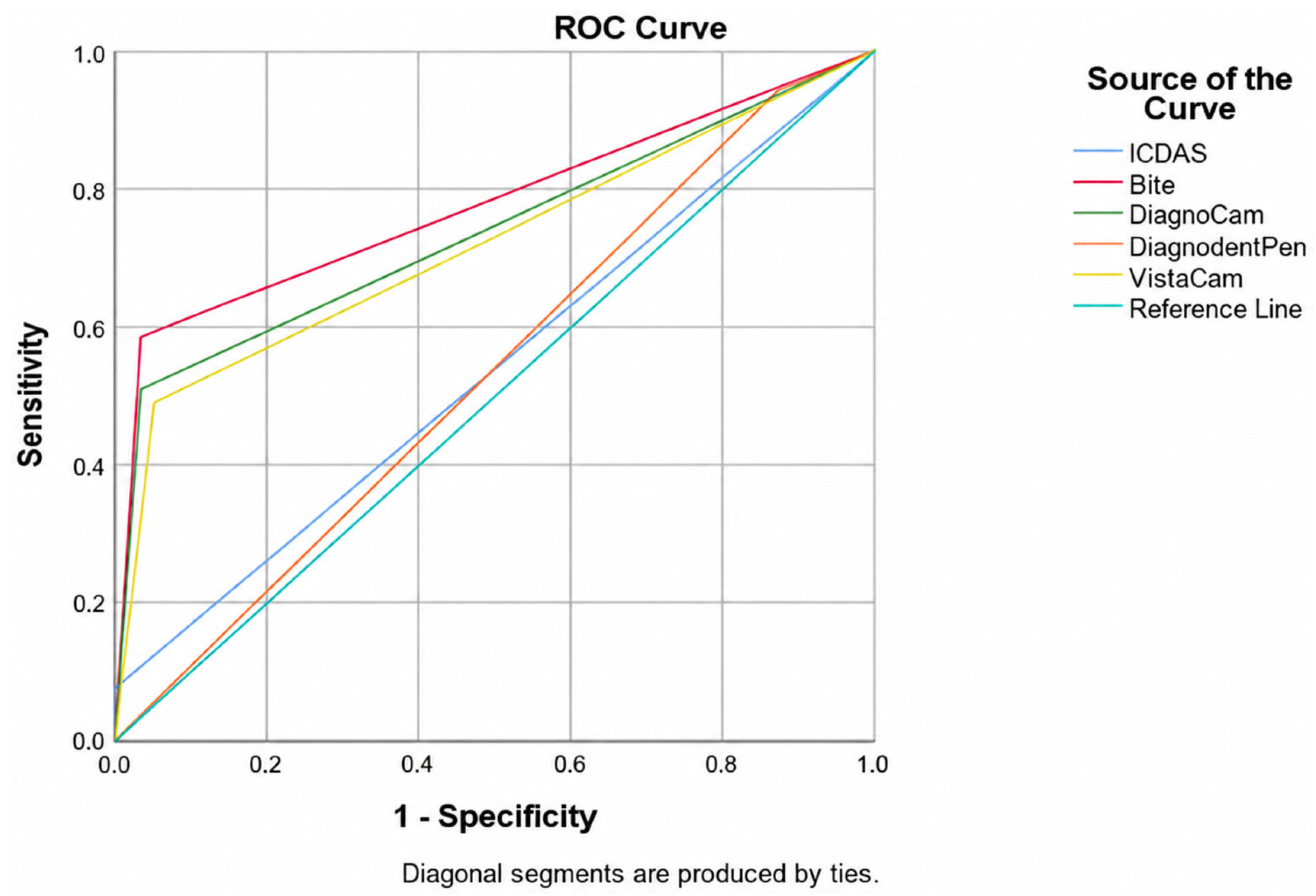
| Visual examination | 7.5 | 100.0 | 100.0 | 78.1 | 78.5 | 0.538 |
| Bite-wing radiography | 58.5 | 96.6 | 83.8 | 88.5 | 87.7 | 0.775 |
| DIAGNOcam | 50.9 | 96.6 | 81.8 | 86.7 | 86.0 | 0.738 |
| VistaCam iX-Proxi head | 49.1 | 94.9 | 74.3 | 86.0 | 84.2 | 0.720 |
| DIAGNOdent Pen | 94.3 | 12.6 | 24.6 | 88.0 | 31.6 | 0.535 |
| Caries Diagnostic Methods | Sensitivity (%) | Specificity (%) | PPV (%) | NPV (%) | Accuracy (%) | |
|---|---|---|---|---|---|---|
| Enamel | Visual examination | 100.0 | 0.0 | 98.9 | 0.0 | 98.9 |
| Bite-wing radiography | 97.7 | 0.0 | 98.8 | 0.0 | 96.6 | |
| DIAGNOcam | 98.3 | 0.0 | 98.8 | 0.0 | 97.1 | |
| VistaCam iX-Proxi head | 94.8 | 0.0 | 98.8 | 0.0 | 93.7 | |
| DIAGNOdent Pen | 12.8 | 100.0 | 100.0 | 1.3 | 13.8 | |
| Dentin | Visual examination | 7.8 | 100.0 | 100.0 | 6.0 | 7.4 |
| Bite-wing radiography | 60.8 | 33.3 | 93.9 | 4.8 | 57.4 | |
| DIAGNOcam | 52.9 | 0.0 | 90.0 | 0.0 | 50.0 | |
| VistaCam iX-Proxi head | 51.0 | 100.0 | 100.0 | 10.7 | 48.1 | |
| DIAGNOdent Pen | 94.1 | 0.0 | 94.1 | 0.0 | 88.9 |
| Caries Diagnostic Methods | Researchers | Enamel | Dentin | ||
|---|---|---|---|---|---|
| n | % | n | % | ||
| Visual examination | 1st Researcher | 75 | 98.7 | 1 | 1.3 |
| 2nd Researcher | 74 | 97.4 | 2 | 2.6 | |
| 3rd Researcher | 75 | 98.7 | 1 | 1.3 | |
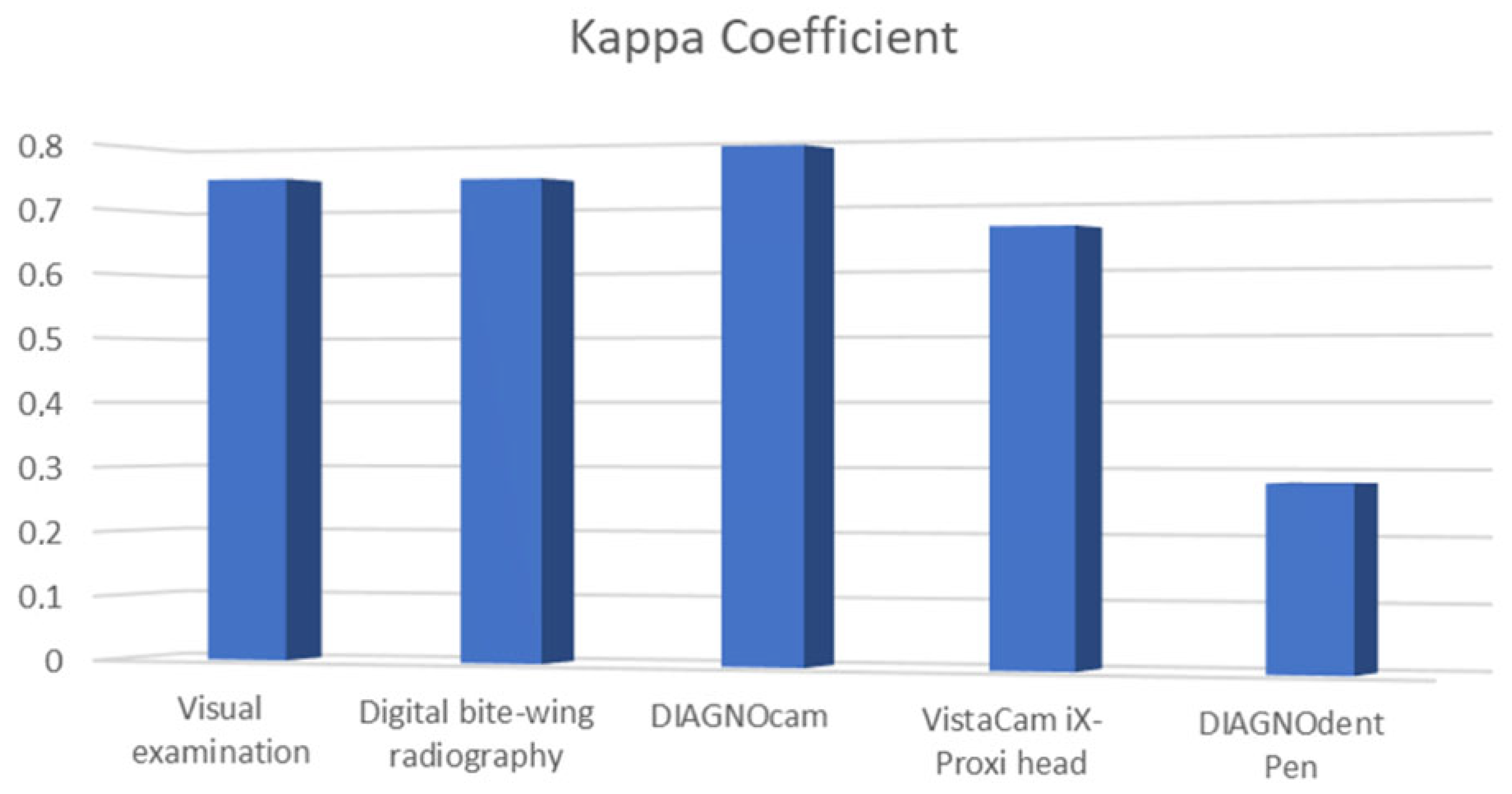
| Kappa coefficient | 0.746 | ||||
| p | 0.000 | ||||
| Digital bite-wing radiography | 1st Researcher | 62 | 81.6 | 14 | 18.4 |
| 2nd Researcher | 64 | 84.2 | 12 | 15.8 | |
| 3rd Researcher | 65 | 85.5 | 11 | 14.5 | |
| Kappa coefficient | 0.742 | ||||
| p | 0.000 | ||||
| DIAGNOcam | 1st Researcher | 66 | 86.8 | 10 | 13.2 |
| 2nd Researcher | 65 | 85.5 | 11 | 14.5 | |
| 3rd Researcher | 64 | 84.2 | 12 | 15.8 | |
| Kappa coefficient | 0.787 | ||||
| p | 0.000 | ||||
| VistaCam iX-Proxi head | 1st researcher | 63 | 82.9 | 13 | 17.1 |
| 2nd researcher | 67 | 88.2 | 9 | 11.8 | |
| 3rd researcher | 63 | 82.9 | 13 | 17.1 | |
| Kappa coefficient | 0.662 | ||||
| p | 0.000 | ||||
| DIAGNOdent Pen | 1st researcher | 7 | 9.2 | 69 | 90.8 |
| 2nd researcher | 3 | 3.9 | 73 | 96.1 | |
| 3rd researcher | 15 | 19.7 | 61 | 80.3 | |
| Kappa coefficient | 0.281 | ||||
| p | 0.000 | ||||
Disclaimer/Publisher’s Note: The statements, opinions and data contained in all publications are solely those of the individual author(s) and contributor(s) and not of MDPI and/or the editor(s). MDPI and/or the editor(s) disclaim responsibility for any injury to people or property resulting from any ideas, methods, instructions or products referred to in the content. |
© 2026 by the authors. Licensee MDPI, Basel, Switzerland. This article is an open access article distributed under the terms and conditions of the Creative Commons Attribution (CC BY) license.
Share and Cite
Bingül, A.; Bala, O.; Akgül, S.; Sarı, C. In Vivo Comparison of Laser Fluorescence, Near-Infrared Transillumination, and Conventional Methods for Approximal Caries Detection. J. Clin. Med. 2026, 15, 7178. https://doi.org/10.3390/jcm15187178
Bingül A, Bala O, Akgül S, Sarı C. In Vivo Comparison of Laser Fluorescence, Near-Infrared Transillumination, and Conventional Methods for Approximal Caries Detection. Journal of Clinical Medicine. 2026; 15(18):7178. https://doi.org/10.3390/jcm15187178
Chicago/Turabian StyleBingül, Arda, Oya Bala, Sinem Akgül, and Ceyda Sarı. 2026. "In Vivo Comparison of Laser Fluorescence, Near-Infrared Transillumination, and Conventional Methods for Approximal Caries Detection" Journal of Clinical Medicine 15, no. 18: 7178. https://doi.org/10.3390/jcm15187178
APA StyleBingül, A., Bala, O., Akgül, S., & Sarı, C. (2026). In Vivo Comparison of Laser Fluorescence, Near-Infrared Transillumination, and Conventional Methods for Approximal Caries Detection. Journal of Clinical Medicine, 15(18), 7178. https://doi.org/10.3390/jcm15187178