1. Introduction
Glaucoma continues to be one of the leading causes of irreversible visual impairment worldwide [
1]. Elevated intraocular pressure is the most important modifiable risk factor and can lead to progressive optic nerve damage and visual field loss [
2]. Primary open angle glaucoma (POAG) is the most common form of glaucoma, whereas pseudoexfoliation glaucoma (PXG) is a secondary open angle glaucoma caused by the accumulation of pseudoexfoliative material in ocular tissues [
3,
4]. In PXG, this material can accumulate in the anterior segment and trabecular meshwork, leading to impaired aqueous outflow and increased IOP. Compared with POAG, PXG is often associated with higher IOP levels, stronger IOP fluctuations, and a more aggressive clinical course [
4].
The first-line treatment usually consists of topical IOP-lowering medication. However, long-term adherence can be difficult, especially in elderly patients, and local side effects may limit the effectiveness of topical therapy [
5]. Filtrating procedures such as trabeculectomy or PreserFlo
® Microshunt provide substantial pressure reduction, but they are more invasive and therefore are associated with a higher rate of postoperative complications [
6,
7]. Therefore, less invasive surgical alternatives have been developed for mild to moderate glaucomas, and minimally invasive glaucoma surgery (MIGS) has gained increasing importance in recent years [
8,
9].
Various MIGS devices are available, differing in their mechanisms of action. The Hydrus
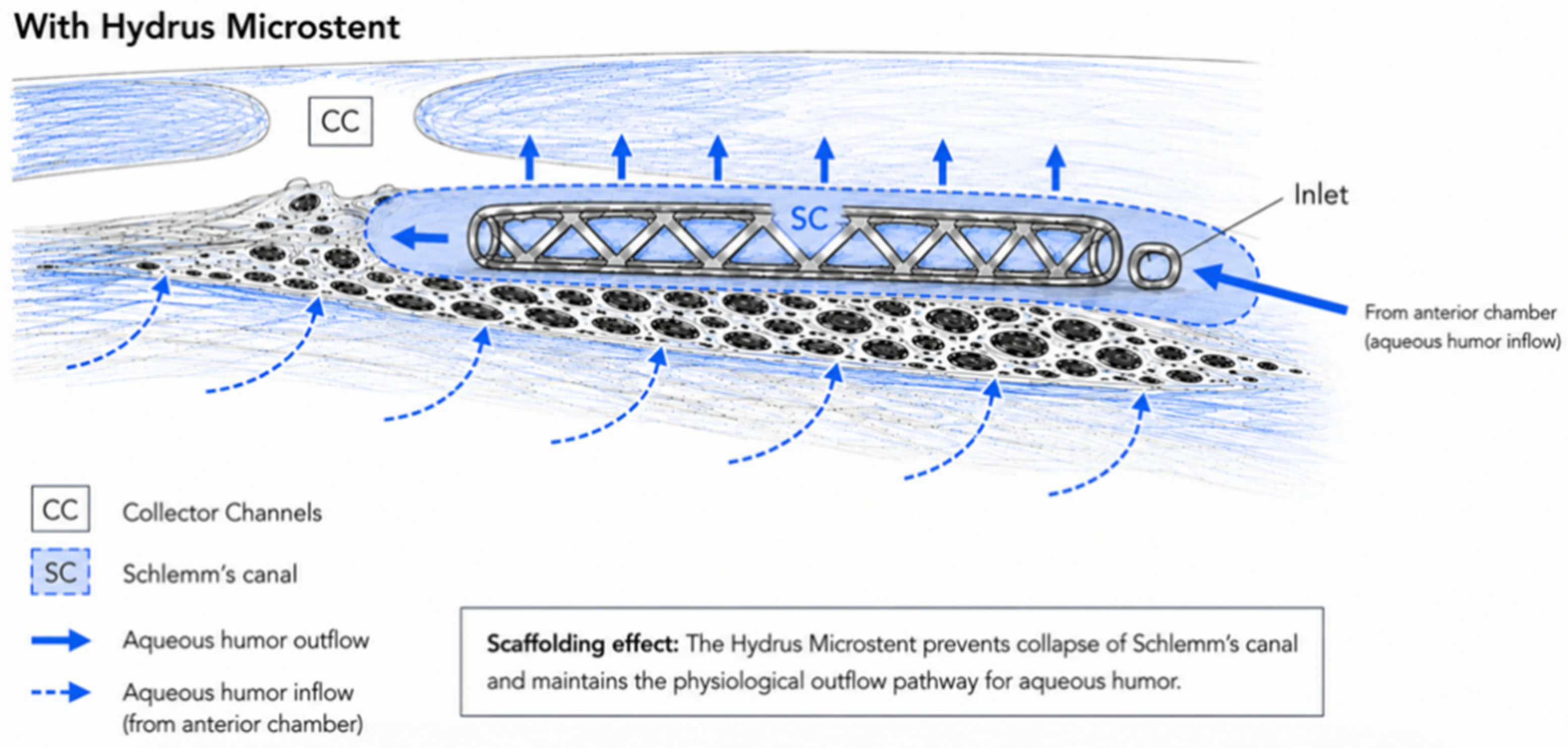
® Microstent is an 8-mm Schlemm’s canal-based device designed to improve aqueous humor outflow through the conventional outflow pathway. By dilating and scaffolding Schlemm’s canal, the device may provide access to several collector channels and reduce outflow resistance [
10]. When implanted in combination with cataract surgery, the Hydrus
® Microstent has shown promising results in lowering IOP and reducing the need for IOP-lowering medication in patients with open angle glaucoma [
11,
12,
13].
Most clinical studies on the Hydrus® Microstent have focused on POAG, while data specifically addressing PXG remain limited. This is clinically relevant because PXG is often more difficult to manage and may present additional surgical challenges, especially in eyes undergoing cataract surgery.
The purpose of the present study was to compare clinical outcomes after Hydrus® Microstent implantation combined with cataract surgery in eyes with PXG and POAG. The main focus was on the evaluation of postoperative IOP, IOP-lowering medication burden, best-corrected visual acuity, surgical success, safety, and the relationship between anterior chamber depth and IOP reduction.
2. Materials and Methods
2.1. Study Design, Inclusion and Exclusion Criteria
This is a single-center, retrospective comparative cohort study conducted at the Department of Ophthalmology, LMU University Hospital Munich, Germany. Consecutive eyes undergoing combined cataract surgery and Hydrus
® Microstent implantation between June 2021 and December 2025 were retrospectively screened. Eligible patients demonstrated ophthalmoscopically detectable glaucomatous optic neuropathy, mild to moderate visual field (VF) loss as defined by Hodappe–Andersone–Parrish criteria, best-corrected visual acuity (BCVA) of 1 or better, Shaffer grade III or IV angle width in all 4 quadrants, and a medicated IOP of 31 mmHg or less. All eyes meeting the predefined inclusion criteria and with sufficient follow-up data were included. Baseline characteristics are summarized in
Table 1.
Exclusion criteria were age younger than 18 years at the time of surgery, previous glaucoma surgery in the respective eye, silicone oil tamponade, simultaneous intravitreal injection therapy at the time of surgery, or selective laser trabeculoplasty (SLT) within 90 days before surgery and corneal dystrophy. Both eyes of one patient could be included if both eyes fulfilled the inclusion criteria.
The study adhered to the tenets of the Declaration of Helsinki and was approved by the local ethics committee (Ethics ID: 25-0296). All patients provided written consent prior to any study-related procedure.
2.2. Baseline Measurements
Epidemiological data were obtained from each patient, including age, gender, previous ocular comorbidities or procedures and objective refraction-based Snellen chart visual acuity, which was later converted to logMAR for analysis.
Intraocular pressure was measured using Goldmann applanation tonometry. Standard automated perimetry was performed before surgery using a Humphrey Visual Field Analyzer (Carl Zeiss AG, Oberkochen, Germany), and mean deviation (MD) and pattern standard deviation (PSD) values were recorded when available. Anterior chamber depth and lens thickness were obtained from the preoperative biometric assessment.
Postoperative data were collected at predefined retrospective intervals, including day 1, day 2, 1–4 weeks, 4–6 weeks, 3–4 months, 4–6 months, and 8–14 months (mean 12.5 ± 2.4 months for POAG and mean 11.6 ± 2.9 months for PXG) after surgery. All data were extracted from the hospital’s electronic medical record system.
2.3. Surgery
The implantation of the Hydrus® Microstent was performed in combination with routine phacoemulsification and intraocular lens implantation under topical or peribulbar anesthesia. All procedures were performed by one experienced glaucoma surgeon at the Department of Ophthalmology, LMU University Hospital Munich, Germany (MM). The Hydrus® Microstent device (Alcon Inc., Fort Worth, TX, USA) is preloaded in a sterile, hand-held delivery cannula. The delivery cannula has a slight curvature to follow the anatomical contour of the anterior chamber angle and Schlemm’s canal.
After completion of cataract surgery, a clear corneal incision was used to introduce the cannula into the anterior chamber. For accurate placement of the device, a surgical gonioprism was used, and the patient’s head position and the surgical microscope were adjusted to allow visualization of the nasal angle. An ophthalmic viscosurgical device was injected to deepen the anterior chamber and widen the angle.
Under direct gonioscopic visualization, the cannula tip was advanced towards the trabecular meshwork and used to access Schlemm’s canal according to the manufacturer’s instructions for use. Using the integrated delivery mechanism, the Hydrus® Microstent was inserted and advanced along approximately 90 degrees of Schlemm’s canal. The proximal part of the stent remained visible in the anterior chamber. After correct positioning was confirmed, the injector was withdrawn and the ophthalmic viscosurgical device was removed.
All surgeries were performed as part of routine clinical care and perioperative management followed the routine clinical standards of the Department of Ophthalmology, LMU University Hospital Munich.
2.4. Peri- and Postoperative Management
In both groups, dexagentamicin 5 mL eye drops 4×/d were given for 7 days. In patients who are at risk for developing macular edema nepafenac 1 mg/mL eye drops 3×/d were prescribed additionally for 4 weeks.
Postoperative medication adjustments were guided by the individual target IOP, taking into account pachymetry and glaucoma severity. Ocular hypotensive medications were added when the follow-up IOP exceeded 18 mmHg or when there was evidence of structural or functional disease progression. In patients at increased risk of macular edema (e.g., those with an epiretinal membrane or a history of central retinal vein occlusion), prostaglandin analogues were avoided during the first postoperative month. During this period, patients were treated with either a topical carbonic anhydrase inhibitor or an alpha-adrenergic agonist, alone or in combination with timolol 0.5%. After the first postoperative month, patients requiring continued IOP-lowering therapy were switched to prostaglandin analogues, with additional medications subsequently introduced according to the IOP response.
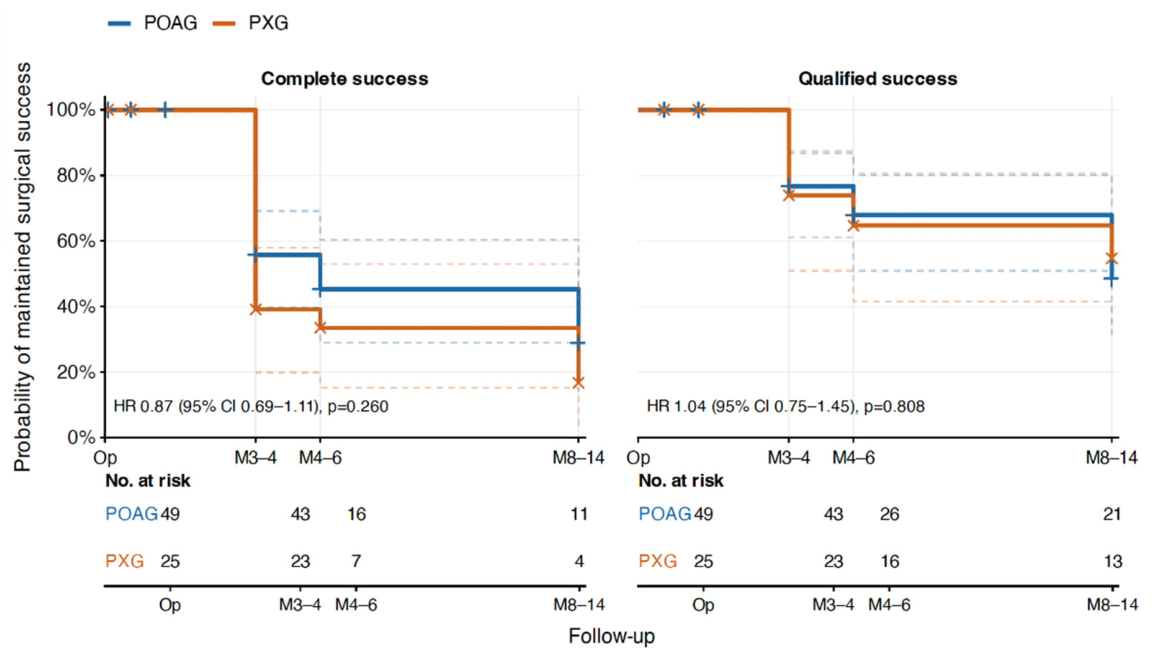
Surgical success was evaluated at two postoperative intervals: 4–6 months and 8–14 months. The 8–14-month interval was considered the final follow-up endpoint, whereas the 4–6-month interval was analyzed as a secondary mid-term endpoint. Complete success was defined as an IOP reduction of at least 20% from baseline without IOP-lowering medications and without secondary glaucoma surgery, surgical revision, or rescue operation before or at the respective postoperative endpoint. Qualified success was defined as an IOP reduction of at least 20% from baseline irrespective of the use of IOP-lowering medications and without secondary glaucoma surgery, surgical revision, or rescue operation. Medication-free status was analyzed separately and defined as no use of IOP-lowering medications at the respective postoperative time point. Anterior chamber depth and lens thickness were analyzed as additional baseline parameters to evaluate their potential impact on postoperative IOP reduction.
2.5. Statistical Analysis
Statistical analyses were performed in R (R Version 4.4.2, R Foundation for Statistical Computing, Vienna, Austria). Continuous variables are reported as mean ± standard deviation and categorical variables as counts and percentages. Because some patients contributed both eyes, analyses were selected to account for within-patient correlation. Baseline age and sex were assessed at the patient level; age was compared using an independent-samples
t-test and sex using Fisher’s exact test. Eye-level continuous baseline characteristics were compared between POAG and PXG using generalized estimating equations [
14] with a Gaussian distribution, exchangeable working correlation structure, and patient-clustered robust standard errors.
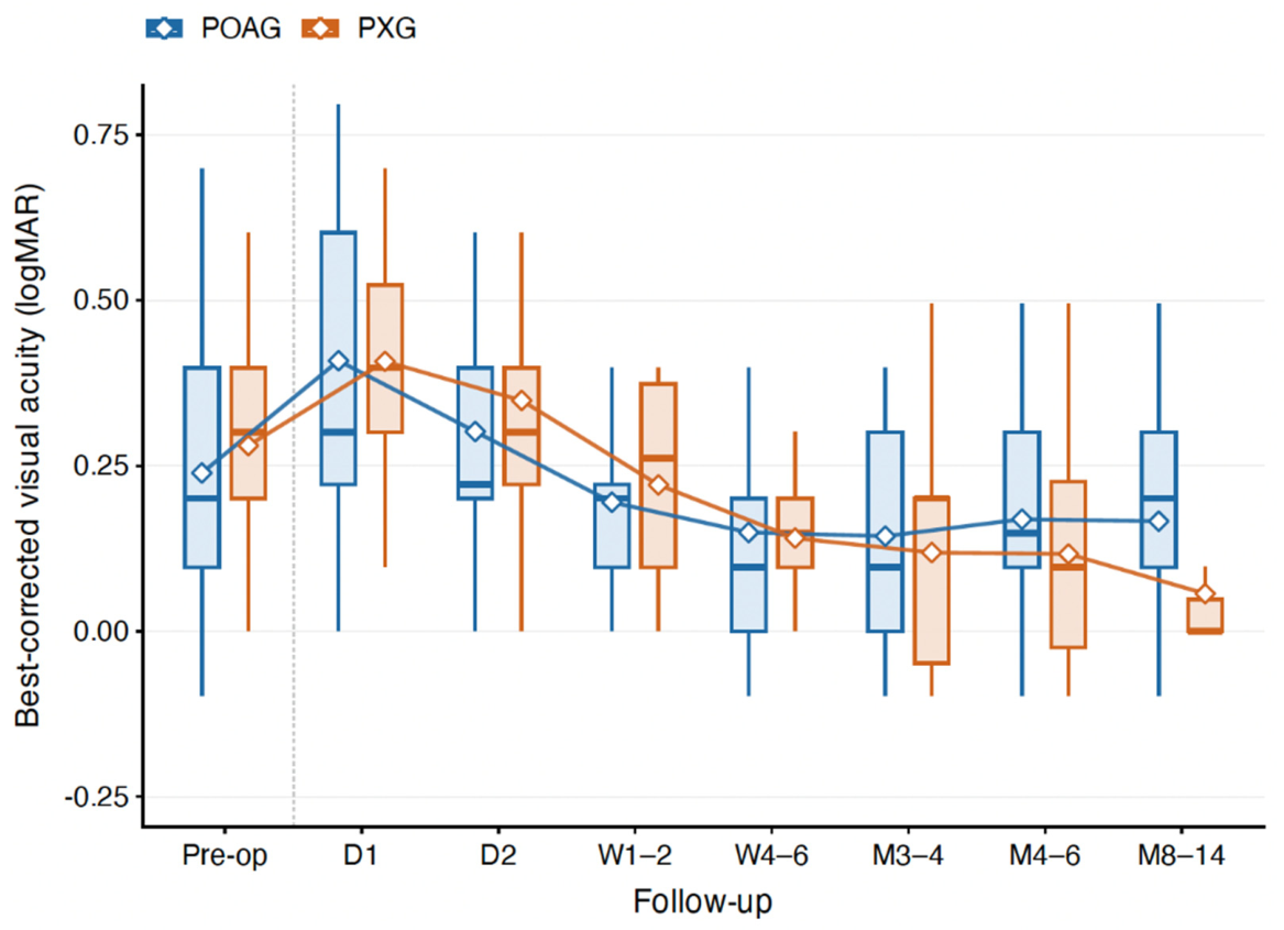
Longitudinal changes in IOP, number of topical IOP-lowering medications, and best-corrected visual acuity (BCVA, logMAR) were analyzed using linear mixed-effects models including glaucoma subtype, postoperative time, and their interaction as fixed effects, with age included as a covariate. Random intercepts for patient and eye were included to account for correlation between fellow eyes and repeated measurements within the same eye, respectively. All available observations were included without single-value imputation of missing follow-up measurements. Overall effects of glaucoma subtype, time, and the glaucoma subtype-by-time interaction were assessed using type III F-tests with Satterthwaite approximation of denominator degrees of freedom. Estimated marginal means and corresponding 95% confidence intervals were derived from the fitted models for prespecified between-group and longitudinal contrasts.
Surgical success was evaluated using time-to-event methods. Qualified success was defined as an IOP reduction of at least 20% from the preoperative value, whereas complete success additionally required the absence of topical IOP-lowering medication. To reduce misclassification caused by isolated IOP fluctuations around the success threshold, criterion-based failure was defined as failure to meet the respective success criterion at two consecutive evaluable postoperative visits. The time of the first of the two consecutive unsuccessful visits was assigned as the failure time. Because no subsequent assessment was available after the final 8–14-month visit, failure at this final scheduled interval was accepted without confirmatory follow-up. Eyes without documented failure were censored at their last evaluable visit. As follow-up was recorded in predefined clinical intervals rather than at uniform exact time points, nominal times of 3.5, 5, and 11 months were used for the 3–4-, 4–6-, and 8–14-month visits, respectively. Kaplan–Meier curves were used to estimate the probability of maintained qualified and complete surgical success. Differences between POAG and PXG were assessed using Cox proportional-hazards models with patient-clustered robust standard errors to account for inclusion of fellow eyes; hazard ratios are reported with 95% confidence intervals.
The proportion of eyes free of topical IOP-lowering medication at 4–6 and 8–14 months was analyzed separately as a secondary binary outcome using logistic GEE with patient-level clustering and an exchangeable working correlation structure. As a sensitivity analysis addressing bilateral inclusion, all principal longitudinal and survival analyses were repeated after randomly selecting one eye per patient using a prespecified fixed random seed. In addition, a more conservative survival analysis in which a single unsuccessful visit was sufficient to define failure was performed to assess the robustness of the Kaplan–Meier findings to the failure definition.
As this was a retrospective study of an available clinical cohort, no a priori sample-size calculation was performed. Effect estimates are therefore presented together with 95% confidence intervals, and absence of statistical significance was not interpreted as evidence of equivalence. All statistical tests were two-sided, and a p value <0.05 was considered statistically significant.
4. Discussion
The present retrospective comparative cohort study provides the first real-world evidence on the clinical outcomes of Hydrus® Microstent implantation combined with cataract surgery in eyes with pseudoexfoliation glaucoma compared to primary open angle glaucoma. In the present study, both the POAG and the PXG group showed a significant postoperative reduction in intraocular pressure and in the number of IOP-lowering medications. Importantly, no statistically significant between-group differences were observed in the longitudinal course regarding IOP or medication use. Moreover, surgical success rates, the rate of postoperative complications, and additional glaucoma-related interventions were not significantly different between groups. These results suggest that Hydrus® Microstent implantation combined with cataract surgery may achieve similar outcomes in PXG and POAG eyes.
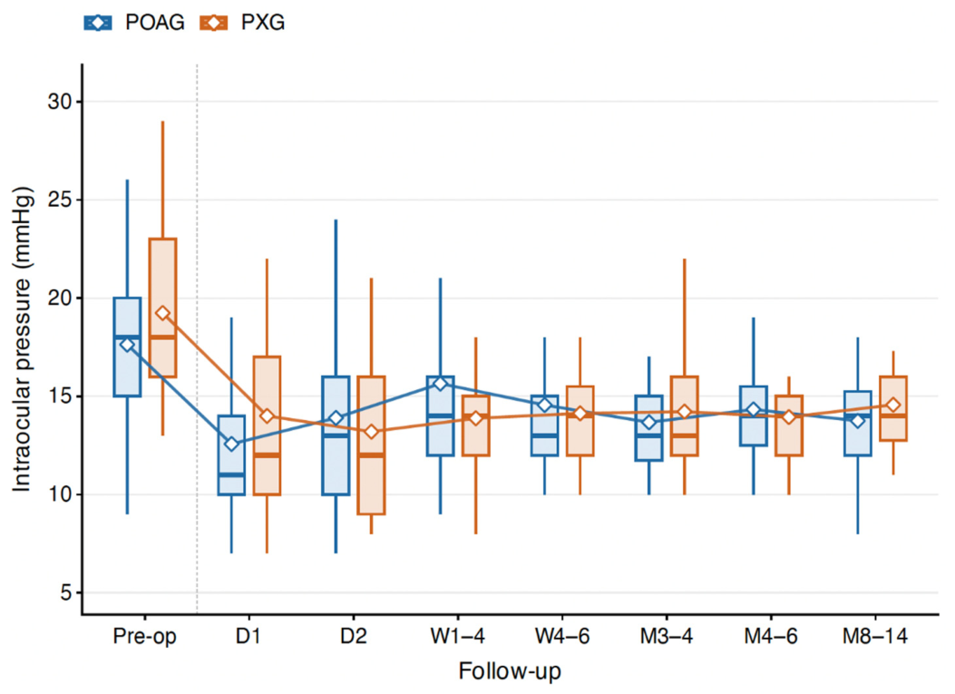
The development of IOP reported in the present study is in line with previous clinical studies evaluating the Hydrus
® Microstent in POAG [
13]. In the present cohort, mean IOP decreased significantly from baseline to the final 8–14-month follow-up in both groups. The mean IOP reduction was −4.0 mmHg in POAG eyes and −5.1 mmHg in PXG eyes, with no significant difference in the magnitude of IOP reduction between groups. The magnitude of IOP reduction observed in the present study was slightly lower than that reported in the HORIZON trial for POAG (mean reduction: 8.7 mmHg). However, this difference should be interpreted in light of the substantially lower baseline IOP in our real-world cohort, as no preoperative medication washout was performed. Conversely, the observed IOP reduction is consistent with findings from other real-world studies evaluating implant-based MIGS combined with cataract surgery. In particular, the meta-analysis by Kahale et al. reported a mean IOP reduction of 4.7 mmHg following combined cataract surgery and iStent implantation, which is comparable to the results of the present study.
In the HORIZON study, cataract surgery alone reached similar IOP reduction compared to cataract surgery + MIGS; however, the combination reached better reduction in the number of medications.
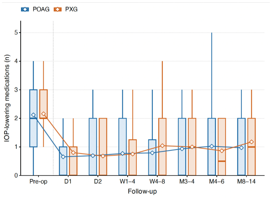
That is to say that, in addition to achieving IOP reduction, one of the principal clinical advantages of MIGS procedures is the reduction in glaucoma medication burden. In the present study, the number of IOP-lowering medications decreased significantly in both groups from baseline to 8–14 months, with a mean reduction in drops of −1.2 medications (
p < 0.001) for POAG and −1.1 medications for PEX. Comparable reductions in medication burden have been reported in previous studies evaluating the Hydrus
® Microstent, including both randomized controlled trials and real-world analyses, supporting the role of Hydrus
® implantation combined with cataract surgery as an effective strategy to reduce dependence on topical IOP-lowering medication [
12,
13,
15,
16]. Notably, the magnitude of medication reduction observed in the present study is consistent with the findings of the HORIZON trial, in which patients undergoing combined Hydrus
® implantation and cataract surgery achieved a mean reduction of 1.2 glaucoma medications. This is clinically relevant because reducing topical therapy may improve treatment adherence, decrease ocular surface-related side effects, and reduce the overall burden of chronic glaucoma care, particularly in elderly patients [
17,
18].
The similar reductions in IOP and medication burden observed in POAG and PXG eyes is particularly noteworthy, as PXG is generally regarded as more difficult to treat due to its higher baseline IOP, stronger IOP fluctuations, faster progression, and more pronounced alterations of the conventional aqueous outflow pathway [
4]. Despite these characteristics, PXG eyes achieved similar reductions in IOP and medication compared to POAG eyes. In PXG, pseudoexfoliative material accumulates in the anterior segment and especially in the trabecular meshwork (TM) [
19]. The pseudoexfoliative material induces profound structural and functional alterations of the TM, including cellular dysfunction with following reduced phagocytic capacity and further accumulation of pigment granules within the outflow system. In addition, PEX is associated with chronic low-grade inflammation: Elevated concentrations of inflammatory cytokines that stimulate abnormal extracellular matrix remodeling and fibrosis within the trabecular meshwork have been identified in the aqueous humor of affected eyes, further increasing outflow resistance [
20]. Iris pigment dispersion may additionally lead to obstruction of the trabecular meshwork and may contribute to postoperative intraocular pressure spikes after angle-based surgery. In the present study, despite PEX as a risk factor for complications within MIGS and cataract surgery, no significant difference in effectiveness was observed between POAG and PEX patients. Histopathological findings may provide a mechanistic explanation for the efficacy of the Hydrus
® Microstent in PXG. Rasmussen et al. demonstrated by electron microscopy that pseudoexfoliative material can induce focal collapse and splitting of Schlemm’s canal, thereby increasing outflow resistance [
18]. Owing to its scaffolding effect, the Hydrus
® Microstent may help maintain or restore Schlemm’s canal patency, potentially facilitating aqueous humor outflow despite these pathological changes (
Figure 5) [
21]. The microstent might furthermore prevent the development of an early rise in IOP after cataract surgery in PEX. Importantly, as in POAG, the pathological alterations in PXG extend beyond the trabecular meshwork and involve the entire conventional aqueous outflow pathway, including the distal collector channels [
21]. While trabecular MIGS can improve aqueous outflow at the level of the trabecular meshwork and Schlemm’s canal, persistent dysfunction of the distal outflow system still limits further intraocular pressure reduction in both glaucoma subtypes.
Although cataract extraction likely reduces ongoing iris–lens friction and thus new pigment release, pre-existing pseudoexfoliative fibrillin deposits, chronically elevated aqueous inflammatory mediators, and peristromal fibrosis may still progressively compromise stent patency and distal collector function beyond the 8–14-month window [
22]. The present 14-month follow-up provides valuable insights into the mid-term performance of the stent; however, it cannot exclude the possibility of late stent occlusion or a subsequent increase in intraocular pressure, particularly in patients with PEX.
Another important finding of this study was the favorable safety profile during the analyzed follow-up period. No postoperative complications were documented in the available dataset, and no additional glaucoma-related postoperative interventions were required in either group up to the final 8–14-month follow-up. This is relevant because PXG eyes can be more difficult to treat surgically due to pseudoexfoliation-related anterior segment changes (see above). These findings are in line with previous Hydrus
® studies, which also reported a favorable safety profile when the Hydrus
® Microstent was implanted in combination with cataract surgery [
11,
12,
16]. However, because the present study was retrospective and included a limited number of eyes, the safety results should be interpreted carefully.
This study has several limitations. First, it was a retrospective single-center study, which may limit the generalizability of the results also due to the limited sample size. Second, the PXG group was slightly older than the POAG group, reflecting the epidemiology of pseudoexfoliation syndrome. Importantly, there is no evidence that advanced age affects the IOP-lowering efficacy of trabecular MIGS. Therefore, the age difference between groups is unlikely to have biased the observed outcomes. The third limitation of the present study is the limited availability of BCVA data at the final follow-up visits. Therefore, long-term changes in visual acuity could not be assessed comprehensively. However, BCVA was not a primary endpoint of the study.
Despite these limitations, the present study adds first real-world data on Hydrus® Microstent implantation combined with cataract surgery in PXG. In this cohort, PXG eyes showed comparable clinical outcomes to POAG eyes regarding IOP reduction, medication reduction, BCVA development, surgical success, and documented safety outcomes. These findings suggest that combined cataract surgery and Hydrus® Microstent implantation may be a useful treatment option in selected patients with PXG. Larger prospective studies with longer follow-up are needed to confirm these results.




 ,
,