Review

Overview of Non-Cirrhotic Portal Hypertension in Pediatric Patients

by
Ambika Walecha
1,†,
Senthilkumar Sankararaman
2,*,† and
Kadakkal Radhakrishnan
2
1
Lady Hardinge Medical College, New Delhi 110001, India
2
Cleveland Clinic Children’s Hospital, Cleveland, OH 44122, USA
*
Author to whom correspondence should be addressed.
These authors contributed equally to this work.
J. Clin. Med. 2026, 15(17), 6901; https://doi.org/10.3390/jcm15176901
Submission received: 5 July 2026 / Revised: 31 August 2026 / Accepted: 4 September 2026 / Published: 6 September 2026

Abstract

Non-cirrhotic portal hypertension (NCPHT) is defined as portal hypertension (PHT) occurring in the absence of cirrhosis. Major etiological causes of NCPHT include immunological disorders, chronic infections, exposure to medications or toxins, prothrombotic conditions, and several genetic syndromes, highlighting that NCPHT is not a single disease but a shared phenotype arising from diverse underlying pathways. NCPHT is frequently misdiagnosed, largely due to inconsistent nomenclature and limited scientific literature. The broader term non-cirrhotic portal fibrosis (NCPF) or porto-sinusoidal vascular disease (PSVD) includes patients in a preclinical stage who demonstrate histological features similar to NCPHT but lack clinical evidence of PHT. Early detection is linked to a favorable prognosis and improved clinical outcomes. Management strategies in pediatrics continue to rely on extrapolations from adult practice, with sparse evidence to guide pediatric care. Liver biopsy remains the cornerstone of diagnosis, demonstrating nodular regenerative hyperplasia, obliterative portal venopathy, or incomplete septal fibrosis. Management focuses on prophylactic and symptomatic care, with endoscopic therapy for controlling variceal bleeding. Porto-systemic shunts and, ultimately, liver transplantation therapies may be needed for advanced stages. A pressing need exists for standardized diagnostic criteria and multicenter studies to define natural history, refine risk stratification, and evaluate therapeutic approaches in the pediatric population. This review provides an overview of the pediatric causes of NCPHT, outlines the current understanding of pathophysiology, and discusses the clinical presentations and management strategies, while highlighting existing research gaps.

1. Introduction

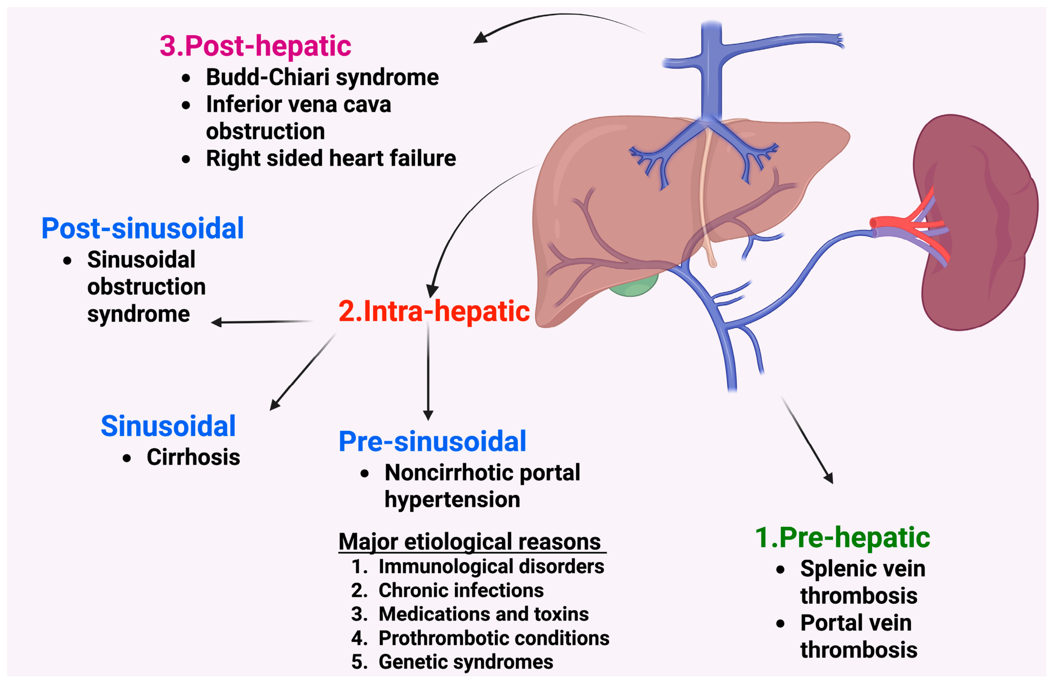
Portal venous pressure depends on splanchnic blood inflow and the resistance opposing this flow [1]. It is expressed as the pressure gradient between the portal vein (reflecting blood inflow into the liver) and the inferior vena cava (demonstrating venous outflow from the liver) [1]. In healthy participants, this hepatic venous pressure gradient (HVPG) ranges between 1 and 5 mm Hg. Portal hypertension (PHT) is defined as a gradient above 5 mm Hg [1,2,3,4,5,6,7]. The complications of PHT, such as variceal bleeding, usually develop when this gradient is above 10 mm Hg [8,9,10]. PHT can generally be classified as prehepatic, hepatic, and posthepatic based on the anatomical level of obstruction (Figure 1) [1,11,12,13].
Non-cirrhotic portal hypertension (NCPHT) includes a heterogeneous group of chronic vascular diseases that are characterized by elevated pressure in the portal circulation in the absence of cirrhosis [11,14,15]. Causes of NCPHT can be categorized as prehepatic (e.g., splenic or portal vein thrombosis, also referred to as extrahepatic portal vein obstruction), intrahepatic, and posthepatic (e.g., Budd–Chiari syndrome, inferior vena cava obstruction, and cardiac dysfunction) [11,13,16]. Intrahepatic causes of NCPHT can be further divided into presinusoidal and postsinusoidal (e.g., sinusoidal obstruction syndrome) [15]. The major etiological reasons for intrahepatic, presinusoidal NCPHT include immunological disorders, chronic infections, exposure to drugs (medications) or toxins, prothrombotic hematological conditions, and several genetic syndromes (Figure 1) [15,17].
With advances in molecular and genetic testing methods, an increasing number of cases previously labeled as “idiopathic NCPHT” are now attributable to specific underlying disorders, resulting in a progressively smaller proportion of truly idiopathic cases compared to historical literature in this field. Further, these disorders are frequently overlooked due to their rarity, non-specific clinical presentation, heterogeneity of the underlying conditions, and the lack of a unified approach in nomenclature and sparse management guidelines in the pediatric population [18,19,20]. As other causes of PHT, such as splenic/portal vein thrombosis and cirrhosis of the liver, are relatively more common and well described in the pediatric population, we aim to focus only on intrahepatic, presinusoidal disorders of NCPHT and hereafter will simply refer to them as NCPHT [21,22,23,24].
In this narrative review, we outline the current understanding of the pathophysiology and etiology of NCPHT, highlighting the key genetic associations, discussing the clinical presentation and management strategies, and reviewing existing research gaps. In this review, we intend to provide a broader overview of this relatively rare, heterogeneous group of diseases predisposing to NCPHT. Key differences between NCPHT and cirrhosis are also detailed in this review.
Clinicians should be aware of various terminologies that overlap with NCPHT [25]. In most Asian countries, the terms non-cirrhotic portal fibrosis (NCPF) and idiopathic portal hypertension (IPH) have been in use to describe intrahepatic disorders characterized by PHT in the absence of cirrhosis and without an identifiable cause [26]. The term “NCPHT” necessitates the inclusion of only patients with established PHT in the advanced stages. However, in the early disease process, identical microscopic changes can be demonstrated without clinically evident PHT, and this terminology does not account for these patients. Some of the prominent histological changes of NCPHT were historically referred to as nodular regenerative hyperplasia (NRH) and obliterative portal venopathy (OPV). NRH is considered an outcome of intrahepatic vasculopathy, arising from ongoing hepatocyte injury and regeneration, leading to nodule formation that compresses adjacent sinusoids and small veins, or to perisinusoidal fibrosis, changes that may progress to PHT [27]. In OPV, the pathological hallmark is injury to the portal venules and sinusoids in the absence of cirrhosis [12,28]. Other overlapping terminologies that describe the histological features include “hepato-portal sclerosis”, “non-cirrhotic portal fibrosis” and “incomplete septal fibrosis” [29]. An umbrella term, “porto-sinusoidal vascular disease” (PSVD), was suggested by De Gottardi and colleagues, highlighting that all lesions involved portal venules or sinusoids without the clinical requirement of PHT [25,30,31]. Slight differences in all these terminologies across major clinical practice guidelines do exist, and clinicians should be aware of these changes [31,32,33].
We searched PubMed using the following keywords: “non-cirrhotic portal hypertension”, “non-cirrhotic portal fibrosis”, “idiopathic portal hypertension”, “porto-sinusoidal vascular disease”, “nodular regenerative hyperplasia”, and “obliterative portal venopathy”. We prioritized the inclusion of pediatric studies, supplemented by adult data where pediatric literature remains sparse.

2. Epidemiology

Given the rarity of NCPHT and the widespread heterogeneity in its nomenclature, accurately determining its epidemiology in all age groups remains difficult. It is estimated that NCPHT accounts for approximately 15–34% of all cases of PHT, with a noticeably higher prevalence in Japan and the Indian subcontinent, where it constitutes nearly one-third of all cases across all age groups [34,35]. The overall prevalence is declining, likely due to improvements in sanitation and hygiene [36]. In Western countries, it generally represents up to 5–16% of all PHT cases [16,20,37].
Epidemiological patterns also show geographic variation in gender distribution across age groups. Most adult cohorts from the West and the Indian subcontinent report a male predominance, whereas Japanese cohorts demonstrate a higher prevalence among females. The mean age of patients in the Indian cohort is 25–35 years, whereas Japanese and Western cohorts report a higher mean age of 36–56 years [20,37]. Single-center pediatric studies noted an average age between 5 and 12 years [38,39]. To our knowledge, no large-scale, population-based studies have been published specifically assessing NCPHT in the pediatric population.

3. Pathophysiology

A dual theory has been postulated to unravel the largely unknown and poorly characterized pathogenesis of this complex disease [12]. The first theory, popularly referred to as the “intrahepatic vascular obstruction” (resistance-initiated) theory, states that NCPHT can be triggered by any factor that injures the intrahepatic vascular bed. These include hypercoagulable states such as thrombophilia or antiphospholipid antibodies; T-cell-mediated autoimmune endothelial damage; and infections that may lead to microthrombosis [19,27,40,41]. This injury to the vascular bed may promote the occlusion of small portal veins, leading to liver ischemia. As a result, some areas of the liver undergo atrophy, whereas other regions develop compensatory hypertrophy. This structural distortion raises resistance to portal blood flow, ultimately leading to elevated portal pressures [40].
The second theory postulates overexpression of iNOS and eNOS in patients with NCPHT. These enzymes are responsible for nitric oxide (NO) synthesis; their overactivity results in sustained local NO overproduction. NO, being a potent vasodilator, causes dilation of the splenic sinusoids, resulting in progressive splenomegaly and a marked increase in splenic blood pooling. The enlarged and hyperperfused spleen augments portal inflow, thereby contributing directly to elevated portal pressures. As per this hypothesis, in NCPHT, splenomegaly may not simply be a passive consequence of vascular congestion but could be a pathogenic driver of the disease process. However, due to the limited and inconclusive nature of current evidence, this hypothesis remains speculative and requires further validation through robust studies [41].

4. Underlying Etiological Conditions Implicated in NCPHT

The underlying etiology of NCPHT is broadly categorized into five major groups: (i) immunological disorders, (ii) prothrombotic conditions, (iii) chronic infections, (iv) genetic syndromes, and (v) exposure to medications or toxins [17,41]. Although these associations are mostly better characterized in adult cohorts, it is reasonable to assume that pediatric NCPHT arises through a similar multifactorial interplay.

4.1. Immune-Mediated and Autoimmune Disorders

Immunological changes, such as decreased CD8+ cells and an altered CD4/CD8 ratio, are well described in OPV and play a crucial role in the pathogenesis of NCPHT [37]. Corroborating evidence for immune-mediated pathogenesis also comes from liver involvement in pediatric common variable immunodeficiency (CVID). CVID is associated with immune dysfunction that often manifests as NRH [42,43]. Persistent cytotoxic CD8+ T-cell infiltration targeting sinusoidal endothelial cells, along with alterations in the intestinal microbiome leading to translocation of inflammatory bacteria and endotoxins, constitutes the immunological basis for the development of liver pathological features such as PSVD and NRH in CVID patients [42]. These combined effects of endothelial-directed cytotoxicity and a disrupted gut–liver axis can ultimately lead to significant liver dysfunction. Similar mechanisms have been described in people (both pediatric and adult patients) with inborn errors of immunity [44], including ataxia telangiectasia [45], adenosine deaminase 2 (ADA2) deficiency [46], and chronic granulomatous disease [47]. Immunodeficiency-associated hepatic histology and clinical outcomes are noted in Table 1.

4.2. Prothrombotic Disorders

One of the major causes of PHT in the pediatric age group is extrahepatic portal vein obstruction (EHPVO), which is most often due to portal vein thrombosis triggered by factors such as umbilical sepsis, catheter-related infections, or neonatal interventions [22]. However, this represents extrahepatic thrombosis and is distinct from NCPHT, which is an intrahepatic disorder [17]. Nevertheless, both conditions can share an underlying prothrombotic state, resulting in a spectrum of manifestations [12]. The unifying hypothesis proposed by Sarin and Kumar suggests that a major thrombotic event early in life tends to occlude the main portal vein, leading to EHPVO, whereas repeated microthrombotic events later in life result in the gradual obliteration of small and medium intrahepatic portal venous branches, leading to NCPHT [49]. Supporting this concept, findings from a Western cohort showed that many adult patients with NCPHT have occult prothrombotic disorders, including myeloproliferative diseases, protein C/S deficiency, and antiphospholipid antibodies. These conditions promote tiny, recurrent microthrombi in intrahepatic portal venules, progressively narrowing or blocking them and producing OPV, one of the hallmarks of NCPHT [50].

4.3. Chronic Infections

Abdominal infections, particularly in early life, have long been implicated in the pathogenesis of OPV and the broader PSVD/NCPHT spectrum, especially in socioeconomically disadvantaged settings where recurrent bacterial exposure is common [19]. In infants, abdominal infections at birth, including omphalitis, neonatal sepsis, and recurrent diarrheal illnesses, are thought to trigger portal pyemia and pylephlebitis, setting off cycles of endothelial injury, microthrombi, and sclerosis of small- and medium-sized portal vein branches. This ultimately evolves into the characteristic vascular remodeling of OPV [37]. Experimental animal studies have supported this infectious hypothesis, as injecting dead E. coli or non-pathogenic colon bacilli into the portal circulation of animals reproduced IPH-like changes [40].

4.4. Genetic Syndromes

Many syndromes have been implicated in NCPHT, and the common ones are described here.

4.4.1. Turner Syndrome

Recent studies identified liver function abnormalities and three principal patterns of liver involvement, namely steatosis, fibrosis, biliary lesions, and vascular-mediated architectural changes such as NRH and focal nodular hyperplasia, in Turner syndrome in both pediatric and adult patients [51,52]. The authors suggested that Turner syndrome is characterized by congenital vascular malformations and OPV, which alter the intrahepatic blood flow and promote the development of NRH. Another study in adults with Turner syndrome revealed that approximately one-fourth of the cases showed NCPHT-like histology, with three patients progressing to clinically significant PHT [53].

4.4.2. Adams–Oliver Syndrome

Adams–Oliver syndrome is a congenital vascular disorder; patients typically present with aplasia cutis, cutis marmorata, and limb anomalies such as syndactyly and absent phalanges. Several pediatric cases have reported the presence of NCPHT among Adams–Oliver syndrome patients, and experts highlight that Adams–Oliver syndrome and NCPHT share a common mechanism involving congenital microvascular abnormalities and a predisposition to thrombosis, which may be modulated genetically [54,55].

4.4.3. Prolidase Deficiency

Prolidase deficiency leads to impaired wound healing due to its important role in collagen turnover. Reduced activity of the enzyme leads to characteristic clinical features such as facial dysmorphism, recurrent infections, and splenomegaly, and a potential link to vascular-mediated liver injury [56]. In contrast, excess prolidase activity has been linked to several fibrotic liver diseases, whereas its deficiency could potentially contribute to vascular-mediated liver injury. These effects may reflect impaired angiogenesis and aberrant inflammatory signaling, such as nuclear factor kappa B (NF-κB) activation. Consistent with these findings, a recent case series in adults also suggested that prolidase deficiency may predispose to portal venous abnormalities and NCPHT [57].

4.4.4. Short Telomere Disorders

Dyskeratosis congenita and the broader telomere biology disorders represent a group of conditions marked by markedly shortened telomeres. These are clinical syndromes arising from telomere dysfunction, with hallmark physical features including nail dystrophy, oral leukoplakia, reticular skin, and bone marrow failure. Younger individuals typically present with bone marrow failure, immunodeficiency, mucocutaneous stigmata, and gastrointestinal inflammation, whereas adults typically have isolated marrow failure, pulmonary fibrosis, or liver disease. In a recent cohort of 58 patients (both pediatric and adult age groups) with dyskeratosis congenita/telomere biology disorder, around 10 patients (17.2%) developed clinically significant PHT over a seven-year follow-up [58]. Detailed histology revealed that three patients had a definite non-cirrhotic PHT pattern, characterized by nodular regenerative hyperplasia with OPV and periportal or bridging fibrosis. In addition, a subset of patients showed mild periportal/pericellular fibrosis or hemosiderosis with clinically established PHT. These constellations of findings are compatible with early or presumed PSVD/NCPHT. Overall, the study highlighted that patients with dyskeratosis congenita/telomere biology disorders, particularly those with severe multisystem phenotypes and autosomal recessive/X-linked recessive or TERF1-interacting nuclear factor 2 variants, may develop NCPHT, with a spectrum of histological changes [58].

4.4.5. Cystic Fibrosis

Liver involvement in the form of advanced cystic fibrosis (CF)-related disease (aCFLD), characterized by cirrhosis of the liver, has been reported in approximately 5–10% of people with CF, with onset of presentation in the first decade of life [59,60]. On the other hand, in the later adult age group, NCPHT has been documented in several case studies [59,61,62,63]. Platelet activation, a pro-inflammatory state characterized by vasculitis (endothelialitis), and recurrent infections (pulmonary and intestinal) have been postulated to be risk factors for NCPHT along with CF transmembrane conductance regulator (CFTR) dysfunction in CF [59].

4.4.6. The Genetic and Epigenetic Landscape

Numerous genetic and epigenetic studies have identified molecular defects that may lead to progressive injury and remodeling of the portal microvasculature, which are detailed in Table 2. These pathways outline a genetic landscape in which vascular, metabolic, and immune factors interact to give rise to an NCPHT phenotype. On a case-by-case basis, genetic screening for these abnormalities, especially in early-onset, syndromic, or familial cases, may support a timely underlying diagnosis and informed, individualized care. Future research should validate these associations and evaluate potential therapeutic targets for this disorder.

4.5. Drugs and Toxins

Medications such as 6-thioguanine have been implicated in causing NCPHT through their ability to cause microvascular injury and induce NRH. For example, Rawat et al. followed 10 children with 6-thioguanine toxicity and found that most had splenomegaly, thrombocytopenia, and PHT long after stopping the drug [72]. Moulik and Taj similarly reported that 8 of 11 children on 6-thioguanine developed features of PHT over 10 years [73]. These studies underscore the importance of long-term surveillance in patients treated with thiopurines. Other implicated drugs/toxins in NCPHT pathogenesis from adult cohorts include arsenic compounds [74], other chemotherapy medications such as oxaliplatin [75], and antiretroviral therapies [76], though the pediatric data remain sparse.

5. Clinical Approach to NCPHT

5.1. Clinical Manifestations

In pediatric patients, NCPHT commonly presents in the first two decades, whereas in adults it is typically present in the third or fourth decade of life [16,49,77]. Asymptomatic splenomegaly and complications of splenomegaly, such as hypersplenism, are more commonly reported in children than in adults [27,78,79]. Liver function is generally preserved, and in later stages, patients may show derangements in liver enzymes, prothrombin time, and albumin [43]. Episodes of variceal bleeding also occur, and this complication may increase with age [16,79]. Nearly half of patients develop ascites following variceal bleeding or infection, which is easier to manage compared to cirrhosis [80]. A small subset of patients may also develop portal vein thrombosis and, rarely, hepatic encephalopathy [19]. Features such as reduced physical activity, abdominal discomfort from splenomegaly, and psychosocial concerns are well described in children with EHPVO, and similar issues may also be observed in pediatric NCPHT, although dedicated studies in this population are limited [78,81].

5.2. Diagnostic Approach to NCPHT

NCPHT should be suspected in patients who present with signs of PHT but maintain preserved liver synthetic function and lack a defined cause of chronic liver disease. A thorough and systematic diagnostic workup integrating clinical assessment, laboratory studies, imaging, and histology is essential to exclude alternative etiologies and to support an accurate diagnosis of NCPHT. Owing to the diagnostic challenges, many criteria have been proposed for adults with NCPHT, including the Japanese criteria, the Asian Pacific Association for the Study of Liver recommendations, and the framework by Schouten et al., which are listed below [41].
The Japanese criteria emphasized splenomegaly, anemia, and signs of PHT, with no evidence of cirrhosis, hematologic disease, parasitic infection, or vascular obstruction [82].
The Asian Pacific Association for the Study of Liver criteria required preserved liver function, normal hepatic and portal vein patency, and histologic exclusion of parenchymal liver disease, while ruling out causes such as viral hepatitis, metabolic-associated liver disease, and schistosomiasis [31].
Schouten et al. proposed a comprehensive set of criteria that incorporated five components for diagnosis, including clinical PHT, histological absence of cirrhosis, exclusion of chronic parenchymal liver disease, exclusion of other causes of NCPHT, and normal vascular patency [19].
A fundamental diagnostic challenge is to differentiate NCPHT from cirrhosis, given their similarity in clinical presentation and potential for misdiagnosis [83]. The spectrum of phenotypic concordance between NCPHT and cirrhosis includes splenomegaly and variceal bleeding. Sometimes, patients are mislabeled as having cryptogenic cirrhosis. Routine imaging modalities cannot reliably differentiate NCPHT from early cirrhosis. Since no imaging finding is pathognomonic, liver biopsy remains a key diagnostic modality [83]. The salient differences between NCPHT and PHT secondary to cirrhosis are noted in Table 3.

5.3. Laboratory Investigations

Complete blood count may show pancytopenia due to hypersplenism, and anemia can be worsened by recurrent variceal bleeding [12,37]. Serum albumin, liver enzymes, and prothrombin time are usually normal in early stages [12,37]. The laboratory investigations that are commonly performed to evaluate the underlying causes for NCPHT and cirrhosis with PHT are detailed in Table 4.

5.4. Radiological Tests

Abdominal ultrasound with Doppler interrogation of the portal vasculature is the first-line radiological test in NCPHT. Imaging typically shows a normal-sized liver and echotexture, and splenomegaly. The characteristic portal venous abnormalities may include portal vein thickening (>3 mm) with echogenic walls and smooth intrahepatic radicles [12,84]. Patients with NRH show nodularity of the liver, which is often confused with cirrhotic nodules. Contrast-enhanced cross-sectional imaging (computer tomography and magnetic resonance imaging) is further helpful to delineate intrahepatic portal vein abnormalities (non-visualization, reduced caliber, occlusive thrombosis), focal nodular liver changes, and possible perfusion defects, which may sometimes help to differentiate NCPHT from cirrhosis.
Elastography methods (transient elastography, shear wave elastography, and magnetic resonance elastography) can be used to measure liver stiffness. Even though liver stiffness measurements tend to be lower in NCPHT when compared to cirrhosis, patients with NRH may show elevated readings (sometimes up to ~22 kPa), which overlap with measurements from early cirrhosis. As liver stiffness measurement correlates poorly with fibrosis in NCPHT, it may create false reassurance or lead to misclassification. Conversely, in a pediatric study, splenic stiffness measurements were noted to be useful in INCPHT due to congestion in the spleen and alterations in splenic architecture, resulting in markedly elevated splenic stiffness [85].
Interventional hemodynamic studies with HVPG can be utilized [86]. HVPG measures pressure across the hepatic sinusoids and is therefore most reliable in sinusoidal causes of portal hypertension, particularly cirrhosis. As NCPHT is a presinusoidal disorder, the HVPG typically remains near normal values. However, in up to 30% of patients, particularly those with NRH, HVPG may be >10 mm Hg if the predominant cause of NRH is sinusoidal, thus making its diagnostic utility challenging. Unlike adults, no definitive link has been established between the degree of fibrosis, probability of complications, and HVPG; its utility remains limited for pediatric PHT. There are several limitations to using HVPG as a surrogate for portal hypertension in children. Because most adult portal hypertension is related to cirrhosis, HVPG is widely used to assess portal hypertension and is clinically significant. In contrast, Ebel et al. noted that INCPH may not accurately reflect the severity or predict complications of INCPH [86].

5.5. Endoscopy

Upper gastrointestinal and anorectal endoscopic evaluations commonly reveal prominent esophageal and anorectal varices, respectively, often discordant with preserved hepatic synthetic function [12,84]. Esophageal varices are commonly seen in 80–90% of cases of NCPHT, and compared to cirrhosis, both esophageal and anorectal varices tend to be generally larger.

5.6. Histopathological Examination of the Liver

Given that the definition of NCPHT requires the exclusion of cirrhosis and demonstration of characteristic vascular lesions, histological evaluation forms the anchor of diagnosis, invariably requiring liver biopsy for confirmation. However, the lesions in NCPHT can be subtle and can be easily overlooked, requiring an expert pathologist to ensure accurate recognition [20]. While gross examination may reveal an enlarged, normal, or shrunken liver, definitive diagnosis is established based on characteristic microscopic lesions. Histologically, lesions can be divided into specific and non-specific changes. The specific lesions include OPV, progressive narrowing of the portal vein lumen, NRH, alternating hyperplastic hepatocytes with atrophic plates, and incomplete septal fibrosis, with intersection of hepatic parenchyma by incomplete fibrotic bands near portal tracts and central veins [17].
As NCPHT is patchy in nature, liver biopsy can either underestimate or overestimate the disease severity. Some of the non-specific features that may support the diagnosis of NCPHT include herniated portal vein branches, hypervascular or periportal thin-walled vessels within or around portal tracts, sinusoidal dilatation, and mild perisinusoidal fibrosis. When such changes are identified in the absence of clinically evident PHT, they may represent a preclinical stage of the disease spectrum (referred to as NCPF, which is discussed in an earlier section) [31,37].

6. Management of NCPHT

Management of NCPHT in children is largely extrapolated from studies conducted in adults due to a lack of clinical trials in the pediatric age group [87,88]. Due to a lack of effective therapies targeting the central vascular pathology, the management of NCPHT primarily revolves around symptomatic treatment and prevention of complications. A major focus of management is the prevention of variceal bleeding [12]. Overall, the prognosis of NCPHT remains much better than that of cirrhosis with PHT. In a single-center pediatric study which included 40 patients with PHT (24 with cirrhosis and 16 with NCPHT), hepatopulmonary syndrome was noted in 16% of patients with cirrhosis, but none of the patients with NCPHT had hepatopulmonary syndrome [89]. Management by an interdisciplinary team consisting of a hepatologist/gastroenterologist, pathologist, hematologist, geneticist, radiologist, surgeon, immunologist, infectious disease specialist, clinical pharmacist, registered nurse, registered dietitian, and social worker/case manager is often needed based on the underlying condition. Compared to adults, the centers managing these complications may not be available in proximity to pediatric patient families, and management decisions should be thoughtfully made after detailed discussion with the families.

6.1. Medical Therapies

6.1.1. Beta-Blockers

The pharmacological benefit of beta-blockers in PHT is due to decreased portal pressure through a reduction in portal and collateral blood flow [1]. Beta-blockers are routinely used in adults for primary and secondary prophylaxis of PHT, but their use in pediatric patients is still controversial [88,90,91]. The contraindications of beta-blockers include asthma, other pulmonary disorders, and cardiac block [92,93]. Also, clinicians should be aware of the fact that beta-blockers attenuate the sympathetic compensatory response during a bleeding episode, predisposing to hypotension, bradycardia, and masking of shock, and this may delay presentation to an emergency department [93]. Also, unlike in adults, there is a lack of target heart rate dosing guidelines in pediatrics. Hence, the utility of beta-blockers requires further research and robust clinical trials to determine their efficacy and to better define guidelines for managing pediatric NCPHT [94].

6.1.2. Anticoagulation

Routine use of anticoagulants is not recommended for NCPHT. Instead, it is strictly reserved for cases secondarily complicated by portal vein thrombosis [95]. According to the Baveno consensus guidelines for adults with non-cirrhotic portal vein thrombosis, low molecular weight heparin is preferred as a first-line therapy if a patient with NCPHT develops an acute, sudden-onset clot [5]. Conversely, rivaroxaban is considered safe and effective for long-term suppression in adults if an NCPHT patient has chronic clotting tendencies or permanent thrombotic risk factors [96]. A large European retrospective cohort study in adults evaluating long-term outcomes associated with IPH documented portal vein thrombosis in approximately 40% of the patients [97]. Comorbidities, notably HIV infection, were associated with a fivefold increased risk of developing PVT. In non-HIV patients, variceal bleeding at diagnosis and higher serum bilirubin were linked to PVT. Overall, approximately half of anticoagulated patients achieved complete or partial recanalization, a rate comparable to that reported in cirrhosis [97]. These findings suggested a possible role of PHT severity and associated immune and prothrombotic mechanisms in thrombosis development [97]. Complementing these observations, a recent case report in an adult described a novel targeted approach in a patient with paroxysmal nocturnal hemoglobinuria complicated by NCPHT, using eculizumab (a C5 inhibitor), which not only controlled hemolysis but also resolved PHT symptoms [98].

6.2. Endoscopic Therapy

Given the reliance on symptomatic management, endoscopic therapy remains the key driving force in care for managing life-threatening variceal bleeding. A recent study evaluating endoscopic treatment options, including variceal ligation and sclerotherapy, found that both modalities have comparable success in eradicating esophageal varices and controlling bleeding [18]. However, endoscopic variceal ligation could offer notable advantages, including fewer treatment sessions, a lower risk of complications, and a reduced likelihood of re-bleeding. Notably, the study also highlighted that a sequential approach, endoscopic variceal ligation followed by endoscopic sclerotherapy, outperforms either modality alone. This combined strategy reduces the overall number of sessions, limits exposure to sclerosant, and results in fewer complications. However, children, especially those under two years, have anatomical constraints limiting the feasibility of endoscopic variceal ligation; thus, endoscopic sclerotherapy remains the more practical choice [18].

6.3. Interventional Radiological/Surgical Interventions

The literature on the surgical management of NCPHT in both adults and pediatric patients remains sparse. Most robust literature is derived from management of PHT from cirrhosis and extrahepatic portal vein thrombosis [1]. Also, many studies across all ages noted that patients with NCPHT may develop portal vein thrombosis as a secondary complication [14,99].

6.3.1. Portosystemic Shunts and Surgical Interventions

Transjugular intrahepatic portosystemic shunt (TIPS) is an effective therapeutic option for patients with severe or refractory complications of NCPHT [100]. Compared with adult patients with cirrhosis, those with NCPHT generally may experience better outcomes following TIPS, including lower rates of hepatic encephalopathy, decompensated hepatic insufficiency, and mortality [80]. However, rates of recurrent variceal bleeding and shunt dysfunction appear to be comparable between the two groups. Careful patient selection remains crucial, with preserved renal function and the absence of significant comorbidities being important considerations before TIPS placement [95]. TIPS is technically challenging due to small vessel caliber and possible vascular anomalies in young children [101]. In a single-center study involving 26 children with a mean age of 5.2 years, nearly 50% underwent surgical interventions [99]. Seven had surgical shunts (initial shunts included a Rex shunt in four patients and a splenorenal shunt in three patients) [99]. One patient had TIPS, and four had splenectomy. The other half were managed conservatively. A recent meta-analysis underscores that TIPS placement can be highly successful in pediatric patients in centers of excellence, despite unique technical challenges [102]. These anatomical and technical limitations can be effectively circumvented by adapting modifications of general tools, using intravascular ultrasound, and implementing pediatric-specific modified techniques by appropriate specialized teams [102]. For pediatric patients with predominant spleen-related issues, a non-selective portosystemic shunt with splenectomy may be a reasonable option, though it carries risks of long-term complications of splenectomy, including increased infections [18].

6.3.2. Liver Transplantation

Liver transplantation is an option in a subset of NCPHT patients with life-threatening complications such as decompensated liver failure, hepatic encephalopathy, and hepatopulmonary syndrome. Early data from adult studies showed that patients with severe NCPHT achieved good outcomes after liver transplantation [103]. Interestingly, some were misdiagnosed as having cirrhosis, but explant livers showed NRH and focal fibrosis, highlighting how frequently NCPHT is mistaken for cirrhosis [103]. A recent case report in an adult exemplified that severe NCPHT can coexist with EHPVO, with successful management of both through liver transplantation and vascular grafts [104]. In a recent multicenter study in adults, 79 patients with PVSD requiring liver transplantation for various conditions, such as refractory ascites, encephalopathy, or hepatorenal syndrome, were analyzed [105]. The study reported worse post-transplant outcomes in those with elevated baseline bilirubin, renal impairment, and advanced PHT. Overall, these findings suggest that liver transplantation can effectively reverse NCPHT in carefully selected patients.

6.4. Extrahepatic Management and Emerging Targeted Therapies

In certain inherited disorders associated with NCPHT, such as short telomere syndromes, the management of extrahepatic complications becomes critical. Allogeneic hematopoietic stem cell transplant is the only curative option for bone marrow failure in these syndromes, but it carries a significant risk with advanced disease and pre-existing organ dysfunction [106]. Reduced-intensity conditioning and meticulous pre-transplant evaluation are important for reducing toxicity. While a hematopoietic stem cell transplant can reverse marrow failure, outcomes in the post-transplant setting remain limited by substantial mortality from pulmonary fibrosis, infections, and worsening of hepatic involvement [106]. Interestingly, emerging research has highlighted the role of aberrant tyrosine kinase signaling in RASopathy, a group of disorders characterized by abnormal regulation of the RAS-MAPK pathway and phenotypes overlapping with Noonan syndrome and myeloproliferative disorder. Casitas B-lineage lymphoma (CBL) protein acts as a critical E3 ubiquitin ligase and functions as a negative regulator of multiple tyrosine kinases, and its loss-of-function results in augmented downstream signaling, providing a theoretical rationale for targeted kinase inhibition, including spleen tyrosine kinase [107]. Recently, a case report described NCPHT as a novel vascular manifestation in a child with CBL RASopathy, further expanding the phenotypic spectrum of this disorder. Further studies are required to elucidate the role of tyrosine kinase-directed therapies and to define their potential relevance in managing vascular and hepatic manifestations associated with RASopathy [107].

7. Research Gaps and Future Research Directions

Most pediatric data are derived from single-center cohorts, and prospective multicenter studies are urgently needed. Age-specific cutoffs for noninvasive liver and spleen stiffness measurements in children are lacking and needed. Management is mostly symptomatic, with PHT (pharmacological/endoscopic therapies vs. radiological/surgical interventions), and interventions targeting the pathophysiology need much research.

8. Conclusions

NCPHT in the pediatric population is a relatively rare condition with limited scientific literature largely due to its heterogeneous nature and inconsistent nomenclature. The broad etiological spectrum includes immunological disorders, prothrombotic conditions, chronic infections, genetic syndromes, and exposure to medications or toxins, highlighting that NCPHT is not a single disease but a shared phenotype arising from diverse pathways. Early detection is linked to a favorable prognosis and improved clinical outcomes. Management strategies for children continue to rely on extrapolations from adult practice, with sparse evidence to guide pediatric care. A pressing need exists for standardized diagnostic criteria and multicenter studies to define natural history, refine risk stratification, and evaluate therapeutic approaches in the pediatric population. Addressing these evidence gaps is essential for earlier recognition and mechanism-based treatment in children with NCPHT.

Author Contributions

Conceptualization, A.W. and K.R.; methodology, writing—original draft preparation, A.W., S.S., and K.R.; writing—review and editing, S.S., and K.R. All authors have read and agreed to the published version of the manuscript.

Funding

This research received no external funding.

Institutional Review Board Statement

Not applicable.

Informed Consent Statement

Not applicable.

Data Availability Statement

No new data were created or analyzed in this study. Data sharing is not applicable to this study.

Conflicts of Interest

The authors declare no conflicts of interest.

References

  1. Kaplan, D.E.; Ripoll, C.; Thiele, M.; Fortune, B.E.; Simonetto, D.A.; Garcia-Tsao, G.; Bosch, J. AASLD Practice Guidance on risk stratification and management of portal hypertension and varices in cirrhosis. Hepatology 2024, 79, 1180–1211. [Google Scholar] [CrossRef] [Scilit]
  2. Mendizabal, M.; Cançado, G.G.L.; Albillos, A. Evolving portal hypertension through Baveno VII recommendations. Ann. Hepatol. 2024, 29, 101180. [Google Scholar] [CrossRef] [Scilit]
  3. Sanyal, A.J.; Bosch, J.; Blei, A.; Arroyo, V. Portal hypertension and its complications. Gastroenterology 2008, 134, 1715–1728. [Google Scholar] [CrossRef] [Scilit]
  4. Bloom, S.; Kemp, W.; Lubel, J. Portal hypertension: Pathophysiology, diagnosis and management. Intern. Med. J. 2015, 45, 16–26. [Google Scholar] [CrossRef] [Scilit]
  5. De Franchis, R.; Bosch, J.; Garcia-Tsao, G.; Reiberger, T.; Ripoll, C.; Abraldes, J.G.; Albillos, A.; Baiges, A.; Bajaj, J.; Bañares, R.; et al. Baveno VII–renewing consensus in portal hypertension. J. Hepatol. 2022, 76, 959–974. [Google Scholar] [CrossRef] [Scilit]
  6. Xia, S.; He, Z.Y.; Wu, X.N.; Tong, X.F.; Li, M.; Wu, S.S.; Zhang, W.; Jia, J.D.; Wang, B.Q.; You, H. Etiology Control for Reducing Hepatic Vein Pressure Gradient in Patients With Cirrhosis and Portal Hypertension: A Systematic Review and Meta-Analysis. J. Dig. Dis. 2025, 26, 31–43. [Google Scholar] [CrossRef] [Scilit]
  7. Dong, J.; Zhang, Y.; Wu, Y.-F.; Yue, Z.-D.; Fan, Z.-H.; Zhang, C.-Y.; Liu, F.-Q.; Wang, L. Computed tomography perfusion in differentiating portal hypertension: A correlation study with hepatic venous pressure gradient. World J. Gastrointest. Surg. 2023, 15, 664–673. [Google Scholar] [CrossRef] [Scilit]
  8. De Franchis, R.; Faculty, B.V. Expanding consensus in portal hypertension: Report of the Baveno VI Consensus Workshop: Stratifying risk and individualizing care for portal hypertension. J. Hepatol. 2015, 63, 743–752. [Google Scholar]
  9. Garcia-Tsao, G.; Bosch, J. Management of varices and variceal hemorrhage in cirrhosis. N. Engl. J. Med. 2010, 362, 823–832. [Google Scholar] [CrossRef] [Scilit]
  10. Siramolpiwat, S. Transjugular intrahepatic portosystemic shunts and portal hypertension-related complications. World J. Gastroenterol. WJG 2014, 20, 16996–17010. [Google Scholar] [CrossRef] [Scilit]
  11. Mironova, M.; Gopalakrishna, H.; Rodriguez, G.M.V.; Majeed, N.A.; Hitawala, A.A.; Fuss, I.J.; Bergerson, J.R.E.; Faust, A.J.; Laurin, J.M.; Norman-Wheeler, J.; et al. Prospective evaluation of patients with non-cirrhotic portal hypertension: A single centre study. Aliment. Pharmacol. Ther. 2024, 59, 1527–1538. [Google Scholar] [CrossRef] [Scilit]
  12. Khanna, R.; Sarin, S.K. Non-cirrhotic portal hypertension–diagnosis and management. J. Hepatol. 2014, 60, 421–441. [Google Scholar] [CrossRef] [Scilit]
  13. Kassam, A.F.; Goddard, G.R.; Johnston, M.E.; Cortez, A.R.; Trout, A.T.; Jenkins, T.M.; Miethke, A.G.; Campbell, K.M.; Bezerra, J.A.; Balistreri, W.F.; et al. Natural course of pediatric portal hypertension. Hepatol. Commun. 2020, 4, 1346–1352. [Google Scholar] [CrossRef] [Scilit]
  14. Gioia, S.; Nardelli, S.; Pasquale, C.; Pentassuglio, I.; Nicoletti, V.; Aprile, F.; Merli, M.; Riggio, O. Natural history of patients with non cirrhotic portal hypertension: Comparison with patients with compensated cirrhosis. Dig. Liver Dis. 2018, 50, 839–844. [Google Scholar] [CrossRef] [Scilit]
  15. Borhani, A.A.; Venkatesh, S.K.; Pugliesi, R.A.; Haghshomar, M.; Catania, R.; Boike, J.R.; Thornburg, B.; Dasyam, A.K.; Furlan, A.; Miller, F.H. Noncirrhotic portal hypertension: Current concepts and overview of causes, pathophysiology, and imaging findings. Radiographics 2025, 45, e240210. [Google Scholar] [CrossRef] [Scilit]
  16. Jarasvaraparn, C.; Ferrante-Gennaro, G.E.; Tolliver, K.M.; Molleston, J.P. Clinical presentation; management, and outcomes for noncirrhotic portal hypertension in children. J. Pediatr. Gastroenterol. Nutr. 2026, 82, 751–759. [Google Scholar] [CrossRef] [Scilit]
  17. Di Giorgio, A.; Matarazzo, L.; Sonzogni, A.; Nicastro, E.; Pietrobattista, A.; Cananzi, M.; Gaio, P.; Sciveres, M.; Di Leo, G.; Iorio, R.; et al. Paediatric porto-sinusoidal vascular disease: Two different clinical phenotypes with subtle histological differences. Liver Int. 2023, 43, 1523–1536. [Google Scholar] [CrossRef] [Scilit]
  18. Sarma, M.S.; Seetharaman, J. Pediatric non-cirrhotic portal hypertension: Endoscopic outcome and perspectives from developing nations. World J. Hepatol. 2021, 13, 1269–1288. [Google Scholar] [CrossRef] [Scilit]
  19. Schouten, J.N.; Verheij, J.; Seijo, S. Idiopathic non-cirrhotic portal hypertension: A review. Orphanet J. Rare Dis. 2015, 10, 67. [Google Scholar] [CrossRef] [Scilit]
  20. Riggio, O.; Gioia, S.; Pentassuglio, I.; Nicoletti, V.; Valente, M.; d’Amati, G. Idiopathic noncirrhotic portal hypertension: Current perspectives. Hepatic Med. Evid. Res. 2016, 8, 81–88. [Google Scholar] [CrossRef] [Scilit]
  21. Sooraj, K.; Shivani, F.; Khan, M.H.; Kumar, R.R.; Bai, S.; Hussaini, H.; Rakesh, F.; Jamil, A.; Arshad, H.; Naz, S. Frequency of causes of portal hypertension in children. Cureus 2022, 14, e25934. [Google Scholar] [CrossRef] [Scilit]
  22. Grama, A.; Pîrvan, A.; Sîrbe, C.; Burac, L.; Ştefănescu, H.; Fufezan, O.; Bordea, M.A.; Pop, T.L. Extrahepatic portal vein thrombosis, an important cause of portal hypertension in children. J. Clin. Med. 2021, 10, 2703. [Google Scholar] [CrossRef] [Scilit]
  23. Duché, M.; Ducot, B.; Ackermann, O.; Guérin, F.; Jacquemin, E.; Bernard, O. Portal hypertension in children: High-risk varices, primary prophylaxis and consequences of bleeding. J. Hepatol. 2017, 66, 320–327. [Google Scholar] [CrossRef] [Scilit]
  24. Yayla, E.N.S.; Sarı, S.; Kaya, N.G.; Gürkan, Ö.E.; Sözen, H.; Özen, İ.O.; Dalgıç, A.; Dalgıç, B. Portal hypertension in children: A tertiary center experience in Turkey. Pediatr. Gastroenterol. Hepatol. Nutr. 2023, 26, 301–311. [Google Scholar] [CrossRef] [Scilit]
  25. De Gottardi, A.; Rautou, P.-E.; Schouten, J.; Rubbia-Brandt, L.; Leebeek, F.; Trebicka, J.; Murad, S.D.; Vilgrain, V.; Hernandez-Gea, V.; Nery, F.; et al. Porto-sinusoidal vascular disease: Proposal and description of a novel entity. Lancet Gastroenterol. Hepatol. 2019, 4, 399–411. [Google Scholar] [CrossRef] [Scilit]
  26. Hernandez-Gea, V.; Paradis, V.; Guindi, M.; Alves, V.A.; Aqul, A.; Cerda, E.; Murad, S.D.; Das, P.; Di Giorgio, A.; de Freitas, L.A.; et al. A multisociety consensus statement on a new common definition and diagnostic criteria for PSVD or NCPF. Hepatol. Int. 2026, 20, 995–1008. [Google Scholar] [CrossRef] [Scilit]
  27. Ziol, M.; Poirel, H.; Kountchou, G.N.; Boyer, O.; Mohand, D.; Mouthon, L.; Tepper, M.; Guillet, J.-G.; Guettier, C.; Raphael, M.; et al. Intrasinusoidal cytotoxic CD8+ T cells in nodular regenerative hyperplasia of the liver. Hum. Pathol. 2004, 35, 1241–1251. [Google Scholar] [CrossRef] [Scilit]
  28. Kmeid, M.; Liu, X.; Ballentine, S.; Lee, H. Idiopathic non-cirrhotic portal hypertension and porto-sinusoidal vascular disease: Review of current data. Gastroenterol. Res. 2021, 14, 49–65. [Google Scholar] [CrossRef] [Scilit]
  29. Valla, D.-C.; Cazals-Hatem, D. Vascular liver diseases on the clinical side: Definitions and diagnosis, new concepts. Virchows Arch. 2018, 473, 3–13. [Google Scholar] [CrossRef] [Scilit]
  30. Verheij, J.; Schouten, J.N.L.; Komuta, M.; Nevens, F.; Hansen, B.E.; Janssen, H.L.; Roskams, T. Histological features in western patients with idiopathic non-cirrhotic portal hypertension. Histopathology 2013, 62, 1083–1091. [Google Scholar] [CrossRef] [Scilit]
  31. Shukla, A.; Rockey, D.C.; Kamath, P.S.; Kleiner, D.E.; Singh, A.; Vaidya, A.; Koshy, A.; Goel, A.; Dökmeci, A.K.; Meena, B.; et al. Non-cirrhotic portal fibrosis/idiopathic portal hypertension: APASL recommendations for diagnosis and management. Hepatol. Int. 2024, 18, 1684–1711. [Google Scholar] [CrossRef] [Scilit]
  32. Northup, P.G.; Garcia-Pagan, J.C.; Garcia-Tsao, G.; Intagliata, N.M.; Superina, R.A.; Roberts, L.N.; Lisman, T.; Valla, D.C. Vascular liver disorders, portal vein thrombosis, and procedural bleeding in patients with liver disease: 2020 practice guidance by the American Association for the Study of Liver Diseases. Hepatology 2021, 73, 366–413. [Google Scholar] [CrossRef] [Scilit]
  33. Rautou, P.-E.; Moga, L.; Hernandez-Gea, V.; Ageno, W.; Murad, S.D.; Garcia-Pagan, J.-C.; Guido, M.; McLin, V.; Tripathi, D.; Vilgrain, V. EASL Clinical Practice Guidelines on vascular diseases of the liver. J. Hepatol. 2025, 84, 399–456. [Google Scholar] [CrossRef] [Scilit]
  34. Sarin, S.K.; Kumar, A.; Chawla, Y.K.; Baijal, S.S.; Dhiman, R.K.; Jafri, W.; Lesmana, L.A.; Mazumder, D.G.; Omata, M.; Qureshi, H.; et al. Noncirrhotic portal fibrosis/idiopathic portal hypertension: APASL recommendations for diagnosis and treatment. Hepatol. Int. 2007, 1, 398–413. [Google Scholar] [CrossRef] [Scilit]
  35. Goel, A.; Ramakrishna, B.; Zachariah, U.; Sajith, K.; Burad, D.K.; Kodiatte, T.A.; Keshava, S.N.; Balasubramanian, K.; Elias, E.; Eapen, C. What makes non-cirrhotic portal hypertension a common disease in India? Analysis for environmental factors. Indian J. Med. Res. 2019, 149, 468–478. [Google Scholar] [CrossRef] [Scilit]
  36. Ohfuji, S.; Furuichi, Y.; Akahoshi, T.; Kage, M.; Obara, K.; Hashizume, M.; Matsuura, T.; Fukushima, W.; Nakamura, Y. Japanese periodical nationwide epidemiologic survey of aberrant portal hemodynamics. Hepatol. Res. 2019, 49, 890–901. [Google Scholar] [CrossRef] [Scilit]
  37. Aggarwal, S.; Fiel, M.I.; Schiano, T.D. Obliterative portal venopathy: A clinical and histopathological review. Dig. Dis. Sci. 2013, 58, 2767–2776. [Google Scholar] [CrossRef] [Scilit]
  38. Ozturk, H.; Sari, S.; Gurkan, O.E.; Dalgic, B. Prevalence of portal vein thrombosis in pediatric patients with cirrhosis and intrahepatic non-cirrhotic portal hypertension. Dig. Liver Dis. 2025, 57, 1451–1454. [Google Scholar] [CrossRef] [Scilit]
  39. Girard, C.; Laborde, N.; Marbach, C.; Mas, E.; Bureau, C.; Broué, P. Porto-sinusoidal vascular disease: A pediatric study of 30 patients. J. Pediatr. Gastroenterol. Nutr. 2022, 74, e132–e137. [Google Scholar]
  40. Nakanuma, Y.; Tsuneyama, K.; Makoto, O.; Katayanagi, K. Pathology and pathogenesis of idiopathic portal hypertension with an emphasis on the liver. Pathol.-Res. Pract. 2001, 197, 65–76. [Google Scholar] [CrossRef] [Scilit]
  41. Schouten, J.N.; Garcia-Pagan, J.C.; Valla, D.C.; Janssen, H.L. Idiopathic noncirrhotic portal hypertension. Hepatology 2011, 54, 1071–1081. [Google Scholar] [CrossRef] [Scilit]
  42. Song, J.; Lleo, A.; Yang, G.X.; Zhang, W.; Bowlus, C.L.; Gershwin, M.E.; Leung, P.S.C. Common variable immunodeficiency and liver involvement. Clin. Rev. Allergy Immunol. 2018, 55, 340–351. [Google Scholar] [CrossRef] [Scilit]
  43. Szczawińska-Popłonyk, A.; Bekalarska, J.; Jęch, K.; Knobloch, N.; Łukasik, O.; Ossowska, A.; Ruducha, J.; Wysocka, Z. The burden of non-infectious organ-specific immunopathology in pediatric common variable immunodeficiency. Int. J. Mol. Sci. 2025, 26, 2653. [Google Scholar] [CrossRef] [Scilit]
  44. Pulvirenti, F.; Pentassuglio, I.; Milito, C.; Valente, M.; De Santis, A.; Conti, V.; d’Amati, G.; Riggio, O.; Quinti, I. Idiopathic non cirrhotic portal hypertension and spleno-portal axis abnormalities in patients with severe primary antibody deficiencies. J. Immunol. Res. 2014, 2014, 672458. [Google Scholar] [CrossRef] [Scilit]
  45. Milligan, K.L.; Schirm, K.; Leonard, S.; Hussey, A.A.; Agharahimi, A.; Kleiner, D.E.; Fuss, I.; Lingala, S.; Heller, T.; Rosenzweig, S.D. Ataxia telangiectasia associated with nodular regenerative hyperplasia. J. Clin. Immunol. 2016, 36, 739–742. [Google Scholar] [CrossRef] [Scilit][Green Version]
  46. Aksentijevich, I.; Sampaio Moura, N.; Barron, K. Adenosine Deaminase 2 Deficiency. In GeneReviews®; University of Washington: Seattle, WA, USA, 1993. [Google Scholar]
  47. Feld, J.J.; Hussain, N.; Wright, E.C.; Kleiner, D.E.; Hoofnagle, J.H.; Ahlawat, S.; Anderson, V.; Hilligoss, D.; Gallin, J.I.; Liang, T.J.; et al. Hepatic involvement and portal hypertension predict mortality in chronic granulomatous disease. Gastroenterology 2008, 134, 1917–1926. [Google Scholar] [CrossRef] [Scilit]
  48. Sparchez, M.; Damian, L.; Socaciu, M.A.; Fufezan, O.; Sparchez, Z. Hepatic Vascular Involvement in Adenosine Deaminase 2 Deficiency (DADA2): Case Reports and Literature Review. Diagnostics 2026, 16, 189. [Google Scholar] [CrossRef] [Scilit]
  49. Sarin, S.K.; Kumar, A. Noncirrhotic portal hypertension. Clin. Liver Dis. 2006, 10, 627–651. [Google Scholar] [CrossRef] [Scilit]
  50. Hillaire, S.; Bonte, E.; Denninger, M.-H.; Casadevall, N.; Cadranel, J.-F.; Lebrec, D.; Valla, D.; Degott, C. Idiopathic non-cirrhotic intrahepatic portal hypertension in the West: A re-evaluation in 28 patients. Gut 2002, 51, 275–280. [Google Scholar] [CrossRef] [Scilit]
  51. Twohig, P.; Li, L.; Danford, D.; Craft, M.; Yetman, A.T. Prevalence of hepatic steatosis and fibrosis in Turner syndrome: A prospective case–control study. Liver Int. 2024, 44, 1309–1315. [Google Scholar] [CrossRef] [Scilit]
  52. Calanchini, M.; Moolla, A.; Tomlinson, J.W.; Cobbold, J.F.; Grossman, A.; Fabbri, A.; Turner, H.E. Liver biochemical abnormalities in Turner syndrome: A comprehensive characterization of an adult population. Clin. Endocrinol. 2018, 89, 667–676. [Google Scholar] [CrossRef] [Scilit]
  53. Roulot, D.; Degott, C.; Chazouillères, O.; Oberti, F.; Calès, P.; Carbonell, N.; Benferhat, S.; Bresson-Hadni, S.; Valla, D. Vascular involvement of the liver in Turner’s syndrome. Hepatology 2004, 39, 239–247. [Google Scholar] [CrossRef] [Scilit]
  54. Girard, M.; Amiel, J.; Fabre, M.; Pariente, D.; Lyonnet, S.; Jacquemin, E. Adams–Oliver syndrome and hepatoportal sclerosis: Occasional association or common mechanism? Am. J. Med. Genet. Part A 2005, 135, 186–189. [Google Scholar] [CrossRef] [Scilit]
  55. Pouessel, G.; Dieux-Coeslier, A.; Wacrenier, A.; Fabre, M.; Gottrand, F. Association of Adams–Oliver syndrome and hepatoportal sclerosis: An additional case. Am. J. Med. Genet. Part A 2006, 140, 1028–1029. [Google Scholar] [CrossRef] [Scilit]
  56. Rossignol, F.; Wang, H.; Ferreira, C. Prolidase Deficiency. In GeneReviews®; Adam, M.P., Feldman, J., Mirzaa, G.M., Pagon, R.A., Wallace, S.E., Amemiya, A., Eds.; University of Washington: Seattle, WA, USA, 1993. Available online: http://www.ncbi.nlm.nih.gov/books/NBK299584/ (accessed on 25 August 2026).
  57. Gopalakrishna, H.; Asif, B.; Rai, A.; Conjeevaram, H.S.; Mironova, M.; Kleiner, D.E.; Freeman, A.F.; Heller, T. Chronic liver disease in patients with prolidase deficiency: A case series. Case Rep. Gastroenterol. 2024, 18, 49–57. [Google Scholar] [CrossRef] [Scilit]
  58. Vittal, A.; Niewisch, M.R.; Bhala, S.; Kudaravalli, P.; Rahman, F.; Hercun, J.; Kleiner, D.E.; Savage, S.A.; Koh, C.; Heller, T.; et al. Progression of liver disease and portal hypertension in dyskeratosis congenita and related telomere biology disorders. Hepatology 2023, 78, 1777–1787. [Google Scholar] [CrossRef] [Scilit]
  59. Hillaire, S.; Cazals-Hatem, D.; Bruno, O.; de Miranda, S.; Grenet, D.; Poté, N.; Soubrane, O.; Erlinger, S.; Lacaille, F.; Mellot, F.; et al. Liver transplantation in adult cystic fibrosis: Clinical, imaging, and pathological evidence of obliterative portal venopathy. Liver Transplant. 2017, 23, 1342–1347. [Google Scholar] [CrossRef] [Scilit]
  60. Kasper, V.L.; Assis, D.N. Pathophysiology of cystic fibrosis liver disease. Pediatr. Pulmonol. 2024, 59, S98–S106. [Google Scholar] [CrossRef] [Scilit]
  61. Wu, H.; Vu, M.; Dhingra, S.; Ackah, R.; Goss, J.A.; Rana, A.; Quintanilla, N.; Patel, K.; Leung, D.H. Obliterative portal venopathy without cirrhosis is prevalent in pediatric cystic fibrosis liver disease with portal hypertension. Clin. Gastroenterol. Hepatol. 2019, 17, 2134–2136. [Google Scholar] [CrossRef] [Scilit]
  62. ONeill, J.P.; Clifford, C.S.; Nolan, N.P.; McCormick, P.A. Non-cirrhotic portal hypertension (obliterative portal venopathy) is the predominant form of chronic liver disease in cystic fibrosis. Ir. J. Med. Sci. 2023, 192, 1657–1660. [Google Scholar] [CrossRef] [Scilit]
  63. Witters, P.; Libbrecht, L.; Roskams, T.; De Boeck, K.; Dupont, L.; Proesmans, M.; Vermeulen, F.; Maleux, G.; Monbaliu, D.; Pirenne, J.; et al. Liver disease in cystic fibrosis presents as non-cirrhotic portal hypertension. J. Cyst. Fibros. 2017, 16, e11–e13. [Google Scholar] [CrossRef] [Scilit]
  64. Tang, S.; Bai, L.; Zhang, W.; Song, W.; Liu, H.; Li, L.; Liang, C.; Duan, Z.; Zheng, S. A HRG novel mutation associated with idiopathic portal hypertension: Case report and literature review. Iliver 2022, 1, 90–95. [Google Scholar] [CrossRef] [Scilit]
  65. Koot, B.G.; Alders, M.; Verheij, J.; Beuers, U.; Cobben, J.M. A de novo mutation in KCNN3 associated with autosomal dominant idiopathic non-cirrhotic portal hypertension. J. Hepatol. 2016, 64, 974–977. [Google Scholar] [CrossRef] [Scilit]
  66. Vilarinho, S.; Sari, S.; Yilmaz, G.; Stiegler, A.L.; Boggon, T.J.; Jain, D.; Akyol, G.; Dalgic, B.; Günel, M.; Lifton, R.P. Recurrent recessive mutation in deoxyguanosine kinase causes idiopathic noncirrhotic portal hypertension. Hepatology 2016, 63, 1977–1986. [Google Scholar] [CrossRef] [Scilit]
  67. Vispo, E.; Cevik, M.; Rockstroh, J.K.; Barreiro, P.; Nelson, M.; Scourfield, A.; Boesecke, C.; Wasmuth, J.-C.; Soriano, V.; European Network of Clinical Trials (NEAT). Genetic determinants of idiopathic noncirrhotic portal hypertension in HIV-infected patients. Clin. Infect. Dis. 2013, 56, 1117–1122. [Google Scholar]
  68. Ye, X.; Quan, X.; Guo, X.; Wang, Z.; Wu, H. Idiopathic non-cirrhotic portal hypertension in a patient with Talaromyces marneffei infection: A case report. BMC Infect. Dis. 2023, 23, 125. [Google Scholar] [CrossRef] [Scilit]
  69. Chen, X.-L.; Serrano, D.; Ghobadi, F.; Mayhue, M.; Hoebe, K.; Ilangumaran, S.; Ramanathan, S. TCR and IL-7 signaling are altered in the absence of functional GTPase of the immune associated nucleotide binding protein 5 (GIMAP5). PLoS ONE 2016, 11, e0151837. [Google Scholar] [CrossRef] [Scilit]
  70. Drzewiecki, K.; Choi, J.; Brancale, J.; Leney-Greene, M.A.; Sari, S.; Dalgiç, B.; Aksu, A.Ü.; Şahin, G.E.; Ozen, A.; Baris, S.; et al. GIMAP5 maintains liver endothelial cell homeostasis and prevents portal hypertension. J. Exp. Med. 2021, 218, e20201745. [Google Scholar] [CrossRef] [Scilit]
  71. Schulteis, R.D.; Chu, H.; Dai, X.; Chen, Y.; Edwards, B.; Haribhai, D.; Williams, C.B.; Malarkannan, S.; Hessner, M.J.; Glisic-Milosavljevic, S.; et al. Impaired survival of peripheral T cells, disrupted NK/NKT cell development, and liver failure in mice lacking Gimap5. Blood J. Am. Soc. Hematol. 2008, 112, 4905–4914. [Google Scholar] [CrossRef] [Scilit]
  72. Rawat, D.; Gillett, P.M.; Devadason, D.; Wilson, D.C.; McKiernan, P.J. Long-term follow-up of children with 6-thioguanine-related chronic hepatoxicity following treatment for acute lymphoblastic leukaemia. J. Pediatr. Gastroenterol. Nutr. 2011, 53, 478–479. [Google Scholar] [CrossRef] [Scilit]
  73. Moulik, N.R.; Taj, M.M. Long-term risk of portal hypertension and related complications in children treated with 6-thioguanine for acute lymphoblastic leukemia: A single-center experience. Pediatr. Blood Cancer 2017, 64. [Google Scholar] [CrossRef] [Scilit]
  74. Nevens, F.; Fevery, J.; Van Steenbergen, W.; Sciot, R.; Desmet, V.; De Groote, J. Arsenic and non-cirrhotic portal hypertension: A report of eight cases. J. Hepatol. 1990, 11, 80–85. [Google Scholar]
  75. Ye, J.; Xie, Y.; Xu, Y.; Chen, N.; Tu, Y. Case report: Oxaliplatin-induced idiopathic non-cirrhotic portal hypertension: A case report and literature review. Front. Med. 2023, 10, 1285064. [Google Scholar] [CrossRef] [Scilit]
  76. Schouten, J.N.L.; Van der Ende, M.E.; Koëter, T.; Rossing, H.H.M.; Komuta, M.; Verheij, J.; van der Valk, M.; Hansen, B.E.; Janssen, H.L.A. Risk factors and outcome of HIV-associated idiopathic noncirrhotic portal hypertension. Aliment. Pharmacol. Ther. 2012, 36, 875–885. [Google Scholar] [CrossRef] [Scilit]
  77. Prasad, D.; Sarma, M.S.; Yachha, S.K.; Srivastava, A.; Poddar, U. Pediatric non-cirrhotic portal fibrosis: Role of endoscopic management in determining long-term outcome. Hepatol. Int. 2020, 14, 281–287. [Google Scholar] [CrossRef] [Scilit]
  78. Sarin, S.K.; Bansal, A.; Sasan, S.; Nigam, A. Portal-vein obstruction in children leads to growth retardation. Hepatology 1992, 15, 229–233. [Google Scholar] [CrossRef] [Scilit]
  79. Chapin, C.A.; Bass, L.M. Cirrhosis and portal hypertension in the pediatric population. Clin. Liver Dis. 2018, 22, 735–752. [Google Scholar] [CrossRef] [Scilit]
  80. Lv, Y.; Li, K.; He, C.; Luo, B.; Zhang, B.; Liu, H.; Wang, Z.; Guo, W.; Wang, Q.; Chen, H.; et al. TIPSS for variceal bleeding in patients with idiopathic non-cirrhotic portal hypertension: Comparison with patients who have cirrhosis. Aliment. Pharmacol. Ther. 2019, 49, 926–939. [Google Scholar] [CrossRef] [Scilit]
  81. Menon, P.; Rao, K.L.N.; Bhattacharya, A.; Thapa, B.R.; Chowdhary, S.K.; Mahajan, J.K.; Samujh, R. Extrahepatic portal hypertension: Quality of life and somatic growth after surgery. Eur. J. Pediatr. Surg. 2005, 15, 82–87. [Google Scholar] [CrossRef] [Scilit]
  82. Furuichi, Y.; Kage, M.; Ohta, M.; Ohfuji, S.; Sasaki, H.; Hidaka, H.; Yoshida, H.; Kanto, T.; Kusano, H.; Akahoshi, T.; et al. Guidelines for the diagnosis and treatment of idiopathic portal hypertension, extrahepatic portal obstruction, and Budd–Chiari syndrome in Japan. Hepatol. Res. 2024, 54, 952–969. [Google Scholar] [CrossRef] [Scilit]
  83. Nicoară-Farcău, O.; Rusu, I.; Stefănescu, H.; Tanțău, M.; Badea, R.I.; Procopeț, B. Diagnostic challenges in non-cirrhotic portal hypertension-porto sinusoidal vascular disease. World J. Gastroenterol. 2020, 26, 3000. [Google Scholar] [CrossRef] [Scilit]
  84. Glatard, A.-S.; Hillaire, S.; d’Assignies, G.; Cazals-Hatem, D.; Plessier, A.; Valla, D.C.; Vilgrain, V. Obliterative portal venopathy: Findings at CT imaging. Radiology 2012, 263, 741–750. [Google Scholar] [CrossRef] [Scilit]
  85. Upadhyay, P.; Khanna, R.; Sood, V.; Lal, B.B.; Patidar, Y.; Alam, S. Splenic stiffness is the best predictor of clinically significant varices in children with portal hypertension. J. Pediatr. Gastroenterol. Nutr. 2023, 76, 364–370. [Google Scholar] [CrossRef] [Scilit]
  86. Ebel, N.H.; Carlin, K.; Shaffer, M.L.; Shivaram, G.; Hawkins, M.; Lane, E.R.; Cooper, K.; Lindquester, W.S.; Gadodia, G.; Murray, K.F. Hepatic venous pressure gradient measurements in children: Correlation with hepatic histology and clinical indicators of portal hypertension. J. Pediatr. Gastroenterol. Nutr. 2019, 68, 788–792. [Google Scholar]
  87. Gugig, R.; Rosenthal, P. Management of portal hypertension in children. World J. Gastroenterol. WJG 2012, 18, 1176–1184. [Google Scholar] [CrossRef] [Scilit]
  88. Ling, S.C.; Walters, T.; McKiernan, P.J.; Schwarz, K.B.; Garcia-Tsao, G.; Shneider, B.L. Primary prophylaxis of variceal hemorrhage in children with portal hypertension: A framework for future research. J. Pediatr. Gastroenterol. Nutr. 2011, 52, 254–261. [Google Scholar] [CrossRef] [Scilit]
  89. Sari, S.; Oguz, D.; Sucak, T.; Dalgic, B.; Atasever, T. Hepatopulmonary syndrome in children with cirrhotic and non-cirrhotic portal hypertension: A single-center experience. Dig. Dis. Sci. 2012, 57, 175–181. [Google Scholar] [CrossRef] [Scilit]
  90. Di Giorgio, A.; De Angelis, P.; Cheli, M.; Vajro, P.; Iorio, R.; Cananzi, M.; Riva, S.; Maggiore, G.; Indolfi, G.; Calvo, P.L.; et al. Etiology, presenting features and outcome of children with non-cirrhotic portal vein thrombosis: A multicentre national study. Dig. Liver Dis. 2019, 51, 1179–1184. [Google Scholar] [CrossRef] [Scilit]
  91. Shneider, B.L.; de Ville de Goyet, J.; Leung, D.H.; Srivastava, A.; Ling, S.C.; Duché, M.; McKiernan, P.; Superina, R.; Squires, R.H.; Bosch, J.; et al. Primary prophylaxis of variceal bleeding in children and the role of MesoRex bypass: Summary of the Baveno VI Pediatric Satellite Symposium. Hepatology 2016, 63, 1368–1380. [Google Scholar] [CrossRef] [Scilit]
  92. Garcia-Tsao, G. The use of nonselective beta blockers for treatment of portal hypertension. Gastroenterol. Hepatol. 2017, 13, 617. [Google Scholar]
  93. Rabiee, A.; Garcia-Tsao, G.; Tapper, E.B. Nonselective Beta-Blockers in Portal Hypertension: Why, When, and How? Clin. Liver Dis. 2022, 19, 118–123. [Google Scholar] [CrossRef] [Scilit]
  94. Cifuentes, L.I.; Gattini, D.; Torres-Robles, R.; Gana, J.C. Beta-blockers versus placebo or no intervention for primary prophylaxis of oesophageal variceal bleeding in children with chronic liver disease or portal vein thrombosis. Cochrane Database Syst. Rev. 2021, CD011973. [Google Scholar] [CrossRef] [Scilit]
  95. Hernández-Gea, V.; Baiges, A.; Turon, F.; Garcia-Pagán, J.C. Idiopathic portal hypertension. Hepatology 2018, 68, 2413–2423. [Google Scholar] [CrossRef] [Scilit]
  96. Willington, A.J.; Tripathi, D. Current concepts in the management of non-cirrhotic non-malignant portal vein thrombosis. World J. Hepatol. 2024, 16, 751–765. [Google Scholar] [CrossRef] [Scilit]
  97. Siramolpiwat, S.; Seijo, S.; Miquel, R.; Berzigotti, A.; Garcia-Criado, A.; Darnell, A.; Turon, F.; Hernandez-Gea, V.; Bosch, J.; Garcia-Pagán, J.C. Idiopathic portal hypertension: Natural history and long-term outcome. Hepatology 2014, 59, 2276–2285. [Google Scholar] [CrossRef] [Scilit]
  98. Alexopoulou, A.; Mani, I.; Tiniakos, D.G.; Kontopidou, F.; Tsironi, I.; Noutsou, M.; Pantelidaki, H.; Dourakis, S.P. Successful treatment of noncirrhotic portal hypertension with eculizumab in paroxysmal nocturnal hemoglobinuria: A case report. World J. Hepatol. 2019, 11, 483–488. [Google Scholar] [CrossRef] [Scilit]
  99. Wojcik, B.M.; Zahid, S.; Cai, S.; Englesbe, M.J. Progression of noncirrhotic portal hypertension in a pediatric population. J. Clin. Exp. Hepatol. 2011, 1, 169–176. [Google Scholar] [CrossRef] [Scilit]
  100. Regnault, D.; d’Alteroche, L.; Nicolas, C.; Dujardin, F.; Ayoub, J.; Perarnau, J.M. Ten-year experience of transjugular intrahepatic portosystemic shunt for noncirrhotic portal hypertension. Eur. J. Gastroenterol. Hepatol. 2018, 30, 557–562. [Google Scholar] [CrossRef] [Scilit]
  101. AlRamahi, R.; Marra, P.; Chewning, R.H.; Shin, D.S.; Chick, J.F.; Monroe, E.J. Pediatric Portal Hypertension Interventions: Ectopic Varices, Portal Vein Recanalization, Portosystemic Shunt Creation, and Maintenance of Surgical Shunts. In Semin Intervent Radiol; 2025; pp. 279–288. [Google Scholar]
  102. Raissi, D.; Brahmbhatt, S.; Yu, Q.; Jiang, L.; Liu, C. Transjugular intrahepatic portosystemic shunt for pediatric portal hypertension: A meta-analysis. J. Clin. Imaging Sci. 2023, 13, 18. [Google Scholar] [CrossRef] [Scilit]
  103. Krasinskas, A.M.; Eghtesad, B.; Kamath, P.S.; Demetris, A.J.; Abraham, S.C. Liver transplantation for severe intrahepatic noncirrhotic portal hypertension. Liver Transplant. 2005, 11, 627–634. [Google Scholar] [CrossRef] [Scilit]
  104. Kawachi, S.; Chiba, N.; Nakagawa, M.; Kobayashi, T.; Hikita, K.; Sano, T.; Tomita, K.; Hirano, H.; Abe, Y.; Obara, H.; et al. Living donor liver transplantation for idiopathic portal hypertension with extrahepatic portal vein stenosis and splenic artery aneurysms: A case report and review of the literature. BMC Surg. 2020, 20, 257. [Google Scholar] [CrossRef] [Scilit]
  105. Magaz, M.; Giudicelli-Lett, H.; Nicoară-Farcău, O.; Rajoriya, N.; Goel, A.; Raymenants, K.; Hillaire, S.; Crespo, G.; Téllez, L.; Elkrief, L.; et al. Liver transplantation for porto-sinusoidal vascular liver disorder: Long-term outcome. Transplantation 2023, 107, 1330–1340. [Google Scholar] [CrossRef] [Scilit]
  106. Barbaro, P.; Vedi, A. Survival after hematopoietic stem cell transplant in patients with dyskeratosis congenita: Systematic review of the literature. Biol. Blood Marrow Transplant. 2016, 22, 1152–1158. [Google Scholar] [CrossRef] [Scilit]
  107. Yadav, S.; Haseley, A.; Talati, R.; Radhakrishnan, K.; Lesmana, H. eP289: Non-cirrhotic portal hypertension: Expanding the phenotype of CBL syndrome-related vasculopathy. Genet. Med. 2022, 24, S183. [Google Scholar] [CrossRef] [Scilit]
Figure 1. Anatomical classification of portal hypertension based on the level of vascular obstruction. Portal hypertension is categorized into prehepatic, intrahepatic, and posthepatic causes. The major etiological reasons for non-cirrhotic portal hypertension are also listed. Created in BioRender. Sankararaman, S. (2026) https://BioRender.com/mxufm74, accessed on 25 August 2026.
Jcm 15 06901 g001
Table 1. Immunodeficiency-associated hepatic histology and clinical outcomes.
ImmunodeficiencyHepatic HistologyClinical Significance and Outcome
CVID and XLA–cohort of 123 adult patients with primary antibody deficiencies (117 CVID, 6 XLA; mean age ~50 years). Approximately one-sixth of patients with CVID had features of NCPHT [44]Histological findings included sinusoidal changes such as endothelialization, congestion, and dilatation, and parenchymal micronodular formationNCPHT was associated with more severe clinical and immunological phenotypes. The authors recommend active evaluation for NCPHT in patients with primary antibody deficiencies, while also considering primary antibody deficiency in patients presenting with otherwise unexplained NCPHT
AT–rare pediatric association reported in a 6-year-old child [45]NRHNRH represents an uncommon
hepatic manifestation of AT and may occur in addition to the more recognized hepatic abnormalities, including dyslipidemia, elevated liver enzymes, and hepatic steatosis
ADA2 deficiency–two pediatric cases with hepatic involvement [48]NRH consistent with hepatic vascular involvementAlthough neurological and
hematological manifestations are well recognized in ADA2 deficiency, hepatic vascular involvement should be considered in patients with unexplained elevations in liver enzymes
Chronic granulomatous disease–cohort of 194 patients (mean age 19.3 years among surviving patients) evaluating the impact of NRH on survival [47]NRHDevelopment of NRH was associated with poorer prognosis and increased mortality, suggesting that NRH may represent an important marker of disease severity in CGD
ADA2—adenosine deaminase 2, AT—ataxia-telangiectasia, CGD—chronic granulomatous disease, CVID—common variable immunodeficiency, NCPHT—non-cirrhotic portal hypertension, NRH—nodular regenerative hyperplasia, XLA—X-linked agammaglobulinemia.
Table 2. Genes implicated in NCPHT and their associated pathogenic pathways.
GeneGene Function Clinical Impact in Abnormal Gene Function
HRG [64]Coagulation, fibrinolysis, immune modulationMicrovascular injury, disruption in endothelial integrity, and NCPHT
KCNN3 (SK3 Channel) [65]Endothelial hyperpolarization; regulation of vascular toneHepatic microcirculatory dysfunction leading to NCPHT
DGUOK [66]Mitochondrial DNA maintenance; purine salvage pathwayMitochondrial dysfunction due to impaired ATP binding; similar mechanism has been implicated in didanosine hepatotoxicity
NT5C2 and XDH [67]Enzyme involved in purine metabolism; influence handling of nucleoside analogs (including didanosine)Associated with NCPHT in HIV-positive patients on didanosine
DOCK8 [68] Immune regulation; leukocyte migration and survivalReported in NCPHT with hyper-IgE syndrome and invasive fungal infections
GIMAP5 [69,70,71]Lymphocyte survival and endothelial cell homeostasisDirect endothelial remodeling and development of NCPHT: capillarization of liver sinusoidal endothelial cells leads to an impaired endothelial phenotype.
ATP—adenosine triphosphate; DGUOK—Deoxyguanosine kinase; DOCK8—dedicator of cytokinesis 8; GIMAP5—GTPase, immune-associated protein 5; HRG—Histidine-rich glycoprotein; HIV—human immunodeficiency virus; IgE—immunoglobulin E; KCNN3—Potassium calcium activated channel subfamily N member 3; SK3—small-conductance calcium activated potassium channel 3; NCPHT—non-cirrhotic portal hypertension; NT5C2—5′-Nucleotidase, cytosolic II; XDH—Xanthine dehydrogenase.
Table 3. Salient differences between cirrhotic and non-cirrhotic portal hypertension.
FeaturesIdiopathic Non-Cirrhotic Portal HypertensionCirrhosis with Portal Hypertension
SplenomegalyEarly feature and asymptomatic splenomegaly. Size can be massiveOften present but usually less pronounced in early stages
Ascites, jaundice, and encephalopathy Typically absent until the late stages. May present after a major episode of variceal bleed or post-shunt surgery.Relatively common and may occur earlier
Gastrointestinal (variceal) bleeding CommonCommon
Stigmata of chronic liver disease (palmar erythema, spider nevi, gynecomastia, telangiectasia)AbsentCommon
Overall health statusPreserved well until late stagesOften impaired earlier in the disease course
Liver function testsUsually preserved until late stagesOften impaired
Liver pathologyWrinkled, irregular macroscopic liver surface, septal fibrosis, nodular regenerative hyperplasiaDisrupted architecture; fibrosis with regenerative nodules and other features as detailed earlier
Table 4. Laboratory tests that are commonly performed to evaluate the underlying causes for NCPHT and cirrhosis with portal hypertension.
TestsComments
Infectious disease workup
  • Viral panel for hepatitis B and C viruses, and HIV testing, stool and ova testing for schistosomiasis.
Immunological labs
  • IgG, ANA, smooth muscle antibodies, anti-LKM to evaluate autoimmune hepatitis
Metabolic conditions predisposing to chronic liver disease
  • Serum iron, iron-binding capacity, and ferritin (hemochromatosis), ceruloplasmin and 24 h urinary copper (for Wilson disease), alpha-1 antitrypsin phenotype
Thrombophilia screen to evaluate EHPVO and also for NCPHT
  • Factor V Leiden, protein C/S, antiphospholipid antibodies
Genetic evaluation
  • Targeted gene panel, whole exome sequencing, and whole genome sequencing as appropriate to evaluate NCPHT vs. other chronic liver disorders as noted earlier in the review
ANA—antinuclear antibody; anti-HCV—hepatitis C virus antibody; anti-LKM—anti-liver kidney microsomal antibody; EHPVO—extrahepatic portal vein obstruction; HBsAg—hepatitis B surface antigen; HIV—human immunodeficiency virus; IgG—immunoglobulin G; INR—international normalized ratio; PT—prothrombin time.
Disclaimer/Publisher’s Note: The statements, opinions and data contained in all publications are solely those of the individual author(s) and contributor(s) and not of MDPI and/or the editor(s). MDPI and/or the editor(s) disclaim responsibility for any injury to people or property resulting from any ideas, methods, instructions or products referred to in the content.