Can Ultrasound Texture Analysis Differentiate Liver Metastases According to the Histopathological Origin of the Primary Tumor?

Abstract
1. Introduction
2. Materials and Methods
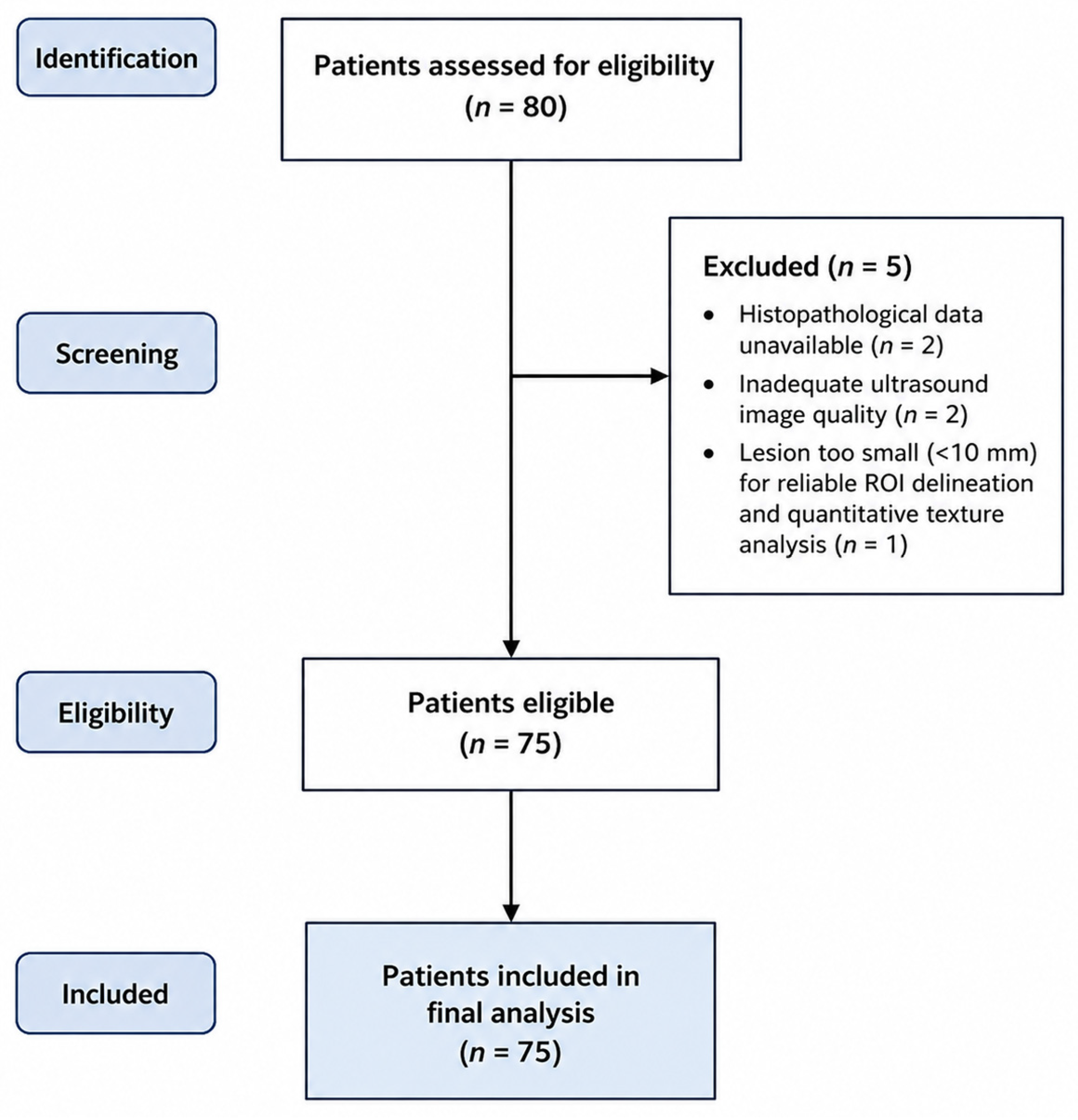
2.1. Study Population
2.2. Ultrasonographic Examination
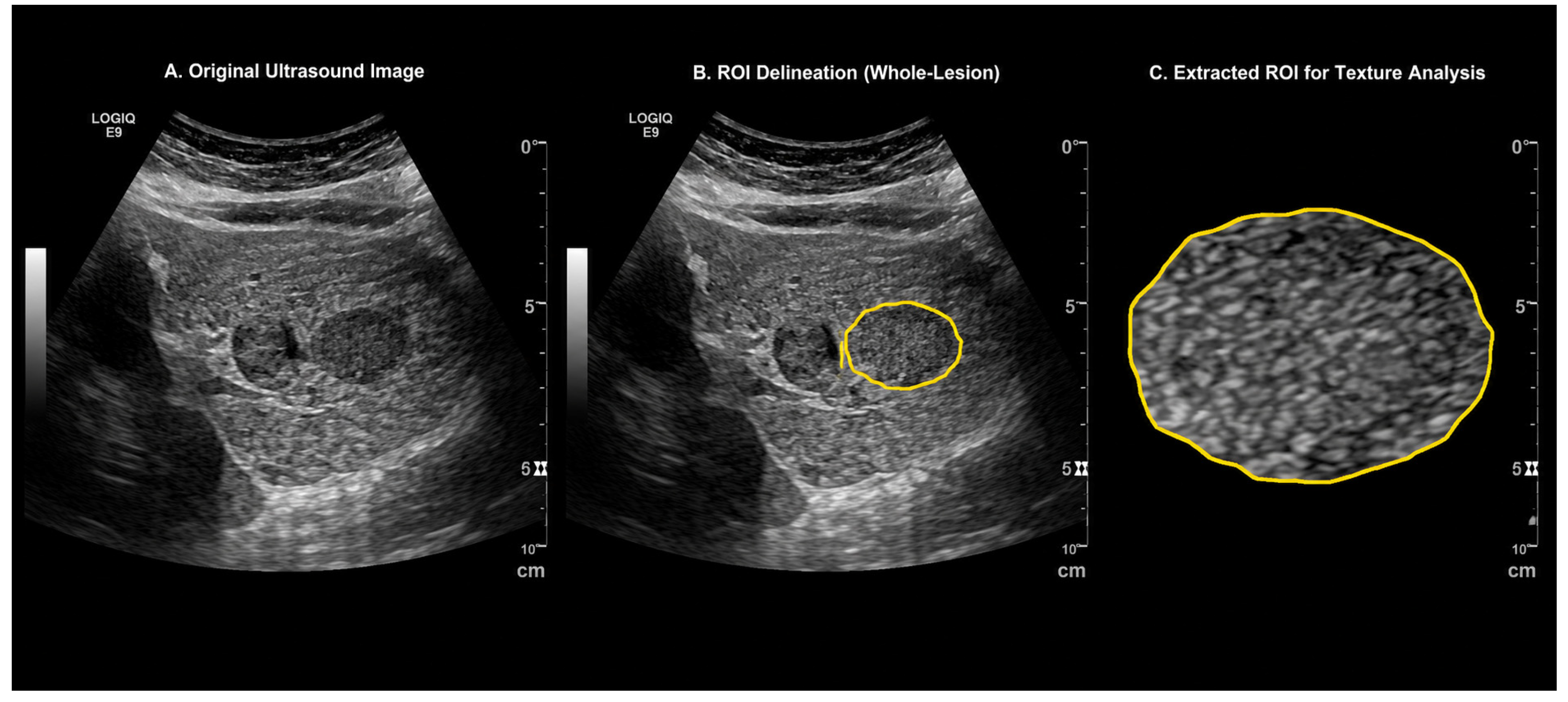
2.3. Texture Analysis
2.4. Statistical Analysis
2.5. Use of Generative Artificial İntelligence
3. Results
4. Discussion
Author Contributions
Funding
Institutional Review Board Statement
Informed Consent Statement
Data Availability Statement
Acknowledgments
Conflicts of Interest
References
- Disibio, G.; French, S.W. Metastatic patterns of cancers: Results from a large autopsy study. Arch. Pathol. Lab. Med. 2008, 132, 931–939. [Google Scholar] [CrossRef] [Scilit] [PubMed]
- Geoghegan, J.G.; Scheele, J. Treatment of colorectal liver metastases. Br. J. Surg. 1999, 86, 158–169. [Google Scholar] [CrossRef] [Scilit] [PubMed]
- Ibragimova, M.K.; Tsyganov, M.M.; Kravtsova, E.A.; Tsydenova, I.A.; Litviakov, N.V. Organ-Specificity of Breast Cancer Metastasis. Int. J. Mol. Sci. 2023, 24, 15625. [Google Scholar] [CrossRef] [Scilit] [PubMed]
- Roth, S.; Michalski, C.; Hoheisel, J.D. A systemic look at pancreatic cancer patients: Predicting metastasis by studying the liver. Signal Transduct. Target. Ther. 2024, 9, 246. [Google Scholar] [CrossRef] [Scilit] [PubMed]
- Ying, X.; Ma, N.; Zhang, X.; Guo, H.; Liu, Y.; Chen, B.; Zhao, S.; Wu, S.; Li, W.; Wang, L.; et al. Research progress on the molecular mechanisms of hepatic metastasis in lung cancer: A narrative review. Ann. Palliat. Med. 2021, 10, 4806–4822. [Google Scholar] [CrossRef] [Scilit] [PubMed]
- Sirody, J.; Kaji, A.H.; Hari, D.M.; Chen, K.T. Patterns of gastric cancer metastasis in the United States. Am. J. Surg. 2022, 224, 445–448. [Google Scholar] [CrossRef] [Scilit] [PubMed]
- Ma, J.; Yu, Q.; Van Ha, T. Image-Guided Liver Biopsy: Perspectives from Interventional Radiology. Semin. Interv. Radiol. 2024, 41, 500–506. [Google Scholar] [CrossRef] [Scilit] [PubMed]
- Thomaides-Brears, H.B.; Alkhouri, N.; Allende, D.; Harisinghani, M.; Noureddin, M.; Reau, N.S.; French, M.; Pantoja, C.; Mouchti, S.; Cryer, D.R.H. Incidence of Complications from Percutaneous Biopsy in Chronic Liver Disease: A Systematic Review and Meta-Analysis. Dig. Dis. Sci. 2022, 67, 3366–3394. [Google Scholar] [CrossRef] [Scilit] [PubMed]
- Choi, J.H. Histological and Molecular Evaluation of Liver Biopsies: A Practical and Updated Review. Int. J. Mol. Sci. 2025, 26, 7729. [Google Scholar] [CrossRef] [Scilit] [PubMed]
- Marusyk, A.; Polyak, K. Tumor heterogeneity: Causes and consequences. Biochim. ET Biophys. Acta BBA Rev. Cancer 2010, 1805, 105–117. [Google Scholar] [CrossRef] [Scilit] [PubMed]
- Lubner, M.G.; Smith, A.D.; Sandrasegaran, K.; Sahani, D.V.; Pickhardt, P.J. CT texture analysis: Definitions, applications, biologic correlates, and challenges. Radiographics 2017, 37, 1483–1503. [Google Scholar] [CrossRef] [Scilit] [PubMed]
- Gillies, R.J.; Kinahan, P.E.; Hricak, H. Radiomics: Images are more than pictures, they are data. Radiology 2016, 278, 563–577. [Google Scholar] [CrossRef] [Scilit] [PubMed]
- Ganeshan, B.; Miles, K.A.; Young, R.C.; Chatwin, C.R. Texture analysis in non-contrast enhanced CT: Impact of malignancy on texture in apparently disease-free areas of the liver. Eur. J. Radiol. 2009, 70, 101–110. [Google Scholar] [CrossRef] [Scilit] [PubMed]
- Granata, V.; Fusco, R.; De Muzio, F.; Cutolo, C.; Setola, S.V.; Dell’aVersana, F.; Grassi, F.; Belli, A.; Silvestro, L.; Ottaiano, A.; et al. Radiomics and machine learning analysis based on magnetic resonance imaging in the assessment of liver mucinous colorectal metastases. Radiol. Medica 2022, 127, 763–772. [Google Scholar] [CrossRef] [Scilit] [PubMed]
- Hanahan, D. Hallmarks of Cancer: New Dimensions. Cancer Discov. 2022, 12, 31–46. [Google Scholar] [CrossRef] [Scilit] [PubMed]
- Friedrich-Rust, M.; Klopffleisch, T.; Nierhoff, J.; Herrmann, E.; Vermehren, J.; Schneider, M.D.; Zeuzem, S.; Bojunga, J. Contrast-Enhanced Ultrasound for the differentiation of benign and malignant focal liver lesions: A meta-analysis. Liver Int. 2013, 33, 739–755. [Google Scholar] [CrossRef] [Scilit] [PubMed]
- Choi, J.M.; Yu, J.-S.; Cho, E.-S.; Kim, J.H.; Chung, J.-J. Texture Analysis of Hepatocellular Carcinoma on Magnetic Resonance Imaging: Assessment for Performance in Predicting Histopathologic Grade. J. Comput. Assist. Tomogr. 2020, 44, 901–910. [Google Scholar] [CrossRef] [Scilit] [PubMed]
- Oh, J.; Lee, J.M.; Park, J.; Joo, I.; Yoon, J.H.; Lee, D.H.; Ganeshan, B.; Han, J.K. Hepatocellular Carcinoma: Texture Analysis of Preoperative Computed Tomography Images Can Provide Markers of Tumor Grade and Disease-Free Survival. Korean J. Radiol. 2019, 20, 569–579. [Google Scholar] [CrossRef] [Scilit] [PubMed]
- Mostafavi, L.; Homayounieh, F.; Lades, F.; Primak, A.; Muse, V.; Harris, G.J.; Kalra, M.K.; Digumarthy, S.R. Correlation of Radiomics with Treatment Response in Liver Metastases. Acad. Radiol. 2024, 31, 3133–3141. [Google Scholar] [CrossRef] [Scilit] [PubMed]
- Lubner, M.G.; Stabo, N.; Lubner, S.J.; del Rio, A.M.; Song, C.; Halberg, R.B.; Pickhardt, P.J. CT textural analysis of hepatic metastatic colorectal cancer: Pre-treatment tumor heterogeneity correlates with pathology and clinical outcomes. Abdom. Imaging 2015, 40, 2331–2337. [Google Scholar] [CrossRef] [Scilit] [PubMed]
- Becker, A.S.; Schneider, M.A.; Wurnig, M.C.; Wagner, M.; Clavien, P.A.; Boss, A. Radiomics of liver MRI predict metastases in mice. Eur. Radiol. Exp. 2018, 2, 11. [Google Scholar] [CrossRef] [Scilit] [PubMed]
- Beckers, R.C.; Lambregts, D.M.; Schnerr, R.S.; Maas, M.; Rao, S.-X.; Kessels, A.G.; Thywissen, T.; Beets, G.L.; Trebeschi, S.; Houwers, J.B.; et al. Whole liver CT texture analysis to predict the development of colorectal liver metastases—A multicentre study. Eur. J. Radiol. 2017, 92, 64–71. [Google Scholar] [CrossRef] [Scilit] [PubMed]
- Janisch, M.; Adelsmayr, G.; Müller, H.; Holzinger, A.; Janek, E.; Talakic, E.; Fuchsjäger, M.; Schöllnast, H. Non-contrast-enhanced CT texture analysis of primary and metastatic pancreatic ductal adenocarcinomas: Value in assessment of histopathological grade and differences between primary and metastatic lesions. Abdom. Radiol. 2022, 47, 4151–4159. [Google Scholar] [CrossRef] [Scilit] [PubMed]
- Feig, C.; Gopinathan, A.; Neesse, A.; Chan, D.S.; Cook, N.; Tuveson, D.A. The pancreas cancer microenvironment. Clin. Cancer Res. 2012, 18, 4266–4276. [Google Scholar] [CrossRef] [Scilit] [PubMed]
- Gao, Y.; Shen, S.; Wang, Y.; Tian, M. Organ Crosstalk: The Role of Spleen. Phenomics 2025, 5, 192–207. [Google Scholar] [CrossRef] [Scilit] [PubMed]
- Qi, Z.; Qi, P.; Jiang, X.; Qi, P. Progress in intestinal homeostasis and mechanisms of pancreatic cancer. Phenomics 2025, 5, 446–456. [Google Scholar] [CrossRef] [Scilit] [PubMed]
- Castellano, G.; Bonilha, L.; Li, L.M.; Cendes, F. Texture analysis of medical images. Clin. Radiol. 2004, 59, 1061–1069. [Google Scholar] [CrossRef] [Scilit] [PubMed]
- Azoulay, A.; Cros, J.; Vullierme, M.-P.; de Mestier, L.; Couvelard, A.; Hentic, O.; Ruszniewski, P.; Sauvanet, A.; Vilgrain, V.; Ronot, M. Morphological imaging and CT histogram analysis to differentiate pancreatic neuroendocrine tumor grade 3 from neuroendocrine carcinoma. Diagn. Interv. Imaging 2020, 101, 821–830. [Google Scholar] [CrossRef] [Scilit] [PubMed]
- Berbís, M.Á.; Godino, F.P.; Rodríguez-Comas, J.; Nava, E.; García-Figueiras, R.; Baleato-González, S.; Luna, A. Radiomics in CT and MR imaging of the liver and pancreas: Tools with potential for clinical application. Abdom. Radiol. 2024, 49, 322–340. [Google Scholar] [CrossRef] [Scilit] [PubMed]



| Organ Metastasis | p | ||||||||||||
|---|---|---|---|---|---|---|---|---|---|---|---|---|---|
| Colon (n: 25) 1 | Pancreas (n: 25) 2 | Breast (n: 25) 3 | |||||||||||
| Age | Mean ± sd | 67.6 | ± | 12.3 | 69.3 | ± | 5.3 | 63.7 | ± | 7.7 | 0.073 | K | |
| Median | 70.0 | 69.0 | 61.0 | ||||||||||
| Gender | Male | n-% | 19 | 76.0% | 16 | 64.0% | 0 | 0.0% | 0.000 | X2 | |||
| Female | n-% | 6 3 | 24.0% | 9 3 | 36.0% | 25 | 100% | ||||||
| Mean of Histogram | Mean ± sd | 76.7 | ± | 13.7 | 123.5 | ± | 18.4 | 55.5 | ± | 20.5 | 0.000 | K | |
| Median | 80.7 2 | 123.7 | 56.6 1,2 | ||||||||||
| Standard Deviation of Histogram | Mean ± sd | 19.8 | ± | 5.0 | 19.2 | ± | 5.0 | 17.1 | ± | 5.2 | 0.136 | K | |
| Median | 18.4 | 20.3 | 15.6 | ||||||||||
| Median of Histogram | Mean ± sd | 76.0 | ± | 13.8 | 123.1 | ± | 18.6 | 55.4 | ± | 21.0 | 0.000 | K | |
| Median | 80.0 2 | 121.0 | 58.0 1,2 | ||||||||||
| Minimum of Histogram | Mean ± sd | 20.2 | ± | 17.8 | 65.6 | ± | 25.8 | 11.2 | ± | 12.7 | 0.000 | K | |
| Median | 17.0 2 | 66.0 | 5.0 2 | ||||||||||
| Maximum of Histogram | Mean ± sd | 156.8 | ± | 30.3 | 194.0 | ± | 26.0 | 117.8 | ± | 38.7 | 0.000 | K | |
| Median | 154.0 2 | 201.0 | 120.0 1,2 | ||||||||||
| Mean Absolute Deviation of Histogram | Mean ± sd | 15.7 | ± | 4.1 | 15.5 | ± | 4.0 | 13.8 | ± | 4.2 | 0.167 | K | |
| Median | 14.9 | 16.2 | 12.8 | ||||||||||
| Median Absolute Deviation of Histogram | Mean ± sd | 13.1 | ± | 3.9 | 13.3 | ± | 3.4 | 11.8 | ± | 3.6 | 0.197 | K | |
| Median | 13.0 | 14.0 | 11.0 | ||||||||||
| Variance of Histogram | Mean ± sd | 415.1 | ± | 235.0 | 393.8 | ± | 190.1 | 319.1 | ± | 194.8 | 0.136 | K | |
| Median | 340.1 | 410.3 | 243.8 | ||||||||||
| Range of Histogram | Mean ± sd | 136.6 | ± | 36.2 | 128.3 | ± | 36.7 | 106.6 | ± | 37.1 | 0.015 | K | |
| Median | 124.0 | 132.0 | 89.0 1,2 | ||||||||||
| Interquartile Range of Histogram | Mean ± sd | 26.1 | ± | 7.8 | 26.7 | ± | 6.8 | 23.5 | ± | 7.4 | 0.199 | K | |
| Median | 25.0 | 27.0 | 21.0 | ||||||||||
| Most Frequent Value of Histogram | Mean ± sd | 71.9 | ± | 14.4 | 125.7 | ± | 19.9 | 53.4 | ± | 22.4 | 0.000 | K | |
| Median | 79.0 2 | 126.0 | 58.0 1,2 | ||||||||||
| Size %L of Histogram | Mean ± sd | 15.7 | ± | 2.2 | 16.6 | ± | 1.3 | 16.4 | ± | 1.7 | 0.255 | K | |
| Median | 16.0 | 16.7 | 16.6 | ||||||||||
| Size %M of Histogram | Mean ± sd | 68.9 | ± | 3.2 | 67.3 | ± | 2.1 | 68.3 | ± | 2.2 | 0.088 | A | |
| Median | 68.7 | 67.1 | 68.4 | ||||||||||
| Size %U of Histogram | Mean ± sd | 15.4 | ± | 1.4 | 16.1 | ± | 1.3 | 15.4 | ± | 1.3 | 0.096 | A | |
| Median | 15.5 | 16.3 | 15.4 | ||||||||||
| Organ Metastasis | p | ||||||||||||
|---|---|---|---|---|---|---|---|---|---|---|---|---|---|
| Colon (n: 25) 1 | Pancreas (n: 25) 2 | Breast (n: 25) 3 | |||||||||||
| Kurtosis of Histogram | Mean ± sd | 3.37 | ± | 0.85 | 2.96 | ± | 0.57 | 2.99 | ± | 0.54 | 0.063 | K | |
| Median | 3.04 | 2.85 | 2.91 | ||||||||||
| Skewness of Histogram | Mean ± sd | 0.27 | ± | 0.36 | 0.10 | ± | 0.32 | 0.20 | ± | 0.28 | 0.167 | A | |
| Median | 0.19 | 0.10 | 0.13 | ||||||||||
| Smoothness of Histogram | Mean ± sd | 0.003 | ± | 0.001 | 0.003 | ± | 0.002 | 0.004 | ± | 0.002 | 0.136 | K | |
| Median | 0.003 | 0.002 | 0.004 | ||||||||||
| Root-Mean-Square Level of Histogram | Mean ± sd | 79.4 | ± | 13.4 | 125.1 | ± | 18.3 | 58.3 | ± | 20.5 | 0.000 | K | |
| Median | 82.1 2 | 125.8 | 58.2 1,2 | ||||||||||
| Root-Sum-of-Squares Level of Histogram | Mean ± sd | 103.01 | ± | 47.45 | 180.53 | ± | 117.37 | 51.17 | ± | 32.23 | 0.000 | K | |
| Median | 101.51 2 | 143.25 | 53.92 1,2 | ||||||||||
| 1st Percentile of Histogram | Mean ± sd | 34.6 | ± | 17.1 | 81.3 | ± | 20.8 | 19.9 | ± | 16.2 | 0.000 | K | |
| Median | 36.0 2 | 78.0 | 17.0 1,2 | ||||||||||
| 3rd Percentile of Histogram | Mean ± sd | 41.4 | ± | 16.4 | 87.9 | ± | 19.9 | 25.1 | ± | 16.3 | 0.000 | K | |
| Median | 42.0 2 | 86.0 | 21.0 1,2 | ||||||||||
| 5th Percentile of Histogram | Mean ± sd | 45.4 | ± | 15.6 | 91.8 | ± | 19.4 | 28.0 | ± | 17.1 | 0.000 | K | |
| Median | 46.0 2 | 90.0 | 24.0 1,2 | ||||||||||
| 10th Percentile of Histogram | Mean ± sd | 52.0 | ± | 14.9 | 98.3 | ± | 18.4 | 33.5 | ± | 17.1 | 0.000 | K | |
| Median | 50.0 2 | 98.0 | 30.0 1,2 | ||||||||||
| 25th Percentile of Histogram | Mean ± sd | 63.3 | ± | 14.4 | 109.8 | ± | 18.6 | 43.4 | ± | 18.4 | 0.000 | K | |
| Median | 63.0 2 | 110.0 | 48.0 1,2 | ||||||||||
| 75th Percentile of Histogram | Mean ± sd | 89.4 | ± | 14.6 | 136.4 | ± | 19.4 | 66.9 | ± | 22.8 | 0.000 | A | |
| Median | 90.0 2 | 135.0 | 66.0 1,2 | ||||||||||
| 90th Percentile of Histogram | Mean ± sd | 102.0 | ± | 15.7 | 148.2 | ± | 20.1 | 77.9 | ± | 24.9 | 0.000 | A | |
| Median | 103.0 2 | 148.0 | 77.0 1,2 | ||||||||||
| 95th Percentile of Histogram | Mean ± sd | 110.4 | ± | 16.6 | 154.8 | ± | 20.4 | 84.4 | ± | 26.6 | 0.000 | A | |
| Median | 110.0 2 | 153.0 | 80.0 1,2 | ||||||||||
| 97th Percentile of Histogram | Mean ± sd | 115.9 | ± | 17.7 | 159.1 | ± | 21.2 | 88.8 | ± | 27.8 | 0.000 | A | |
| Median | 117.0 2 | 156.0 | 84.0 1,2 | ||||||||||
| 99th Percentile of Histogram | Mean ± sd | 126.9 | ± | 19.4 | 167.3 | ± | 21.8 | 97.0 | ± | 30.9 | 0.000 | A | |
| Median | 129.0 2 | 162.0 | 92.0 1,2 | ||||||||||
| Entropy of Histogram | Mean ± sd | 5.69 | ± | 0.32 | 5.77 | ± | 0.41 | 5.40 | ± | 0.52 | 0.014 | K | |
| Median | 5.68 | 5.82 | 5.36 1,2 | ||||||||||
| Uniformity of Histogram | Mean ± sd | 0.20 | ± | 0.06 | 0.23 | ± | 0.06 | 0.21 | ± | 0.07 | 0.337 | A | |
| Median | 0.20 | 0.23 | 0.21 | ||||||||||
Disclaimer/Publisher’s Note: The statements, opinions and data contained in all publications are solely those of the individual author(s) and contributor(s) and not of MDPI and/or the editor(s). MDPI and/or the editor(s) disclaim responsibility for any injury to people or property resulting from any ideas, methods, instructions or products referred to in the content. |
© 2026 by the authors. Licensee MDPI, Basel, Switzerland. This article is an open access article distributed under the terms and conditions of the Creative Commons Attribution (CC BY) license.
Share and Cite
Karakucuk, S.N.; Baykara, M.; Demir, M.; İsler, A. Can Ultrasound Texture Analysis Differentiate Liver Metastases According to the Histopathological Origin of the Primary Tumor? J. Clin. Med. 2026, 15, 6815. https://doi.org/10.3390/jcm15176815
Karakucuk SN, Baykara M, Demir M, İsler A. Can Ultrasound Texture Analysis Differentiate Liver Metastases According to the Histopathological Origin of the Primary Tumor? Journal of Clinical Medicine. 2026; 15(17):6815. https://doi.org/10.3390/jcm15176815
Chicago/Turabian StyleKarakucuk, Seda Nida, Murat Baykara, Mehmet Demir, and Ali İsler. 2026. "Can Ultrasound Texture Analysis Differentiate Liver Metastases According to the Histopathological Origin of the Primary Tumor?" Journal of Clinical Medicine 15, no. 17: 6815. https://doi.org/10.3390/jcm15176815
APA StyleKarakucuk, S. N., Baykara, M., Demir, M., & İsler, A. (2026). Can Ultrasound Texture Analysis Differentiate Liver Metastases According to the Histopathological Origin of the Primary Tumor? Journal of Clinical Medicine, 15(17), 6815. https://doi.org/10.3390/jcm15176815