Early Detection of Major Fetal Structural Anomalies in the First Trimester: A Retrospective Single-Center Study in an Unselected Population


Abstract
1. Introduction
2. Materials and Methods
2.1. Study Design and Population
2.2. Inclusion and Exclusion Criteria
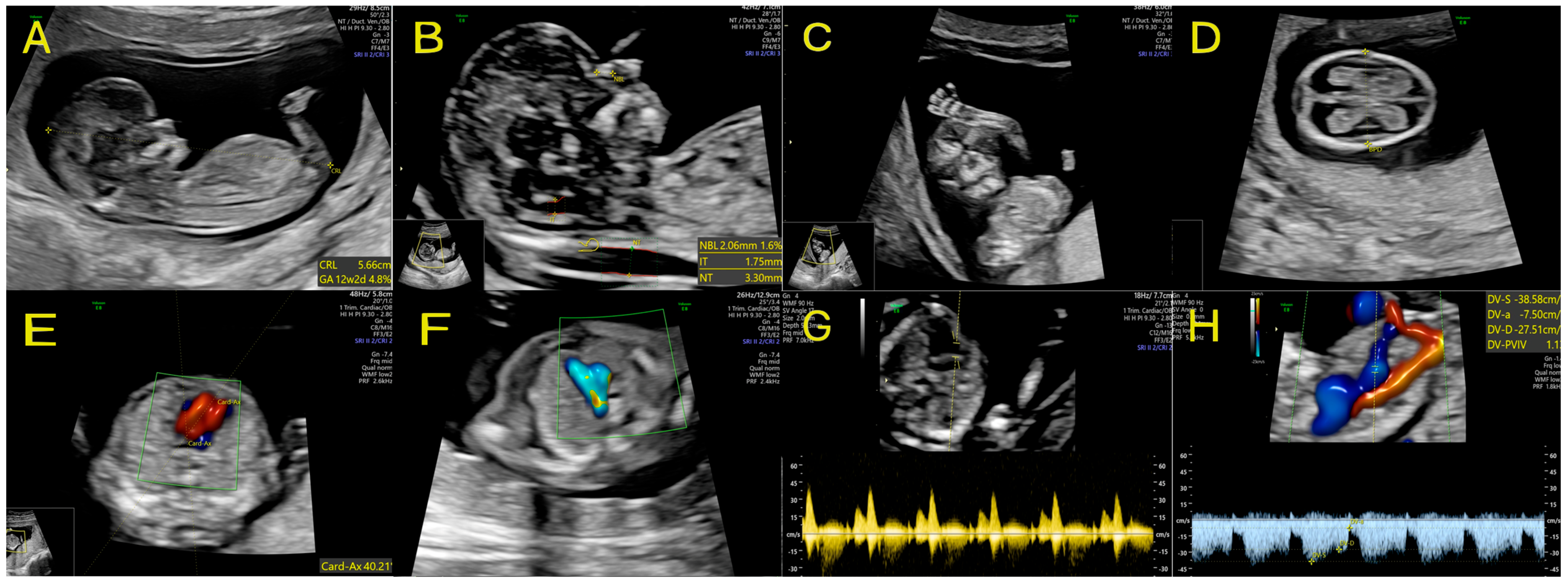
2.3. Ultrasound Examination Protocol
2.4. Combined First-Trimester Screening and Risk Assessment
2.5. Maternal Characteristics and Data Collection
2.6. Statistical Analysis
2.7. Ethical Approvals
3. Results
3.1. Study Population and Baseline Characteristics
3.2. Prevalence and Spectrum of Suspected Anomalies
3.3. Confirmation Rate and Follow-Up Outcomes
- -
- Excluding the single case lost to follow-up from the denominator yielded a confirmation rate of 87.5% (14/16; 95% CI: 61.7–98.4%).
- -
- Explicitly classifying the lost-to-follow-up case as a false positive maintained the confirmation rate at 82.4% (14/17; 95% CI: 56.6–96.2%).
3.4. Nuchal Translucency and Statistical Associations
4. Discussion
4.1. The Value of a First-Trimester Anatomical Protocol
4.2. Strengths and Limitations
4.3. Clinical Implications and Future Research Directions
5. Conclusions
Author Contributions
Funding
Institutional Review Board Statement
Informed Consent Statement
Data Availability Statement
Conflicts of Interest
References
- Almli, L.M.; Ely, D.M.; Ailes, E.C.; Abouk, R.; Grosse, S.D.; Isenburg, J.L.; Waldron, D.B.; Reefhuis, J. Infant Mortality Attributable to Birth Defects—United States, 2003–2017. Morb. Mortal. Wkly. Rep. 2020, 69, 25–29. [Google Scholar] [CrossRef] [Scilit]
- World Health Organization. Congenital Anomalies: Fact Sheet. Available online: https://www.who.int/news-room/fact-sheets/detail/birth-defects (accessed on 10 July 2026).
- Karim, J.N.; Roberts, N.W.; Salomon, L.J.; Papageorghiou, A.T. Systematic review of first-trimester ultrasound screening for detection of fetal structural anomalies and factors that affect screening performance. Ultrasound Obstet. Gynecol. 2017, 50, 429–441. [Google Scholar] [CrossRef] [Scilit]
- Rossi, A.C.; Prefumo, F. Accuracy of ultrasonography at 11-14 weeks of gestation for detection of fetal structural anomalies: A systematic review. Obstet. Gynecol. 2013, 122, 1160–1167. [Google Scholar] [CrossRef] [Scilit]
- Vayna, A.M.; Veduta, A.; Duta, S.; Panaitescu, A.M.; Stoica, S.; Buinoiu, N.; Nedelea, F.; Peltecu, G. Diagnosis of Fetal Structural Anomalies at 11 to 14 Weeks. J. Ultrasound Med. 2018, 37, 2063–2073. [Google Scholar] [CrossRef] [Scilit]
- Syngelaki, A.; Hammami, A.; Bower, S.; Zidere, V.; Akolekar, R.; Nicolaides, K.H. Diagnosis of fetal non-chromosomal abnormalities on routine ultrasound examination at 11-13 weeks’ gestation. Ultrasound Obstet. Gynecol. 2019, 54, 468–476. [Google Scholar] [CrossRef] [Scilit]
- Buijtendijk, M.F.; Bet, B.B.; Leeflang, M.M.; Shah, H.; Reuvekamp, T.; Goring, T.; Docter, D.; Timmerman, M.G.; Dawood, Y.; Lugthart, M.A.; et al. Diagnostic accuracy of ultrasound screening for fetal structural abnormalities during the first and second trimester of pregnancy in low-risk and unselected populations. Cochrane Database Syst. Rev. 2024, 5, CD014715. [Google Scholar] [CrossRef] [Scilit]
- Centers for Disease Control and Prevention. Birth Defects: Tracking and Research. Available online: https://www.cdc.gov/birth-defects/tracking/index.html (accessed on 24 July 2026).
- EUROCAT. EUROCAT Guide 1.5: Instructions for the Registration of Congenital Anomalies; Joint Research Centre, European Commission: Brussels, Belgium, 2022. [Google Scholar]
- Salomon, L.J.; Alfirevic, Z.; Berghella, V.; Bilardo, C.M.; Chalouhi, G.E.; Da Silva Costa, F.; Hernandez-Andrade, E.; Malinger, G.; Munoz, H.; Paladini, D.; et al. ISUOG Practice Guidelines (updated): Performance of the routine mid-trimester fetal ultrasound scan. Ultrasound Obstet. Gynecol. 2022, 59, 840–856. [Google Scholar] [CrossRef] [Scilit]
- Karim, J.N.; Broughan, J.M.; Aldridge, N.; Pandya, P.; McHugh, A.; Papageorghiou, A.T.; ACCEPTS Study Group. Impact of first-trimester ultrasound on early detection of major fetal anomalies: Nationwide population-based study of over 1 million pregnancies. PLoS Med. 2025, 22, e1004709. [Google Scholar] [CrossRef] [Scilit]
- Grande, M.; Arigita, M.; Borobio, V.; Jimenez, J.M.; Fernandez, S.; Borrell, A. First-trimester detection of structural abnormalities and the role of aneuploidy markers. Ultrasound Obstet. Gynecol. 2012, 39, 157–163. [Google Scholar] [CrossRef] [Scilit]
- Syngelaki, A.; Chelemen, T.; Dagklis, T.; Allan, L.; Nicolaides, K.H. Challenges in the diagnosis of fetal non-chromosomal abnormalities at 11-13 weeks. Prenat. Diagn. 2011, 31, 90–102. [Google Scholar] [CrossRef] [Scilit]
- Salomon, L.J.; Alfirevic, Z.; Bilardo, C.M.; Chalouhi, G.E.; Ghi, T.; Kagan, K.O.; Lau, T.K.; Papageorghiou, A.T.; Raine-Fenning, N.J.; Stirnemann, J.; et al. ISUOG practice guidelines: Performance of first-trimester fetal ultrasound scan. Ultrasound Obstet. Gynecol. 2013, 41, 102–113. [Google Scholar] [CrossRef] [Scilit]
- Fetal Medicine Foundation. Protocol and Certification Materials for the 11–14 Week Scan. Available online: https://fetalmedicine.org (accessed on 15 June 2025).
- Iliescu, D.; Tudorache, S.; Comanescu, A.; Antsaklis, P.; Cotarcea, S.; Novac, L.; Cernea, N.; Antsaklis, A. Improved detection rate of structural abnormalities in the first trimester using an extended examination protocol. Ultrasound Obstet. Gynecol. 2013, 42, 300–309. [Google Scholar] [CrossRef] [Scilit]
- Lust, E.E.R.; Bronsgeest, K.; Henneman, L.; Crombag, N.; Bilardo, C.M.; Galjaard, R.H.; Sikkel, E.; van der Hout, S.; Coumans, A.; Elvan-Taşpınar, A.; et al. Introduction of a nationwide first-trimester anomaly scan in the Dutch national screening program. Am. J. Obstet. Gynecol. 2025, 232, 396.e1–396.e19. [Google Scholar] [CrossRef] [Scilit]
- Borowski, D.; Pietryga, M.; Basta, P.; Cnota, W.; Czuba, B.; Dubiel, M.; Fuchs, T.; Huras, H.; Iciek, R.; Jaczyńska, R.; et al. Rekomendacje Sekcji Ultrasonografii Polskiego Towarzystwa Ginekologów i Położników w zakresie przesiewowej diagnostyki ultrasonograficznej w ciąży o przebiegu prawidłowym—2020 rok. Ginekol. Perinatol. Prakt. 2020, 5, 63–75. [Google Scholar]
- Fetal Medicine Foundation. The 11–14 week scan: FMF algorithm for combined screening. Available online: https://fetalmedicine.org (accessed on 15 June 2025).
- Sieroszewski, P.; Haus, O.; Zimmer, M.; Wielgos, M.; Latos-Bielenska, A.; Borowiec, M.; Borowski, D.; Cnota, W.; Czuba, B.; Dubiel, M.; et al. Recommendations for prenatal diagnostics of the Polish Society of Gynaecologists and Obstetricians and the Polish Society of Human Genetics. Ginekol. Pol. 2022, 93, 427–437. [Google Scholar] [CrossRef] [Scilit]
- Fetal Medicine Foundation. Screening for Preeclampsia and Fetal Growth Restriction. Available online: https://fetalmedicine.org (accessed on 15 June 2025).
- Rolnik, D.L.; Wright, D.; Poon, L.C.; O’Gorman, N.; Syngelaki, A.; de Paco Matallana, C.; Akolekar, R.; Cicero, S.; Janga, D.; Singh, M.; et al. Aspirin versus Placebo in Pregnancies at High Risk for Preterm Preeclampsia. N. Engl. J. Med. 2017, 377, 613–622. [Google Scholar] [CrossRef] [Scilit]
- Kenkhuis, M.J.A.; Bakker, M.; Bardi, F.; Fontanella, F.; Bakker, M.K.; Fleurke-Rozema, J.H.; Bilardo, C.M. Effectiveness of 12-13-week scan for early diagnosis of fetal congenital anomalies in the cell-free DNA era. Ultrasound Obstet. Gynecol. 2018, 51, 463–469. [Google Scholar] [CrossRef] [Scilit]
- Carvalho, M.H.; Brizot, M.L.; Lopes, L.M.; Chiba, C.H.; Miyadahira, S.; Zugaib, M. Detection of fetal structural abnormalities at the 11-14 week ultrasound scan. Prenat. Diagn. 2002, 22, 1–4. [Google Scholar] [CrossRef] [Scilit]
- EUROCAT. EUROCAT Interactive Prevalence Charts and Tables. Available online: https://eu-rd-platform.jrc.ec.europa.eu/eurocat/eurocat-data/prevalence_en (accessed on 15 June 2025).
- Dolk, H.; Loane, M.; Garne, E. The prevalence of congenital anomalies in Europe. Adv. Exp. Med. Biol. 2010, 686, 349–364. [Google Scholar] [CrossRef] [Scilit]
- Khoshnood, B.; Loane, M.; Walle, H.; Arriola, L.; Addor, M.-C.; Barisic, I.; Béres, J.; Bianchi, F.; Dias, C.; Draper, E.; et al. Long term trends in prevalence of neural tube defects in Europe: Population based study. BMJ 2015, 351, h5949. [Google Scholar] [CrossRef] [Scilit]
- Volpe, N.; Dall’Asta, A.; Di Pasquo, E.; Frusca, T.; Ghi, T. First-trimester fetal neurosonography: Technique and diagnostic potential. Ultrasound Obstet. Gynecol. 2021, 57, 204–214. [Google Scholar] [CrossRef] [Scilit]
- Paladini, D.; Volpe, P. Ultrasound of Congenital Fetal Anomalies: Differential Diagnosis and Prognostic Indicators, 2nd ed.; CRC Press: Boca Raton, FL, USA, 2014. [Google Scholar]
- Ungureanu, D.R.; Drăgușin, R.C.; Căpitănescu, R.G.; Zorilă, L.; Ofițeru, A.M.I.; Marinaș, C.; Pătru, C.L.; Comănescu, A.C.; Comănescu, M.C.; Sîrbu, O.C.; et al. First Trimester Ultrasound Detection of Fetal Central Nervous System Anomalies. Brain Sci. 2023, 13, 118. [Google Scholar] [CrossRef] [Scilit]
- Nicolaides, K.H. Screening for fetal aneuploidies at 11 to 13 weeks. Prenat. Diagn. 2011, 31, 7–15. [Google Scholar] [CrossRef] [Scilit]
- Martins Santana, E.F.; Araujo Júnior, E.; Tonni, G.; Costa, F.D.S.; Meagher, S. Acrania-exencephaly-anencephaly sequence phenotypic characterization using two- and three-dimensional ultrasound between 11 and 13 weeks and 6 days of gestation. J. Ultrason. 2018, 18, 240–246. [Google Scholar] [CrossRef] [Scilit]
- Nizard, J.; Bernard, J.P.; Ville, Y. Fetal cystic malformations of the posterior fossa in the first trimester of pregnancy. Fetal Diagn. Ther. 2005, 20, 146–151. [Google Scholar] [CrossRef] [Scilit]
- Malinger, G.; Paladini, D.; Haratz, K.K.; Monteagudo, A.; Pilu, G.L.; Timor-Tritsch, I.E. ISUOG Practice Guidelines (updated): Sonographic examination of the fetal central nervous system. Part 1: Performance of screening examination and indications for targeted neurosonography. Ultrasound Obstet. Gynecol. 2020, 56, 476–484. [Google Scholar] [CrossRef] [Scilit]
- Chaoui, R.; Benoit, B.; Mitkowska-Wozniak, H.; Heling, K.S.; Nicolaides, K.H. Assessment of intracranial translucency (IT) in the detection of spina bifida at the 11-13-week scan. Ultrasound Obstet. Gynecol. 2009, 34, 249–252. [Google Scholar] [CrossRef] [Scilit]
- Popa, A.I.; Cernea, N.; Marinaș, M.C.; Comănescu, M.C.; Sîrbu, O.C.; Popa, D.G.; Pătru, L.; Pădureanu, V.; Pătru, C.L. Ultrasound Screening in the First and Second Trimester of Pregnancy for the Detection of Fetal Cardiac Anomalies in a Low-Risk Population. Diagnostics 2025, 15, 769. [Google Scholar] [CrossRef] [Scilit]
- Huhta, J.C. First-trimester screening for congenital heart disease. Curr. Opin. Cardiol. 2016, 31, 72–77. [Google Scholar] [CrossRef] [Scilit]
- Duta, S.; Veduta, A.; Vayna, A.M.; Panaitescu, A.; Nedelea, F.; Peltecu, G. The outcome of structural heart defects diagnosed in the first trimester of pregnancy. J. Matern. Fetal Neonatal Med. 2021, 34, 1389–1394. [Google Scholar] [CrossRef] [Scilit]
- Nawara-Baran, A.; Radzymińska-Chruściel, B. On Outcome of Five Consecutive Pregnancies with Heart Defects Detected in 1st Trimester of Pregnancy. Prenat. Cardiol. 2015, 2015, 13–17. [Google Scholar] [CrossRef] [Scilit]
- Wiechec, M.; Knafel, A.; Nocun, A. Prenatal detection of congenital heart defects at the 11- to 13-week scan using a simple color Doppler protocol including the 4-chamber and 3-vessel and trachea views. J. Ultrasound Med. 2015, 34, 585–594. [Google Scholar] [CrossRef] [Scilit]
- Wiechec, M.; Nocun, A.; Wiercinska, E.; Beithon, J.; Knafel, A. First trimester tricuspid regurgitation and fetal abnormalities. J. Perinat. Med. 2015, 43, 597–603. [Google Scholar] [CrossRef] [Scilit]
- Volpe, P.; Marasini, M.; Caruso, G.; Lituania, M.; Marzullo, A.; Volpe, G.; Gentile, M. Prenatal diagnosis of ductus venosus agenesis and its association with cytogenetic/congenital anomalies. Prenat. Diagn. 2002, 22, 995–1000. [Google Scholar] [CrossRef] [Scilit]
- Berg, C.; Kamil, D.; Geipel, A.; Kohl, T.; Knöpfle, G.; Hansmann, M.; Gembruch, U. Absence of ductus venosus-importance of umbilical venous drainage site. Ultrasound Obstet. Gynecol. 2006, 28, 275–281. [Google Scholar] [CrossRef] [Scilit]
- Takita, H.; Hasegawa, J.; Arakaki, T.; Hamada, S.; Tokunaka, M.; Nakamura, M.; Matsuoka, R.; Sekizawa, A. Outcomes in the absence of the ductus venosus diagnosed in the first trimester. J. Matern. Fetal Neonatal Med. 2018, 31, 253–257. [Google Scholar] [CrossRef] [Scilit]
- Desselle, C.; Herve, P.; Toutain, A.; Lardy, H.; Sembely, C.; Perrotin, F. Pentalogy of Cantrell: Sonographic assessment. J. Clin. Ultrasound 2007, 35, 216–220. [Google Scholar] [CrossRef] [Scilit]
- Sarkar, P.; Bastin, J.; Katoch, D.; Pal, A. Pentalogy of Cantrell: Diagnosis in the first trimester. J. Obstet. Gynaecol. 2005, 25, 812–813. [Google Scholar] [CrossRef] [Scilit]
- Türkçapar, A.F.; Sargın Oruc, A.; Öksüzoglu, A.; Danışman, N. Diagnosis of pentalogy of cantrell in the first trimester using transvaginal sonography and color Doppler. Case Rep. Obstet. Gynecol. 2015, 2015, 179298. [Google Scholar] [CrossRef] [Scilit]
- Cantrell, J.R.; Haller, J.A.; Ravitch, M.M. A syndrome of congenital defects involving the abdominal wall, sternum, diaphragm, pericardium, and heart. Surg. Gynecol. Obstet. 1958, 107, 602–614. [Google Scholar]
- Abuhamad, A.; Chaoui, R. First Trimester Ultrasound Diagnosis of Fetal Abnormalities, 1st ed.; Wolters Kluwer Heath: Philadelphia, PA, USA, 2018. [Google Scholar]
- Cianci, M.C.; Fusi, G.; Morini, F.; Severi, E.; Morabito, A.; Grimaldi, C. Fetal treatment of sacrococcygeal teratoma: State of the art. Front. Pediatr. 2025, 13, 1410269. [Google Scholar] [CrossRef] [Scilit]
- el-Qarmalawi, M.A.; Saddik, M.; el Abdel Hadi, F.; Muwaffi, R.; Nageeb, K. Diagnosis and management of fetal sacrococcygeal teratoma. Int. J. Gynaecol. Obstet. 1990, 31, 275–281. [Google Scholar] [CrossRef] [Scilit]
- Roman, A.S.; Monteagudo, A.; Timor-Tritsch, I.; Rebarber, A. First-trimester diagnosis of sacrococcygeal teratoma: The role of three-dimensional ultrasound. Ultrasound Obstet. Gynecol. 2004, 23, 612–614. [Google Scholar] [CrossRef] [Scilit]
- Sepulveda, W.; Wong, A.E.; Martinez-Ten, P.; Perez-Pedregosa, J. Retronasal triangle: A sonographic landmark for the screening of cleft palate in the first trimester. Ultrasound Obstet. Gynecol. 2010, 35, 7–13. [Google Scholar] [CrossRef] [Scilit]
- Hui, L.; Bianchi, D.W. Noninvasive Prenatal DNA Testing: The Vanguard of Genomic Medicine. Annu. Rev. Med. 2017, 68, 459–472. [Google Scholar] [CrossRef] [Scilit]
- Jayashankar, S.S.; Nasaruddin, M.L.; Hassan, M.F.; Dasrilsyah, R.A.; Shafiee, M.N.; Ismail, N.A.S.; Alias, E. Non-Invasive Prenatal Testing (NIPT): Reliability, Challenges, and Future Directions. Diagnostics 2023, 13, 2570. [Google Scholar] [CrossRef] [Scilit]
- Karadzov Orlic, N.; Egic, A.; Damnjanovic-Pazin, B.; Lukic, R.; Joksic, I.; Mikovic, Z. Screening performance of congenital heart defects in first trimester using simple cardiac scan, nuchal translucency, abnormal ductus venosus blood flow and tricuspid regurgitation. Congenit. Heart Dis. 2019, 14, 1094–1101. [Google Scholar] [CrossRef] [Scilit]
- Karim, J.N.; Bradburn, E.; Roberts, N.; Papageorghiou, A.T.; ACCEPTS Study. First-trimester ultrasound detection of fetal heart anomalies: Systematic review and meta-analysis. Ultrasound Obstet. Gynecol. 2022, 59, 11–25. [Google Scholar] [CrossRef] [Scilit]
- Novotná, M.; Hašlík, L.; Svabík, K.; Zižka, Z.; Belošovičová, H.; Břešťák, M.; Calda, P. Detection of fetal major structural anomalies at the 11-14 ultrasound scan in an unselected population. Ceska Gynekol. 2012, 77, 330–335. [Google Scholar]
- Nicolaides, K.H. First-trimester screening for chromosomal abnormalities. Semin. Perinatol. 2005, 29, 190–194. [Google Scholar] [CrossRef] [Scilit]
- Bilardo, C.M.; Chaoui, R.; Hyett, J.A.; Kagan, K.O.; Karim, J.N.; Papageorghiou, A.T.; Poon, L.C.; Salomon, L.J.; Syngelaki, A.; Nicolaides, K.H.; et al. ISUOG Practice Guidelines (updated): Performance of 11–14-week ultrasound scan. Ultrasound Obstet. Gynecol. 2023, 61, 127–143. [Google Scholar] [CrossRef] [Scilit]
- Bronsgeest, K.; Lust, E.E.R.; Henneman, L.; Crombag, N.; Bilardo, C.M.; Stemkens, D.; Galjaard, R.H.; Sikkel, E.; van der Hout, S.H.; Bekker, M.N.; et al. Current practice of first-trimester ultrasound screening for structural fetal anomalies in developed countries. Prenat. Diagn. 2023, 43, 873–880. [Google Scholar] [CrossRef] [Scilit]
- World Federation for Ultrasound in Medicine and Biology. WFUMB policy and statements on safety of ultrasound. Ultrasound Med. Biol. 2013, 39, 926–929. [Google Scholar] [CrossRef] [Scilit]
- Salvesen, K.; Abramowicz, J.; Ter Haar, G.; Miloro, P.; Sinkovskaya, E.; Dall’Asta, A.; Maršál, K.; Lees, C.; Board of the International Society of Ultrasound in Obstetrics and Gynecology (ISUOG). ISUOG statement on the safe use of Doppler for fetal ultrasound examination in the first 13 + 6 weeks of pregnancy (updated). Ultrasound Obstet. Gynecol. 2021, 57, 1020. [Google Scholar] [CrossRef] [Scilit]
- Quarello, E.; Lafouge, A.; Fries, N.; Salomon, L.J.; CFEF. Basic heart examination: Feasibility study of first-trimester systematic simplified fetal echocardiography. Ultrasound Obstet. Gynecol. 2017, 49, 224–230. [Google Scholar] [CrossRef] [Scilit]
- Khalil, A.; Nicolaides, K.H. Fetal heart defects: Potential and pitfalls of first-trimester detection. Semin. Fetal Neonatal Med. 2013, 18, 251–260. [Google Scholar] [CrossRef] [Scilit]
- Rasiah, S.V.; Publicover, M.; Ewer, A.K.; Khan, K.S.; Kilby, M.D.; Zamora, J. A systematic review of the accuracy of first-trimester ultrasound examination for detecting major congenital heart disease. Ultrasound Obstet. Gynecol. 2006, 28, 110–116. [Google Scholar] [CrossRef] [Scilit]
- Asoglu, M.R.; Yao, R.; Seger, L.; Turan, O.M.; Turan, S. Applicability of Standardized Early Fetal Heart Examination in the Obese Population. J. Ultrasound Med. 2019, 38, 1269–1277. [Google Scholar] [CrossRef] [Scilit]
- Duan, J.; Ding, R.; Yu, Y.; Li, M.; Ruan, Y.; Hu, Y.; He, Y.; Sun, Z. Global and regional burden of congenital birth defects, 1990-2021: Persistent healthcare disparities and emerging challenges from non-fatal health burden. BMJ Public Health 2025, 3, e001608. [Google Scholar] [CrossRef] [Scilit]
- Fonseca, A.; Nazaré, B.; Canavarro, M.C. Medical information concerning an infant’s congenital anomaly: Successful communication to support parental adjustment and transition. Disabil. Health J. 2016, 9, 150–156. [Google Scholar] [CrossRef] [Scilit]
- Davey, B.T.; Lee, J.H.; Manchester, A.; Gunnlaugsson, S.; Ohannessian, C.M.; Rodrigues, R.; Popp, J. Maternal Reaction and Psychological Coping After Diagnosis of Congenital Heart Disease. Matern. Child. Health J. 2023, 27, 671–679. [Google Scholar] [CrossRef] [Scilit]
- Luz, R.; George, A.; Spitz, E.; Vieux, R. Breaking bad news in prenatal medicine: A literature review. J. Reprod. Infant. Psychol. 2017, 35, 14–31. [Google Scholar] [CrossRef] [Scilit]
- Karapanos, I.; Greco, E.; Fisher, J.; Hindes, I.; Fisher, S.; Begum, Y.; Dooley, J.; Jovanović, N.; Iliodromiti, S. Communicating Unexpected News in the Fetal Medicine Setting: Experiences of Pregnant Individuals with Mental Health Conditions (The UNDERSTAND study). BMC Pregnancy Childbirth 2026, 26, 591. [Google Scholar] [CrossRef] [Scilit]
- Mangin-Heimos, K.S.; Strube, M.; Taylor, K.; Galbraith, K.; O’Brien, E.; Rogers, C.; Lee, C.K.; Ortinau, C. Trajectories of Maternal and Paternal Psychological Distress After Fetal Diagnosis of Moderate-Severe Congenital Heart Disease. J. Pediatr. Psychol. 2023, 48, 305–316. [Google Scholar] [CrossRef] [Scilit]
- Oftedal, A.; Bekkhus, M.; Haugen, G.; Hjemdal, O.; Czajkowski, N.O.; Kaasen, A. Long-Term Impact of Diagnosed Fetal Anomaly on Parental Traumatic Stress, Resilience, and Relationship Satisfaction. J. Pediatr. Psychol. 2023, 48, 181–192. [Google Scholar] [CrossRef] [Scilit]



| Category | Parameters Evaluated |
|---|---|
| Biometry & Dating | CRL, pregnancy dating |
| Nuchal region | NT measurement |
| Face | Nasal bone, facial profile, orbits, lips, retronasal triangle |
| Heart | 4CV, OTV, CF Doppler, cardiac axis |
| Venous flow | DV PI |
| Doppler | UtA Doppler |
| Maternal factors | Mean arterial pressure, cervical length |
| Biochemistry & Risk | Free β-hCG, PAPP-A, PlGF; combined risk for trisomy 21/18/13, PE, FGR |
| Central nervous system | Cranium shape, brain structures (butterfly sign), third ventricle, cerebral aqueduct (of Sylvius), IT, posterior fossa |
| Spine | Vertebrae (longitudinal and axial), intact overlying skin |
| Abdominal wall | Abdominal wall integrity, umbilical cord insertion |
| Abdomen | Stomach, kidneys, bladder |
| Limbs | Presence of all 4 limbs, hands, feet |
| Placenta & Cord | Size and texture, three-vessel cord |
| Variable | n (%) or Mean ± SD |
|---|---|
| Age (years) | 30.7 ± 5.1 |
| BMI (kg/m2) | 24.8 ± 4.7 |
| Parity | |
| Nulliparous | 156 (31.2%) |
| Multiparous | 344 (68.8%) |
| Mode of conception | |
| Spontaneous | 498 (99.6%) |
| ART (Assisted Reproductive Technology) | 2 (0.4%) |
| Chronic medical conditions | |
| Hypertension | 9 (1.8%) |
| Diabetes mellitus | 7 (1.4%) |
| Smoking status | |
| Non-smoker | 479 (95.8%) |
| Smoking history | 21 (4.2%) |
| - Current smoker | 6 (28.6%) |
| - Former smoker | 15 (71.4%) |
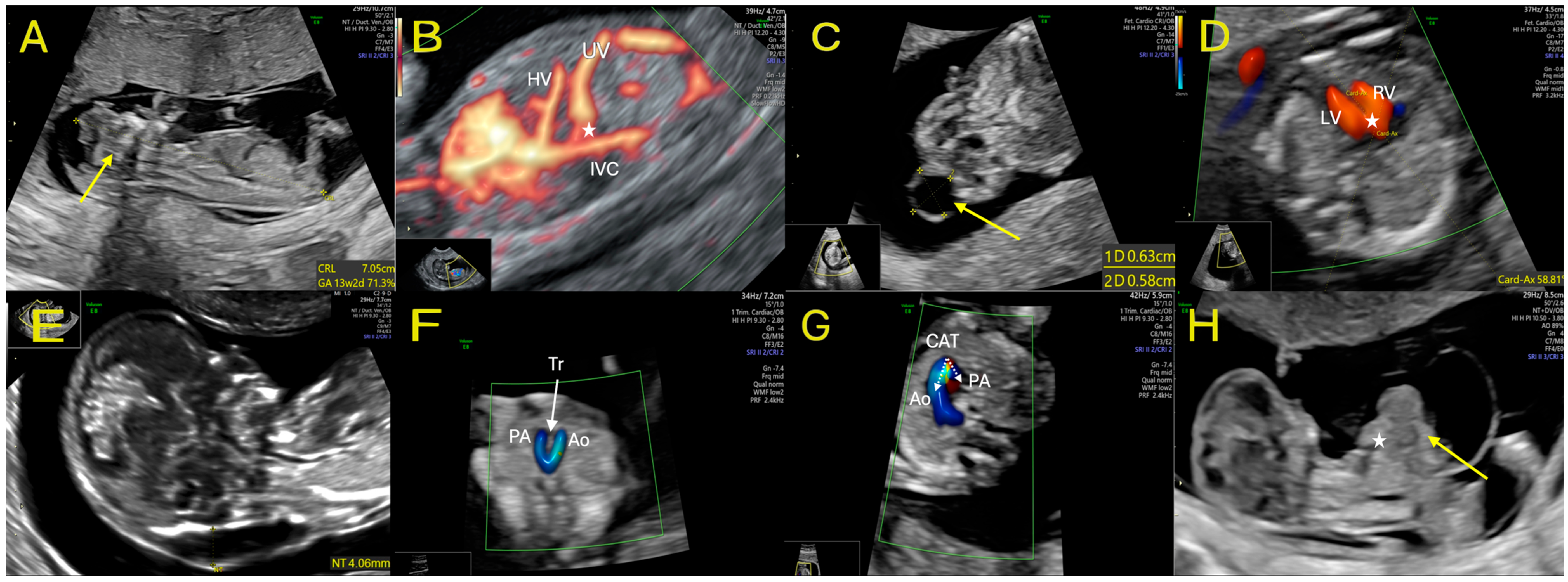
| Case | Medical History | NT (mm) | NB | TR | DV (PVIV) | 4CH | 3VTV | Fetal Anatomy | Risk of Aneuploidy | Post-Test Referral | Prenatal Genetic Testing & Results | Verification of Diagnosis | Fetal Outcome |
|---|---|---|---|---|---|---|---|---|---|---|---|---|---|
| 1 | Age 36 GI Low risk group BMI 23 | 1.9 | Hypoplastic | + | 0.83 | Normal | Normal | Acrania/exencephaly | T21 1:203 T18 1:449 T13 1:1271 | No | No | Yes | ToP |
| 2 | Age 33 GII PII Low risk group BMI 22 | 1.00 | Hypoplastic | + | 0.74 | Normal | Normal | Acrania/exencephaly | T21 1:8394 T18 < 1:20,000 T13 1:3231 | No | No | Yes | ToP |
| 3 | Age 29 GI Low-risk group BMI 34 | 1.1 | Hypoplastic | + | 1.09 | Incorrect cardiac axis | Normal | Acrania/exencephaly | T21 1:3401 T18 1:4920 T13 1:8751 | No | No | Yes | ToP |
| 4 | Age 21 GI Maternal smoking BMI 30 | 1.71 | + | + | Agenesis of DV (intrahepatic type) | Normal | Normal | No other anomalies detected | T21 < 1: 20,000 T18 < 1:20,000 T13 < 1:20,000 | Fetal echocardiography | No | Yes | Healthy live birth |
| 5 | Age 34 GII PII Low risk group BMI 21 | 1.33 | + | + | Agenesis of DV (intrahepatic with thin connection to IVC) | Normal | Normal | No other anomalies detected | T21 1:724 T18 1:2377 T13 1:1780 | Fetal echocardiography | No | Yes | Live birth (with postnatal ASD) |
| 6 | Age 26 GII PII Low risk group BMI 34 | 1.6 | + | + | 1.04 | Normal | Normal | Sacrococcygeal teratoma | T21 1:17,593 T18 < 1:20,000 T13 < 1:20,000 | Genetic counseling | Normal karyotype | Yes | Attempted intrauterine therapy, fetal demise |
| 7 | Age 34 GII PII Low risk group BMI 23 | 2.5 | + | + | 1.07 | Normal | Abnormal | No other anomalies detected | T21 1:1807 T18 1:11,850 T13 < 1:20,000 | Fetal echocardiography | No | False positive | Healthy live birth |
| 8 | Age 29 GII PII Low risk group BMI 33 | 4.8 | + | + | 1.01 | Incorrect cardiac axis | Abnormal | No other anomalies detected | T21 > 1:4 T18 1:277 T13 1:1916 | Genetic counseling | Normal karyotype | False positive | Healthy live birth |
| 9 | Age 34 GIV PIII Low risk group BMI 35 | 6.9 | Hypoplastic | + | 1.81 reversed flow | AVSD | RAA | No other anomalies detected | T21 > 1:4 T18 1:467 T13 1:168 | Fetal echocardiography, genetic counseling | Recommended, not performed | Yes | Intrauterine demise |
| 10 | Age 26 GI Low risk group BMI 23 | 1.74 | + | + | 0.95 | Normal | RAA | No other anomalies detected | T21 1:18,670 T18 < 1:20,000 T13 < 1:20,000 | Fetal echocardiography | Recommended | Yes | Live birth (isolated anomaly) |
| 11 | Age 29 GIII PII Low risk group BMI 22 | 1.8 | + | Not evaluable | 0.98 | Incorrect cardiac axis | CAT | No other anomalies detected | T21 1:3007 T18 1:5912 T13 < 1:20,000 | Fetal echocardiography | Recommended | Yes (additional dysplastic PV and ACC) | Pregnancy outcome unknown |
| 12 | Age 29 GIV PIII Maternal smoking BMI 20 | 2.3 | + | Regurgitation | 0.82 | Normal | Normal | Reversed flow in aorta (3VV) | T21 1:414 T18 1:1841 T13 1:34 | Fetal echocardiography, genetic counseling | Normal karyotype | Yes (additional VSD, VM, ACC | Live birth |
| 13 | Age 34 GIII PIII Low risk group BMI 35 | 1.8 | + | + | 1.24 | Normal | Normal | PFC, abnormal IT | T21 1:1497 T18 1:15,684 T13 < 1:20,000 | No | No | Yes transitory | Healthy live birth |
| 14 | Age 32 GIII PII Low risk group BMI 21 | 3.4 | + | + | 1.42 | Normal | Normal | Interrupted RT, megacystis, DUT | T21 1:21 T18 1:10 T13 1:10 | Genetic counseling | Recommended | Yes | Pregnancy outcome unknown |
| 15 | Age 28 GII PII Low risk group BMI 20 | 1.8 | + | + | 1.06 | Incorrect cardiac axis | Abnormal outflows (suspicion of TOF) | No other anomalies detected | T21 1:1346 T18 < 1:20,000 T13 1:8199 | Fetal echocardiography | No | Lost to follow-up | Lost to follow-up |
| 16 | Age 39 GII PII Low risk group BMI 21 | 1.8 | Hypoplastic | Not evaluable | Not evaluable | Ectopia cordis | Ectopia cordis | Omphalocele, LSD, DPD, ADD-(POC), PFC, interrupted RT | T21 1:363 T18 >1:4 T13 1:10 | Genetic counseling | Recommended | Yes | Intrauterine demise |
| 17 | Age 29 GII PII Low risk group BMI 33 | 4.1 | Hypoplastic | + | Reversed flow | RV > LV single ventricular inflow | Abnormal | Maxillary gap | T21 >1:4 T18 1:12 T13 1:101 | Fetal echocardiography, genetic counseling | 46,XX,der(3)t(1;3) (trisomy 1q32 and monosomy 3q29)” | Yes (complex cardiac defect) VSD, HLHS | Termination of pregnancy |
Disclaimer/Publisher’s Note: The statements, opinions and data contained in all publications are solely those of the individual author(s) and contributor(s) and not of MDPI and/or the editor(s). MDPI and/or the editor(s) disclaim responsibility for any injury to people or property resulting from any ideas, methods, instructions or products referred to in the content. |
© 2026 by the authors. Licensee MDPI, Basel, Switzerland. This article is an open access article distributed under the terms and conditions of the Creative Commons Attribution (CC BY) license.
Share and Cite
Korniluk, M.; Korniluk, A.; Szewczyk, G. Early Detection of Major Fetal Structural Anomalies in the First Trimester: A Retrospective Single-Center Study in an Unselected Population. J. Clin. Med. 2026, 15, 6560. https://doi.org/10.3390/jcm15176560
Korniluk M, Korniluk A, Szewczyk G. Early Detection of Major Fetal Structural Anomalies in the First Trimester: A Retrospective Single-Center Study in an Unselected Population. Journal of Clinical Medicine. 2026; 15(17):6560. https://doi.org/10.3390/jcm15176560
Chicago/Turabian StyleKorniluk, Maciej, Andrzej Korniluk, and Grzegorz Szewczyk. 2026. "Early Detection of Major Fetal Structural Anomalies in the First Trimester: A Retrospective Single-Center Study in an Unselected Population" Journal of Clinical Medicine 15, no. 17: 6560. https://doi.org/10.3390/jcm15176560
APA StyleKorniluk, M., Korniluk, A., & Szewczyk, G. (2026). Early Detection of Major Fetal Structural Anomalies in the First Trimester: A Retrospective Single-Center Study in an Unselected Population. Journal of Clinical Medicine, 15(17), 6560. https://doi.org/10.3390/jcm15176560