Acute Ischemic Stroke in 2026: From Time to Penumbra—An Updated Narrative Review of Reperfusion Strategies with a Latin American Implementation Perspective
 , , ,
, , ,  ,
,
Abstract
1. Introduction
2. Methods
3. Results
- Prehospital stroke recognition and early clinical identification, including validated screening tools and triage strategies for timely diagnosis and referral, particularly relevant in resource-constrained environments.
- Current classification and etiological evaluation of ischemic stroke, including established entities such as ESUS (Embolic Stroke of Undetermined Source) and recent advances in etiological stratification.
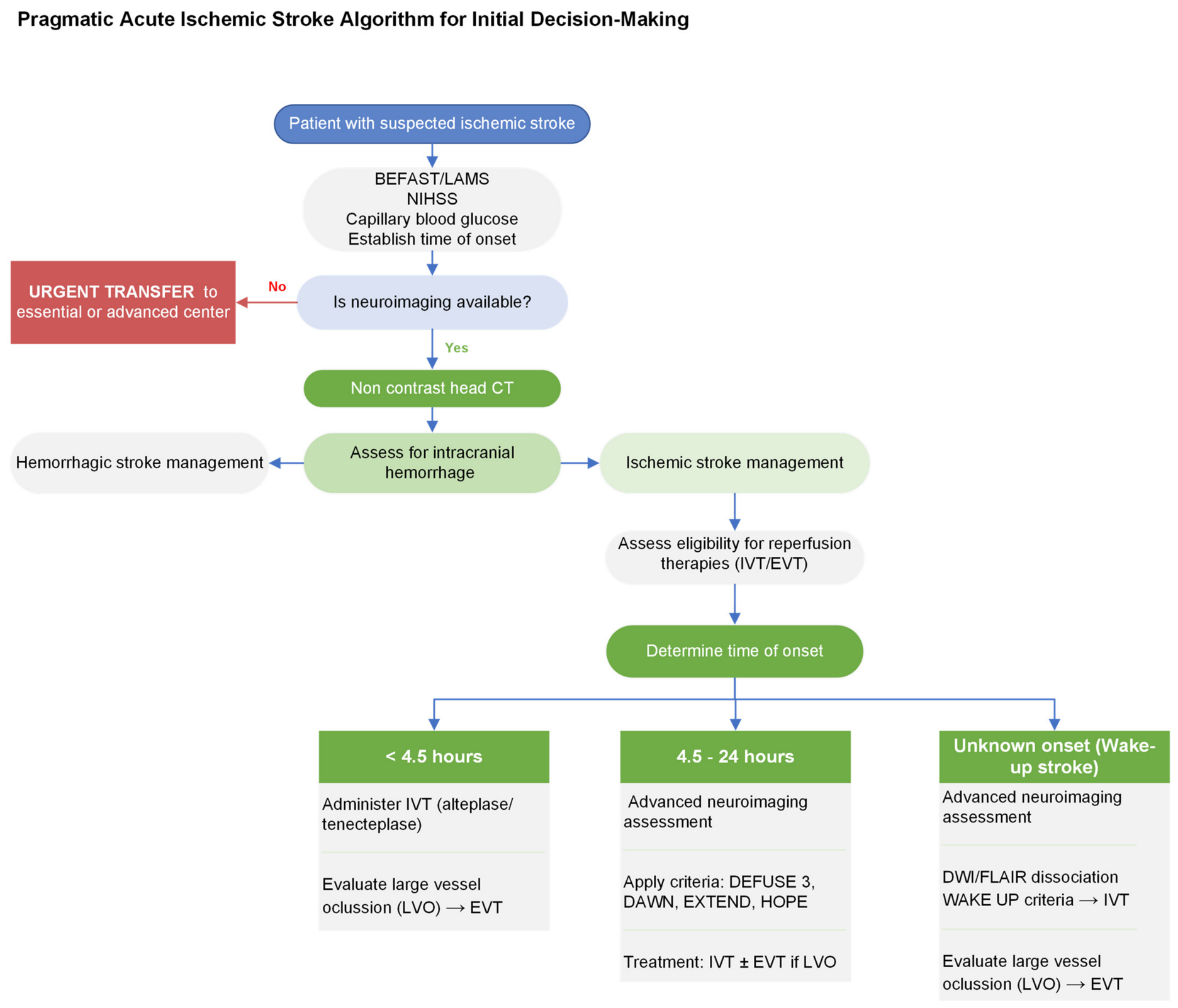
- Thrombolysis in extended time windows, supported by recent clinical trials and its applicability in settings using advanced imaging-based selection.
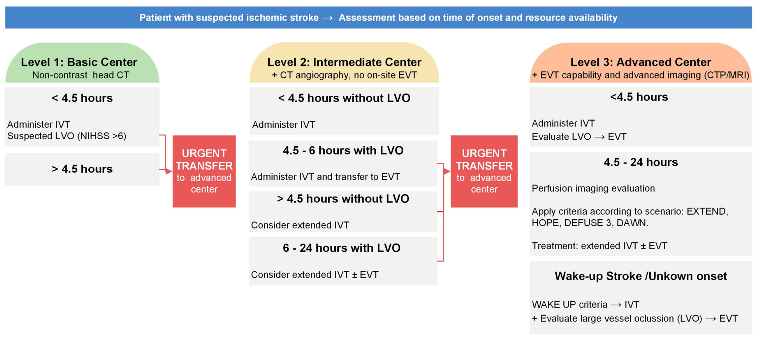
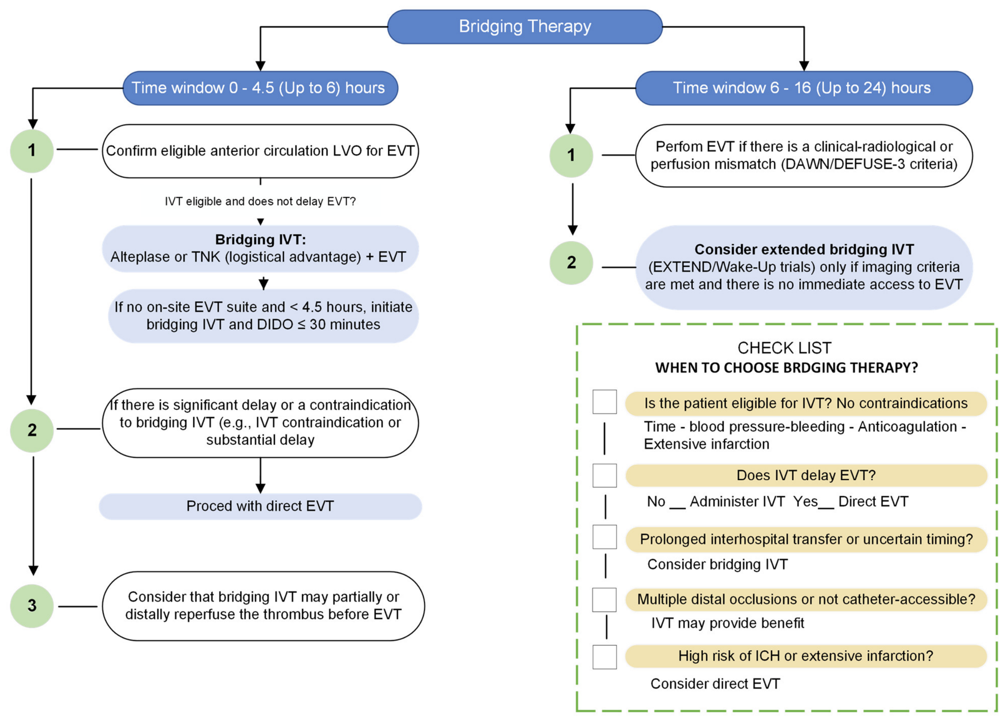
- Endovascular therapy, including extended therapeutic windows and the evolving role of combined strategies (bridging therapy vs. direct EVT), with a focus on comparative efficacy and patient selection.
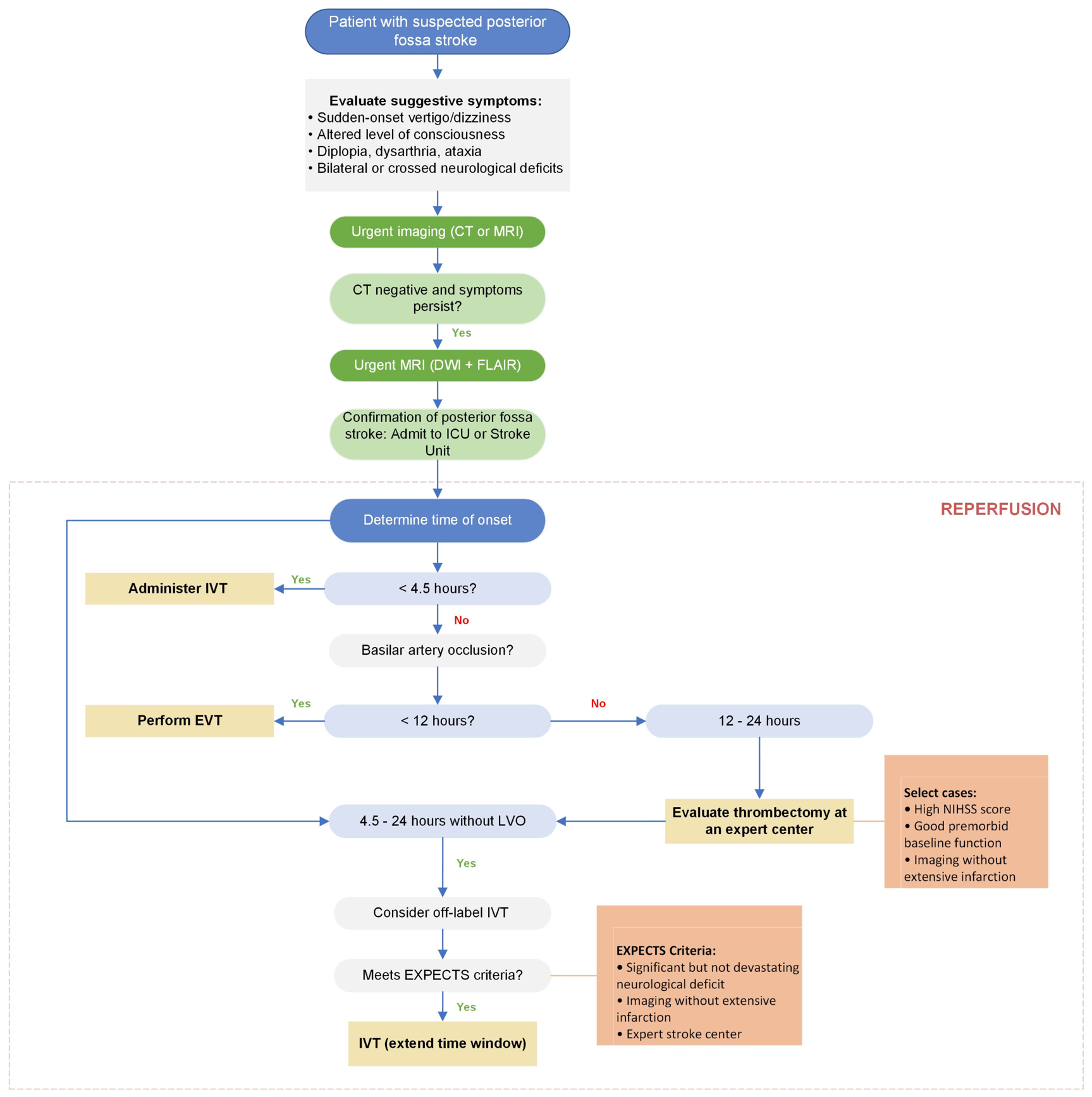
- Posterior circulation stroke, emphasizing contemporary reperfusion evidence and the pathophysiological and diagnostic particularities of this vascular territory.
3.1. Prehospital Stroke Recognition and Early Clinical Identification
3.2. Current Classification and Etiological Evaluation of Ischemic Stroke
- Large-artery atherosclerosis (LAA): Associated with ≥50% stenosis of the carotid or major cerebral arteries; accounts for approximately 15–20% of cases.
- Cardioembolic (CE): Resulting from embolism originating in the heart (e.g., atrial fibrillation, intracavitary thrombi, prosthetic valves, endocarditis), typically characterized by sudden onset, larger infarct volumes, and poorer prognosis; represents 20–30% of cases.
- Small-vessel occlusion (lacunar): Caused by lipohyalinosis or microatheromatosis affecting penetrating arteries, leading to infarcts <15 mm in regions such as the basal ganglia or pons; often associated with lacunar syndromes; accounts for 20–25% of cases.
- Other determined etiologies: Includes less common causes such as arterial dissections, vasculitis, prothrombotic states, genetic conditions, or infections; represents <5% of cases.
- Undetermined etiology (cryptogenic or multiple): Defined when no cause is identified despite comprehensive evaluation, or when multiple potential mechanisms coexist without a clear predominance; reported in up to 30–35% of cases.
3.3. Thrombolysis in the Era of Extended Windows
- WAKE-UP (NEJM 2018): This trial included 503 patients with stroke of unknown onset (typically upon awakening), selected using MRI demonstrating a diffusion-FLAIR mismatch (i.e., a lesion visible on DWI but not on FLAIR), which serves as a surrogate marker of early time window (<4.5–6 h) rather than direct assessment of penumbral tissue. Alteplase administered within 4.5 h from symptom recognition significantly improved functional independence (mRS 0–1: 53.3% vs. 41.8%; OR 1.62; p = 0.003), with an absolute difference of approximately 11–12%. Although symptomatic intracranial hemorrhage was more frequent in the treatment group, the difference was not statistically significant (2.0% vs. 0.4%), and no increase in mortality was observed (4.1% vs. 1.2%; p = 0.07). These findings support the use of imaging-based selection in patients with unknown onset, but within a time-surrogate framework rather than true penumbra-based selection [36].
- EXTEND (NEJM 2019): This trial evaluated 225 patients treated between 4.5 and 9 h after symptom onset (or with wake-up stroke), selected based on perfusion mismatch criteria (core <70 mL, penumbra >10 mL, mismatch ratio ≥1.2) using CT perfusion (CTP) or MRI (PWI/DWI). Alteplase significantly increased functional independence (mRS 0–2: 35.4% vs. 29.5%; adjusted RR 1.44; p = 0.04), although it was associated with a higher rate of symptomatic intracranial hemorrhage (6.2% vs. 0.9%), without a significant increase in mortality [37]. These results demonstrated the feasibility of extending thrombolysis up to 9 h using imaging-based selection, although the magnitude of benefit was more modest compared with the standard time window (NNT = 17) [38].
- HOPE (JAMA 2025): This recent trial evaluated the safety and efficacy of alteplase administered up to 24 h after symptom onset in imaging-selected patients. A total of 372 patients were included, meeting viability criteria similar to those used in EXTEND (core <70 mL, penumbra/core ratio ≥1.2, and mismatch volume >10 mL), and excluding candidates for thrombectomy. Alteplase was associated with higher rates of functional independence (mRS 0–1 at 90 days: 49.5% vs. 35.5%; absolute difference 13.9%; p = 0.004), with a symptomatic intracranial hemorrhage rate of 3.8% and no increase in mortality. Although most treated patients were within the first 9 h, the trial supports the safety and efficacy of imaging-guided thrombolysis within an extended window of up to 24 h [39].
- Meta-analysis (Stroke 2025): These findings were further supported by a meta-analysis published in Stroke (2025), which included eight randomized trials (excluding HOPE) and 1742 patients treated with thrombolysis beyond 4.5 h (up to 24 h), excluding those undergoing thrombectomy. The pooled results confirmed the clinical benefit of an imaging-based selection strategy: thrombolysis increased rates of excellent recovery (mRS 0–1: 44% vs. 36%; OR 1.43; p = 0.0005) and improved functional outcomes (mRS 0–2; OR 1.36; p = 0.002). Greater benefit was observed with perfusion-based selection (OR 1.45; 95% CI 1.08–1.94) compared with DWI–FLAIR mismatch selection (OR 1.34; 95% CI 0.94–1.91). Additionally, the effect appeared more pronounced with tenecteplase (OR 1.47; 95% CI 1.06–2.04) than with alteplase (OR 1.38; 95% CI 1.08–1.78). Symptomatic intracranial hemorrhage rates were below 5–6%, and no significant differences in mortality were observed, supporting the concept that imaging-defined tissue viability can supersede strict time-based treatment thresholds [40].
Tenecteplase and Single-Bolus Administration
- EXTEND-IA TNK (NEJM 2018): This trial included patients with large-vessel occlusions (LVO) eligible for thrombectomy and demonstrated that tenecteplase at a dose of 0.25 mg/kg achieved higher rates of pre-thrombectomy reperfusion compared with alteplase (22% vs. 10%; OR 2.6; 95% CI 1.1–5.9). Tenecteplase was also associated with improved functional outcomes at 90 days, reflected by a more favorable mRS distribution (OR 1.7; 95% CI 1.0–2.8). Rates of symptomatic intracranial hemorrhage (sICH) were low and similar in both groups (1%) [43].
- EXTEND-IA TNK II (JAMA 2020): This trial compared tenecteplase at doses of 0.25 mg/kg versus 0.4 mg/kg and found no significant differences in reperfusion or functional outcomes (mRS at 90 days: RR 1.03; 95% CI 0.66–1.61), supporting 0.25 mg/kg as the optimal dose [44]. In larger trials, including TRACE-2 (Lancet 2023) and ATTEST-2 (Lancet 2024), tenecteplase at 0.25 mg/kg demonstrated non-inferiority to alteplase in terms of functional outcomes, with comparable rates of symptomatic intracranial hemorrhage (3–4%) and no differences in mortality at 90 days [45,46].
- TIMELESS (NEJM 2024): This trial included 458 patients with large-vessel occlusion (LVO) and imaging evidence of salvageable tissue treated between 4.5 and 24 h after symptom onset. Tenecteplase demonstrated a favorable safety profile (sICH: 3.2% vs. 2.3%) and achieved higher rates of recanalization compared with standard care (76.7% vs. 63.9%). A trend toward improved functional outcomes was observed (mRS 0–2: 45.9% vs. 31.4%), although this difference did not reach statistical significance [47].
- TRACE-III (NEJM 2024): this trial showed that patients without access to endovascular thrombectomy, tenecteplase administered between 4.5 and 24 h after symptom onset was associated with reduced disability (mRS 0–1: 33.0% vs. 24.2%; RR 1.37; 95% CI 1.04–1.81; p = 0.03), with an acceptable safety profile (sICH: 3.0% vs. 0.8%) [48].
3.4. Endovascular Therapy in Ischemic Stroke: Extended Windows and Bridging Therapy
3.4.1. Extended Windows for Endovascular Therapy (6–24 h)
- DAWN (NEJM 2018): This trial included 206 patients with occlusion of the terminal internal carotid artery or proximal middle cerebral artery (MCA) treated between 6 and 24 h from last known well. Patients were selected based on a clinical–imaging mismatch (severe neurological deficit with a small infarct core). Eligibility criteria included: age ≥80 years with NIHSS ≥10 and core volume <21 mL; or age <80 years with NIHSS ≥10 and core <31 mL (or NIHSS ≥20 and core <51 mL). The trial was stopped early due to efficacy, demonstrating functional independence (mRS 0–2) in approximately 50% of patients undergoing thrombectomy versus 13–19% with medical therapy alone (OR 4.5), without significant increases in mortality or symptomatic intracranial hemorrhage, supporting benefit up to 24 h in highly selected patients [51].
- DEFUSE 3 (NEJM 2018): This trial included 182 patients treated between 6 and 16 h after symptom onset with ICA or MCA M1 occlusion, selected using perfusion imaging (CT or MRI with RAPID software: core <70 mL, penumbra ≥15 mL, mismatch ratio ≥1.8). Similarly to DAWN, the trial was stopped early after demonstrating benefit. Thrombectomy significantly improved functional outcomes, with independence (mRS 0–2) achieved in 45% of patients compared with 17% in the medical therapy group (p < 0.001; NNT= 3–4) [52].
- Occlusion location: The strongest evidence supports EVT in anterior circulation large-vessel occlusions (LVO), particularly involving the internal carotid artery (ICA) or proximal middle cerebral artery (MCA). For basilar artery occlusion (posterior circulation), the evidence base has evolved more recently and is addressed in a dedicated section below. Earlier guidelines suggested that EVT may be considered in this context due to the high mortality associated with untreated basilar artery occlusion, although this recommendation was based on lower levels of evidence (conditional recommendation, Class IIa/B) [11,50].
- Large ischemic cores: Recent trials have expanded EVT eligibility to include patients with large infarct cores (e.g., >70 mL or ASPECTS 3–5), a group previously considered to have a poor prognosis.
- (I)
- SELECT2 (NEJM 2023): included 352 patients with anterior circulation LVO and large infarcts defined by ASPECTS 3–5 on non-contrast CT or infarct volume ≥50 mL on CTP or MRI (low ADC). EVT significantly reduced disability, with mRS 0–3 achieved in 46% of patients compared with 30% in the medical therapy group (OR 1.97; 95% CI 1.21–3.20; p = 0.003), without a significant increase in symptomatic intracranial hemorrhage or mortality, although treatment effects were attenuated with increasing infarct volume [53].
- (II)
- ANGEL-ASPECT (NEJM 2023): an Asian multicenter trial including 456 patients, defined large infarct cores using ASPECTS 3–5 on CT or volumes of 70–100 mL on CTP/MRI, without requiring formal penumbral mismatch. EVT improved the overall functional outcome distribution at 90 days (OR 1.37; 95% CI 1.11–1.69; p = 0.004), with a number needed to treat (NNT) of 8 to reduce one level of disability [54]. Subsequent analyses suggest that better outcomes may be associated with concordant clinical–imaging profiles (e.g., high NIHSS with large core) and favorable perfusion parameters, such as a hypoperfusion-to-core ratio ≥1.8 and penumbra volume ≥15 mL [55]. However, these findings should be interpreted cautiously, as treatment effects are smaller than in patients with small infarct cores and may depend on careful patient selection.
- Other selection considerations: Current guidelines recommend considering baseline functional status, typically defined as a pre-stroke mRS of 0–1, and a minimum NIHSS score of ≥6. There is no strict upper limit for NIHSS; patients with very severe deficits (NIHSS >20) may still benefit from early intervention, although some centers place greater emphasis on imaging to exclude extensive established infarction [50]. Age alone should not be considered an exclusion criterion, as benefit has been demonstrated in patients older than 80 years, albeit with lower rates of functional independence [50]. Regarding imaging criteria, an ASPECTS ≥6 is generally required in early time windows. In extended-window scenarios, however, advanced imaging with CT perfusion or MRI is often necessary to quantify infarct core and penumbral tissue, thereby guiding patient selection more precisely [10,11,50].
3.4.2. Endovascular Therapy Alone vs. Combined with IV Thrombolysis (Bridging Therapy)
3.5. Management of Posterior Fossa Stroke (Basilar Artery Occlusion)
3.5.1. Endovascular Therapy in Posterior Fossa Stroke: Current Evidence
- BASICS (NEJM 2021): This trial included 300 patients, most treated within 6 h of symptom onset. Functional independence (mRS 0–3 at 90 days) was achieved in 44% of patients undergoing thrombectomy compared with 38% receiving medical therapy (RR 1.18; 95% CI 0.92–1.50; p = 0.17). The lack of a statistically significant benefit may be partly explained by trial design factors, including the inclusion of patients with lower stroke severity (NIHSS <10) and a relatively high rate of spontaneous recanalization, which may have attenuated the observed treatment effect [66].
- BEST (Lancet 2020): This Chinese randomized trial was stopped early after enrolling 131 patients (target 344) due to recruitment challenges and a high rate of treatment crossover. Favorable outcomes (mRS 0–3) were observed in 42% of patients undergoing thrombectomy compared with 32% receiving medical therapy (adjusted RR 1.74; 95% CI 0.81–3.74), although this difference did not reach statistical significance [67].
- ATTENTION (NEJM 2022): This trial included 340 patients with acute basilar artery occlusion (BAO) and NIHSS ≥10 treated within ≤12 h from symptom onset (median 5 h). Functional independence (mRS 0–3) was achieved in 46% of patients undergoing EVT compared with 23% receiving medical therapy (RR 2.06; p < 0.001), leading to early termination of the trial for efficacy. Mortality was also significantly reduced (37% in the EVT group vs. 55% in the medical therapy group; RR 0.66; 95% CI 0.52–0.82), with low rates of symptomatic intracranial hemorrhage (5% vs. 0%) [68].
- BAOCHE (NEJM 2022): This trial extended the treatment window to 24 h (median 11 h) and included 217 patients with acute BAO, NIHSS ≥10, and no evidence of extensive infarction. Functional independence (mRS 0–3) was achieved in 46% of patients undergoing EVT compared with 24% receiving medical therapy (p < 0.001). Mortality was lower in the EVT group (31% vs. 42%), although this difference did not reach statistical significance, likely due to sample size limitations. Importantly, patient selection excluded cases with chronic occlusion or complete brainstem infarction [69].
3.5.2. Role of IV Thrombolysis in Posterior Circulation Stroke
4. Discussion
5. Conclusions
Author Contributions
Funding
Data Availability Statement
Conflicts of Interest
Abbreviations
| ASPECTS | Alberta Stroke Program Early CT Score |
| BAO | Basilar Artery Occlusion |
| BP | Blood Pressure |
| COPE | Committee on Publication Ethics |
| CT | Computed Tomography |
| CTA | CT Angiography |
| CTP | CT Perfusion |
| CTP/CTA mismatch | Perfusion–angiography mismatch (marker of viable ischemic penumbra) |
| DIDO | Door-In Door-Out time |
| DM | Diabetes Mellitus |
| DNT | Door-to-Needle time |
| DPN | Door-to-Puncture time |
| EMS | Emergency Medical Services |
| EVT | Endovascular Therapy (mechanical thrombectomy) |
| AF | Atrial Fibrillation |
| FLAIR | Fluid-Attenuated Inversion Recovery |
| HTN | Hypertension |
| SU | Stroke Unit |
| ICMJE | International Committee of Medical Journal Editors |
| IVT/rtPA | Intravenous Thrombolysis/Recombinant Tissue Plasminogen Activator |
| LMICs | Low- and Middle-Income Countries |
| LVO | Large Vessel Occlusion |
| mRS | Modified Rankin Scale |
| MRI | Magnetic Resonance Imaging |
| NIHSS | National Institutes of Health Stroke Scale |
| SVO | Video Monitoring System (if applicable in stroke units or monitoring protocols) |
| TICI | Thrombolysis in Cerebral Infarction (angiographic reperfusion grade) |
| TIA | Transient Ischemic Attack |
| TNK | Tenecteplase |
References
- Feigin, V.L.; Brainin, M.; Norrving, B.; Martins, S.O.; Pandian, J.; Lindsay, P.; Grupper, M.F.; Rautalin, I. World Stroke Organization: Global Stroke Fact Sheet 2025 (based on GBD 2021). Int. J. Stroke 2025, 20, 132–144. [Google Scholar] [CrossRef] [Scilit] [PubMed]
- Saver, J.L. Time is brain—Quantified. Stroke 2006, 37, 263–266. [Google Scholar] [CrossRef] [Scilit]
- GBD 2021 Stroke Collaborators. Global and national burden of stroke 1990–2021: A systematic analysis for the Global Burden of Disease Study 2021. Lancet Neurol. 2024, 23, 973–1003. [Google Scholar] [CrossRef] [Scilit]
- World Health Organization. The Top 10 Causes of Death; World Health Organization: Geneva, Switzerland, 7 August 2024. Available online: https://www.who.int/news-room/fact-sheets/detail/the-top-10-causes-of-death (accessed on 9 October 2025).
- Bayona, H.; Amaya, P.; Bustos, J.L.; Giraldo-Tapias, L.M.; Betancourt, A.; Vallejo, C.; Rivillas, J.; Llanos-Leyton, N.; Mantilla, K.; Alvarez, T.F.; et al. Quality indicators of stroke care in Colombia based on the RES-Q registry. J. Stroke Cerebrovasc. Dis. 2025, 34, 108249. [Google Scholar] [CrossRef] [Scilit] [PubMed]
- Hammond, G.; Luke, A.A.; Elson, L.; Towfighi, A.; Joynt Maddox, K.E. Urban-Rural Inequities in Acute Stroke Care and In-HospitalMortality. Stroke 2020, 51, 2131–2138. [Google Scholar] [CrossRef] [Scilit] [PubMed]
- Hacke, W.; Kaste, M.; Bluhmki, E.; Brozman, M.; Dávalos, A.; Guidetti, D.; Larrue, V.; Lees, K.R.; Medeghri, Z.; Machnig, T.; et al. Thrombolysis with alteplase 3 to 4.5 hours after acute ischemic stroke. N. Engl. J. Med. 2008, 359, 1317–1329. [Google Scholar] [CrossRef] [Scilit] [PubMed]
- Goyal, M.; Menon, B.K.; van Zwam, W.H.; Dippel, D.W.J.; Mitchell, P.J.; Demchuk, A.M.; Dávalos, A.; Majoie, C.B.L.M.; van der Lugt, A.; de Miquel, M.A.; et al. Endovascular thrombectomy after large-vessel ischemic stroke: A meta-analysis of individual patient data from five randomised trials. Lancet 2016, 387, 1723–1731. [Google Scholar] [CrossRef] [Scilit] [PubMed]
- Powers, W.J.; Rabinstein, A.A.; Ackerson, T.; Adeoye, O.M.; Bambakidis, N.C.; Becker, K.; Biller, J.; Brown, M.; Demaerschalk, B.M.; Hoh, B.; et al. 2019 Update to the 2018 Guidelines for the Early Management of Acute Ischemic Stroke. Stroke 2019, 50, e344–e418. [Google Scholar] [CrossRef] [Scilit] [PubMed]
- Prabhakaran, S.; Gonzalez, N.R.; Zachrison, K.S.; Adeoye, O.; Alexandrov, A.W.; Ansari, S.A.; Chapman, S.; Czap, A.L.; Dumitrascu, O.M.; Ishida, K.; et al. 2026 Guideline for the Early Management of Patients with Acute Ischemic Stroke: A Guideline from the American Heart Association/American Stroke Association. Stroke 2026, 57, STR.0000000000000513. [Google Scholar] [CrossRef] [Scilit] [PubMed]
- Turc, G.; Tsivgoulis, G.; Audebert, H.J.; Boogaarts, H.; Bhogal, P.; De Marchis, G.M.; Fonseca, A.C.; Khatri, P.; Mazighi, M.; Pérez de la Ossa, N.; et al. European Stroke Organisation (ESO)–European Society for Minimally Invasive Neurological Therapy (ESMINT) expedited recommendation on intravenous thrombolysis prior to mechanical thrombectomy in acute ischaemic stroke. Eur. Stroke J. 2022, 7, I–XXVI. [Google Scholar] [CrossRef] [Scilit] [PubMed]
- Venema, E.; Duvekot, M.H.C.; Lingsma, H.F.; Rozeman, A.D.; Moudrous, W.; Vermeij, F.H.; Biekart, M.; van der Lugt, A.; Kerkhoff, H.; Dippel, D.W.J.; et al. Prehospital triage of patients with suspected stroke symptoms (PRESTO): Protocol of a prospective observational study. BMJ Open 2019, 9, e028810. [Google Scholar] [CrossRef] [Scilit] [PubMed]
- Widimsky, P.; Snyder, K.; Sulzenko, J.; Hopkins, L.N.; Stetkarova, I. Acute ischaemic stroke: Recent advances in reperfusion treatment. Eur. Heart J. 2023, 44, 1205–1215. [Google Scholar] [CrossRef] [Scilit] [PubMed]
- Kazadi Kabanda, I.; Kiangebeni Ngonzo, C.; Emeka Bowamou, C.-K.; Divengi Nzambi, J.-P.; Kiatoko Ponte, N.; Tuyinama Madoda, O.; Nkodila Natuhoyila, A.; M’Buyamba-Kabangu, J.-R.; Longo-Mbenza, B.; Banzulu Bomba, D.; et al. Stroke signs knowledge and factors associated with a delayed hospital arrival of patients with acute stroke in Kinshasa. Heliyon 2024, 10, e28311. [Google Scholar] [CrossRef] [Scilit] [PubMed]
- Correia, P.N.; Roy, J.; Haumesser, C.; Touma, L.; Jacquin, G.; Poppe, A.Y.; Deschaintre, Y.; Veilleux Carpentier, A. Extended Delays in Recognition of Stroke Symptoms and Stroke Code Activation for In-Hospital Strokes: The DELAY Study. Can. J. Neurol. Sci. 2025, 52, 502–509. [Google Scholar] [CrossRef] [Scilit] [PubMed]
- Farokhfar, M.; Almani, M.S.P. Study on delay factors and time to hospital arrival after acute stroke in patients at Shahid Rajaei hospital, Tonekabon (2022–2023). BMC Neurol. 2025, 25, 340. [Google Scholar] [CrossRef] [Scilit] [PubMed]
- Darkhabani, M.Z.; Homa-Bonell, J.K.; Thoreson, L.; Bobholz, J.A.; Spaulding, D.; Engebose, M. BE FAST Versus FAST: A Randomized Pilot Trial Comparing Retention of Stroke Symptoms Between 2 Mnemonics. J. Am. Heart Assoc. 2024, 13, e035696. [Google Scholar] [CrossRef] [Scilit] [PubMed]
- Hilditch, M.; Brand, C.; Devlin, S.; Boyd, A.; Venema, E. BE-FAST vs FAST in prehospital stroke recognition: A systematic review. Br. J. Community Nurs. 2025, 30, 439–447. [Google Scholar] [CrossRef] [Scilit] [PubMed]
- Arthur, K.C.; Huang, S.; Gudenkauf, J.C.; Mohseni, A.; Wang, R.; Aslan, A.; Nabi, M.; Hoseinyazdi, M.; Johnson, B.; Patel, N.; et al. Assessing the Relationship between LAMS and CT Perfusion Parameters in Acute Ischemic Stroke Secondary to Large Vessel Occlusion. J. Clin. Med. 2023, 12, 3374. [Google Scholar] [CrossRef] [Scilit] [PubMed]
- Carbonera, L.A.; Souza, A.C.D.; Rodrigues, M.D.S.; Mottin, M.D.; Nogueira, R.G.; Martins, S.C.O. FAST-ED scale for prehospital triage of large vessel occlusion: Results in the field. Arq. Neuro-Psiquiatr. 2022, 80, 885–892. [Google Scholar] [CrossRef] [Scilit] [PubMed]
- Pornpanit, C.; Loymai, P.; Uransilp, N.; Srivilaithon, W. Accuracy of FAST-ED for Assessment Large Vessel Occlusion of Acute Ischemic Stroke in Emergency Department. Open Access Emerg. Med. 2024, 16, 203–210. [Google Scholar] [CrossRef] [Scilit] [PubMed]
- Zang, J.; Bai, Q.; Xiong, X.; He, P.; Sun, J.; Gong, X. Early identification of stroke symptoms and risk factors using the BE FAST method: Benefits of early intervention in high-risk populations. Front. Neurol. 2025, 16, 1630384. [Google Scholar] [CrossRef] [Scilit] [PubMed]
- Baser, Y.; Zarei, H.; Gharin, P.; Baradaran, H.R.; Sarveazad, A.; Roshdi Dizaji, S.; Yousefifard, M. Cincinnati Prehospital Stroke Scale (CPSS) as a Screening Tool for Early Identification of Cerebral Large Vessel Occlusions; a Systematic Review and Meta-analysis. Arch. Acad. Emerg. Med. 2024, 12, e38. [Google Scholar] [CrossRef] [Scilit] [PubMed]
- Truong, A.T.L.; Tran, H.T.M.; Phan, L.D.; Nguyen, H.B.T.; Nguyen, T.Q.; Le, T.V.S.; Chiem, D.N.; Nguyen, H.; Nguyen, T.N.; Nguyen, T.H. Predictive Value of the Prehospital RACE Scale for Large Vessel Occlusion in Acute Stroke Patients. Cerebrovasc. Dis. Extra 2024, 15, 39–47. [Google Scholar] [CrossRef] [Scilit] [PubMed]
- Almunif, T.M.; Alkaabba, A.F.; Alomran, K.W.; Alanazi, A.M.; Alharbi, F.N.; Alshahrani, S.S.; Alsaeed, N.M.; Alabdulkader, R.A.; Alibraheem, S.A.; Alzahrani, K.S.; et al. Early Recognition and Referral of Acute Stroke in Primary and Emergency Care: A Systematic Review. West. J. Emerg. Med. 2026, 27. [Google Scholar] [CrossRef] [Scilit] [PubMed]
- Radu, R.A.; Terecoasă, E.O.; Băjenaru, O.A.; Tiu, C. Etiologic classification of ischemic stroke: Where do we stand? Clin. Neurol. Neurosurg. 2017, 159, 93–106. [Google Scholar] [CrossRef] [Scilit] [PubMed]
- Rathburn, C.M.; Mun, K.T.; Sharma, L.K.; Saver, J.L. TOAST stroke subtype classification in clinical practice: Implications for the Get With The Guidelines-Stroke nationwide registry. Front. Neurol. 2024, 15, 1375547. [Google Scholar] [CrossRef] [Scilit] [PubMed]
- Meschia, J.F. Diagnostic evaluation of stroke etiology. Continuum 2023, 29, 412–424. [Google Scholar] [CrossRef] [Scilit] [PubMed]
- Amarenco, P.; Bogousslavsky, J.; Caplan, L.R.; Donnan, G.A.; Wolf, M.E.; Hennerici, M.G. The ASCOD phenotyping of ischemic stroke (updated ASCO phenotyping). Cerebrovasc. Dis. 2013, 36, 1–5. [Google Scholar] [CrossRef] [Scilit] [PubMed]
- Hart, R.G.; Catanese, L.; Perera, K.S.; Ntaios, G.; Connolly, S.J. Embolic stroke of undetermined source: A systematic review and clinical update. Stroke 2017, 48, 867–872. [Google Scholar] [CrossRef] [Scilit] [PubMed]
- Gladstone, D.J.; Spring, M.; Dorian, P.; Panzov, V.; Thorpe, K.E.; Hall, J.; Vaid, H.; O’Donnell, M.; Laupacis, A.; Côté, R.; et al. Atrial fibrillation in patients with cryptogenic stroke (EMBRACE). N. Engl. J. Med. 2014, 370, 2467–2477. [Google Scholar] [CrossRef] [Scilit] [PubMed]
- Sanna, T.; Diener, H.C.; Passman, R.S.; Di Lazzaro, V.; Bernstein, R.A.; Morillo, C.A.; Rymer, M.M.; Thijs, V.; Rogers, T.; Beckers, F.; et al. Cryptogenic stroke and underlying atrial fibrillation (CRYSTAL AF). N. Engl. J. Med. 2014, 370, 2478–2486. [Google Scholar] [CrossRef] [Scilit] [PubMed]
- Hart, R.G.; Sharma, M.; Mundl, H.; Kasner, S.E.; Bangdiwala, S.I.; Berkowitz, S.D.; Swaminathan, B.; Lavados, P.; Wang, Y.; Wang, Y.; et al. Rivaroxaban for stroke prevention after ESUS (NAVIGATE-ESUS). N. Engl. J. Med. 2018, 378, 2191–2201. [Google Scholar] [CrossRef] [Scilit] [PubMed]
- Diener, H.-C.; Sacco, R.L.; Easton, J.D.; Granger, C.B.; Bernstein, R.A.; Uchiyama, S.; Kreuzer, J.; Cronin, L.; Cotton, D.; Grauer, C.; et al. Dabigatran for prevention of stroke after ESUS (RE-SPECT ESUS). N. Engl. J. Med. 2019, 380, 1906–1917. [Google Scholar] [CrossRef] [Scilit] [PubMed]
- Martins, S.C.O.; Lavados, P.; Secchi, T.L.; Brainin, M.; Ameriso, S.; Gongora-Rivera, F.; Sacks, C.; Cantú-Brito, C.; Alvarez Guzman, T.F.; Pérez-Romero, G.E.; et al. Fighting Against Stroke in Latin America: A Joint Effort of Medical Professional Societies and Governments. Front. Neurol. 2021, 12, 743732. [Google Scholar] [CrossRef] [Scilit] [PubMed]
- Thomalla, G.; Simonsen, C.Z.; Boutitie, F.; Andersen, G.; Berthezene, Y.; Cheng, B.; Cheripelli, B.; Cho, T.-H.; Fazekas, F.; Fiehler, J.; et al. MRI-guided thrombolysis for stroke with unknown time of onset (WAKE-UP). N. Engl. J. Med. 2018, 379, 611–622. [Google Scholar] [CrossRef] [Scilit] [PubMed]
- Ma, H.; Campbell, B.C.V.; Parsons, M.W.; Churilov, L.; Levi, C.R.; Hsu, C.; Kleinig, T.J.; Wijeratne, T.; Curtze, S.; Dewey, H.M.; et al. Thrombolysis guided by perfusion imaging up to 9 hours after onset (EXTEND). N. Engl. J. Med. 2019, 380, 1795–1803. [Google Scholar] [CrossRef] [Scilit] [PubMed]
- Bersano, A.; Burgin, W.S.; Molina, C.A. The changing landscape of intravenous thrombolysis for acute ischemic stroke. J. Clin. Med. 2024, 13, 2152. [Google Scholar] [CrossRef] [Scilit] [PubMed]
- Zhou, Y.; He, Y.; Campbell, B.C.V.; Liebeskind, D.S.; Yuan, C.; Chen, H.; Zhang, Y.; Yi, T.; Luo, Z.; Zhang, Z.; et al. Alteplase for acute ischemic stroke at 4.5 to 24 hours: The HOPE randomized clinical trial. JAMA 2025, 334, 788–798. [Google Scholar] [CrossRef] [Scilit] [PubMed]
- Günkan, A.; Ferreira, M.Y.; Vilardo, M.; Scarcia, L.; Bocanegra-Becerra, J.E.; Cardoso, L.J.C.; Paleare, L.F.F.; Almeida, G.O.; Semione, G.; Ferreira, C.; et al. Thrombolysis beyond the 4.5-hour window for acute ischemic stroke: A meta-analysis. Stroke 2025, 56, 580–590. [Google Scholar] [CrossRef] [Scilit] [PubMed]
- Seetge, J.; Cséke, B.; Karádi, Z.N.; Bosnyák, E.; Szapáry, L. Bridging the gap: Improving acute ischemic stroke outcomes with intravenous thrombolysis prior to mechanical thrombectomy. Neurol. Int. 2024, 16, 1189–1202. [Google Scholar] [CrossRef] [Scilit] [PubMed]
- Campbell, B.C.V.; Mistry, E.A. Tenecteplase for stroke—Opening the window. N. Engl. J. Med. 2024, 390, 677–679. [Google Scholar] [CrossRef] [Scilit] [PubMed]
- Campbell, B.C.V.; Mitchell, P.J.; Churilov, L.; Yassi, N.; Kleinig, T.J.; Dowling, R.J.; Yan, B.; Bush, S.J.; Dewey, H.M.; Thijs, V.; et al. Tenecteplase versus alteplase before thrombectomy (EXTEND-IA TNK). N. Engl. J. Med. 2018, 378, 1573–1582. [Google Scholar] [CrossRef] [Scilit] [PubMed]
- Campbell, B.C.V.; Mitchell, P.J.; Churilov, L.; Yassi, N.; Kleinig, T.J.; Dowling, R.J.; Yan, B.; Bush, S.J.; Thijs, V.; Scroop, R.; et al. Tenecteplase dose on cerebral reperfusion before thrombectomy (EXTEND-IA TNK Part 2). JAMA 2020, 323, 1257–1265. [Google Scholar] [CrossRef] [Scilit] [PubMed]
- Muir, K.W.; Ford, G.A.; Ford, I.; Wardlaw, J.M.; McConnachie, A.; Greenlaw, N.; Mair, G.; Sprigg, N.; Price, C.I.; MacLeod, M.J.; et al. Tenecteplase versus alteplase for acute stroke (ATTEST-2). Lancet 2024, 403, 1945–1956. [Google Scholar] [CrossRef] [Scilit] [PubMed]
- Wang, Y.; Li, S.; Pan, Y.; Li, H.; Parsons, M.W.; Campbell, B.C.V.; Schwamm, L.H.; Fisher, M.; Che, F.; Dai, H.; et al. Tenecteplase versus alteplase in acute ischaemic cerebrovascular events (TRACE-2). Lancet 2023, 401, 1024–1036. [Google Scholar] [CrossRef] [Scilit] [PubMed]
- Albers, G.W.; Jumaa, M.; Purdon, B.; Zaidi, S.F.; Streib, C.; Shuaib, A.; Sangha, N.; Kim, M.; Froehler, M.T.; Schwartz, N.E.; et al. Tenecteplase for stroke at 4.5 to 24 hours with perfusion-imaging selection (TIMELESS). N. Engl. J. Med. 2024, 390, 701–711. [Google Scholar] [CrossRef] [Scilit] [PubMed]
- Xiong, Y.; Campbell, B.C.V.; Schwamm, L.H.; Meng, X.; Jin, A.; Parsons, M.W.; Fisher, M.; Jiang, Y.; Che, F.; Wang, L.; et al. Tenecteplase for ischemic stroke at 4.5 to 24 hours without thrombectomy (TRACE-III). N. Engl. J. Med. 2024, 391, 203–212. [Google Scholar] [CrossRef] [Scilit] [PubMed]
- Nguyen, T.N.; Abdalkader, M.; Fischer, U.; Qiu, Z.; Nagel, S.; Chen, H.-S.; Miao, Z.; Khatri, P. Endovascular management of acute stroke. Lancet 2024, 403, 1209–1223. [Google Scholar] [CrossRef] [Scilit] [PubMed]
- European Stroke Organisation. ESO Guideline on the acute management of basilar artery occlusion. Eur. Stroke J. 2024, 9, 133–154. [Google Scholar] [CrossRef] [Scilit] [PubMed]
- Nogueira, R.G.; Jadhav, A.P.; Haussen, D.C.; Bonafe, A.; Budzik, R.F.; Bhuva, P.; Yavagal, D.R.; Ribo, M.; Cognard, C.; Hanel, R.A.; et al. Thrombectomy 6 to 24 hours after stroke with mismatch between deficit and infarct (DAWN). N. Engl. J. Med. 2018, 378, 11–21. [Google Scholar] [CrossRef] [Scilit] [PubMed]
- Albers, G.W.; Marks, M.P.; Kemp, S.; Christensen, S.; Tsai, J.P.; Ortega-Gutierrez, S.; McTaggart, R.A.; Torbey, M.T.; Kim-Tenser, M.; Leslie-Mazwi, T.; et al. Thrombectomy for stroke at 6 to 16 hours with selection by perfusion imaging. N. Engl. J. Med. 2018, 378, 708–718. [Google Scholar] [CrossRef] [Scilit] [PubMed]
- Sarraj, A.; Hassan, A.E.; Abraham, M.G.; Ortega-Gutierrez, S.; Kasner, S.E.; Hussain, M.S.; Chen, M.; Blackburn, S.; Sitton, C.W.; Churilov, L.; et al. Trial of endovascular thrombectomy for large ischemic strokes (SELECT2). N. Engl. J. Med. 2023, 388, 1259–1271. [Google Scholar] [CrossRef] [Scilit] [PubMed]
- Huo, X.; Ma, G.; Tong, X.; Zhang, X.; Pan, Y.; Nguyen, T.N.; Yuan, G.; Han, H.; Chen, W.; Wei, M.; et al. Trial of endovascular therapy for acute ischemic stroke with large infarct (ANGEL-ASPECT). N. Engl. J. Med. 2023, 388, 1272–1283. [Google Scholar] [CrossRef] [Scilit] [PubMed]
- Zheng, L.; Nie, X.; Wang, M.; Liu, X.; Duan, W.; Zhang, Z.; Liu, J.; Wei, Y.; Wen, M.; Yang, Z.; et al. Post-hoc analysis of the ANGEL-ASPECT trial. eClinicalMedicine 2024, 74, 102595. [Google Scholar] [CrossRef] [Scilit] [PubMed]
- Lin, C.H.; Lee, M.; Ovbiagele, B.; Liebeskind, D.S.; Sanz-Cuesta, B.; Saver, J.L. Endovascular thrombectomy in acute stroke with a large ischemic core: A systematic review and meta-analysis of randomized controlled trials. PLoS Med. 2025, 22, e1004484. [Google Scholar] [CrossRef] [Scilit] [PubMed]
- Alemseged, F.; Nguyen, T.N.; Mont’Alverne, F.; Liu, X.; Schonewille, W.J.; Nogueira, R.G. Endovascular therapy for basilar artery occlusion. Stroke 2023, 54, 2412–2424. [Google Scholar] [CrossRef] [Scilit] [PubMed]
- Yang, P.; Zhang, Y.; Zhang, L.; Zhang, Y.; Treurniet, K.M.; Chen, W.; Peng, Y.; Han, H.; Wang, J.; Wang, S.; et al. Endovascular thrombectomy with or without intravenous alteplase in acute stroke (DIRECT-MT). N. Engl. J. Med. 2020, 382, 1981–1993. [Google Scholar] [CrossRef] [Scilit] [PubMed]
- Zi, W.; Qiu, Z.; Li, F.; Sang, H.; Wu, D.; Luo, W.; Liu, S.; Yuan, J.; Song, J.; Shi, Z.; et al. Effect of endovascular treatment alone vs intravenous alteplase plus endovascular treatment on functional independence in patients with acute ischemic stroke: The DEVTrandomized clinical trial. JAMA 2021, 325, 234–243. [Google Scholar] [CrossRef] [Scilit] [PubMed]
- Suzuki, K.; Matsumaru, Y.; Takeuchi, M.; Morimoto, M.; Kanazawa, R.; Takayama, Y.; Kamiya, Y.; Shigeta, K.; Okubo, S.; Hayakawa, M.; et al. Effect of mechanical thrombectomy without vs with intravenous thrombolysis on functional outcome (SKIP). JAMA 2021, 325, 244–253. [Google Scholar] [CrossRef] [Scilit] [PubMed]
- LeCouffe, N.E.; Kappelhof, M.; Treurniet, K.M.; Rinkel, L.A.; Bruggeman, A.E.; Berkhemer, O.A.; Wolff, L.; van Voorst, H.; Tolhuisen, M.L.; Dippel, D.W.; et al. A randomized trial of intravenous alteplase before endovascular treatment for stroke (MR CLEAN–NO IV). N. Engl. J. Med. 2021, 385, 1833–1844. [Google Scholar] [CrossRef] [Scilit] [PubMed]
- Fischer, U.; Kaesmacher, J.; Strbian, D.; Eker, O.; Cognard, C.; Plattner, P.S.; Bütikofer, L.; Mordasini, P.; Deppeler, S.; Pereira, V.M.; et al. SWIFT DIRECT Collaborators. Thrombectomy alone versus intravenous alteplase plus thrombectomy in patients with stroke (SWIFT DIRECT). Lancet 2022, 400, 104–115. [Google Scholar] [CrossRef] [Scilit] [PubMed]
- Kolahchi, Z.; Rahimian, N.; Momtazmanesh, S.; Hamidianjahromi, A.; Shahjouei, S.; Mowla, A. Direct Mechanical Thrombectomy Versus Prior Bridging Intravenous Thrombolysis in Acute Ischemic Stroke: A Systematic Review and Meta-Analysis. Life 2023, 13, 185. [Google Scholar] [CrossRef] [Scilit] [PubMed]
- Huang, J.; Wang, M.; Li, F.; Kong, W.; Liu, D.; Li, H.; Zhou, P.; Yan, Z.; Wang, Y.; Song, J.; et al. Clinical predictors for early mortality of patients with acute basilar artery occlusion. Cerebrovasc. Dis. 2023, 52, 202–209. [Google Scholar] [CrossRef] [Scilit] [PubMed]
- Cao, J.; Mo, Y.; Chen, R.; Shao, H.; Xuan, J.; Peng, Y.; Zhu, X. Predictors of functional outcome and mortality in endovascular treatment for acute basilar artery occlusion. Front. Neurol. 2021, 12, 731300. [Google Scholar] [CrossRef] [Scilit] [PubMed]
- Langezaal, L.C.M.; van der Hoeven, E.J.R.J.; Mont’Alverne, F.J.A.; de Carvalho, J.J.F.; Lima, F.O.; Dippel, D.W.J.; van der Lugt, A.; Lo, R.T.H.; Boiten, J.; Lycklama à Nijeholt, G.J.; et al. Endovascular therapy for stroke due to basilar-artery occlusion (BASICS). N. Engl. J. Med. 2021, 384, 1910–1920. [Google Scholar] [CrossRef] [Scilit] [PubMed]
- Liu, X.; Dai, Q.; Ye, R.; Zi, W.; Liu, Y.; Wang, H.; Zhu, W.; Ma, M.; Yin, Q.; Li, M.; et al. Endovascular treatment vs standard medical treatment for vertebrobasilar artery occlusion (BEST). Lancet Neurol. 2019, 18, 1153–1163. [Google Scholar] [CrossRef] [Scilit] [PubMed]
- Tao, C.; Nogueira, R.G.; Zhu, Y.; Sun, J.; Han, H.; Yuan, G.; Wen, C.; Zhou, P.; Chen, W.; Zeng, G.; et al. Trial of endovascular treatment of acute basilar-artery occlusion (ATTENTION). N. Engl. J. Med. 2022, 387, 1361–1372. [Google Scholar] [CrossRef] [Scilit] [PubMed]
- Jovin, T.G.; Li, C.; Wu, L.; Wu, C.; Chen, J.; Jiang, C.; Shi, Z.; Gao, Z.; Song, C.; Chen, W.; et al. Trial of thrombectomy 6 to 24 hours after stroke due to basilar-artery occlusion (BAOCHE). N. Engl. J. Med. 2022, 387, 1373–1384. [Google Scholar] [CrossRef] [Scilit] [PubMed]
- Sairanen, T.; Strbian, D.; Soinne, L.; Silvennoinen, H.; Salonen, O.; Artto, V.; Koskela, I.; Häppölä, O.; Kaste, M.; Lindsberg, P.J. Helsinki Stroke Thrombolysis Registry (HSTR) Group. Intravenous thrombolysis of basilar artery occlusion: Predictors of recanalization and outcome. Stroke 2011, 42, 2175–2179. [Google Scholar] [CrossRef] [Scilit] [PubMed]
- Räty, S.; Virtanen, P.; Ritvonen, J.; Georgiopoulos, G.; Sairanen, T.; Lindsberg, P.J.; Strbian, D. IV thrombolysis in basilar artery occlusion: Outcomes and comparison with endovascular thrombectomy. Neurology 2024, 102, e209249. [Google Scholar] [CrossRef] [Scilit] [PubMed]
- Francalanza, I.; Ciacciarelli, A.; Caragliano, A.A.; Casella, C.; Cotroneo, M.; Dell’Aera, C.; Fazio, M.C.; Grillo, F.; Pitrone, A.; Vinci, S.L.; et al. Acute stroke treatment in patients with basilar artery occlusion: A single-center observational study. Cerebrovasc. Dis. Extra 2019, 9, 90–97. [Google Scholar] [CrossRef] [Scilit] [PubMed]
- Yan, S.; Zhou, Y.; Lansberg, M.G.; Liebeskind, D.S.; Yuan, C.; Yu, H.; Chen, F.; Chen, H.; Zhang, B.; Mao, L.; et al. Alteplase for posterior circulation ischemic stroke at 4.5 to 24 hours (EXPECTS). N. Engl. J. Med. 2025, 392, 1288–1296. [Google Scholar] [CrossRef] [Scilit] [PubMed]
- Botelho, A.; Rios, J.; Fidalgo, A.P.; Ferreira, E.; Nzwalo, H. Organizational Factors Determining Access to Reperfusion Therapies in Ischemic Stroke-Systematic Literature Review. Int. J. Environ. Res. Public Health 2022, 19, 16357. [Google Scholar] [CrossRef] [Scilit] [PubMed]
- Wiyarta, E.; Fisher, M.; Kurniawan, M.; Hidayat, R.; Geraldi, I.P.; Khan, Q.A.; Widyadharma, I.P.E.; Badshah, A.; Pandian, J.D. Global Insights on Prehospital Stroke Care: A Comprehensive Review of Challenges and Solutions in Low- and Middle-Income Countries. J. Clin. Med. 2024, 13, 4780. [Google Scholar] [CrossRef] [Scilit] [PubMed]
- Sarmiento, R.J.; Wagner, A.; Sheriff, A.; Taralson, C.; Moniz, N.; Opsahl, J.; Jeerakathil, T.; Buck, B.; Sevcik, W.; Shuaib, A.; et al. Workflow and Short-Term Functional Outcomes in Simultaneous Acute Code Stroke Activation and Stroke Reperfusion Therapy. NeuroSci 2024, 5, 291–300. [Google Scholar] [CrossRef] [Scilit] [PubMed]
- Saceleanu, V.M.; Toader, C.; Ples, H.; Covache-Busuioc, R.-A.; Costin, H.P.; Bratu, B.-G.; Dumitrascu, D.-I.; Bordeianu, A.; Corlatescu, A.D.; Ciurea, A.V. Integrative Approaches in Acute Ischemic Stroke: From Symptom Recognition to Future Innovations. Biomedicines 2023, 11, 2617. [Google Scholar] [CrossRef] [Scilit] [PubMed]



| Study | Time Window | Imaging-Based Selection | Key Result |
|---|---|---|---|
| NINDS (1995) | <3 h | Non-contrast CT (exclude hemorrhage) | mRS 0–1: 39% vs. 26% (OR 1.7); sICH 6.4% vs. 0.6% |
| ECASS III (2008) | 3–4.5 h | CT without extensive infarction | mRS 0–1: 52.4% vs. 45.2% (OR 1.34; p = 0.04); sICH 2.4% vs. 0.2% |
| WAKE-UP (2018) | Unknown onset (wake-up) | MRI DWI-FLAIR mismatch (time-surrogate marker of <4.5 h | mRS 0–1: 53% vs. 42% (OR 1.61; p = 0.003); sICH 2.0% vs. 0.4%; no ↑ mortality |
| EXTEND (2019) | 4.5–9 h/wake-up | CTP or MRI PWI/DWI (penumbra >10 mL, core <70 mL, ratio ≥1.2) | mRS 0–2: 35.4% vs. 29.5% (RR 1.44; p = 0.04); sICH 6.2% vs. 0.9%; no ↑ mortality |
| HOPE (2025) | Up to 24 h (≈85–90% treated before 9 h) | CT perfusion–core mismatch (same criteria as EXTEND) | mRS 0–1 at 90 days: 49.5% vs. 35.5%(absolute diff 13.9%; p = 0.004); sICH 3.8%; no ↑ mortality |
| Meta-analysis (2025) | 4.5–24 h (8 RCTs, ≈1742 patients; HOPE not included) | Advanced imaging (CTP, MRI PWI/DWI, DWI–FLAIR mismatch) | mRS 0–1: 44% vs. 36% (OR 1.43; p < 0.001); mRS 0–2: similar benefit (OR ≈ 1.36; p = 0.002) |
Disclaimer/Publisher’s Note: The statements, opinions and data contained in all publications are solely those of the individual author(s) and contributor(s) and not of MDPI and/or the editor(s). MDPI and/or the editor(s) disclaim responsibility for any injury to people or property resulting from any ideas, methods, instructions or products referred to in the content. |
© 2026 by the authors. Licensee MDPI, Basel, Switzerland. This article is an open access article distributed under the terms and conditions of the Creative Commons Attribution (CC BY) license.
Share and Cite
Ordoñez, D.A.S.; Urbano, J.L.Z.; Ucros, H.E.V.; Suarez, A.G.C.; Bastidas, A.E.A.; Bolaños, J.D.C.; López Delgado, D.S.; Cajigas, A.C.V.; Rivera-Lozada, O.; Bonilla Asalde, C.; et al. Acute Ischemic Stroke in 2026: From Time to Penumbra—An Updated Narrative Review of Reperfusion Strategies with a Latin American Implementation Perspective. J. Clin. Med. 2026, 15, 6496. https://doi.org/10.3390/jcm15166496
Ordoñez DAS, Urbano JLZ, Ucros HEV, Suarez AGC, Bastidas AEA, Bolaños JDC, López Delgado DS, Cajigas ACV, Rivera-Lozada O, Bonilla Asalde C, et al. Acute Ischemic Stroke in 2026: From Time to Penumbra—An Updated Narrative Review of Reperfusion Strategies with a Latin American Implementation Perspective. Journal of Clinical Medicine. 2026; 15(16):6496. https://doi.org/10.3390/jcm15166496
Chicago/Turabian StyleOrdoñez, Danilo Alejandro Solarte, Jose Leonel Zambrano Urbano, Harold Enrique Vasquez Ucros, Ana Gabriela Cruz Suarez, Angie Estefanía Arcos Bastidas, Juan David Camacho Bolaños, Darío S. López Delgado, Angela Catalina Vallejo Cajigas, Oriana Rivera-Lozada, Cesar Bonilla Asalde, and et al. 2026. "Acute Ischemic Stroke in 2026: From Time to Penumbra—An Updated Narrative Review of Reperfusion Strategies with a Latin American Implementation Perspective" Journal of Clinical Medicine 15, no. 16: 6496. https://doi.org/10.3390/jcm15166496
APA StyleOrdoñez, D. A. S., Urbano, J. L. Z., Ucros, H. E. V., Suarez, A. G. C., Bastidas, A. E. A., Bolaños, J. D. C., López Delgado, D. S., Cajigas, A. C. V., Rivera-Lozada, O., Bonilla Asalde, C., & Barboza, J. J. (2026). Acute Ischemic Stroke in 2026: From Time to Penumbra—An Updated Narrative Review of Reperfusion Strategies with a Latin American Implementation Perspective. Journal of Clinical Medicine, 15(16), 6496. https://doi.org/10.3390/jcm15166496