Review

Structural and Functional Treatment of Persistent Spinal Pain Syndrome Type 2: A Fibrosis-Stratified Network Meta-Analysis of Epidural Adhesiolysis and Neuromodulation


by
Wolfgang Auffermann
1,2,*,
Mohammed Al Jumaily
3,4 and
Alina Auffermann
5,*
1
Department of Interventional Radiology, Dr. Sulaiman Al Habib Hospital, Dubai 505005, United Arab Emirates
2
Department of Biomedical Engineering, Hamburg University of Applied Sciences (HAW Hamburg), 21033 Hamburg, Germany
3
American Spine Center, Dubai P.O. Box 99227, United Arab Emirates
4
Department of Health and Applied Social Sciences, John Moores University, Liverpool L3 5AH, UK
5
German Clinic, Al Razi Medical Complex, Dubai Healthcare City, Dubai 505005, United Arab Emirates
*
Authors to whom correspondence should be addressed.
J. Clin. Med. 2026, 15(16), 6480; https://doi.org/10.3390/jcm15166480
Submission received: 8 July 2026 / Revised: 8 August 2026 / Accepted: 17 August 2026 / Published: 21 August 2026
(This article belongs to the Special Issue Spine Surgery and Postoperative Management)

Abstract

Background/Objectives: Persistent spinal pain syndrome type 2 (PSPS-T2) is treated with two mechanistically distinct interventional families: epidural adhesiolysis (EA), directed at structural epidural pathology, and neuromodulation (NM), directed at modulation of nociceptive signaling. Epidural fibrosis is a biologically plausible candidate for treatment stratification, but whether it independently modifies treatment response remains unproven. This study reviewed the available evidence to determine both the comparative findings and the limitations imposed by the current evidence architecture. Methods: A PROSPERO-registered systematic review and frequentist random-effects network meta-analysis were performed. Studies were classified at the study level, rather than the individual-patient level, as fibrosis-positive (structurally confirmed) or unselected (without confirmed fibrosis). Treatment effects were synthesized separately within each stratum, and an exploratory study-level meta-regression evaluated fibrosis status as a potential moderator. Results: The final dataset comprised 87 studies including 33,504 patients: 40 fibrosis-positive studies (n = 4180) and 47 fibrosis-unselected studies (n = 29,324). Treatment family and fibrosis status were almost completely confounded. EA was evaluated almost exclusively in fibrosis-positive cohorts, whereas NM was evaluated almost exclusively in fibrosis-unselected studies. Although pooled improvements in pain (−0.97 vs. −0.63) and disability (−11.01 vs. −8.46) were greater in fibrosis-positive studies, these differences cannot be attributed independently to fibrosis. Residual heterogeneity remained substantial (I2 71–92%). Peripheral nerve field stimulation and EA protocols incorporating steroid, hyaluronidase, and hypertonic saline achieved the highest probability score for pain relief. All probability scores for the treatments should be interpreted as exploratory. Conclusions: Discordance between EA and NM in PSPS-T2 stems from structurally different populations, precluding pooled comparative classifications or claims that fibrosis modifies treatment effect. While fibrosis remains a promising candidate stratifier, proving it requires prospective trials that stratify randomization to EA versus NM following standardized fibrosis assessment.

1. Introduction

1.1. The Clinical Problem

Persistent spinal pain syndrome type 2 (PSPS-T2), formerly failed back surgery syndrome (FBSS), affects 20–40% of patients after lumbar spine surgery [1,2,3]. Despite technically successful procedures, these patients experience disabling back and leg pain, leading to opioid dependence, unemployment, and profound quality-of-life impairment [4,5,6]. Two interventional treatments have emerged: epidural adhesiolysis (EA) and neuromodulation (NM) [4,7,8]. EA and NM are characterized by fundamentally distinct mechanisms of action. EA is conceptually aligned with structural pathology because it aims to disrupt adhesions, improve target-site access, and restore nerve-root mobility. NM, in contrast, is aligned with functional modulation of nociceptive signaling and network-level pain processing. Yet, despite decades of clinical use, no evidence-based framework exists to guide treatment selection. This gap has led to conflicting practice patterns and suboptimal outcomes for many patients. Spinal disorders and chronic pain syndromes together constitute one of the largest contributors to years lived with disability in high-income settings, and the rehabilitation evidence base for these conditions has been systematically catalogued, underlining both the scale of the burden and the limitations of current treatment evidence [9,10].

1.2. Epidural Fibrosis as a Biologically Active Structural Lesion

Among the myriad etiologies of PSPS-T2, epidural fibrosis—scar tissue formation in the epidural space following laminectomy—represents a common yet frequently overlooked mechanism [11,12]. Ross and colleagues [13] demonstrated that patients with extensive epidural scarring are 3.2 times more likely to experience recurrent radicular pain. In symptomatic FBSS patients undergoing epiduroscopy, significant epidural fibrosis was identified in 91.0% and severe fibrosis in 83.3%, whereas magnetic resonance imaging (MRI) detected fibrosis in only 16.1%, underscoring that clinically relevant postoperative fibrosis may be substantially underrecognized when ascertainment relies on conventional imaging alone [12].
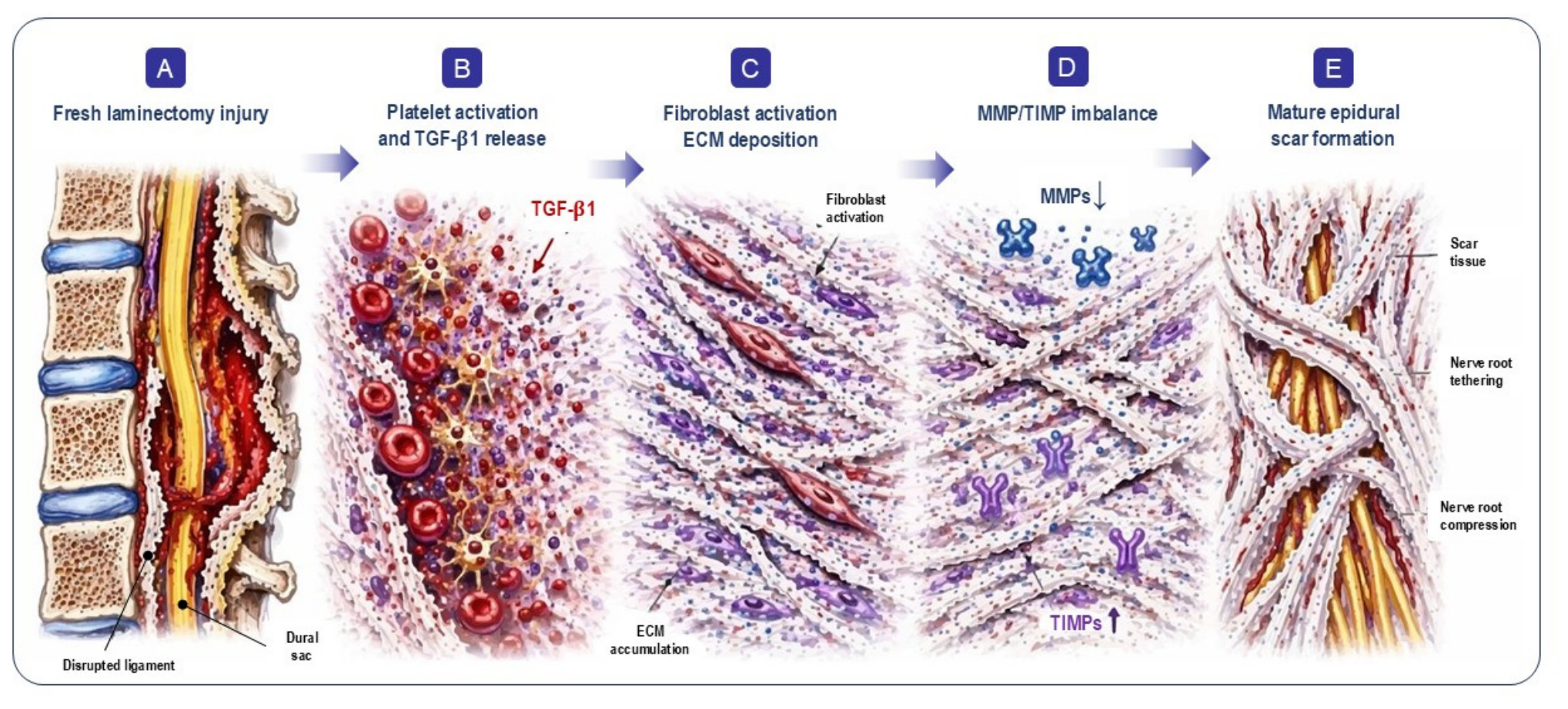
Epidural fibrosis is not merely mechanical scarring, but a dynamic biological process driven by dysregulated wound healing [11,14,15,16]. Following laminectomy, the disruption of normal epidural tissue planes initiates a cascade of inflammatory and fibrotic responses. Within hours, platelet degranulation releases transforming growth factor-β1 (TGF-β1), a central regulator of fibrosis, which stimulates fibroblast proliferation and differentiation into myofibroblasts [15,16]. These myofibroblasts deposit excessive extracellular matrix components including collagen type I and III, fibronectin, and hyaluronic acid [17,18].
The fibrotic response is further shaped by imbalance between matrix metalloproteinases (MMPs) and their inhibitors (TIMPs), favoring extracellular matrix accumulation over degradation [19,20]. Preclinical post-laminectomy data further support that epidural fibrosis is a biologically active process rather than inert scar tissue [21]. The resulting scar tethers nerve roots, restricts their normal mobility, compromises microcirculation, and traps inflammatory mediators including interleukin-1β (IL-1β) and tumor necrosis factor-α (TNF-α) [22,23,24]; how far these findings translate to human postoperative fibrosis has not been established directly.
This proposed cascade—from surgical injury through TGF-β1 activation and matrix accumulation to mature scar—positions epidural fibrosis as a biologically active structural lesion rather than an inert radiological finding, and provides the rationale for examining it as a candidate stratifier of treatment response. It is a mechanistic rationale drawn largely from preclinical work, and is presented here as motivation for the question addressed rather than as established human pathophysiology (Figure 1).

1.3. Studies Without Confirmed Fibrosis

In the absence of confirmed fibrosis, the predominant pain mechanism cannot be assumed. This represents a study-level classification rather than a patient-level, fibrosis-unselected phenotype. By including cohorts where fibrosis was unassessed, unconfirmed, or unreported, this category encompasses a broader post-surgical population rather than a homogeneous biologic subgroup. In these populations, persistent pain may reflect a complex interplay of residual structural contributors, neuropathic mechanisms, central sensitization, or pathomechanical dysfunction—factors that were not systematically differentiated at the study level [25,26,27,28,29,30].
Consequently, these findings should not be interpreted as a literal exclusion of clinically relevant epidural fibrosis. Because fibrosis ascertainment is highly method-dependent, this comparator stratum likely reflects differences in diagnostic intensity, study design, or operator expertise rather than a pure biologic absence of the condition.

1.4. Epidural Adhesiolysis: A Proposed Multi-Modal Structurally Directed Strategy

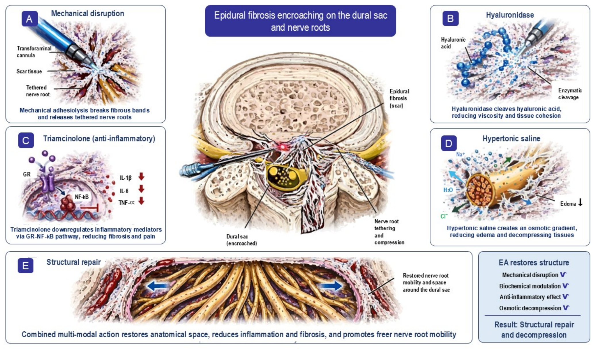
The biologically active structural character of epidural fibrosis has direct therapeutic implications, because some interventions attempt to modify the lesion itself rather than merely modulate pain perception downstream. In this framework, EA is aligned with structural pathology, because it is intended to disrupt adhesions, improve target-site access, restore nerve-root mobility, and deliver medication directly to the scar-nerve interface. Specifically, the procedure combines mechanical disruption with the delivery of three pharmacologic agents (hyaluronidase, corticosteroid and hypertonic saline), each contributing a distinct therapeutic mechanism [31,32,33].
These mechanisms are hypothesized to act as a multimodal, structurally directed strategy: mechanical disruption of adhesions, enzymatic degradation of the fibrotic matrix, anti-inflammatory suppression of ongoing fibrosis, and osmotic decompression of neural elements (Figure 2). These combined effects are thought to improve target-site access and nerve-root mobility, although scar reformation and variability in long-term durability remain recognized clinical realities.

1.5. Neuromodulation Without Structural Repair: A Hypothesis

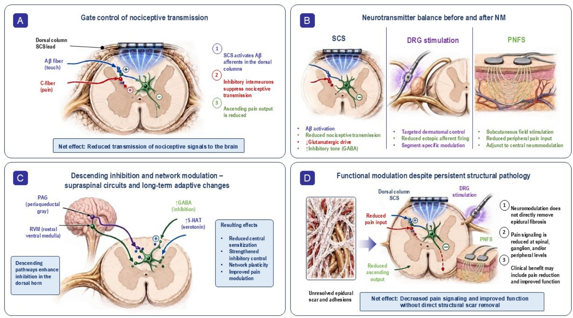
Unlike adhesiolysis, NM is not designed to physically remove or alter epidural scar tissue; its rationale rests instead on altering how pain signals are transmitted and processed at the spinal and brain levels. Since its introduction by Shealy and colleagues [34], spinal cord stimulation (SCS) has worked by delivering electrical current to the dorsal columns, engaging large-diameter Aβ fibers. The gate control framework proposed by Melzack and Wall [35] holds that this Aβ activation dampens incoming nociceptive signals at the dorsal horn through GABA- and glycine-releasing interneurons [36]. Put simply, SCS uses controlled electrical pulses to interrupt or blunt pain signaling before it reaches higher brain centers. This mechanistic basis helps explain why NM can be effective across broad postoperative pain populations even without confirmed fibrosis, since its benefit does not hinge on physically clearing scar tissue or freeing a tethered nerve root. Newer stimulation paradigms—tonic, burst, and 10 kHz high-frequency—appear to add further effects, including long-term depression of excitatory synaptic activity and strengthened descending inhibitory control [37,38], likely via changes in glutamatergic signaling through AMPA and NMDA receptor pathways [39,40]. Ultimately, this functional form of pain modulation leaves structural tethering unresolved, marking a clear mechanistic contrast with the structural-repair approach of adhesiolysis (Figure 3).

1.6. Structural Confirmation of Fibrosis: Imaging and Procedural Techniques

Accurate structural confirmation of epidural fibrosis is essential if fibrosis is to be evaluated as a candidate stratifier of treatment response rather than treated as a speculative explanatory construct. A diagnosis depends on multimodal structural confirmatory procedures, including contrast-enhanced MRI, epidurography, epiduroscopy, computed tomography (CT)/myelography and direct intraoperative visualization. Contrast-enhanced MRI remains the most familiar noninvasive postoperative technique, particularly for distinguishing enhancing scar from nonenhancing recurrent disc material [13,41]. A critical interpretive premise is that studies lacking confirmed fibrosis cannot be assumed to represent truly fibrosis-absent populations.
Patient-level fibrosis stratification has not been feasible in previous comparative work. The included studies report fibrosis status at cohort level rather than for individual participants; ascertainment methods differ between studies and are not uniformly reported; individual-participant data were not available; and no comparative trial has prospectively stratified randomization by structurally confirmed fibrosis. Classification at study level is therefore a constraint imposed by the evidence base rather than an analytic preference.

1.7. Study Objectives and Hypothesis

These biological and therapeutic distinctions motivate a testable hypothesis: that treatment-response patterns in PSPS-T2 may differ according to whether epidural fibrosis is present, such that structurally directed interventions (EA) align most closely with structurally confirmed fibrosis and functionally directed interventions (NM) align most closely with broader post-surgical populations. This is regarded as a hypothesis to be tested by future prospective work rather than one that can be confirmed by the existing literature. The objectives of the present review were therefore threefold: (1) to synthesize, within fibrosis-defined strata, the comparative effects of EA and NM variants against control; (2) to map the joint distribution of treatment family and fibrosis status across the evidence base, so as to define which comparative questions the literature can and cannot answer; and (3) to examine, as an explicitly exploratory analysis, whether study-level fibrosis status is associated with treatment-effect magnitude, recognizing in advance that fibrosis status and treatment family may be confounded.

2. Materials and Methods

2.1. Study Design and Registration

The present study constitutes the first part of a multi-stage evaluation stemming from a comprehensive systematic review. This review and network meta-analysis was prospectively registered with PROSPERO (CRD420261291928) and conducted according to PRISMA-NMA guidelines [42]. The original registered review framework was broader and addressed comparative interventional evidence in PSPS-T2/FBSS rather than fibrosis as an a priori stratification factor. The fibrosis-focused framing presented in this manuscript emerged during mature dataset review and manuscript-level specialization and was therefore interpretive and exploratory rather than the sole original registered objective. Protocol-related refinements, terminology changes, comparator clarifications, study-level consolidation rules, and sensitivity logic are documented in Supplementary File S1.

2.2. Search Strategy

A systematic search was performed in Medline, PubMed, EMBASE, Cochrane Central Register of Controlled Trials, and Scopus from database inception to 8 February 2026, as well a complementary citation search in Research Gate and Google Scholar (citation and research group tracking). The review included randomized controlled trials (RCTs) (tier 1), prospective cohort studies (tier 2) and retrospective studies (tier 3). No language restrictions were applied. The search strategy (Boolean logic) included various combinations of keywords related to failed back surgery syndrome OR postlaminectomy syndrome OR persistent spinal pain syndrome AND neuromodulation OR spinal cord stimulation OR epidural adhesiolysis OR fibrosis. Full search strings, including the specific Boolean operators, are provided in in Supplementary File S1. The Cochrane Central Register of Controlled Trials indexes records derived from trial registries as well as from bibliographic databases and registry-derived records were therefore screened as part of the primary search. A dedicated systematic search of grey literature—conference abstracts, dissertations, and regulatory agency documents—was not performed, and eligibility was restricted to peer-reviewed publications.

2.3. Eligibility Criteria

Studies were included if they: (1) enrolled adults (≥18 years) with PSPS-T2/FBSS and/or epidural fibrosis; (2) evaluated NM or EA; (3) reported pain outcomes (VAS/NRS) or functional outcomes (ODI); and (4) provided data for effect size calculation. Exclusion criteria included: (1) patients with no history of surgery or fibrotic causes of radicular pain; (2) treatments outside the spine; (3) multiple treatments carried out simultaneously or in a crossover design; and (4) single-center retrospective case series (<100 patients), reviews, editorials, case reports and conference contributions.

2.4. Study Selection and Data Extraction

Two reviewers independently screened titles, abstracts, and full texts. Disagreements were resolved by consensus or third reviewer. Data were extracted using standardized forms: study characteristics (author, year, country, design), patient demographics, intervention details, fibrosis confirmation method, and outcomes. The EA and NM abbreviations used are listed in Table 1.

2.5. The Critical Distinction: EA vs. EA_Std

A methodological nuance critical to this analysis is that many randomized controlled trials of EA include a control arm receiving epidural steroid injection without mechanical adhesiolysis. We designate this as standard EA (EA_Std)—representing the control condition, not true EA (Table 1). In our primary analysis, EA_Std arms are excluded from the EA treatment group. Only studies or study arms that utilize epidural lysis of adhesions are classified as EA.

2.6. Classification and Description of Active Treatment Options

Mechanical Adhesiolysis (EA_Mechanical): Under fluoroscopic or endoscopic guidance, a steerable catheter (2.1 mm diameter preferred) is advanced into the epidural space. The catheter is manipulated through fibrotic tissue, physically disrupting adhesions that tether nerve roots [43]. This mechanical lysis restores nerve root mobility—a critical determinant of functional capacity.
Hyaluronidase (EA_Hyal): Hyaluronidase might act as a hydrolytic enzyme that could depolymerize hyaluronic acid, potentially reduce tissue viscosity and hypothetically facilitate steroid penetration [44]. This response assumes hyaluronidase might degrade scars, downregulate TGF-β1, and synergize with mechanical lysis to disrupt adhesion [20].
Corticosteroid (EA_Std): Corticosteroids have proven molecular anti-inflammatory pathways, but their ability to reduce fibrosis is largely hypothetical and clinically unsupported. While cellular pathways like nuclear factor-κB (NF-κB) suppression are well-established [45], clinical evidence shows monotherapy fails to halt progressive fibrotic diseases [15].
Hypertonic Saline (EA_HyperSal): Hypertonic saline likely contributes through osmotic and neurophysiologic mechanisms, specifically by reducing perineural edema and exerting differential nerve conduction blocks. This action preferentially affects small unmyelinated C-fibers over larger myelinated fibers while restoring impaired microcirculation and relieving neural compression fibers [46,47,48].
Conventional low-frequency stimulation (SCS_LF) is categorized as a purely functional modality. Unlike structural repairs, its efficacy relies on the segmental and supraspinal modulation of nociceptive input [34,35]. By leveraging both classical gate-control mechanisms and the recruitment of descending inhibitory pathways, SCS_LF addresses pain at the level of neural processing [36].
Burst stimulation (SCS_Burst) represents a modified neuromodulatory strategy intended to alter pain processing with less reliance on continuous paresthesia. Mechanistically, burst-pattern stimulation is thought to influence both segmental nociceptive gating and broader affective-attentional dimensions of pain processing, thereby extending NM beyond simple tonic dorsal-column activation [36,38].
High-frequency and subperception stimulation (SCS_HF10/SCS_SUBP) are best understood as non-structural pain-modulation strategies. While their effects depend less on classic paresthesia-linked gating and more on the modulation of excitatory–inhibitory balance within spinal circuits, they remain mechanistically aligned with the broader NM family. This alignment holds even if the specific waveform delivery differs significantly from conventional low-frequency or burst protocols [36,37,49].
Dorsal root ganglion stimulation (DRG) represents a more segmentally focused neuromodulatory approach, targeting sensory processing closer to the affected dermatome rather than relying on broader dorsal-column field effects. Conceptually, DRG remains within the same functional-modulation family; its therapeutic logic seeks to alter afferent pain signaling and segmental hypersensitivity [4,50].
Peripheral nerve field stimulation and related subcutaneous approaches (SCS_PNFS) extend NM beyond the dorsal columns by targeting nociceptive input more peripherally within the painful field itself. PNFS remains a functional-modulation strategy, often serving as an adjunct to conventional SCS rather than a structural intervention. Its inclusion in the NM family is justified by its shared therapeutic goal [51,52].

2.7. Fibrosis Classification

Studies were classified at the study level as fibrosis-positive when the analyzed cohort or subgroup was explicitly described as having structurally confirmed epidural fibrosis via one or more direct assessment methods reported in the study, including contrast-enhanced MRI [13], epidurography [53,54], epiduroscopy [12], CT/myelography, intraoperative confirmation, or equivalent procedure-relevant structural documentation [55]. A single explicit structural confirmation method was sufficient for fibrosis-positive classification. Studies were classified as studies without confirmed fibrosis (unselected) when fibrosis was not structurally assessed, not explicitly reported, or not required for eligibility. This category was not interpreted as proving absence of fibrosis. Where studies used mixed or partially specified ascertainment methods, classification favored the most explicit structurally documented study-level phenotype available (fibrosis-positive) in the publication set.
Classification was necessarily performed at study level rather than for individual participants: individual-participant fibrosis data were not available from the included reports, and ascertainment methods were not uniformly described across studies. Study-level classification therefore reflects a constraint imposed by the available evidence rather than an analytic preference, and its interpretive consequences are considered in the Limitations.

2.8. Quality Assessment

Risk of bias was assessed using the Cochrane Risk of Bias 2.0 tool for RCTs [56] and the ROBINS-I tool for non-randomized and retrospective cohort studies [57]. Studies were categorized as having low, moderate, serious, or critical risk of bias. Potential publication bias and study-level heterogeneity were investigated using contour-enhanced comparison-adjusted funnel plots, and Egger’s test was applied to statistically evaluate asymmetry. To evaluate the stability and robustness of the pooled network meta-analysis estimates, a sensitivity analysis was conducted by restricting the model to include only RCTs (tier 1) and, separately, RCTs + non-RCTs (tier 1 + 2). These results were compared with the primary network analysis (which included all studies) to determine the influence of the retrospective data.

2.9. Outcome Measures

This study recorded all defined post-treatment outcomes at 3, 6, 12, and 24 months for RCTs, and at the longest available time point after treatment for non-randomized studies. Primary outcomes were as follows: (1) Pain intensity, measured on a 0–10 cm Visual Analogue Scale (VAS) or 0–10 Numeric Rating Scale (NRS) capturing average pain. If both back and leg pain were recorded, the pain corresponding to the patient’s predominant symptom was selected. (2) Functional disability, measured by the Oswestry Disability Index (ODI, 0–100%) [58]. When nonstandard functional disability measures were reported, harmonized directional transformations were performed where methodologically justifiable to permit inclusion in pooled comparative analyses. Secondary outcome was the responder outcome, defined as the proportion of participants achieving a clinically meaningful reduction of ≥50% in pain intensity from baseline to post-intervention. Where non-standard functional instruments were reported, directional harmonization preserved relative improvement rather than assuming numerical equivalence between scales, and responder analyses combined studies using ≥50% and ≥30% thresholds. These transformations introduce uncertainty in a predictable direction: harmonized disability values should be read as approximate rather than as true ODI measurements, and inclusion of the lower responder threshold may modestly inflate pooled response rates while contributing to heterogeneity. Because only a small number of studies required either transformation, the influence on pooled estimates is expected to be modest; this is nevertheless flagged as a source of uncertainty in the Limitations.

2.10. Network Meta-Analysis and Evidence Geometry

Primary network analyses were performed using a frequentist random-effects framework in R using the netmeta package (RSTUDIO, Version: 2026.01.2+418), which synthesizes direct and indirect evidence within a unified graph-theoretical approach [59]. Because the retained subgroup networks differed materially in their available comparator structures, family-level estimates were interpreted within strata rather than as direct cross-stratum comparative-effectiveness estimates. Fibrosis status was assigned at the study level. Consequently, family-level groups were stratified as positive for studies with confirmed fibrosis and unselected for those where fibrosis was not assessed or confirmed. Studies were included in the network meta-analysis only if they documented the comparative efficacy of at least two treatments (including a control group). Different technical approaches to the same treatment were not considered as separate comparison groups in this analysis.
Multi-arm studies were handled within the network framework so that within-study correlations were preserved rather than treated as independent pairwise observations. Where a single trial had generated more than one publication, these were consolidated into one study family so that the cohort entered the analysis once; the selection across follow-up timepoint per study is described in Section 2.11. Treatment ranking was summarized using P-scores, which were interpreted as exploratory ranking summaries rather than as definitive comparative-effectiveness evidence. To improve transparency, Supplementary Materials report network geometry, node-splitting inconsistency outputs, heterogeneity statistics, and additional sensitivity analyses categorized by level of evidence. Where available, the supplement also reports the proportion of direct versus indirect evidence contributing to key subgroup estimates. This review used a frequentist exploratory network meta-analysis framework with substantial clinical and methodological heterogeneity across evidence tiers and fibrosis-stratified subgroup architectures.

2.11. Transitivity, Comparator Structure, and Follow-Up

Transitivity was assessed before synthesis. We examined how potential effect modifiers were distributed across comparisons: baseline pain and disability, age, sex, fibrosis ascertainment method, comparator structure, and study design. Baseline pain did not differ significantly between the treatment families and control (p > 0.05). Baseline disability did differ (p < 0.05), indicating some imbalance at entry, and both age and sex acted as significant moderators in meta-regression (p < 0.001). The decisive finding, however, concerned the network itself. Treatment family and fibrosis status are confounded in this dataset, and the two subgroup networks share no active treatment node. Transitivity is therefore not satisfied for any cross-family comparison: an EA-versus-NM contrast would rest on the untestable assumption that the two strata are exchangeable. Quantitative synthesis was accordingly restricted to within-family, within-stratum comparisons against a common control. Where necessary, the cross-family effect was computed.
Each study contributed a single observation to each network. Where a trial reported outcomes at several follow-up intervals, the timepoint closest to the prespecified primary window of six months was selected; where two intervals were equidistant, the earlier was taken. Linked publications reporting different follow-up timepoints from the same underlying trial were consolidated at study-family level, so that no cohort contributed more than once to any analysis. The timepoint retained for each study is recorded in the supplementary duplication audit. Restriction to Tier 1 and to Tiers 1 + 2 were analyzed separately as sensitivity analyses rather than pooled with the primary window.

2.12. Interaction Testing

Because study-level fibrosis status was largely determined by which treatment a study evaluated, a meta-regression using fibrosis status as a moderator cannot in principle separate the influence of fibrosis from that of treatment family; the analysis is therefore reported strictly as an exploration of heterogeneity. Exploratory interaction testing was used to examine whether treatment-response patterns differed according to study-level fibrosis-confirmation status. This interaction analysis was exploratory and distinct from the primary within-stratum family-level network summaries. A mixed-effects meta-regression using fibrosis status as a study-level moderator was performed to assess whether fibrosis status explained treatment-effect variability across the broader evidence base. The omnibus test of moderators (QM) was used to assess subgroup differences, and Cochran’s Q-test (QE) was evaluated to measure residual heterogeneity. If the remaining heterogeneity is high and the comparative structures differ across the various subgroup networks, the moderator analysis will be regarded as supporting evidence and not as the sole criterion for the central hypothesis of this study.

3. Results

3.1. Study Selection and Characteristics

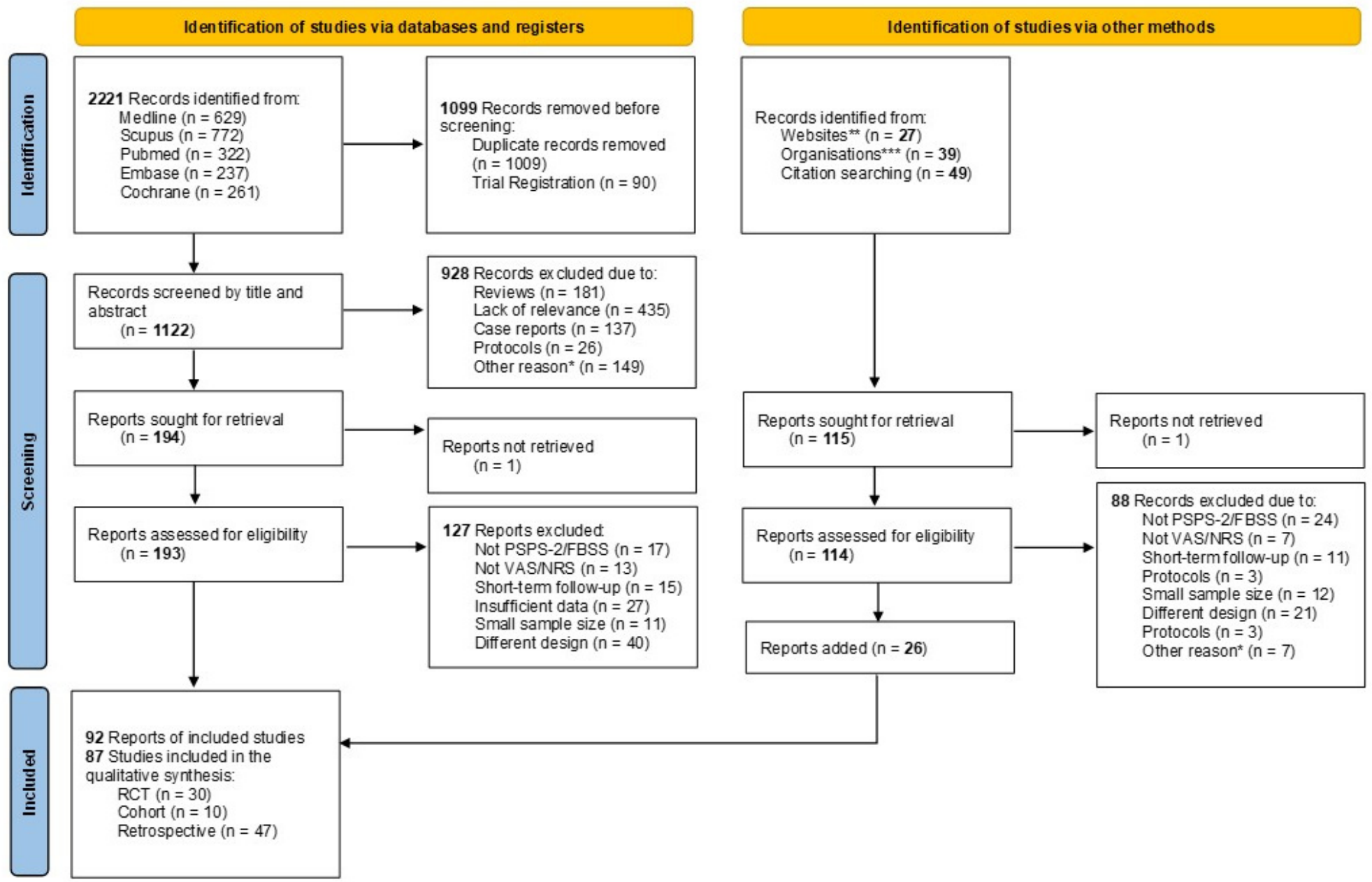
From 2221 records identified, 1122 remained after removing duplicates and initial screening. Ultimately, 87 studies (92 publications) involving 33,504 patients met inclusion criteria (Figure 4). This total comprised 30 RCTs (n = 2400), 10 prospective cohorts (n = 901), and 47 retrospective cohorts or registries (n = 30,203). Across the database, 40 studies (n = 4180) were classified as fibrosis-positive and 47 (n = 29,324) as fibrosis-unselected. EA treatments were more prevalent in the fibrosis-positive group, while NM treatments dominated the fibrosis-unselected group. Only one non-randomized study has performed a direct, head-to-head comparison evaluating the treatment effects of Dorsal Root Ganglion (DRG) NM versus EA with hyaluronidase [59]. Only one study reporting the use of the SCS_SUBP treatment could be included in the meta-regression analysis [60].
As shown in Table 2, the composition of study designs was balanced across both groups. Within RCTs, 16 studies (n = 1055) were fibrosis-positive compared to 14 (n = 1345) without confirmed fibrosis. Prospective cohorts were split evenly (5 studies each, n = 311 positive; n= 590 unselected), while retrospective studies included 19 fibrosis-positive (n = 2814) and 28 fibrosis-unselected (n = 27,389) cohorts. This distribution suggests that confounding with treatment family precludes attributing this signal to fibrosis itself (Supplementary File S2). Among all trials, the higher patient count in the fibrosis-unselected subgroup likely stems from differences in study design and ascertainment practices, rather than providing evidence that fibrosis was absent in these larger cohorts.
Methodological inconsistencies across the included literature present a challenge to synthesis. These include diverse justifications for treatment designs, non-standardized outcome criteria, and clinical variations in study populations (Supplementary File S1). Studies containing mixed chronic lumbar pain populations were retained when postoperative PSPS-T2/FBSS patients represented a clinically relevant component of the cohort and the intervention mechanism remained applicable to the postoperative fibrosis-associated setting. The EA_HyperSal node included protocols using hypertonic saline as well as selected high-volume saline adhesiolysis approaches when saline-mediated hydrodissection or osmotic augmentation formed part of the intended treatment mechanism.

3.2. NMA Results: Primary Within-Stratum Pain Results Versus Control

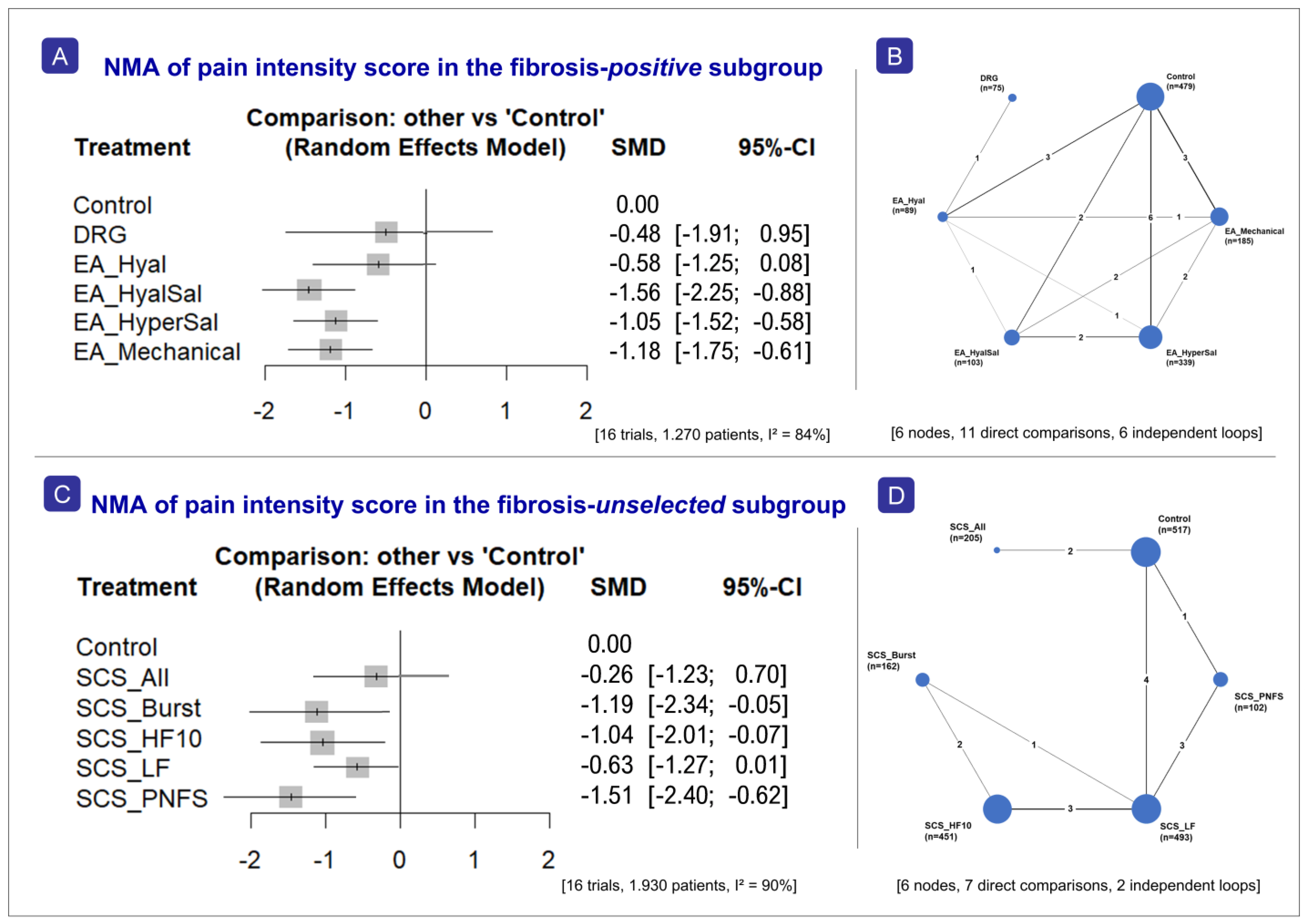
This network meta-analysis shows a mixed pattern of effectiveness across treatments when compared with control (EA_Std, conventional medical management-CMM, placebo/sham), as indicated by standardized mean differences (SMD) (Figure 5).
The analysis yields two implications that are crucial for the interpretation of all the results presented below. Firstly, whilst the within-family and within-stratum networks (Section 3.2, Section 3.3 and Section 3.4) are internally valid, they describe different populations and cannot be combined into a single ranking. Secondly, given the high heterogeneity of the networks (Section 3.5), the structural dependence should lead to a downgrading of the certainty of the evidence. The full list of studies included in the NMA is set out in Supplementary File S2.

3.2.1. Fibrosis-Positive Subgroup

This frequentist network meta-analysis (random-effects model; 16 trials, 1270 patients) indicated that every point estimate favored active treatment over control (negative SMDs) (Figure 5A), with EA_HyalSal, EA_HyperSal, and EA_Mechanical yielding the largest effects (all p < 0.001) and showing the most consistent and statistically significant effects. EA_Hyal (p = 0.085) and DRG (p = 0.508) did not differ significantly from control. Among active treatments, EA_HyalSal tends to perform better than EA_HyperSal and EA_Hyal, while differences between EA_HyperSal and EA_Mechanical are small and non-significant.
The network plot in this subgroup contains six treatment nodes—the largest NMA network in this analysis—with 11 direct comparisons forming a partially connected hub-and-spoke structure, as well as additional edges creating 6 closed loops (Figure 5B). Comparisons involving DRG rest on a single direct edge and are imprecise, with wide confidence intervals crossing zero, indicating no clear differences. Substantial heterogeneity (I2 = 83.7%, 95% CI 74.5–89.6; τ2 = 0.36) and significant between-design inconsistency (Q = 42.83, 9 df, p < 0.001) indicate that study-design variation influences the outcomes. Node-splitting nevertheless identified no individual contrast at which direct and indirect estimates disagreed significantly, so the incoherence is distributed across designs rather than localized to one comparison (Supplementary File S2).
In the fibrosis-positive supportive network, the probability score reveals EA_HyalSal as the most effective treatment (Pscore = 0.986), while control group ranks last (Pscore = 0.033) (Supplementary File S2) These are summaries of probability rankings and do not constitute evidence of any comparative superiority. The comparison-adjusted funnel plot for this network is visually asymmetric, but the asymmetry is not statistically supported by Egger’s test (p = 0.45). While larger, more precise studies cluster closer to the center, smaller studies tend to report more extreme effects, suggesting that the overall treatment effect may be somewhat overestimated.

3.2.2. Fibrosis-Unselected Subgroup

For the fibrosis-unselected subgroup (16 trials, 1930 patients), the network meta-analysis indicates that all NM modalities reduce the outcome compared to control (negative SMDs) (Figure 5C). Among the interventions, SCS_PNFS appears the most effective (largest negative SMD), followed by SCS_Burst and SCS_HF10. SCS_Burst, SCS_HF10, SCS_LF, and particularly SCS_PNFS demonstrate statistically significant benefits (p < 0.05), while SCS_All shows a non-significant effect, with its confidence interval crossing zero (p = 0.092). The network plot reveals a connected evidence structure of six nodes joined by 7 distinct direct comparisons, with SCS_LF acting as the principal connector (Figure 5D); two independent closed loops are present. Control versus SCS_HF10 and control versus SCS_Burst carry no direct evidence and are estimated indirectly, with correspondingly wide intervals.
Heterogeneity in this network is substantial (I2 = 89.6%, 95% CI 83.8–93.4; τ2 = 0.45) (Supplementary File S2). Both within-design heterogeneity (Q = 96.37, 9 df, p < 0.001) and between-design inconsistency (Q = 9.78, 2 df, p = 0.008) are statistically significant, suggesting that differences in study design and disagreement between direct and indirect evidence affect the robustness of the estimates. The netsplit analysis confirms significant inconsistencies, most notably in the SCS_HF10:SCS_LF and SCS_Burst:SCS_HF10 comparisons, where direct and indirect estimates disagree significantly. Additionally, several direct comparisons show very high heterogeneity (I2 78.8–97.5%).
The funnel plot shows pronounced asymmetry, suggesting significant heterogeneity or publication bias (Supplementary File S2). Egger’s test on the comparison-adjusted estimates is not significant (p = 0.085). The probability score test places SCS_PNFS first (Pscore =0.823) and control last (Pscore =0.009). These are exploratory summaries, and not evidence of comparative superiority (Supplementary File S2).
Taken together, these results indicate a differential pattern of within-stratum treatment-response behavior across fibrosis-defined study strata, but they do not constitute definitive evidence of EA-versus-NM comparative superiority within either stratum. Because cross-family EA-versus-NM comparisons were predominantly indirect, node-level rankings and representative time-window signals were treated as supportive rather than sovereignty-bearing analyses.

3.3. NMA Results: Primary Within-Stratum ODI Results Versus Control

This network meta-analysis (12 trials) shows a mixed pattern of effectiveness across treatments compared with control (EA_Std, CMM, placebo/sham)—specifically, although results varied, most evaluated treatments produced a significant improvement in ODI.

3.3.1. Fibrosis-Positive Subgroup

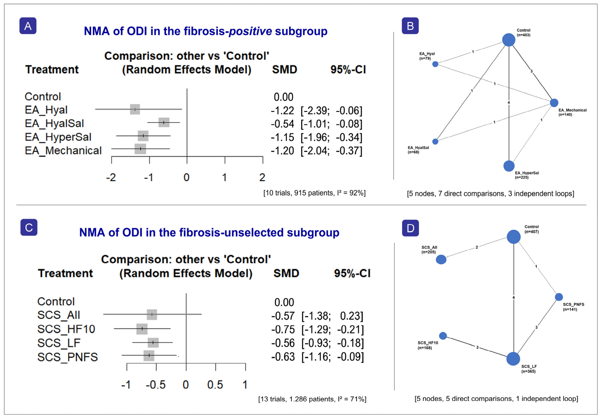
The analysis (10 trials, 915 patients) shows that all evaluated treatments (EA_Hyal, EA_HyalSal, EA_HyperSal, and EA_Mechanical) significantly improve ODI compared with the control group, as indicated by negative SMD values (Figure 6A). Among them, EA_Hyal demonstrates the largest effect (p = 0.05), followed closely by EA_Mechanical (p = 0.005) and EA_HyperSal (p = 0.005), while EA_HyalSal shows a comparatively smaller effect (p = 0.05). The network plot for the fibrosis-positive subgroup comprises five nodes joined by 7 distinct direct comparisons (Figure 6B). Control is the busiest node, but direct EA-versus-EA evidence is also present, linking EA_Mechanical to each of EA_Hyal, EA_HyalSal and EA_HyperSal. The network contains 3 independent closed loops.
Heterogeneity across this network is the highest of the six primary networks (I2 = 91.9%, 95% CI 86.5–95.2; τ2 = 0.67). Both the within-design heterogeneity test (Q = 19.98, 3 df, p < 0.001) and the between-design inconsistency test (Q = 66.74, 4 df, p < 0.001) are statistically significant, indicating disagreement between direct and indirect evidence that materially limits the robustness of the comparisons (Supplementary File S2).
The netsplit analysis also show that all treatments outperform the control in the network estimates, with EA_Hyal, EA_HyperSal, and EA_Mechanical demonstrating consistent and statistically significant effects, while EA_HyalSal also shows a benefit, but with some inconsistency between direct and indirect evidence. Notably, for EA_Hyal and EA_HyalSal versus control, the direct and indirect estimates differ in magnitude—and even direction in the case of EA_HyalSal—suggesting potential inconsistency in the network. In contrast, EA_HyperSal and EA_Mechanical versus control display close agreement between direct and network estimates, indicating more reliable evidence (Supplementary File S2).
The funnel plot shows asymmetry, and Egger’s test on the comparison-adjusted estimates is statistically significant (p = 0.018), indicating small-study effects or publication bias in this network. The probability score test reveals EA_Hyal as the most effective treatment (Pscore = 0.785), while the control group ranks last (Pscore = 0.000) (Supplementary File S2). The three leading P-scores are best described as indistinguishable rather than ordered (Supplementary File S2).

3.3.2. Fibrosis-Unselected Subgroup

In the fibrosis-unselected subgroup (13 trials, 1.286 patients), the network plot consisted of five treatment nodes: control, SCS_PNFS, SCS_LF, SCS_HF10, and SCS_All (Figure 6C). All active nodes improve ODI relative to control, as reflected by negative SMD values. However, SCS_All does not reach statistical significance. SCS_HF10 shows the strongest and statistically significant effect, followed by SCS_PNFS, SCS_All, and SCS_LF, all not separable from one another. Notably, there was no direct edge connecting control and SCS_HF10, but the network is connected and that contrast is estimable through indirect evidence (Figure 6D)—joined by 5 distinct direct comparisons forming one independent closed loop.
Heterogeneity here is the lowest of the six primary networks (I2 = 71.1%, 95% CI 44.9–84.8; τ2 = 0.12), indicating moderate-to-high variability in effect sizes (Supplementary File S2). Inconsistency between direct and indirect evidence is minimal: the between-designs test is not statistically significant (Q = 0.13, 1 df, p = 0.722), indicating that the network estimates are coherent despite the observed heterogeneity.
The netsplit plot suggests that, for most comparisons, there is no strong evidence of inconsistency between direct and indirect estimates, as their confidence intervals largely overlap, indicating good agreement. Likewise, SCS_PNFS vs. control shows a stronger effect in the indirect estimate, but the network estimate remains consistent and significant. For contrasts such as SCS_HF10 vs. SCS_LF and SCS_LF vs. SCS_PNFS, the confidence intervals cross zero and overlap considerably, suggesting no meaningful differences and acceptable consistency (Supplementary File S2).
The funnel plot shows only slight asymmetry, and Egger’s test computed on the 13 comparison-adjusted estimates is not significant (p = 0.715), so there is no statistical evidence of small-study effects in this network (Supplementary File S2). The probability score test places SCS_HF10 first (P-score 0.839), while the control group ranks last (Pscore = 0.022) (Supplementary File S2).

3.4. Mixed-Effects Meta-Regression

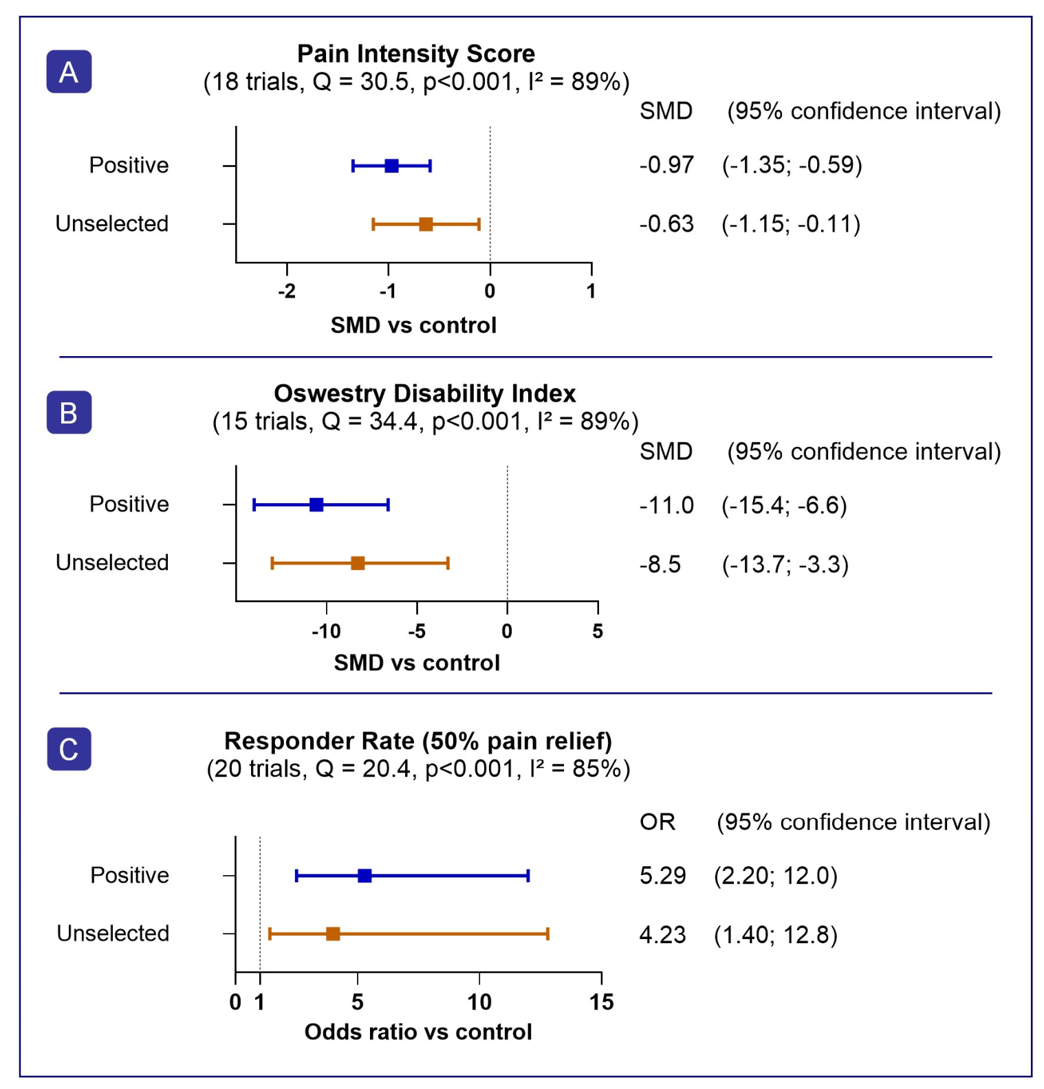
To explore sources of statistical heterogeneity, a mixed-effects meta-regression (categorical subgroup analysis) was performed. Fibrosis status was entered into the model as a moderator. As established in Section 3.2, fibrosis status is confounded with treatment family in this dataset; the moderator terms below therefore index a difference between EA-dominated and NM-dominated evidence and must not be read as isolating an effect of fibrosis. The therapeutic interventions yielded significant improvements across both symptomatic and functional metrics (Figure 7).
Regarding pain outcomes (18 studies, 21 contrasts), results demonstrated substantial residual heterogeneity across the included studies (I2 = 89.49%; QE = 204.45, p < 0.001), which persisted even after accounting for subgroup classifications; the moderator analysis was significant (QM = 63.74, p < 0.0001). Subgroup evaluation confirmed significant pain reductions in both the fibrosis-positive cohorts (pooled effect estimate = −0.9716; 95% CI −1.3538 to −0.5894, p < 0.001) and the unselected subgroup (pooled estimate = −0.6322; 95% CI −1.1522 to −0.1121, p = 0.017) (Figure 7A). The point estimate is larger in the fibrosis-positive stratum, but the two intervals overlap and, as set out in Section 3.3, the moderator indexes which treatment family dominates each stratum rather than an effect of fibrosis.
The analysis of functional recovery via the ODI (15 studies, 16 contrasts) followed a broadly similar pattern. Marked residual variance was again present (I2 = 89.40%; QE = 101.10, p < 0.001), indicating substantial between-study differences beyond random chance, and the moderator test was significant (QM = 34.38, p < 0.001). In fibrosis-positive studies the pooled effect estimate was −11.01 ODI points (95% CI −15.39 to −6.62, p < 0.001); in the unselected it was −8.46 (95% CI −13.65 to −3.28, p = 0.001) (Figure 7B). The two strata are separated by 2.5 ODI points with substantially overlapping intervals.
Furthermore, the mixed-effects meta-regression regarding responder rates (50% pain relief) (20 studies, 23 contrasts) demonstrated substantial residual heterogeneity (I2 = 85.02%; τ2 = 2.121), indicating that a considerable proportion of variability across studies remained unexplained after accounting for the subgroup moderator. This residual heterogeneity test was statistically significant (QE = 156.50 on 21 df, p < 0.001), confirming persistent between-study differences, and the moderator test was significant (QM = 20.43 on 2 df, p < 0.001). Both strata showed significantly higher odds of response than control—fibrosis-positive OR 5.29 (95% CI 2.20 to 12.69; log-OR 1.6653, p < 0.001) and fibrosis-unselected OR 4.23 (95% CI 1.40 to 12.79; log-OR 1.4422, p = 0.011)—but the two intervals overlap almost entirely and the strata are not statistically distinguishable from one another (Figure 7C).

3.5. Inconsistency and Sensitivity Analyses

Sensitivity analyses (Tier 1 and Tiers 1 + 2) closely matched the all-tier network meta-analysis, showing that including retrospective studies (tier 3) did not alter the main benefit trends for pain and disability. Thirty sensitivity refits were performed: each of the six primary networks was re-estimated at 3-, 12- and 24-month follow-up windows and under restriction to Tier 1 and to Tiers 1 + 2. All 30 converged and no treatment node was dropped by the estimator; the tier-restricted networks contain fewer nodes only because fewer studies contribute to them. The EA_HyalSal node was stable for pain (in all five fibrosis-positive scenarios; SCS_PNFS in all five fibrosis-unselected scenarios) and for responder rates (SCS_PNFS in all five fibrosis-unselected scenarios; EA_HyperSal or EA_Mechanical in the fibrosis-positive scenarios). For ODI, the performing node was scenario-dependent, alternating between EA_Mechanical and EA_HyperSal in the fibrosis-positive stratum and between SCS_HF10 and SCS_PNFS in the fibrosis-unselected stratum; these intervention placements should therefore not be read as a rigid hierarchy.
Heterogeneity varied widely across the refits (I2 22.5% to 92.4%; lowest in the Tier-1-only fibrosis-unselected responder network, highest in the 3-month fibrosis-unselected pain network). A detailed description can be found in the attached file (Supplementary File S2).

3.6. Risk of Bias and Publication-Bias Summary

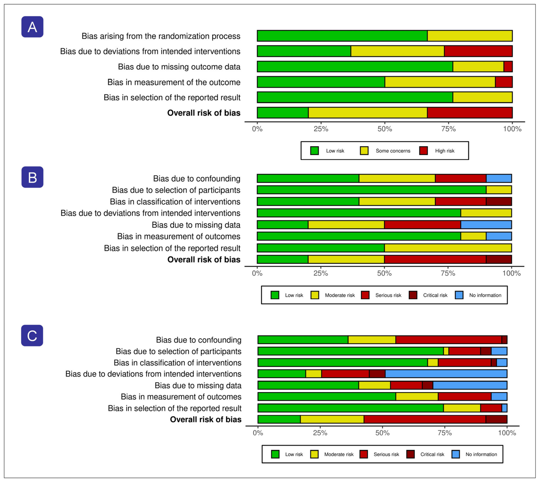
The overall strength of evidence is constrained by methodological limitations—specifically study design, potential selection bias, and inconsistent reporting—which may attenuate the validity of the findings (Figure 8). Therefore, results should be interpreted within these limitations, as they introduce a notable risk of bias and reduce overall certainty. Detailed risk-of-bias assessments (tiers 1–3) and supporting funnel plots are provided in the Supplementary File S2. Table 3 summarizes the key findings by fibrosis stratum of the primary network.

3.7. Evidence Limitations

Across comparisons, certainty was rated low to very low. Several domains contributed. Within-study risk of bias was frequently moderate or high, and heterogeneity was substantial to extreme (I2 71–92% across the primary networks and 85–89% across the three meta-regression models). Several within-family contrasts rested on indirect evidence with wide confidence intervals—most notably control versus SCS_HF10 in the fibrosis-unselected ODI network, which carries no direct edge, although it remains estimable—and localized incoherence was present in the fibrosis-positive ODI network, where the EA_HyalSal contrasts against control and against EA_Mechanical yield direct and indirect estimates of opposite sign (side-split p = 0.002 for both). No comparison in any of the primary networks was disconnected. Small-study effects were detected in the fibrosis-positive ODI network (Egger p = 0.018), but in neither pain network (p = 0.449 and p = 0.085). Beyond these familiar domains lies a more fundamental constraint. Treatment family and fibrosis status are collinear within the randomized evidence, so even a precise within-stratum estimate cannot support an inference about fibrosis acting independently of treatment (Table 4). The body of evidence is therefore best regarded as hypothesis-generating rather than confirmatory, and the treatment’s probability score reported above as exploratory summaries rather than evidence of comparative superiority.

4. Discussion

4.1. Principal Findings

The principal finding of this review is structural rather than comparative: in the existing PSPS-T2 literature, treatment family and fibrosis status are almost entirely confounded, and among randomized trials they are perfectly collinear. EA has been studied almost exclusively in structurally confirmed, fibrosis-positive populations, and NM almost exclusively in unselected post-surgical populations. As a result, the two treatment literatures describe different patients, and the field does not yet contain the evidence needed to compare them within a common fibrosis stratum. Within that constraint, the within-family analyses are informative on their own terms: among fibrosis-positive studies the EA variants incorporating hyaluronidase and hypertonic saline achieved the highest probability score for both pain and disability, and among fibrosis-unselected studies peripheral-nerve-field and high-frequency SCS achieved the highest probability scores. What cannot be inferred from these data is that fibrosis itself determines which treatment works better, because in every randomized comparison the two are indistinguishable.
The contribution of this study is therefore to make that confound explicit and to reframe epidural fibrosis, for now, as a biologically grounded candidate stratifier. Positioning fibrosis in this way is not a retreat from the hypothesis but a precondition for testing it: naming the structure of the evidence identifies exactly the trial that would be needed to move fibrosis from plausible to proven.

4.2. What the Present Analysis Does and Does Not Show

The present analysis characterizes the architecture of the evidence rather than a treatment hierarchy. Direct EA-versus-NM comparisons within fibrosis-defined strata were, as anticipated, entirely absent. The observed association between fibrosis-positive status and larger effect size is real but confounded: because fibrosis-positive studies are EA studies and fibrosis-unselected studies are NM studies, the same data are equally well described as ‘EA trials showed larger effects than NM trials.’ No analysis of this literature can separate the two, and readers should treat any apparent fibrosis effect as inseparable from a treatment effect. However, this distinction matters. The absence of head-to-head stratified trials limits direct claims, making fibrosis a candidate variable worthy of investigation in future PSPS-T2 trial design.
A central limitation of the present analysis is that the comparator stratum should not be interpreted as a true fibrosis-unselected population. Studies without confirmed fibrosis were defined by lack of structural confirmation, not by demonstrated absence of fibrosis. Given the known discrepancy between epiduroscopic and MRI-based fibrosis detection, it is likely that at least some of these cohorts included patients with clinically relevant but unrecognized fibrosis [12]. This misclassification may dilute a true effect-modification signal, but it may also reflect broader study-level differences in diagnostic intensity, case selection, operator expertise, geography, era, and publication patterns. Accordingly, the studies-without-confirmed-fibrosis stratum is best understood as a heterogeneous non-confirmation category rather than as a biologically pure comparator phenotype.
Interpretation of the fibrosis-positive effect sizes also depends on control composition. Injection-only comparators classified as EA_Std were analytically separated from true adhesiolysis because they do not include mechanical adhesiolysis. However, EA_Std may still be more active than inert sham alone. The magnitude of benefit observed for EA-family interventions in fibrosis-positive studies therefore reflects both treatment activity and the retained comparator structure of that subgroup network, rather than a simple placebo-only contrast.

4.3. Biological Rationale for Fibrosis as a Candidate Stratifier

The biological plausibility of the observed pattern rests on the divergent mechanisms of EA and NM, grounded in the molecular pathogenesis of epidural fibrosis and the differing therapeutic targets of structural versus functional treatment. Epidural fibrosis is a biologically active structural lesion. The combination nerve-root tethering, restricted mobility, and persistent profibrotic signaling provides a mechanistic rational for why structurally targeted interventions may perform differently from purely neuromodulatory approaches [22,23,24]. The plausibility of this structural pathway is strengthened by the fact that profibrotic signaling is not static; reactive oxygen species may further amplify TGF-β1-mediated fibroblast activation and matrix deposition. The mechanistic framework remains biologically plausible rather than finally proven.
EA is the interventional strategy most directly aligned with that need. Mechanical adhesiolysis may restore nerve root mobility [33,43], hyaluronidase may degrade hyaluronic acid within the fibrotic matrix and facilitate tissue penetration [20,44], triamcinolone may suppress NF-κB signaling and dampen cytokine-driven fibrosis [15,45], and hypertonic saline is thought to contribute through osmotic and neurophysiologic effects, including reduction in perineural edema and differential susceptibility of unmyelinated fibers in foundational dorsal-root preparations [46,47,48]. Together, these mechanisms make it biologically plausible that structurally dominant fibrosis-positive disease would show a stronger response to structurally directed intervention (Figure 2). Procedural outcome data further suggest that successful postadhesiolysis contrast passage beyond the foramen may act as a correlate of effective structural release and target-site access [142].
By contrast, studies without confirmed fibrosis represent a broader and less structurally resolved population. In these patients, pain is more likely to include altered dorsal horn processing, central sensitization, glial activation, maladaptive neuroplasticity, and impaired descending inhibition [25,26,27,28,29,30]. NM is aligned with this biology because it modulates nociceptive transmission through gate control and network-level functional modulation rather than structural release [35,36,37,38]. In this sense, the observed treatment-response pattern is not only statistically interesting but mechanistically coherent (Figure 3). The statistical supplement does not convert this mechanistic plausibility into definitive universal moderator proof, but it does support the continued testing of a biologically stratified framework.

4.4. Functional Recovery and Durability: A Hypothesized Signature

The functional disability findings sharpen the structural interpretation of the manuscript. If adhesiolysis were acting only as a short-lived analgesic adjunct, one would expect weaker or less differentiated downstream disability effects. Instead, the fibrosis-positive signal identified in the pain analysis extended into ODI, and meta-regression showed that the functional treatment effect was greater in fibrosis-positive studies than in fibrosis-unselected studies. This pattern is biologically plausible: if adhesiolysis reduces scar-related tethering, improves nerve-root mobility, and restores access to the structurally relevant epidural target, downstream gains should also be visible in disability domains such as walking, standing, sitting, and self-care. This interpretation is consistent with prior adhesiolysis literature reporting sustained clinical benefit, including later functional improvement, in selected postoperative radicular pain populations [31,33,82]. It is also supported by procedural studies linking favorable postadhesiolysis contrast distribution with better recovery, suggesting that meaningful benefit depends partly on successful structural access rather than drug delivery alone [142]. In the unselected subgroup, several NM modalities were also favorable for ODI versus control, consistent with the broader SCS literature [37,98,126], but the functional signal was smaller than the fibrosis-positive EA-side pattern.
Taken together, these findings align with the structural-versus-functional framework, indicating that structural release could promote functional persistence if fibrosis drives the pain.

4.5. Structural Confirmation of Fibrosis and Procedure-Relevant Assessment

Structural confirmation of epidural fibrosis is strongly method-dependent. In a prospective epiduroscopic FBSS cohort, clinically significant fibrosis was far more frequently detected by epiduroscopy than by MRI, suggesting that fibrosis may be substantially underrecognized when conventional imaging alone is used [12]. Procedural techniques such as epidurography and epiduroscopy are especially relevant because they can provide direct or procedure-linked evidence of adhesions and target-site access [143]. They can also demonstrate whether adhesions limit target access and whether meaningful structural release has actually been achieved. In this context, fibrosis confirmation should be understood as structurally grounded and multimodal.
Beyond baseline structural confirmation, epidurography may also provide procedure-relevant information during adhesiolysis itself; in observational lumbar neuroplasty data, postadhesiolysis extraforaminal contrast distribution was associated with better functional outcome, suggesting that final contrast spread may serve as a procedural correlate of successful structural release [142]. Advanced diffusion-based techniques such as diffusion tensor imaging remain of research interest as future biomarker approaches, but they were not the dominant basis for fibrosis classification in the present dataset and should therefore be regarded here as future-oriented rather than clinically established tools [144,145].

4.6. Comparison with Previous Literature

The present findings help explain why earlier EA and NM literatures can appear discordant. Major NM trials reported meaningful benefit in broad post-surgical pain populations [49,98,126], whereas many EA studies reported benefit in cohorts enriched for epidural fibrosis or related structural pathology [31,33,81,105]. These findings align with prior postoperative pain literature showing that structurally targeted interventions perform best when scar-related pathology is demonstrable and that NM performs well in broader neuropathic and refractory pain populations [37,82,98]. The present review extends that literature by showing that this apparent discordance is largely an artefact of population selection: the EA and NM evidence bases were built on structurally different patients, so their divergent results reflect who was studied at least as much as what was done. Documenting that separation—rather than resolving it—is what the current evidence permits.

4.7. Implications for Current Clinical Practice

A practical question follows from these findings: should structural confirmation of epidural fibrosis influence treatment selection today? On the present evidence, it should not do so by itself. The association between fibrosis-positive status and larger treatment effects cannot be separated from the treatments delivered in those cohorts, and no randomized comparison has tested both families within a single fibrosis stratum. Where structural assessment is already part of routine postoperative evaluation, documenting the presence, extent and location of fibrosis is clinically reasonable and may inform procedural planning—for example, whether adhesiolysis is technically feasible or whether scarring is likely to obstruct lead placement. It is quite another matter to use fibrosis status as a criterion for choosing between adhesiolysis and neuromodulation, and that step is not supported by the current literature. We regard fibrosis-guided treatment selection as a hypothesis for prospective validation rather than as a present standard of care.

4.8. Implications for Future Prospective Trial Design

The implications of these findings are substantial. Postoperative pain in PSPS-T2 should not be treated as a single undifferentiated syndrome when structurally relevant fibrosis may be dominant in one subgroup and absent, unassessed, or non-dominant in another.
The clinical relevance of this stratified framework is reinforced by the historically poor and declining success of repeated lumbar surgery in PSPS-T2, with commonly cited estimates of approximately 30% after a second operation, 15% after a third, and 5% after a fourth, and by 5-year cohort data showing only 34% successful outcome after repeated lumbosacral operation [119,146].
A similar caution emerges from fusion-specific data in established PSPS-T2, where only 35% of patients reported good outcome after instrumented fusion, further underscoring the need for better pathology-aligned non-fusion treatment strategies [147]. The need for such trials is strengthened not by a single statistically universal moderator result, but by the convergence of biologic plausibility, structurally grounded ascertainment logic, and directionally coherent stratified treatment-response patterns across the current evidence base.
A rigorous next-step design would incorporate standardized fibrosis assessment using multimodal structural criteria, followed by stratified randomization to EA versus NM with prespecified pain and functional outcomes. Such a design would directly test whether the differential pattern observed here reflects true treatment-effect modification rather than the combined influence of heterogeneous populations, variable fibrosis ascertainment, and indirect evidence structure. If confirmed prospectively, that would move PSPS-T2 management from empiric sequencing toward genuinely mechanism-informed treatment selection.
The present study therefore strengthens the rationale for future fibrosis-stratified prospective trials in which fibrosis assessment is standardized and treatment effects are tested explicitly within structurally defined populations. Such studies are justified not only by mechanistic plausibility but by convergent evidence across pain, ODI, responder, family-level, and moderator analyses. Whether EA produces true structural resolution of epidural fibrosis will require confirmation in those future prospective clinical studies. Future trials should prospectively prespecify fibrosis ascertainment, comparator structure, and follow-up windows in a way that allows direct EA-versus-NM testing within fibrosis-defined strata. Such trials should distinguish confirmed fibrosis, confirmed absence of fibrosis where feasible, and unassessed populations, rather than collapsing all non-positive cases into a single comparator category. Only this design will allow the present framework to be tested directly and determine whether structurally targeted benefit also reflects true structural resolution of fibrosis.

4.9. Limitations

Several factors could undermine the validity of these results. First, fibrosis was classified at study level rather than for individual participants, so inferring a patient-level treatment interaction from these data constitutes an ecological inference; aggregated patterns cannot be assumed to hold within studies, and participant-level fibrosis grades were not available from the included reports. Second, network heterogeneity was substantial across the primary and moderator models, so that point estimates should be read as approximate directional summaries rather than as precise comparative quantities. Direct randomized comparisons of EA versus NM within fibrosis-defined strata are unavailable. The stratum of studies without confirmed fibrosis is heterogeneous and likely includes unrecognized epidural fibrosis, as ascertainment was not uniform. Residual heterogeneity persisted in both pain and ODI moderator models. Study design, sampling methods, and variable presentation carry risk of bias. Fibrosis confirmation may also proxy for diagnostic intensity, operator expertise, case selection, and era. Pooling of ODI subgroups was not feasible for all treatment nodes due to sparse data. Additionally, waveform evolution within the NM family and inadequately specified timepoint aggregation contribute heterogeneity that cannot be fully normalized. The dominant limitation, from which most others follow, is the confounding of fibrosis status with treatment family: because only one study provided both treatments within a single fibrosis stratum, this review can map the comparative evidence and quantify within-family effects, but cannot establish fibrosis as an independent treatment-effect modifier. That question is deferred, by necessity, to prospective stratified trials.

5. Conclusions

This systematic review and network meta-analysis shows that, in PSPS-T2, the comparative literature on EA and NM is built on structurally different populations: adhesiolysis has been evaluated almost exclusively where epidural fibrosis is confirmed, and NM almost exclusively where it is not, with the two perfectly confounded among randomized trials. Within fibrosis-defined strata, hyaluronidase- and hypertonic-saline-augmented adhesiolysis showed the highest probability of pain relief in the fibrosis-positive studies, whilst peripheral nerve field stimulation and high-frequency stimulation achieved a high probability in the studies without fibrosis selection, but these strata cannot be joined into a valid head-to-head ranking, and the larger effects seen in fibrosis-positive studies cannot be separated from the treatments delivered there. Epidural fibrosis therefore remains a biologically compelling but still unproven candidate for stratifying interventional treatment. Establishing it as a genuine biomarker for treatment stratification will require prospective studies that hold treatment fixed while varying fibrosis status—optimally, stratified randomization to EA versus NM following standardized multimodal fibrosis assessment. Defining that trial is the most useful contribution the present evidence base can make.

Supplementary Materials

The following supporting information can be downloaded at: https://www.mdpi.com/article/10.3390/jcm15166480/s1, Supplementary File S1: PROSPERO-related amendments, search, study selection, and final dataset architecture; Supplementary File S2: Results [148].

Author Contributions

Conceptualization, W.A.; methodology, W.A.; software, W.A.; validation, W.A., A.A. and M.A.J.; formal analysis, A.A.; investigation, A.A.; resources, W.A.; data curation, A.A.; writing—original draft preparation, W.A.; writing—review and editing, W.A., M.A.J. and A.A.; visualization, A.A.; supervision, W.A.; project administration, W.A. All authors have read and agreed to the published version of the manuscript.

Funding

This research received no external funding.

Institutional Review Board Statement

Not applicable.

Informed Consent Statement

Informed consent was waived for the fully anonymized, non-identifiable MRI image used in the Graphical Abstract.

Data Availability Statement

The data presented in this study are available upon reasonable request from the corresponding author.

Acknowledgments

The authors declare that all graphical pictures, diagrams, and the graphical abstract in this manuscript were solely designed and created by the authors using human effort only. The authors gratefully acknowledge Vanessa Damps for her expert graphic design support and for creating the graphical illustrations used in this manuscript. The authors thank Roseline Adekunle for statistical support, and the librarians of the Mohammed bin Rashed University of Medicine and Health Sciences Library for library and literature-search support. Generative AI was used for limited language editing only. All database searches, reference screening, scientific interpretation, reference verification, and final manuscript editing were performed and approved by the authors.

Conflicts of Interest

The authors declare no conflicts of interest.

Abbreviations

The following abbreviations are used in this manuscript:
CMMConventional medical management
CTComputed tomography
EAEpidural adhesiolysis
FBSSFailed back surgery syndrome
MRIMagnetic resonance imaging
NMNeuromodulation
ODIOswestry Disability Index
PSPS-T2Persistent spinal pain syndrome type 2
SCSSpinal cord stimulation

References

  1. Thomson, S.; Huygen, F.; Prangnell, S.; De Andrés, J.; Baranidharan, G.; Belaïd, H.; Berry, N.; Billet, B.; Cooil, J.; De Carolis, G.; et al. Appropriate Referral and Selection of Patients with Chronic Pain for Spinal Cord Stimulation: European Consensus Recommenda-tions and e-Health Tool. Eur. J. Pain 2020, 24, 1169–1181. [Google Scholar] [CrossRef] [Scilit]
  2. Weir, S.; Samnaliev, M.; Kuo, T.C.; Ni Choitir, C.; Tierney, T.S.; Cumming, D.; Bruce, J.; Manca, A.; Taylor, R.S.; Eldabe, S. The Incidence and Healthcare Costs of Persistent Postoperative Pain Following Lumbar Spine Surgery in the UK: A Cohort Study Using the Clinical Practice Research Datalink (CPRD) and Hospital Episode Statistics (HES). BMJ Open 2017, 7, e017585. [Google Scholar] [CrossRef] [Scilit] [PubMed]
  3. Inoue, S.; Kamiya, M.; Nishihara, M.; Arai, Y.C.P.; Ikemoto, T.; Ushida, T. Prevalence, Characteristics, and Burden of Failed Back Surgery Syndrome: The Influence of Various Residual Symptoms on Patient Satisfaction and Quality of Life as Assessed by a Nationwide Internet Survey in Japan. J. Pain Res. 2017, 10, 811–823. [Google Scholar] [CrossRef] [Scilit] [PubMed]
  4. Amirdelfan, K.; Webster, L.; Poree, L.; Sukul, V.; McRoberts, P. Treatment Options for Failed Back Surgery Syndrome Patients with Refractory Chronic Pain: An Evidence Based Approach. Spine 2017, 42, S41–S52. [Google Scholar] [CrossRef] [Scilit] [PubMed]
  5. Dhatt, S.S.; Gupta, A.; Kumar, V.; Das, S.; Dunga, S.T. Clinical Insights and Statistical Analysis of Failed Back Surgery Syndrome: A Multicentric Retrospective Review. Asian J. Neurosurg. 2025, 21, 78–86. [Google Scholar] [CrossRef] [Scilit] [PubMed]
  6. Baber, Z.; Erdek, M.A. Failed Back Surgery Syndrome: Current Perspectives. J. Pain Res. 2016, 9, 979–987. [Google Scholar] [CrossRef] [Scilit] [PubMed]
  7. Goudman, L.; Russo, M.; Pilitsis, J.G.; Eldabe, S.; Duarte, R.V.; Billot, M.; Roulaud, M.; Rigoard, P.; Moens, M. Treatment Modalities for Patients with Persistent Spinal Pain Syndrome Type II: A Systematic Review and Network Meta-Analysis. Commun. Med. 2025, 5, 63. [Google Scholar] [CrossRef] [Scilit] [PubMed]
  8. van de Minkelis, J.; Peene, L.; Cohen, S.P.; Staats, P.; Al-Kaisy, A.; Van Boxem, K.; Kallewaard, J.W.; Van Zundert, J. 6. Persistent Spinal Pain Syndrome Type 2. Pain Pract. 2024, 24, 919–936. [Google Scholar] [CrossRef] [Scilit] [PubMed]
  9. Santos, J.V.; Padron-Monedero, A.; Bikbov, B.; Grad, D.A.; Plass, D.; Mechili, E.A.; Gazzelloni, F.; Fischer, F.; Sulo, G.; Ngwa, C.H.; et al. The State of Health in the European Union (EU-27) in 2019: A Systematic Analysis for the Global Burden of Disease Study 2019. BMC Public Health 2024, 24, 1374. [Google Scholar] [CrossRef] [Scilit] [PubMed]
  10. Negrini, S.; Imperio, G.; Villafañe, J.H.; Negrini, F.; Zaina, F. Systematic Reviews of Physical and Rehabilitation Medicine Cochrane Contents. Part 1. Disabilities Due to Spinal Disorders and Pain Syndromes in Adults. Eur. J. Phys. Rehabil. Med. 2013, 49, 597–609. [Google Scholar] [PubMed]
  11. Fritsch, E.W.; Heisel, J.; Rupp, S. The Failed Back Surgery Syndrome: Reasons, Intraoperative Findings, and Long-Term Results: A Report of 182 Operative Treatments. Spine 1996, 21, 626–633. [Google Scholar] [CrossRef] [Scilit] [PubMed]
  12. Bosscher, H.A.; Heavner, J.E. Incidence and Severity of Epidural Fibrosis after Back Surgery: An Endoscopic Study. Pain Pract. 2010, 10, 18–24. [Google Scholar] [CrossRef] [Scilit] [PubMed]
  13. Ross, J.S.; Robertson, J.T.; Frederickson, R.C.; Petrie, J.L.; Obuchowski, N.; Modic, M.T.; deTribolet, N. Association between Peridural Scar and Recurrent Radicular Pain after Lumbar Discectomy: Magnetic Resonance Evaluation. ADCON-L European Study Group. Neurosurgery 1996, 38, 855–861. [Google Scholar] [PubMed]
  14. LaRocca, H.; Macnab, I. The Laminectomy Membrane. Studies in Its Evolution, Characteristics, Effects and Prophylaxis in Dogs. J. Bone Joint Surg. Br. 1974, 56B, 545–550. [Google Scholar]
  15. Wynn, T.A.; Ramalingam, T.R. Mechanisms of Fibrosis: Therapeutic Translation for Fibrotic Disease. Nat. Med. 2012, 18, 1028–1040. [Google Scholar] [CrossRef] [Scilit] [PubMed]
  16. Meng, X.M.; Nikolic-Paterson, D.J.; Lan, H.Y. TGF-β: The Master Regulator of Fibrosis. Nat. Rev. Nephrol. 2016, 12, 325–338. [Google Scholar] [CrossRef] [Scilit] [PubMed]
  17. Cooper, R.G.; Freemont, A.J.; Hoyland, J.A.; Jenkins, J.P.; West, C.G.; Illingworth, K.J.; Jayson, M.I.V. Herniated Intervertebral Disc-Associated Periradicular Fibrosis and Vascular Abnormalities Occur without Inflammatory Cell Infiltration. Spine 1995, 20, 591–598. [Google Scholar] [CrossRef] [Scilit] [PubMed]
  18. Hoyland, J.A.; Freemont, A.J.; Jayson, M.I.V. Intervertebral Foramen Venous Obstruction. A Cause of Periradicular Fibrosis? Spine 1989, 14, 558–568. [Google Scholar] [CrossRef] [Scilit] [PubMed]
  19. Visse, R.; Nagase, H. Matrix Metalloproteinases and Tissue Inhibitors of Metalloproteinases: Structure, Function, and Biochemistry. Circ. Res. 2003, 92, 827–839. [Google Scholar] [CrossRef] [Scilit] [PubMed]
  20. Zhou, J. Electrochemical Biosensors for the Detection of Matrix Metalloproteinases. Int. J. Electrochem. Sci. 2022, 17, 221034. [Google Scholar] [CrossRef] [Scilit]
  21. Xu, H.; Liu, C.; Sun, Z.; Guo, X.; Zhang, Y.; Liu, M.; Li, P. CCN5 Attenuates Profibrotic Phenotypes of Fibroblasts through the Smad6-CCN2 Pathway: Potential Role in Epidural FIbrosis. Int. J. Mol. Med. 2015, 36, 123–129. [Google Scholar] [CrossRef] [Scilit] [PubMed]
  22. McCarron, R.F.; Wimpee, M.W.; Hudkins, P.G.; Laros, G.S. The Inflammatory Effect of Nucleus Pulposus. A Possible Element in the Pathogenesis of Low-Back Pain. Spine 1987, 12, 760–764. [Google Scholar] [CrossRef] [Scilit] [PubMed]
  23. Sebaaly, A.; Lahoud, M.J.; Rizkallah, M.; Kreichati, G.; Kharrat, K. Etiology, Evaluation, and Treatment of Failed Back Surgery Syndrome. Asian Spine J. 2018, 12, 574–585. [Google Scholar] [CrossRef] [Scilit] [PubMed]
  24. Omarker, K.; Myers, R.R. Pathogenesis of Sciatic Pain: Role of Herniated Nucleus Pulposus and Deformation of Spinal Nerve Root and Dorsal Root Ganglion. Pain 1998, 78, 99–105. [Google Scholar] [CrossRef] [Scilit] [PubMed]
  25. Woolf, C.J. Central Sensitization: Implications for the Diagnosis and Treatment of Pain. Pain 2011, 152, S2–S15. [Google Scholar] [CrossRef] [Scilit] [PubMed]
  26. Latremoliere, A.; Woolf, C.J. Central Sensitization: A Generator of Pain Hypersensitivity by Central Neural Plasticity. J. Pain 2009, 10, 895–926. [Google Scholar] [CrossRef] [Scilit] [PubMed]
  27. Ji, R.R.; Nackley, A.; Huh, Y.; Terrando, N.; Maixner, W. Neuroinflammation and Central Sensitization in Chronic and Widespread Pain. Anesthesiology 2018, 129, 343–366. [Google Scholar] [CrossRef] [Scilit] [PubMed]
  28. Grace, P.M.; Hutchinson, M.R.; Maier, S.F.; Watkins, L.R. Pathological Pain and the Neuroimmune Interface. Nat. Rev. Immunol. 2014, 14, 217–231. [Google Scholar] [CrossRef] [Scilit] [PubMed]
  29. Kuner, R. Central Mechanisms of Pathological Pain. Nat. Med. 2010, 16, 1258–1266. [Google Scholar] [CrossRef] [Scilit] [PubMed]
  30. Ossipov, M.H.; Dussor, G.O.; Porreca, F. Central Modulation of Pain. J. Clin. Investig. 2010, 120, 3779–3787. [Google Scholar] [CrossRef] [Scilit] [PubMed]
  31. Yousef, A.A.A.M.; EL-Deen, A.S.; Al-Deeb, A.E. The Role of Adding Hyaluronidase to Fluoroscopically Guided Caudal Steroid and Hypertonic Saline Injection in Patients with Failed Back Surgery Syndrome: A Prospective, Double-Blinded, Randomized Study. Pain Pract. 2010, 10, 548–553. [Google Scholar] [CrossRef] [Scilit] [PubMed]
  32. Manchikanti, L.; Rivera, J.J.; Pampati, V.; Damron, K.S.; Beyer, C.D.; Brandon, D.E.; Wilson, S.R. Spinal Endoscopic Adhesiolysis in the Management of Chronic Low Back Pain: A Preliminary Report of a Randomized, Double-Blind Trial. Pain Physician 2003, 6, 259–267. [Google Scholar] [CrossRef] [Scilit]
  33. Rapčan, R.; Kočan, L.; Mláka, J.; Burianek, M.; Kočanová, H.; Rapčanová, S.; Hess, M.; Hammond, A.; Griger, M.; Venglarčık, M.; et al. A Randomized, Multicenter, Double-Blind, Parallel Pilot Study Assessing the Effect of Mechanical Adhesiolysis vs Adhesiolysis with Corticosteroid and Hyaluronidase Administration into the Epidural Space During Epiduroscopy. Pain Med. 2018, 19, 1436–1444. [Google Scholar] [CrossRef] [Scilit] [PubMed]
  34. Shealy, C.N.; Mortimer, J.T.; Reswick, J.B. Electrical Inhibition of Pain by Stimulation of the Dorsal Columns: Preliminary Clinical Report. Anesth. Analg. 1967, 46, 489–491. [Google Scholar] [CrossRef] [Scilit]
  35. Melzack, R.; Wall, P.D. Pain Mechanisms: A New Theory. Science 1965, 150, 971–979. [Google Scholar] [CrossRef] [Scilit] [PubMed]
  36. Linderoth, B.; Foreman, R.D. Conventional and Novel Spinal Stimulation Algorithms: Hypothetical Mechanisms of Action and Comments on Outcomes. Neuromodulation 2017, 20, 525–533. [Google Scholar] [CrossRef] [Scilit] [PubMed]
  37. Kapural, L.; Yu, C.; Doust, M.W.; Gliner, B.E.; Vallejo, R.; Todd Sitzman, B.; Amirdelfan, K.; Morgan, D.M.; Brown, L.L.; Yearwood, T.L.; et al. Novel 10-KHz High-Frequency Therapy (HF10 Therapy) Is Superior to Traditional Low-Frequency Spinal Cord Stimulation for the Treatment of Chronic Back and Leg Pain: The SENZA-RCT Randomized Controlled Trial. Anesthesiology 2015, 123, 851–860. [Google Scholar] [CrossRef] [Scilit] [PubMed]
  38. De Ridder, D.; Vanneste, S.; Plazier, M.; Van Der Loo, E.; Menovsky, T. Burst Spinal Cord Stimulation: Toward Paresthesia-Free Pain Suppression. Neurosurgery 2010, 66, 986–990. [Google Scholar] [CrossRef] [Scilit] [PubMed]
  39. Wang, L.; Zhang, Y.; Dai, J.; Yang, J.; Gang, S. Electroacupuncture (EA) Modulates the Expression of NMDA Receptors in Primary Sensory Neurons in Relation to Hyperalgesia in Rats. Brain Res. 2006, 1120, 46–53. [Google Scholar] [CrossRef] [Scilit] [PubMed]
  40. Wang, T.; Chen, H.Y.; Yang, P.; Zhang, X.; Su, S.Y. Electroacupuncture Induces Analgesia by Regulating Spinal Synaptic Plasticity via the AMPA/NMDA Receptor in a Model of Cervical Spondylotic Radiculopathy: Secondary Analysis of an Experimental Study in Rats. Acupunct. Med. 2025, 43, 38–51. [Google Scholar] [CrossRef] [Scilit] [PubMed]
  41. Budzik, J.F.; Musset, T.; Lefebvre, G.; Legrand, J.; Decaudain, J.; Ducoulombier, V.; Verclytte, S. Imaging Persistent Spinal Pain Syndrome and Spine Surgery Complications: An Interpretation Guide for Radiologists. Insights Imaging 2026, 17, 26. [Google Scholar] [CrossRef] [Scilit] [PubMed]
  42. Page, M.J.; McKenzie, J.E.; Bossuyt, P.M.; Boutron, I.; Hoffmann, T.C.; Mulrow, C.D.; Shamseer, L.; Tetzlaff, J.M.; Akl, E.A.; Brennan, S.E.; et al. The PRISMA 2020 Statement: An Updated Guideline for Reporting Systematic Reviews. BMJ 2021, 372, n71. [Google Scholar] [CrossRef] [Scilit] [PubMed]
  43. Kim, J.Y.; Lee, Y.H.; Yoo, S.; Kim, J.Y.; Joo, M.; Park, H.J. Factors Predicting the Success of Adhesiolysis Using a Steerable Catheter in Lumbar Failed Back Surgery Syndrome: A Retrospective Study. J. Clin. Med. 2021, 10, 913. [Google Scholar] [CrossRef] [Scilit] [PubMed]
  44. Jordan, A.R.; Racine, R.R.; Hennig, M.J.P.; Lokeshwar, V.B. The Role of CD44 in Disease Pathophysiology and Targeted Treatment. Front. Immunol. 2015, 6, 182. [Google Scholar] [CrossRef] [Scilit] [PubMed]
  45. Barnes, P.J. How Corticosteroids Control Inflammation: Quintiles Prize Lecture 2005. Br. J. Pharmacol. 2006, 148, 245–254. [Google Scholar] [CrossRef] [Scilit] [PubMed]
  46. Monteiro Pacheco, A.; Martins Coimbra, R.S.; Kreimeier, U.; Frey, L.; Messmer, K. Hypertonic Volume Therapy: Feasibility in the Prevention and Treatment of Multiple Organ Failure and Sepsis. Sao Paulo Med. J. 1995, 113, 1053–1060. [Google Scholar] [CrossRef] [Scilit] [PubMed]
  47. King, J.S.; Jewett, D.L.; Sundberg, H.R. Differential Blockade of Cat Dorsal Root C Fibers by Various Chloride Solutions. J. Neurosurg. 1972, 36, 569–583. [Google Scholar] [CrossRef] [Scilit] [PubMed]
  48. Jewett, D.L.; King, J.S. Conduction Block of Monkey Dorsal Rootlets by Water and Hypertonic Saline Solutions. Exp. Neurol. 1971, 33, 225–237. [Google Scholar] [CrossRef] [Scilit] [PubMed]
  49. Kapural, L.; Yu, C.; Doust, M.W.; Gliner, B.E.; Vallejo, R.; Sitzman, B.T.; Amirdelfan, K.; Morgan, D.M.; Yearwood, T.L.; Bundschu, R.; et al. Comparison of 10-KHz High-Frequency and Traditional Low-Frequency Spinal Cord Stimulation for the Treatment of Chronic Back and Leg Pain: 24-Month Results from a Multicenter, Randomized, Controlled Pivotal Trial. Neurosurgery 2016, 79, 667–676. [Google Scholar] [CrossRef] [Scilit] [PubMed]
  50. Bretherton, B.; de Ridder, D.; Crowther, T.; Black, S.; Whelan, A.; Baranidharan, G. Men and Women Respond Equally Well to Spinal Cord and Dorsal Root Ganglion Stimulation. Neuromodulation 2022, 25, 1015–1023. [Google Scholar] [CrossRef] [Scilit] [PubMed]
  51. Van Gorp, E.J.J.A.A.; Teernstra, O.P.M.; Gültuna, I.; Hamm-Faber, T.; Bürger, K.; Schapendonk, R.; Willem Kallewaard, J.; Spincemaille, G.; Vonhögen, L.H.; Hendriks, J.C.M.; et al. Subcutaneous Stimulation as ADD-ON Therapy to Spinal Cord Stimulation Is Effective in Treating Low Back Pain in Patients with Failed Back Surgery Syndrome: A Multicenter Randomized Controlled Trial. Neuromodulation 2016, 19, 171–178. [Google Scholar] [CrossRef] [Scilit] [PubMed]
  52. van Heteren, E.P.Z.; van Roosendaal, B.K.W.P.; van Gorp, E.J.J.A.A.; Bronkhorst, E.M.; Kallewaard, J.W.; Wegener, J.T.; Bürger, K.; Teernstra, O.P.M.; Buschman, H.P.J.; Hamm-Faber, T.E.; et al. Spinal Cord Stimulation with Additional Peripheral Nerve/Field Stimulation Versus Spinal Cord Stimulation Alone on Back Pain and Quality of Life in Patients with Persistent Spinal Pain Syndrome. Neuromodulation 2023, 26, 658–665. [Google Scholar] [CrossRef] [Scilit] [PubMed]
  53. Johnson, B.A.; Schellhas, K.P.; Pollei, S.R. Epidurography and Therapeutic Epidural Injections: Technical Considerations and Experience with 5334 Cases. Am. J. Neuroradiol. 1999, 20, 697–705. [Google Scholar]
  54. Zinreich, S.J.; Wang, H.; Updike, M.L.; Kumar, A.J.; Ahn, H.S.; North, R.B.; Rosenbaum, A.E. CT Myelography for Outpatients. An Inpatient/Outpatient Pilot Study to Assess Methodology. Radiology 1985, 157, 387–390. [Google Scholar] [CrossRef] [Scilit] [PubMed]
  55. Sterne, J.A.C.; Savović, J.; Page, M.J.; Elbers, R.G.; Blencowe, N.S.; Boutron, I.; Cates, C.J.; Cheng, H.Y.; Corbett, M.S.; Eldridge, S.M.; et al. RoB 2: A Revised Tool for Assessing Risk of Bias in Randomised Trials. BMJ 2019, 366, l4898. [Google Scholar] [CrossRef] [Scilit] [PubMed]
  56. Sterne, J.A.; Hernán, M.A.; Reeves, B.C.; Savović, J.; Berkman, N.D.; Viswanathan, M.; Henry, D.; Altman, D.G.; Ansari, M.T.; Boutron, I.; et al. ROBINS-I: A Tool for Assessing Risk of Bias in Non-Randomised Studies of Interventions. BMJ 2016, 355, i4919. [Google Scholar] [CrossRef] [Scilit] [PubMed]
  57. Fairbank, J.C.T.; Pynsent, P.B. The Oswestry Disability Index. Spine 2000, 25, 2940–2953. [Google Scholar] [CrossRef] [Scilit] [PubMed]
  58. Balduzzi, S.; Rücker, G.; Nikolakopoulou, A.; Papakonstantinou, T.; Salanti, G.; Efthimiou, O.; Schwarzer, G. Netmeta: An R Package for Network Meta-Analysis Using Frequentist Methods. J. Stat. Softw. 2023, 106, 1–40. [Google Scholar] [CrossRef] [Scilit]
  59. La Grua, M.; Sindaco, G.; Zanella, M.; Grazzini, I.; Musio, A.; Merlini, A.; Paci, V.; Vigneri, S.; Bertone, C.; Pari, G. Is Electrocatheter-Mediated High-Voltage Pulsed Radiofrequency of the Dorsal Root Ganglion an Effective Adjuvant to Epidural Adhesiolysis in the Treatment of Chronic Lumbosacral Radicular Pain? A Retrospective Analysis. OBM Neurobiol. 2023, 7, 171. [Google Scholar] [CrossRef] [Scilit]
  60. North, J.; Loudermilk, E.; Lee, A.; Sachdeva, H.; Kaiafas, D.; Washabaugh, E.; Sheth, S.; Scowcroft, J.; Mekhail, N.; Lampert, B.; et al. Outcomes of a Multicenter, Prospective, Crossover, Randomized Controlled Trial Evaluating Subperception Spinal Cord Stimulation at ≤1.2 KHz in Previously Implanted Subjects. Neuromodulation 2020, 23, 102–108. [Google Scholar] [CrossRef] [Scilit] [PubMed]
  61. Akbas, M.; Elawamy, A.R.; Salem, H.H.; Fouad, A.Z.; Abbas, N.A.; Dagistan, G. Comparison of 3 Approaches to Percutaneous Epidural Adhesiolysis and Neuroplasty in Post Lumbar Surgery Syndrome. Pain Physician 2018, 21, E501–E508. [Google Scholar] [CrossRef] [Scilit]
  62. Akbas, M.; Salem, H.H.; Emara, T.H.; Dinc, B.; Karsli, B. Sensor Driven-Position Adaptive versus Conventional Spinal Cord Stimulation in Failed Back Surgery Syndrome: A Retrospective Case Series. Egypt. J. Neurol. Psychiatry Neurosurg. 2019, 55, 84. [Google Scholar] [CrossRef] [Scilit]
  63. Akbas, M.; Babun, H.; Salem, H.H.; Emara, T.H.; Elmosly, S.; Gunduz, E.; Karsli, B.; Asik, F. One-Year Evaluation of Epiduroscopy in Chronic Back Pain with and without Radiculopathy: A Retrospective Study. Egypt. J. Neurol. Psychiatry Neurosurg. 2020, 56, 4. [Google Scholar] [CrossRef] [Scilit]
  64. Bayerl, S.; Paz-Solis, J.; Matis, G.; Rigoard, P.; Kallewaard, J.W.; Canos-Verdecho, M.A.; Vesper, J.; Llopis, J.E.; Kyriakopoulos, G.; Gulve, A.; et al. Two-Year Outcomes Using Fast-Acting, Sub-Perception Therapy for Spinal Cord Stimulation: A European, Real-World, Multicenter Experience. J. Clin. Med. 2024, 13, 6999. [Google Scholar] [CrossRef] [Scilit] [PubMed]
  65. Bernaerts, L.; Roelant, E.; Lecomte, F.; Moens, M.; Van Buyten, J.P.; Billet, B.; Bryon, B.; Puylaert, M.; Turgay, T.; Malone, M.; et al. Large-Scale Real-World Data on a Multidisciplinary Approach to Spinal Cord Stimulation for Persistent Spinal Pain Syndromes: First Evaluation of the Neuro-Pain® Nationwide Screening and Follow-up Interactive Register. Front. Neurosci. 2024, 18, 1322105. [Google Scholar] [CrossRef] [Scilit] [PubMed]
  66. Bolash, R.; Creamer, M.; Rauck, R.; Vahedifar, P.; Calodney, A.; Fox, I.; Özaktay, C.; Panchal, S.; Vanquathem, N.; Yasin, M. Wireless High-Frequency Spinal Cord Stimulation (10 KHz) Compared with Multiwaveform Low-Frequency Spinal Cord Stimulation in the Management of Chronic Pain in Failed Back Surgery Syndrome Subjects: Preliminary Results of a Multicenter, Prospective Randomized Controlled Study. Pain Med. 2019, 20, 1971–1979. [Google Scholar] [CrossRef] [Scilit] [PubMed]
  67. Burton, C.V. Safety and Clinical Efficacy. Neurosurgery 1977, 1, 214–215. [Google Scholar] [CrossRef] [Scilit]
  68. Campwala, Z.; Datta, P.; DiMarzio, M.; Sukul, V.; Feustel, P.J.; Pilitsis, J.G. Spinal Cord Stimulation to Treat Low Back Pain in Patients with and Without Previous Spine Surgery. Neuromodulation 2021, 24, 1363–1369. [Google Scholar] [CrossRef] [Scilit] [PubMed]
  69. Ceylan, A.; Aşik, İ.; Özgencil, G.E.; Erken, B. Evaluation of the Efficacy of Epiduroscopic Adhesiolysis in Failed Back Surgery Syndrome. Turk. J. Med. Sci. 2019, 49, 249. [Google Scholar] [CrossRef] [Scilit] [PubMed]
  70. Choi, E.J.; Yoo, Y.J.; Lee, P.B.; Kim, Y.C.; Lee, S.C.; Moon, J.Y. A Retrospective Study to Evaluate the Effect of Concentration of Hypertonic Saline on Efficacy and Safety of Epidural Adhesiolysis. Anesth. Analg. 2017, 124, 2021–2029. [Google Scholar] [CrossRef] [Scilit] [PubMed]
  71. Chun-Jing, H.; Hao-Xiong, N.; Jia-Xiang, N. The Application of Percutaneous Lysis of Epidural Adhesions in Patients with Failed Back Surgery Syndrome. Acta Cirúrgica Bras. 2012, 27, 357–362. [Google Scholar] [CrossRef] [Scilit] [PubMed]
  72. Daǧistan, G.; Özdemir, I. Assessment of Efficacy of Percutaneous Epidural Neuroplasty for Lumbar Stenosis and Failed Back Surgery Syndrome: Effective and Safe? Eur. Rev. Med. Pharmacol. Sci. 2023, 27, 11303–11314. [Google Scholar] [CrossRef] [Scilit] [PubMed]
  73. De Andres, J.; Monsalve-Dolz, V.; Fabregat-Cid, G.; Villanueva-Perez, V.; Harutyunyan, A.; Asensio-Samper, J.M.; Sanchis-Lopez, N. Prospective, Randomized Blind Effect-on-Outcome Study of Conventional vs High-Frequency Spinal Cord Stimulation in Patients with Pain and Disability Due to Failed Back Surgery Syndrome. Pain Med. 2017, 18, 2401–2421. [Google Scholar] [CrossRef] [Scilit] [PubMed]
  74. Devulder, J.; Deene, P.; De Laat, M.; Van Bastelaere, M.; Brusselmans, G.; Rolly, G. Nerve Root Sleeve Injections in Patients with Failed Back Surgery Syndrome: A Comparison of Three Solutions. Clin. J. Pain 1999, 15, 132–135. [Google Scholar] [CrossRef] [Scilit] [PubMed]
  75. Ege, F. Evaluation of the Efficacy of Caudal Epidural Neuroplasty in Patients with Lumbar Epidural Fibrosis. Cureus 2024, 16, e52606. [Google Scholar] [CrossRef] [Scilit] [PubMed]
  76. Fawzi, E. Epiduroplasty in failed back surgery syndrome with and without epiduroscopy assistance. J. Sci. Artic. 2016, 18, 11–19. [Google Scholar]
  77. Eldabe, S.S.; Taylor, R.S.; Goossens, S.; Bouche, B.; Gültuna, I.; Green, C.; Tinsley, J.; Luyet, P.P.; Buchser, E. A Randomized Controlled Trial of Subcutaneous Nerve Stimulation for Back Pain Due to Failed Back Surgery Syndrome: The SubQStim Study. Neuromodulation 2019, 22, 519–528. [Google Scholar] [CrossRef] [Scilit] [PubMed]
  78. Funao, H.; Yokosuka, K.; Ukai, J.; Nakanishi, K.; Paku, M.; Tomita, T.; Hoshino, M.; Saito, T.; Ishii, K.; Sato, K. Efficacy of Minimally Invasive Trans-Sacral Canal Plasty between Patients with and without Failed Back Surgery Syndrome. Medicina 2022, 58, 251. [Google Scholar] [CrossRef] [Scilit] [PubMed]
  79. García March, G.; Bordes, V.; Roldán, P.; Real, L.; González Darder, J.M. Estimulación Medular Crónica y Síndrome de Cirugía de Espalda Fallida. Resultados Clínicos Con La Utilización de Electrodos Planos Quirúrgicos. Neurocirugia 2015, 26, 78–83. [Google Scholar] [CrossRef] [Scilit] [PubMed]
  80. Gazzeri, R.; Tribuzi, S.; Leoni, M.L.G.; Micheli, F.; Occhigrossi, F. Evaluation of Lumbar Adhesiolysis Using a Radiofrequency Catheter During Epiduroscopy in the Treatment of Failed Back Surgery Syndrome (FBSS). Surg. Technol. Int. 2023, 43, 301–306. [Google Scholar] [CrossRef] [Scilit] [PubMed]
  81. Gerdesmeyer, L.; Wagenpfeil, S.; Birkenmaier, C.; Veihelmann, A.; Hauschild, M.; Wagner, K.; Al Muderis, M.; Gollwitzer, H.; Diehl, P.; Toepfer, A. Percutaneous Epidural Lysis of Adhesions in Chronic Lumbar Radicular Pain: A Randomized, Double-Blind, Placebo-Controlled Trial. Pain Physician 2013, 16, 185–196. [Google Scholar] [CrossRef] [Scilit]
  82. Gerdesmeyer, L.; Noe, C.; Prehn-Kristensen, A.; Harrasser, N.; Al Muderis, M.; Weuster, M.; Klueter, T. Long-Term Efficacy of Percutaneous Epidural Neurolysis of Adhesions in Chronic Lumbar Radicular Pain: 10 Year Follow-up of a Randomized Controlled Trial. Pain Physician 2021, 24, 359–367. [Google Scholar] [CrossRef] [Scilit]
  83. Gheith, R.; Wortmann, M.; Najjar, M.; Oliver, C.; Whitlow, B.; Raterman, B.; Shackelford, K.R. Real-World Outcomes of Spinal Cord Stimulation: A Consecutive Institutional Experience with 505 Trials, Trial-to-Implant Ratio, Long-Term Efficacy, and Explantation Risk Factors. J. Pain Res. 2025, 18, 6381. [Google Scholar] [CrossRef] [Scilit] [PubMed]
  84. Goudman, L.; De Smedt, A.; Forget, P.; Eldabe, S.; Moens, M. High-Dose Spinal Cord Stimulation Reduces Long-Term Pain Medication Use in Patients with Failed Back Surgery Syndrome Who Obtained at Least 50% Pain Intensity and Medication Reduction During a Trial Period: A Registry-Based Cohort Study. Neuromodulation 2021, 24, 520–531. [Google Scholar] [CrossRef] [Scilit] [PubMed]
  85. Goudman, L.; Van Buyten, J.P.; De Smedt, A.; Smet, I.; Devos, M.; Jerjir, A.; Moens, M. Predicting the Response of High Frequency Spinal Cord Stimulation in Patients with Failed Back Surgery Syndrome: A Retrospective Study with Machine Learning Techniques. J. Clin. Med. 2020, 9, 4131. [Google Scholar] [CrossRef] [Scilit] [PubMed]
  86. Goudman, L.; Moens, M.; Kelly, S.; Young, C.; Pilitsis, J.G. Incidence of Infections, Explantations, and Displacements/Mechanical Complications of Spinal Cord Stimulation During the Past Eight Years. Neuromodulation 2024, 27, 1082–1089. [Google Scholar] [CrossRef] [Scilit] [PubMed]
  87. Hagedorn, J.M.; D’Souza, R.S.; Yadav, A.; George, T.K.; DeTemple, N.; Ovrom, E.; Lam, C.M.; Sayed, D.; Hall, M.; Stephenson, L.; et al. Relationship between Number of Prior Lumbar Spine Surgeries and Outcomes Following Spinal Cord Stimulator Implantation: A Multisite, Retrospective Pooled Analysis. Pain Pract. 2024, 24, 882–890. [Google Scholar] [CrossRef] [Scilit] [PubMed]
  88. Hayek, S.M.; Veizi, E.; Hanes, M. Treatment-Limiting Complications of Percutaneous Spinal Cord Stimulator Implants: A Review of Eight Years of Experience from an Academic Center Database. Neuromodulation Technol. Neural Interface 2015, 18, 603–609. [Google Scholar] [CrossRef] [Scilit] [PubMed]
  89. Heavner, J.E.; Racz, G.B.; Raj, P. Percutaneous Epidural Neuroplasty: Prospective Evaluation of 0.9% NaCl versus 10% NaCl with or without Hyaluronidase. Reg. Anesth. Pain Med. 1999, 24, 202–207. [Google Scholar] [CrossRef] [Scilit] [PubMed]
  90. Hossieni, B.; Dadkhah, P.; Moradi, S.; Hashemi, S.M.; Safdari, F. The Results of Treating Failed Back Surgery Syndrome by Adhesiolysis: Comparing the One- and Three-Day Protocols. Anesthesiol. Pain Med. 2017, 7, e60271. [Google Scholar] [CrossRef] [Scilit] [PubMed]
  91. Joo, E.Y.; Koh, W.U.; Choi, S.-S.; Choi, J.-H.; Ahn, H.-S.; Yun, H.J.; Shin, J.-W. Efficacy of Adjuvant 10% Hypertonic Saline in Transforaminal Epidural Steroid Injection: A Retrospective Analysis. Pain Physician 2017, 20, E107–E114. [Google Scholar] [CrossRef] [Scilit]
  92. Kaijankoski, H.; Nissen, M.; Ikäheimo, T.M.; Von Und Zu Fraunberg, M.; Airaksinen, O.; Huttunen, J. Effect of Spinal Cord Stimulation on Early Disability Pension in 198 Failed Back Surgery Syndrome Patients: Case-Control Study. Neurosurgery 2019, 84, 1225–1232. [Google Scholar] [CrossRef] [Scilit] [PubMed]
  93. Kalagac, F.L.; Šuput, A.; Gusić, N.; Mamontov, P. Epidural Adhesiolysis in the Management of Chronic Low Back Pain in Failed Back Surgery Syndrome and in Lumbar Radicular Pain: First Year of Experience in General Hospital Pula—Croatia, A Randomized Trial. J. Anesth. Ther. 2018, 1, 102. [Google Scholar]
  94. Kapural, L.; Sayed, D.; Kim, B.; Harstroem, C.; Deering, J. Retrospective Assessment of Salvage to 10 KHz Spinal Cord Stimulation (SCS) in Patients Who Failed Traditional SCS Therapy: RESCUE Study. J. Pain Res. 2020, 13, 2861–2867. [Google Scholar] [CrossRef] [PubMed]
  95. Kim, H.J.; Cho, Y.B.; Bae, J.; Kim, S.H. Relationship between Time Elapsed Since Pain Onset and Efficacy of Pain Relief in Patients Undergoing Lumbar Percutaneous Epidural Adhesiolysis. Yonsei Med. J. 2023, 64, 448–454. [Google Scholar] [CrossRef] [Scilit] [PubMed]
  96. Kim, S.B.; Lee, K.W.; Lee, J.H.; Kim, M.A.; An, B.W. The Effect of Hyaluronidase in Interlaminar Lumbar Epidural Injection for Failed Back Surgery Syndrome. Ann. Rehabil. Med. 2012, 36, 466–473. [Google Scholar] [CrossRef] [Scilit] [PubMed]
  97. Kumar, K.; Taylor, R.S.; Jacques, L.; Eldabe, S.; Meglio, M.; Molet, J.; Thomson, S.; O’Callaghan, J.; Eisenberg, E.; Milbouw, G.; et al. The Effects of Spinal Cord Stimulation in Neuropathic Pain Are Sustained: A 24-Month Follow-up of the Prospective Randomized Controlled Multicenter Trial of the Effectiveness of Spinal Cord Stimulation. Neurosurgery 2008, 63, 762–768. [Google Scholar] [CrossRef] [Scilit] [PubMed]
  98. Kumar, K.; Taylor, R.S.; Jacques, L.; Eldabe, S.; Meglio, M.; Molet, J.; Thomson, S.; O’Callaghan, J.; Eisenberg, E.; Milbouw, G.; et al. Spinal Cord Stimulation versus Conventional Medical Management for Neuropathic Pain: A Multicentre Randomised Controlled Trial in Patients with Failed Back Surgery Syndrome. Pain 2007, 132, 179–188. [Google Scholar] [CrossRef] [Scilit] [PubMed]
  99. Kumar, K.; Hunter, G.; Demeria, D. Spinal Cord Stimulation in Treatment of Chronic Benign Pain: Challenges in Treatment Planning and Present Status, a 22-Year Experience. Neurosurgery 2006, 58, 481–494. [Google Scholar] [CrossRef] [Scilit] [PubMed]
  100. Labaran, L.; Jain, N.; Puvanesarajah, V.; Jain, A.; Buchholz, A.L.; Hassanzadeh, H. A Retrospective Database Review of the Indications, Complications, and Incidence of Subsequent Spine Surgery in 12,297 Spinal Cord Stimulator Patients. Neuromodulation 2020, 23, 634–638. [Google Scholar] [CrossRef] [Scilit] [PubMed]
  101. Lee, J.H.; Lee, S.H. Clinical Effectiveness of Percutaneous Adhesiolysis versus Transforaminal Epidural Steroid Injection in Patients with Postlumbar Surgery Syndrome. Reg. Anesth. Pain Med. 2014, 39, 214–218. [Google Scholar] [CrossRef] [Scilit] [PubMed]
  102. Lee, S.I.; Kim, K.T.; Hwang, J.K. Endoscopic and Non-Endoscopic Epidural Adhesiolysis in FBSS Patient. Korean J. Anesthesiol. 2004, 46, 329. [Google Scholar] [CrossRef] [Scilit]
  103. Leong, S.L.; De Ridder, D.; Deer, T.; Vanneste, S. Potential Therapeutic Effect of Low Amplitude Burst Spinal Cord Stimulation on Pain. Neuromodulation 2021, 24, 574–580. [Google Scholar] [CrossRef] [Scilit] [PubMed]
  104. Luikku, A.J.; Martikainen, A.; Ikäheimo, T.-M.; Nissen, M.; von und zu Fraunberg, M.; Jyrkkänen, H.-K.; Huttunen, J. Mortality Outcomes After Spinal Cord Stimulation: A 10-Year Survival Analysis of 330 Patients with Chronic Neuropathic Pain. Neurosurgery 2025, 99, 202–208. [Google Scholar] [CrossRef] [Scilit] [PubMed]
  105. Manchikanti, L.; Rivera, J.J.; Pampati, V.; Damron, K.S.; McManus, C.D.; E Brandon, D.; Wilson, S.R. One Day Lumbar Epidural Adhesiolysis and Hypertonic Saline Neurolysis in Treatment of Chronic Low Back Pain: A Randomized, Double-Blind Trial. Pain Physician 2004, 7, 177–186. [Google Scholar] [CrossRef] [Scilit]
  106. Manchikanti, L.; Boswell, M.V.; Rivera, J.J.; Pampati, V.S.; Damron, K.S.; McManus, C.D.; Brandon, D.E.; Wilson, S.R. A Randomized, Controlled Trial of Spinal Endoscopic Adhesiolysis in Chronic Refractory Low Back and Lower Extremity Pain [ISRCTN 16558617]. BMC Anesthesiol. 2005, 5, 10. [Google Scholar] [CrossRef] [Scilit] [PubMed]
  107. Manchikanti, L.; Singh, V.; A Cash, K.; Pampati, V.; Datta, S. A Comparative Effectiveness Evaluation of Percutaneous Adhesiolysis and Epidural Steroid Injections in Managing Lumbar Post Surgery Syndrome: A Randomized, Equivalence Controlled Trial. Pain Physician 2009, 12, E355–E368. [Google Scholar] [CrossRef] [Scilit]
  108. Manchikanti, L.; Cash, K.A.; McManus, C.D.; Pampati, V. Assessment of Effectiveness of Percutaneous Adhesiolysis in Managing Chronic Low Back Pain Secondary to Lumbar Central Spinal Canal Stenosis. Int. J. Med. Sci. 2012, 10, 50–59. [Google Scholar] [CrossRef] [Scilit] [PubMed]
  109. Manchikanti, L.; Pampati, V.; E Bakhit, C.; Pakanati, R.R. Non-Endoscopic and Endoscopic Adhesiolysis in Post-Lumbar Laminectomy Syndrome: A One-Year Outcome Study and Cost Effectiveness Analysis. Pain Physician 1999, 2, 52–58. [Google Scholar] [CrossRef] [Scilit]
  110. Masopust, V.; Holubová, J.; Skalický, P.; Rokyta, R.; Fricová, J.; Lacman, J.; Netuka, D.; Patríková, J.; Janoušková, K. Neuromodulation in the Treatment of Postoperative Epidural Fibrosis: Comparison of the Extent of Epidural Fibrosis and the Effect of Stimulation. Physiol. Res. 2021, 70, 461. [Google Scholar] [CrossRef] [Scilit] [PubMed]
  111. Mekhail, N.; Armanyous, S.; Templeton, E.; Prayson, N.; Saweris, Y. The Choice of Spinal Cord Stimulation Versus Targeted Drug Delivery in the Management of Chronic Pain: Validation of an Outcomes Predictive Formula. Neuromodulation 2023, 26, 1218–1225. [Google Scholar] [CrossRef] [Scilit] [PubMed]
  112. Mekhail, N.A.; Mathews, M.; Nageeb, F.; Guirguis, M.; Mekhail, M.N.; Cheng, J. Retrospective Review of 707 Cases of Spinal Cord Stimulation: Indications and Complications. Pain Pract. 2011, 11, 148–153. [Google Scholar] [CrossRef] [Scilit] [PubMed]
  113. Metzger, C.S.; Hammond, M.B.; Pyles, S.T.; Washabaugh, E.P.; Waghmarae, R.; Berg, A.P.; North, J.M.; Pei, Y.; Jain, R. Pain Relief Outcomes Using an SCS Device Capable of Delivering Combination Therapy with Advanced Waveforms and Field Shapes. Expert Rev. Med. Devices 2020, 17, 951–957. [Google Scholar] [CrossRef] [Scilit] [PubMed]
  114. Nicolaevich Morozov, I.; Ushakov, A.I. Neuromodulation for Refractory Failed Back Surgery Syndrome Patients. Sovrem. Tehnol. Med. 2015, 7, 90–93. [Google Scholar] [CrossRef] [Scilit]
  115. Muhammad, S.; Roeske, S.; Chaudhry, S.R.; Kinfe, T.M. Burst or High-Frequency (10 KHz) Spinal Cord Stimulation in Failed Back Surgery Syndrome Patients with Predominant Back Pain: One Year Comparative Data. Neuromodulation 2017, 20, 661–667. [Google Scholar] [CrossRef] [Scilit] [PubMed]
  116. Nissen, M.; Ikäheimo, T.M.; Huttunen, J.; Leinonen, V.; Von Und Zu Fraunberg, M. Long-Term Outcome of Spinal Cord Stimulation in Failed Back Surgery Syndrome: 20 Years of Experience with 224 Consecutive Patients. Neurosurgery 2019, 84, 1011–1018. [Google Scholar] [CrossRef] [Scilit] [PubMed]
  117. North, R.B.; Kidd, D.H.; Farrokhi, F.; Piantadosi, S.A. Spinal Cord Stimulation versus Repeated Lumbosacral Spine Surgery for Chronic Pain: A Randomized, Controlled Trial. Neurosurgery 2005, 56, 98–106. [Google Scholar] [CrossRef] [Scilit] [PubMed]
  118. North, R.B.; Kidd, D.H.; Petrucci, L.; Dorsi, M.J. Spinal Cord Stimulation Electrode Design: A Prospective, Randomized, Controlled Trial Comparing Percutaneous with Laminectomy Electrodes: Part II-Clinical Outcomes. Neurosurgery 2005, 57, 990–995. [Google Scholar] [CrossRef] [Scilit] [PubMed]
  119. North, R.B.; Campbell, J.N.; James, C.S.; Conover-Walker, M.K.; Wang, H.; Piantadosi, S.; Rybock, J.D.; Long, D.M. Failed Back Surgery Syndrome: 5-Year Follow-up in 102 Patients Undergoing Repeated Operation. Neurosurgery 1991, 28, 685–690. [Google Scholar] [CrossRef] [Scilit]
  120. Oh, Y.; Shin, D.A.; Kim, D.J.; Cho, W.; Na, T.; Leem, J.G.; Shin, J.W.; Kim, D.H.; Hahm, K.D.; Choi, S.S. Effectiveness of and Factors Associated with Balloon Adhesiolysis in Patients with Lumbar Post-Laminectomy Syndrome: A Retrospective Study. J. Clin. Med. 2020, 9, 1144. [Google Scholar] [CrossRef] [Scilit] [PubMed]
  121. Park, C.H.; Lee, S.H. The Outcome of Pulsed Radiofrequency Treatment According to Electodiagnosis in Patients with Intractable Lumbosacral Radicular Pain. Pain Med. 2019, 20, 1697–1701. [Google Scholar] [CrossRef] [Scilit] [PubMed]
  122. Pérez, C.; Rojo, E.; Margarit, C.; Sanchez, N.; Blanco, T.; Muñoz, M.; Crespo, C.; Ochoa, D. 24-Month Real-World Study of Spinal Cord Stimulation in Failed Back Surgery Patients with Refractory Pain. Pain Physician 2021, 24, 479–488. [Google Scholar] [CrossRef] [Scilit]
  123. Probst, C. Spinal Cord Stimulation in 112 Patients with Epi-/Intradural Fibrosis Following Operation for Lumbar Disc Herniation. Acta Neurochir. 1990, 107, 147–151. [Google Scholar] [CrossRef] [Scilit] [PubMed]
  124. Puylaert, M.; Nijs, L.; Buyse, K.; Vissers, K.; Vanelderen, P.; Nagels, M.; Daenekindt, T.; Weyns, F.; Mesotten, D.; Van Zundert, J.; et al. Long-Term Outcome in Patients with Spinal Cord Stimulation for Failed Back Surgery Syndrome: A 20-Year Audit of a Single Center. Neuromodulation 2023, 26, 1433–1440. [Google Scholar] [CrossRef] [Scilit] [PubMed]
  125. Rauck, R.L.; Loudermilk, E.; Thomson, S.J.; Paz-Solis, J.F.; Bojrab, L.; Noles, J.; Vesper, J.; Atallah, J.; Roth, D.; Hegarty, J.; et al. Long-Term Safety of Spinal Cord Stimulation Systems in a Prospective, Global Registry of Patients with Chronic Pain. Pain Manag. 2023, 13, 115–127. [Google Scholar] [CrossRef] [Scilit] [PubMed]
  126. Rigoard, P.; Basu, S.; Desai, M.; Taylor, R.; Annemans, L.; Tan, Y.; Johnson, M.J.; Van Den Abeele, C.; North, R. Multicolumn Spinal Cord Stimulation for Predominant Back Pain in Failed Back Surgery Syndrome Patients: A Multicenter Randomized Controlled Trial. Pain 2019, 160, 1410–1420. [Google Scholar] [CrossRef] [Scilit] [PubMed]
  127. Rigoard, P.; Billot, M.; Ingrand, P.; Durand-Zaleski, I.; Roulaud, M.; Peruzzi, P.; Dam Hieu, P.; Voirin, J.; Raoul, S.; Page, P.; et al. How Should We Use Multicolumn Spinal Cord Stimulation to Optimize Back Pain Spatial Neural Targeting? A Prospective, Multicenter, Randomized, Double-Blind, Controlled Trial (ESTIMET Study). Neuromodulation 2021, 24, 86–101. [Google Scholar] [CrossRef] [Scilit] [PubMed]
  128. Rigoard, P.; Ounajim, A.; Goudman, L.; Bouche, B.; Roulaud, M.; Page, P.; Lorgeoux, B.; Baron, S.; Nivole, K.; Many, M.; et al. The Added Value of Subcutaneous Peripheral Nerve Field Stimulation Combined with SCS, as Salvage Therapy, for Refractory Low Back Pain Component in Persistent Spinal Pain Syndrome Implanted Patients: A Randomized Controlled Study (CUMPNS Study) Based on 3D-Mapping Composite Pain Assessment. J. Clin. Med. 2021, 10, 5094. [Google Scholar] [CrossRef] [Scilit] [PubMed]
  129. Rigoard, P.; Ounajim, A.; Goudman, L.; Wood, C.; Roulaud, M.; Page, P.; Lorgeoux, B.; Baron, S.; Nivole, K.; Many, M.; et al. Combining Awake Anesthesia with Minimal Invasive Surgery Optimizes Intraoperative Surgical Spinal Cord Stimulation Lead Placement. J. Clin. Med. 2022, 11, 5575. [Google Scholar] [CrossRef] [Scilit] [PubMed]
  130. Rufolo, D.; Costa, C.A.; Bravo, G.; Nosella, P. Pulsed Radiofrequency Therapy at Different Voltages on Dorsal Root Ganglia Using Multifunctional Catheter to Treat Low Back Pain: A Comparative Retrospective Study. Pain Physician 2024, 27, 141–147. [Google Scholar] [CrossRef] [Scilit]
  131. Russo, M.; Verrills, P.; Mitchell, B.; Salmon, J.; Barnard, A.; Santarelli, D. High Frequency Spinal Cord Stimulation at 10 KHz for the Treatment of Chronic Pain: 6-Month Australian Clinical Experience. Pain Physician 2016, 19, 267–280. [Google Scholar] [CrossRef] [Scilit]
  132. Sanders, R.A.; Moeschler, S.M.; Gazelka, H.M.; Lamer, T.J.; Wang, Z.; Wenchun, Q.; Hoelzer, B.C. Patient Outcomes and Spinal Cord Stimulation: A Retrospective Case Series Evaluating Patient Satisfaction, Pain Scores, and Opioid Requirements. Pain Pract. 2016, 16, 899–904. [Google Scholar] [CrossRef] [Scilit] [PubMed]
  133. Sulaiman, A.I.; Abdulmajeed, O.I.; Rashid, H.S. Presentation and management of failed back surgery syndrome by steerable epidural catheter. J. Sulaimani Med. Coll. 2021, 11, 289–296. [Google Scholar] [CrossRef] [Scilit]
  134. Taïbi, T.; Billot, M.; Ounajim, A.; Moens, M.; Goudman, L.; Bouche, B.; Page, P.; Roulaud, M.; David, R.; Rigoard, P. Surgical vs Percutaneous Spinal Cord Stimulation for Persistent Spinal Pain Syndrome Type 2: A Digital Pain Mapping and Pain Coverage Quantitative Assessment Combined with Multidimensional Clinical Outcome Study. Neuromodulation 2026, 29, 139–149. [Google Scholar] [CrossRef] [Scilit] [PubMed]
  135. Takeshima, N.; Miyakawa, H.; Okuda, K.; Hattori, S.; Hagiwara, S.; Takatani, J.; Noguchi, T. Evaluation of the Therapeutic Results of Epiduroscopic Adhesiolysis for Failed Back Surgery Syndrome. Br. J. Anaesth. 2009, 102, 400–407. [Google Scholar] [CrossRef] [Scilit] [PubMed]
  136. Tuijp, S.J.; Van Zundert, J.; De Vooght, P.; Puylaert, M.; Mestrum, R.; Heylen, R.; Vanelderen, P. Does the Use of Epiduroscopic Lysis of Adhesions Reduce the Need for Spinal Cord Stimulation in Failed Back Surgery Syndrome? A Short-Term Pilot Study. Pain Pract. 2018, 18, 839–844. [Google Scholar] [CrossRef] [Scilit] [PubMed]
  137. Turner, J.A.; Hollingworth, W.; Comstock, B.A.; Deyo, R.A. Spinal Cord Stimulation for Failed Back Surgery Syndrome: Outcomes in a Workers’ Compensation Setting. Pain 2010, 148, 14–25. [Google Scholar] [CrossRef] [Scilit] [PubMed]
  138. Van Buyten, J.P.; Van Zundert, J.; Vueghs, P.; Vanduffel, L. Efficacy of Spinal Cord Stimulation: 10 Years of Experience in a Pain Centre in Belgium. Eur. J. Pain 2001, 5, 299–307. [Google Scholar] [CrossRef] [Scilit] [PubMed]
  139. van Gorp, E.J.J.A.A.; Teernstra, O.; Aukes, H.J.; Hamm-Faber, T.; Bürger, K.; Kallewaard, J.W.; Spincemaille, G.; Schapendonk, J.W.L.C.; Vonhögen, L.; Bronkhorst, E.; et al. Long-Term Effect of Peripheral Nerve Field Stimulation as Add-On Therapy to Spinal Cord Stimulation to Treat Low Back Pain in Failed Back Surgery Syndrome Patients: A 12-Month Follow-Up of a Randomized Controlled Study. Neuromodulation 2019, 22, 970–977. [Google Scholar] [CrossRef] [Scilit] [PubMed]
  140. Veihelmann, A.; Devens, C.; Trouillier, H.; Birkenmaier, C.; Gerdesmeyer, L.; Refior, H.J. Epidural Neuroplasty versus Physiotherapy to Relieve Pain in Patients with Sciatica: A Prospective Randomized Blinded Clinical Trial. J. Orthop. Sci. 2006, 11, 365–369. [Google Scholar] [CrossRef] [Scilit] [PubMed]
  141. Witkam, R.L.; Kragt, E.A.M.; Arnts, I.J.J.; Bronkhorst, E.M.; van Dongen, R.; Kurt, E.; Steegers, M.A.H.; van Haren, F.G.A.M.; Maandag, N.J.G.; Gort, C.; et al. Spinal Cord Stimulation for Failed Back Surgery Syndrome: To Trial or Not to Trial? J. Pain 2023, 24, 1298–1306. [Google Scholar] [CrossRef] [Scilit] [PubMed]
  142. Park, S.H.; Ji, G.Y.; Cho, P.G.; Shin, D.A.; Yoon, Y.S.; Kim, K.N.; Oh, C.H. Clinical Significance of Epidurography Contrast Patterns after Adhesiolysis during Lumbar Percutaneous Epidural Neuroplasty. Pain Res. Manag. 2018, 2018, 6268045. [Google Scholar] [CrossRef] [Scilit] [PubMed]
  143. Kallewaard, J.W.; Vanelderen, P.; Richardson, J.; Van Zundert, J.; Heavner, J.; Groen, G.J. Epiduroscopy for Patients with Lumbosacral Radicular Pain. Pain Pract. 2014, 14, 365–377. [Google Scholar] [CrossRef] [Scilit] [PubMed]
  144. Witkam, R.L.; Buckens, C.F.; van Goethem, J.W.M.; Vissers, K.C.P.; Henssen, D.J.H.A. The Current Role and Future Directions of Imaging in Failed Back Surgery Syndrome Patients: An Educational Review. Insights Imaging 2022, 13, 117. [Google Scholar] [CrossRef] [Scilit] [PubMed]
  145. Burmeister, L.S.; Witkam, R.L.; Vissers, K.C.P.; Gotthardt, M.; Henssen, D.J.H.A. Molecular Imaging Techniques in Patients with Persistent Spinal Pain Syndrome Type 2—A Systematic Review and Meta-Analysis. EJNMMI Rep. 2025, 9, 30. [Google Scholar] [CrossRef] [Scilit] [PubMed]
  146. Hazard, R.G. Failed Back Surgery Syndrome: Surgical and Nonsurgical Approaches. Clin. Orthop. Relat. Res. 2006, 443, 228–232. [Google Scholar] [CrossRef] [Scilit] [PubMed]
  147. Arts, M.P.; Kols, N.I.; Onderwater, S.M.; Peul, W.C. Clinical Outcome of Instrumented Fusion for the Treatment of Failed Back Surgery Syndrome: A Case Series of 100 Patients. Acta Neurochir. 2012, 154, 1213–1217. [Google Scholar] [CrossRef] [Scilit] [PubMed]
  148. Higgins, J.P.T.; Li, T.; Deeks, J.J. Chapter 6: Choosing effect measures and computing estimates of effect. In Cochrane Handbook for Systematic Reviews of Interventions; Higgins, J.P.T., Thomas, J., Chandler, J., Cumpston, M., Li, T., Page, M.J., Welch, V.A., Eds.; Version 6.4. Cochrane; 2023; Available online: https://www.cochrane.org/authors/handbooks-and-manuals/handbook/archive/v6.4 (accessed on 1 August 2026).
Figure 1. Preclinical mechanism of molecular pathogenesis of epidural fibrosis. Schematic illustration of the molecular cascade following lumbar laminectomy. (A) Surgical injury disrupts posterior stabilizing structures and epidural fat, (B) triggering platelet degranulation and release of transforming growth factor-β1 (TGF-β1). (C) TGF-β1 activates fibroblasts and promotes differentiation into matrix-producing myofibroblasts, leading to excessive extracellular matrix deposition (ECM). (D) Dysregulation of matrix metalloproteinases (MMPs) and their inhibitors (TIMPs) shifts the balance toward matrix accumulation and impaired remodeling. (E) Progressive scar organization culminates in mature epidural fibrosis with nerve root tethering and compression [11,14,15,16,17,18,19,20,21,22,23,24]. ↓ decrease; ↑ increase. Figure created by the authors.
Jcm 15 06480 g001
Figure 2. Epidural adhesiolysis as a proposed multimodal, structure-guided strategy. A composite schematic illustration of the four complementary mechanisms proposed for epidural adhesiolysis, based on preclinical findings. (A) Mechanical disruption of adhesions; (B) enzymatic reduction of a hyaluronic acid-rich matrix; (C) anti-inflammatory and anti-fibrotic corticosteroid effects; and (D) osmotic and neurophysiologic effects of hypertonic saline. (E) Structurally directed logic of adhesiolysis [31,32,33]. The schematic depicts a mechanistic rationale and not a pathway demonstrated in the included studies. GR = glucocorticoid; NF-kβ = nuclear factor kappa; IL = interleukin; TNF-∝ = tumor necrosis factor alpha; Na+ = sodium; Cl = chloride; H2O = water; ↓ decrease. Figure created by the authors.
Jcm 15 06480 g002
Figure 3. Pre-clinical neuromodulation mechanisms. Schematic illustration of neuromodulation mechanisms of pain inhibition: (A) Gate control through Aβ-fiber activation and dorsal horn inhibitory interneurons. (B) Reduced excitatory transmission before and after stimulation. (C) Neuroplasticity and descending inhibitory modulation involving PAG/RVM pathways and inhibitory neurotransmission. (D) Persistent structural scar with functional modulation of nociceptive transmission, emphasizing that SCS modulates pain without resolving structural fibrosis [34,35,36,37,38,39,40]. Figure created by the authors. The schematic depicts a mechanistic rationale drawn from preclinical literature and is not a pathway demonstrated by the included studies.
Jcm 15 06480 g003
Figure 4. PRISMA 2020 flow diagram of studies through the review process. * Conference contributions, letters, editorials; ** Google Scholar, ResearchGate; *** Mohammed Bin Rashed University of Medicine and Health Sciences Library, Dubai and Khalifa University Library, Abu Dhabi, UAE.
Jcm 15 06480 g004
Figure 5. Network meta-analysis of pain intensity score (0–10 cm). (A) Forest plot for standardized mean differences of treatment options on pain intensity scores for studies in the fibrosis-positive subgroup. (B) Geometry for fibrosis-positive network. (C) Forest plot for standardized mean differences of treatment options on pain intensity scores for studies in the fibrosis-unselected subgroup. (D) Geometry for fibrosis-unselected network. Each study contributes one observation per network, at the follow-up timepoint nearest six months. DRG and SCS_All appear in the primary networks but exist only in Tiers 2–3. The number displayed on each line indicates the total comparison count between two specific nodes.
Jcm 15 06480 g005
Figure 6. Network meta-analysis of ODI. (A) Forest plot for standardized mean differences of treatment options on ODI for studies with confirmed fibrosis. (B) Geometry for fibrosis-positive network. (C) Forest plot for standardized mean differences of treatment options on ODI for studies in the fibrosis-unselected subgroup. (D) Geometry for fibrosis-unselected network. Each study contributes one observation per network, at the follow-up timepoint nearest six months. SCS_All appears in the primary networks but exists only in Tiers 2–3. The number displayed on each line indicates the total comparison count between two specific nodes.
Jcm 15 06480 g006
Figure 7. Mixed-effects meta-regression stratified by fibrosis status. Meta-regression results showing: (A) significant pain reduction in both the fibrosis-positive and fibrosis-unselected strata (18 trials); (B) significant disability (ODI) improvement in both strata, with substantially overlapping intervals (15 trials); (C) significantly increased odds of response in both strata (20 trials). Fibrosis status and treatment family are confounded: the moderator indexes which treatment family dominates the evidence in each stratum and does not isolate an effect of fibrosis.
Jcm 15 06480 g007
Figure 8. Study quality assessment (RoB 2 and ROBINS-I). Risk of bias assessed using: (A) the Revised Cochrane Risk-of-Bias Tool for Randomized Trials and (B,C) the ROBINS-I tool for non-randomized studies (tier 2 + 3). Judgments for RoB 2 domains represent: low risk, some concerns, or high risk. Judgments for ROBINS-I domains represent: low, moderate, serious, or critical risk of bias.
Jcm 15 06480 g008
Table 1. Description of the abbreviations used in the NMA for neuromodulation, epidural adhesiolysis, and control treatment.
Treatment NodeCategoryDefinition
EA_HyalEpidural AdhesiolysisEA with steroid with hyaluronidase 1500 IU with/0.9% saline
EA_HyperSalEpidural AdhesiolysisEA with steroid with hypertonic saline 10%
EA_HyalSalEpidural AdhesiolysisEA with steroid with hyaluronidase with hypertonic saline 10%
EA_MechanicalEpidural AdhesiolysisMechanical Adhesionlysis with epiduroscopy/Catheter
SCS_LFNeuromodulationTraditional Low-Frequency SCS (30 < 60 Hz)
SCS_SUBPNeuromodulationSCS Subperception (500 < 1200 Hz)
SCS_HF10Neuromodulation10 kHz High-Frequency
SCS_BurstNeuromodulationBurstDR™
SCS_PNFSNeuromodulationSCS and peripheral nerve field stimulation/subcutaneous
DRGNeuromodulationDorsal Root Ganglion
SCS_AllNeuromodulationAll SCS * Category
EA_StdControlESI with dexamethasone-hydrophil/with triamcinolone-particulate/steroid with 0.9% saline
CMM ControlConventional medical management
PlaceboControlSham procedure/placebo
* SCS: Spinal Cord Stimulation.
Table 2. Selected articles and characteristics (Tier 1 + 2 + 3). Epidural adhesiolysis (EA) and neuromodulation (NM).
First AuthorYearDesignNTreatmentFibrosis StatusConfirmationMonths
Akbas M [61]2018RCT60EAPositiveMRI/Epidurography6
Akbas M [62]2019Retrospective120NMUnselected 3
Akbas M [63]2020Retrospective148EAPositiveEpiduroscopy12
Bayerl S [64]2024Retrospective154NMUnselected 19
Bernaerts L [65]2024Retrospective6170NMUnselected 6
Bolash R [66]2019RCT99NMUnselected 6
Bretherton B [50]2022Retrospective374NMUnselected 24
Burton CV [67]1977Retrospective198NMUnselected 24
Campwala Z [68]2021Retrospective134NMUnselected 13
Ceylan A [69]2019Cohort82EAPositiveEpiduroscopy12
Choi EJ [70]2017Retrospective543EAPositiveMRI/Epidurography6
Chun-Jing H [71]2012RCT76EAPositiveEpidurography6
Dagistan G [72]2023Retrospective117EAPositiveMRI/Epidurography6
De Andres J [73]2017RCT55NMUnselected 12
Devulder J [74]1999RCT40EAPositiveMRI/Epidurography6
Ege F [75]2024Retrospective72EAPositiveMRI/Epidurography6
El Molla AF [76]2016RCT42EAPositiveMRI/Epiduroscopy6
Eldabe SS [77]2019RCT116NMUnselected 9
Funao H [78]2022Retrospective271EAPositiveEpiduroscopy/Epidurography6
Garcia March G [79]2015Retrospective119NMPositiveMRI56
Gazzeri R [80]2023Retrospective49EAPositiveEpidurography/Epiduroscopy6
Gerdesmeyer L [81,82]2013/2021RCT90EAPositiveCT/MRI/Epidurography120
Gheith R [83]2025Retrospective333NMUnselected 13
Goudman L [84]2021Cohort259NMUnselected 12
Goudman L [85]2020Retrospective119NMUnselected 19
Goudman L [86]2024Retrospective11,934NMUnselected 84
Hagedorn JM [87]2024Retrospective468NMUnselected 12
Hayek SM [88]2015Retrospective203NMUnselected 44
Heavner JE [89]1999RCT59EAPositiveEpidurography12
Hossieni B [90]2017RCT60EAPositiveMR/Epidurography3
Joo EY [91]2017Retrospective246EAPositiveMRI6
Kaijankoski H [92]2019Retrospective198NMUnselected 24
Kalagac FL [93]2018Cohort54EAPositiveMRI3
Kapural L [37,49]2015/2016RCT198NMUnselected 24
Kapural L [94]2020Retrospective105NMUnselected 12
Kim HJ [95]2023Retrospective169EAPositiveCT/MRI/Epidurography6
Kim JY [43]2021Retrospective150EAPositiveMRI/Epidurography3
Kim SB [96]2012RCT43EAPositiveMRI/Epidurography3
Kumar K [97,98]2007/2008RCT100NMUnselected 6
Kumar K [99]2006Retrospective220NMPositiveCT/MRI98
La Grua M [59]2023Cohort99NM vs. EAPositiveMRI1
Labaran L [100]2020Retrospective3104NMUnselected 36
Lee JH [101]2014Retrospective114EAPositiveEpidurography6
Lee SI [102]2004RCT86EAPositiveMRI6
Leong SL [103]2021RCT100NMUnselected 3
Luikku AJ [104]2025Retrospective256NMUnselected 109
Manchikanti L [32]2003RCT39EAPositiveEpidurography6
Manchikanti L [105]2004RCT75EAPositiveCT/MR/Epidurography12
Manchikanti L [106]2005RCT83EAPositiveEpidurography12
Manchikanti L [107,108]2009/2012RCT120EAPositiveCT/MR/Epidurography24
Manchikanti L [109]1999Retrospective120EAPositiveEpidurography12
Masopust V [110]2021Cohort48NMPositiveMRI36
Mekhail N [111]2023Retrospective166NMUnselected 6
Mekhail NA [112]2011Retrospective235NMUnselected 41
Metzger CS [113]2020Retrospective420NMUnselected 12
Morozov IN [114]2015RCT80NMUnselected 6
Muhammad S [115]2017Cohort16NMUnselected 15
Nissen M [116]2019Retrospective175NMUnselected 60
North J [60]2020RCT140NMUnselected 3
North RB [117] 2005 aRCT50NMUnselected 6
North RB [118] 2005 bRCT24NMUnselected 34
North RB [119] 1991Retrospective50NMPositiveCT/Myelography60
Oh Y [120]2020Retrospective147EAPositiveMRI/Epidurography6
Park CH [121]2019Retrospective82NMPositiveEpidurography12
Perez C [122]2021Cohort85NMUnselected 24
Probst C [123]1990Retrospective112NMPositiveCT/Myelogram54
Puylaert M [124]2023Retrospective191NMUnselected 127
Rapcan R [33]2018RCT45EAPositiveMRI/Epiduroscopy12
Rauck RL [125]2023Retrospective652NMUnselected 36
Rigoard P [126]2019RCT218NMUnselected 6
Rigoard P [127]2021 aRCT100NMUnselected 12
Rigoard P [128]2021 bRCT13NMUnselected 3
Rigoard P [129]2022Retrospective108NMUnselected 12
Rufolo D [130]2024Retrospective155NMUnselected 9
Russo M [131]2016Retrospective256NMUnselected 6
Sanders RA [132]2016Retrospective199NMUnselected 12
Sulaiman AI [133]2021Retrospective50EAPositiveMRI/Epidurography3
Taibi T [134]2026Retrospective175NMUnselected 13
Takeshima N [135]2009Cohort28EAPositiveEpiduroscopy6
Tuijp SJ [136]2018Retrospective35EAPositiveMRI/Epiduroscopy6
Turner JA [137]2010Cohort155NMUnselected 24
Van Buyten JP [138]2001Retrospective217NMUnselected 40
van Gorp EJAA [51,139]2016/2019RCT52NMUnselected 12
van Heteren EPZ [52]2023Cohort75NMUnselected 12
Veihelmann A [140]2006RCT99EAPositiveMRI/CT12
Witkam RL [141]2023Retrospective570NMUnselected 36
Yousef AAAM [31]2010RCT38EAPositiveEpidurography12
CT: Computed Tomography; MRI: Magnetic Resonance Imaging; n: Total Population Size.
Table 3. Summary of principal findings by fibrosis stratum.
StratumStudiesPatients
n
Meta-Regression
Network Heterogeneity
Highest-Ranked Node (Exploratory P-Score, Patients n)
PAIN
Fibrosis-positive
Fibrosis-unselected
 
16
 
1.270
 
−0.97
I2 = 89.49% 
EA_HyalSal (0.986, n = 103)
161.930−0.63SCS_PNFS (0.891, n = 102)
ODI
Fibrosis-positive
Fibrosis-unselected
 
10
 
915
 
−11.01
I2 = 89.40% 
EA_Hyal (0.785, n = 79)
131.286−8.46SCS_HF10 (0.839, n = 168)
The pain and ODI estimates shown are the subgroup coefficients from the mixed-effects meta-regression and are not directly comparable across strata, because the retained comparator structures differ between the subgroup networks and because fibrosis status is confounded with treatment family. Heterogeneity values are those of the corresponding networks. P-scores are exploratory ranking summaries and do not denote comparative superiority.
Table 4. Joint distribution of treatment family and fibrosis status across the included evidence.
Treatment FamilyFibrosis-PositiveFibrosis-UnselectedTotal
All study families (N = 87)
Epidural adhesiolysis (EA) only33033
Neuromodulation (NM) only64753
Both EA and NM (single cohort)101
Total404787
Randomized controlled trials (N = 30)
Epidural adhesiolysis (EA)16016
Neuromodulation (NM)01414
Total161430
EA = epidural adhesiolysis; NM = neuromodulation. Counts are study families, derived from the master extraction dataset. The EA/fibrosis-unselected cell is empty in the full dataset and among randomized trials. Among randomized controlled trials, fibrosis status and treatment family are perfectly collinear: every fibrosis-positive trial evaluated EA and every fibrosis-unselected trial evaluated NM, so that no randomized trial delivered both families within one fibrosis stratum. The single study evaluating both families [59] was non-randomized and fibrosis-positive.
Disclaimer/Publisher’s Note: The statements, opinions and data contained in all publications are solely those of the individual author(s) and contributor(s) and not of MDPI and/or the editor(s). MDPI and/or the editor(s) disclaim responsibility for any injury to people or property resulting from any ideas, methods, instructions or products referred to in the content.