1. Introduction
Lower eyelid ectropion is characterized by outward rotation of the eyelid margin, resulting in loss of normal eyelid–globe apposition and impaired protection of the ocular surface. In advanced cases, complete eversion of the lower eyelid leads to conjunctival exposure, chronic epiphora, punctal malposition, keratinization of the palpebral conjunctiva, ocular surface irritation, and substantial functional and cosmetic impairment, significantly affecting patients’ quality of life [
1,
2].
Involutional ectropion is the most common subtype and results from progressive age-related degeneration of the anatomical structures responsible for lower eyelid stability. Degenerative changes involving the medial and lateral canthal tendons, attenuation of the orbicularis oculi muscle, and weakening or disinsertion of the lower eyelid retractors contribute to progressive horizontal and vertical eyelid instability [
3,
4,
5,
6,
7,
8,
9]. In advanced disease, these pathological processes frequently coexist, creating a complex biomechanical disturbance that involves multiple components of the lower eyelid and complicates surgical management.
Several surgical techniques have been described for the treatment of involutional lower eyelid ectropion [
6,
10,
11,
12,
13,
14,
15,
16,
17]. Conventional procedures primarily address horizontal eyelid laxity through lateral tarsal strip (LTS) surgery, wedge resection, or other shortening procedures, often combined with correction of medial eyelid abnormalities. These techniques generally provide satisfactory results in patients with mild or moderate involutional ectropion [
10,
12,
13,
14]. However, their effectiveness may be limited in severe cases characterized by complete lower eyelid eversion, particularly when pathological changes of the lower eyelid retractors contribute to persistent vertical imbalance, maintaining the everted position [
3,
4,
18,
19]. This highlights the need for surgical strategies that address both horizontal laxity and vertical vector forces within the eyelid complex [
1,
16,
17,
18]. During surgical management of advanced ectropion, some patients are found intraoperatively to have marked fibrosis and shortening of the lower eyelid retractors, which produce persistent downward traction despite adequate horizontal eyelid tightening. In such cases, complete release of the retractors may reduce or eliminate this abnormal traction and facilitate restoration of physiological eyelid position. Although this concept is supported by intraoperative observations, its role in the management of severe involutional ectropion has received little attention in the literature.
The present study describes our retrospective experience with surgical management of severe involutional lower eyelid ectropion with complete eyelid eversion. The primary aim was to describe the surgical technique and clinical outcomes associated with complete lower eyelid retractor release in selected patients with intraoperative evidence of retractor fibrosis or shortening. Given the retrospective design and limited number of patients, the findings are intended to generate hypotheses and inform future prospective studies rather than establish comparative treatment efficacy.
2. Materials and Methods
This retrospective single-center case series included consecutive patients who underwent surgical treatment for severe involutional lower eyelid ectropion at the Department of Ophthalmology between January 2024 and December 2025. Medical records, operative reports, and available clinical photographs were reviewed.
Patients were eligible if they presented with severe involutional lower eyelid ectropion characterized by complete fixed eversion of the lower eyelid, complete loss of eyelid–globe apposition, severe lateral canthal tendon laxity clinically manifested by rounding of the lateral canthus, punctal eversion, and persistent exposure of the palpebral conjunctiva. In all patients, the eyelid remained permanently everted and could not maintain spontaneous apposition to the globe. Because all patients presented with end-stage disease characterized by permanent eyelid eversion and complete lateral canthal insufficiency, additional functional tests commonly used in mild or moderate involutional ectropion, such as the snap-back and distraction tests, were not considered clinically informative and therefore were not routinely performed.
Patients with cicatricial, paralytic, congenital, or mechanical ectropion were excluded. Patients with mild or moderate involutional ectropion were also excluded. Although intraoperative fibrosis and shortening of the lower eyelid retractors were observed in several patients, none demonstrated clinical features of primary cicatricial ectropion. Patients with a history of facial burns, periocular trauma, previous lower eyelid surgery causing cicatricial shortening, inflammatory skin disease, or other causes of anterior lamella scarring were excluded.
All surgical procedures were performed under local anesthesia by one of two experienced oculoplastic surgeons (BKE or DM). The operative technique consisted of a combined approach addressing both horizontal and vertical components of eyelid malposition.
Horizontal eyelid instability was corrected using a lateral tarsal strip (LTS) procedure. Following lateral canthotomy and inferior cantholysis, a lateral tarsal strip was fashioned, shortened according to the degree of horizontal laxity, and secured to the periosteum of the lateral orbital rim at the level of the physiological lateral canthus [
11,
12,
13,
14]. In selected revision cases with marked residual horizontal laxity, full-thickness wedge excision was additionally performed. In patients with significant punctal eversion, a medial conjunctival diamond excision was carried out to improve punctal inversion and restore contact with the tear lake [
11,
20].
Vertical correction was performed according to the principles of the Jones procedure [
11,
20]. Following a subciliary skin incision and elevation of a skin–muscle flap, careful dissection was performed to expose the lower eyelid retractors. In patients in whom the retractors appeared mobile without significant fibrosis or shortening, the retractors were reattached or tightened using interrupted absorbable and non-absorbable sutures.
When, in the surgeon’s clinical judgment, dense fibrosis, shortening, reduced mobility of the retractors, and persistent inferior traction prevented passive repositioning of the eyelid despite correction of horizontal laxity, the retractors were completely released from the inferior tarsal border. Fibrotic adhesions were carefully divided until abnormal inferior traction on the lower eyelid was eliminated. Adequate release was confirmed intraoperatively when the lower eyelid could be repositioned against the globe without residual downward traction before horizontal fixation was completed. The retractors were not reattached to the tarsal plate following release, and only skin was closed with interrupted absorbable sutures.
Anterior lamella reconstruction was performed only when clinically significant skin deficiency prevented tension-free eyelid repositioning. In these patients, reconstruction was achieved using either a full-thickness skin graft or a flap harvested from the ipsilateral upper eyelid. A temporary Frost suture was used in selected cases to support the eyelid in the early postoperative period.
Given the limited number of patients and the descriptive nature of the study, only descriptive statistical analyses were performed.
3. Results
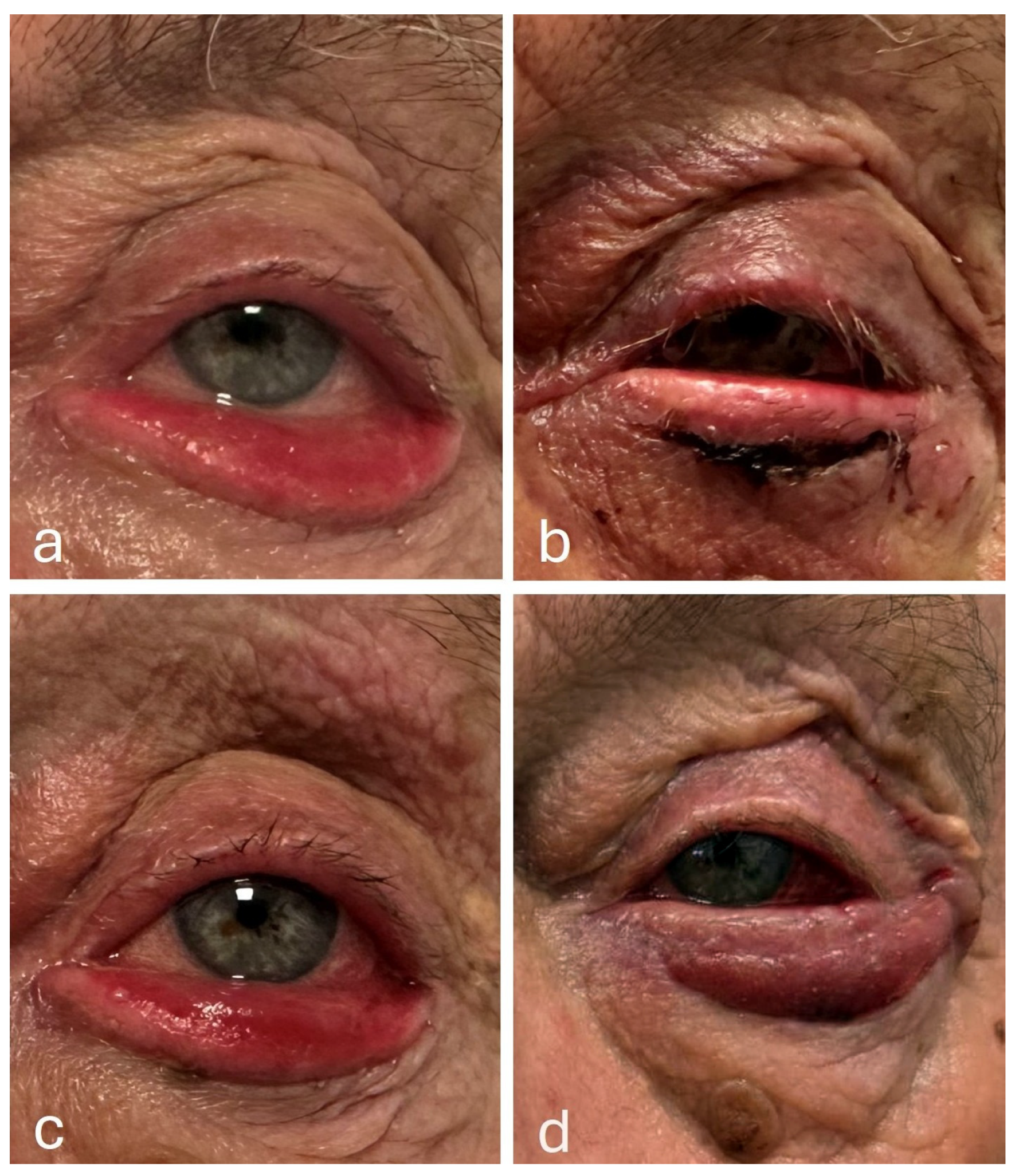
A total of nine consecutive patients with unilateral severe involutional lower eyelid ectropion met the inclusion criteria during the study period. The cohort consisted of six men and three women, with a mean age of 81 years (range, 73–90 years). All patients presented with long-standing lower eyelid ectropion characterized by complete eyelid eversion, conjunctival hyperemia, variable degrees of keratinization, chronic epiphora, and ocular surface irritation (
Figure 1). The duration of symptoms before surgery ranged from 3 to 22 months.
3.1. Primary Surgical Procedures
Horizontal eyelid shortening was performed in all patients using an LTS procedure. Additional medial conjunctival diamond excision was performed in four patients because of marked punctal eversion. Two patients required anterior lamella reconstruction using an ipsilateral upper eyelid transposition skin flap owing to skin deficiency. A temporary Frost suture was placed in two patients during the primary procedure.
Regarding vertical correction, three patients underwent complete lower eyelid retractor release during primary surgery because dense fibrosis, shortening, and persistent inferior traction of the retractors were identified intraoperatively despite correction of horizontal laxity. In the remaining six patients, the retractors were managed by reattachment or tightening according to conventional surgical principles. In some of these patients, no obvious fibrosis or shortening requiring release was recognized during the initial procedure, whereas in others, our surgical strategy had not yet evolved to include complete retractor release as a primary treatment option for advanced disease.
As experience accumulated during the study period, recurrent cases consistently demonstrated marked fibrosis and shortening of the lower eyelid retractors at revision surgery. These intraoperative findings led to a modification of our surgical approach, and complete retractor release subsequently became the preferred technique whenever advanced fibrosis and pathological inferior traction of the retractors were encountered.
Patient characteristics, surgical procedures, recurrences, and final outcomes are summarized in
Table 1 and
Table 2.
3.2. Recurrence and Revision Surgery (Table 2)
During a mean follow-up of 10.5 months (range 6–18 months), early recurrent ectropion developed in 5 of the 6 patients who underwent retractor reattachment or tightening as part of the primary procedure. No recurrence was observed in any patient who underwent complete retractor release during the initial operation.
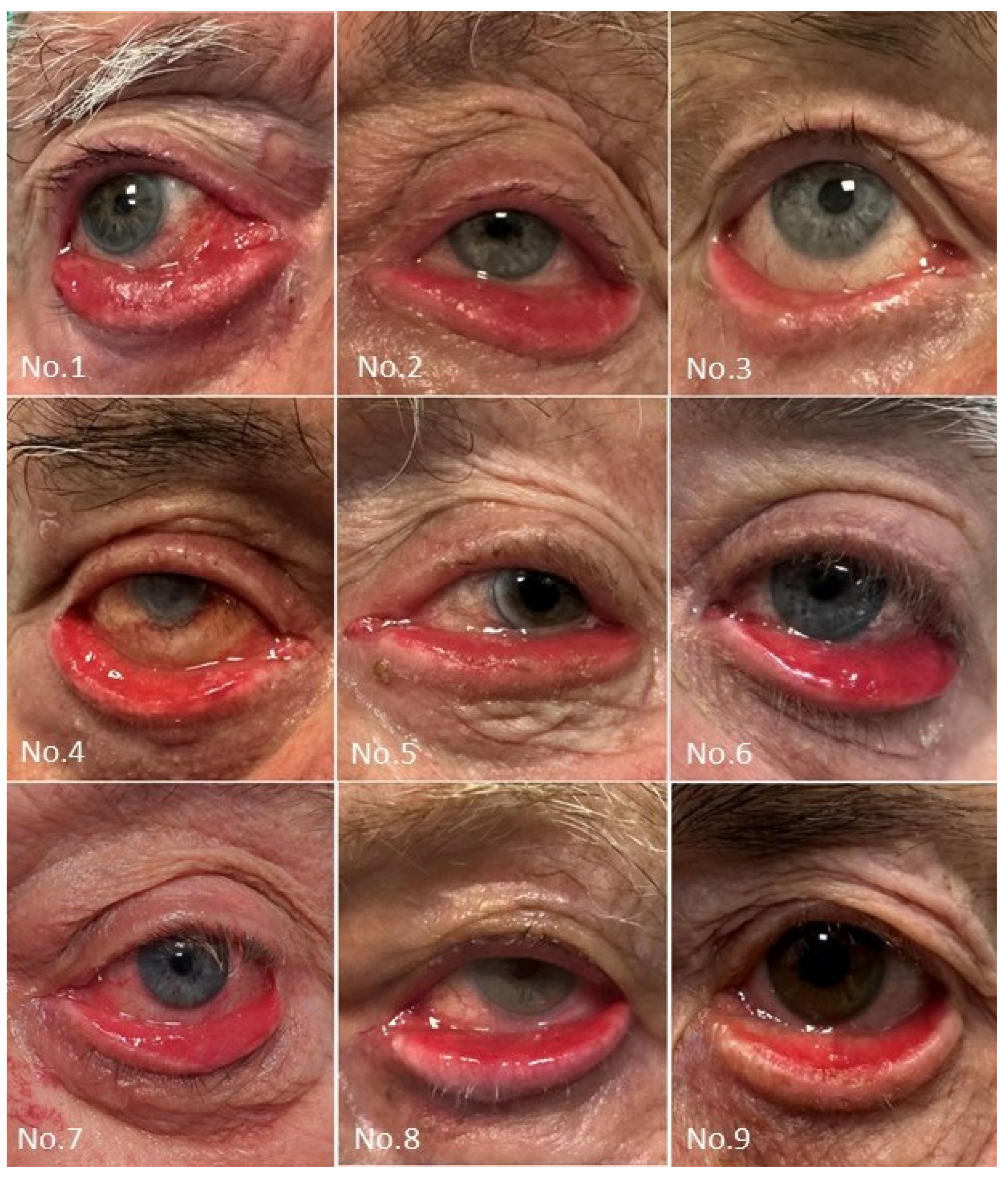
Two patients (No. 2 and No. 7) with recurrent ectropion subsequently underwent two revision surgeries, the latest consisting of complete lower eyelid retractor release in combination with additional horizontal eyelid shortening procedures. In both patients, a stable postoperative eyelid position was maintained throughout the subsequent follow-up period without further recurrence (
Figure 2). One additional patient (No. 8) underwent revision surgery at another institution. Two patients (No. 1 and No. 5) were considered candidates for further revision because of recurrent ectropion but were not reoperated because of significant medical comorbidities.
Overall, five patients underwent complete lower eyelid retractor release, either during the primary procedure (n = 3) or during revision surgery (n = 2). None of these patients developed recurrent ectropion during the available follow-up period.
In addition, all six patients who achieved stable eyelid position without recurrence (four after primary surgery and two after revision surgery) demonstrated satisfactory functional and aesthetic outcomes. Resolution or marked improvement of epiphora, ocular surface irritation, and conjunctival exposure was accompanied by restoration of stable eyelid–globe apposition and satisfactory cosmetic appearance.
Because of the retrospective design, limited sample size, heterogeneous surgical pathways, and absence of standardized functional outcome measures, no comparative statistical analysis between surgical techniques was performed.
4. Discussion
The present study describes our clinical experience with the surgical management of severe involutional lower eyelid ectropion presenting with complete eyelid eversion. Although limited by its retrospective design and small sample size, this case series suggests that complete lower eyelid retractor release may represent a useful adjunctive procedure in selected patients with advanced disease and intraoperative evidence of marked retractor fibrosis and shortening.
Involutional ectropion is generally regarded as a multifactorial disorder resulting from progressive age-related degeneration of the lower eyelid supporting structures. Horizontal eyelid laxity caused by attenuation of the medial and lateral canthal tendons, weakening of the orbicularis oculi muscle, and degeneration or disinsertion of the lower eyelid retractors all contribute to progressive eyelid malposition. Consequently, most established surgical techniques focus primarily on restoring horizontal eyelid stability, usually by means of a lateral tarsal strip procedure combined, when indicated, with retractor reattachment, tightening, or advancement [
6,
10,
11,
12,
13,
14,
15,
16,
17,
18].
The patients included in the present series, however, represented a distinctly different clinical population. All presented with long-standing disease characterized by fixed complete lower eyelid eversion, complete loss of eyelid–globe apposition, punctal eversion, and complete lateral canthal insufficiency manifested by rounding of the lateral canthus. In our opinion, these findings represent the end-stage of involutional ectropion rather than simply a more advanced degree of horizontal eyelid laxity.
We hypothesize that, in such advanced cases, the original involutional process becomes progressively complicated by secondary cicatricial remodeling. Chronic eyelid eversion exposes the palpebral conjunctiva to persistent desiccation, inflammation, and mechanical irritation, promoting progressive fibrosis of the posterior lamella. Histopathological studies have demonstrated degeneration of elastic fibers, collagen remodeling, and structural alterations within the lower eyelid retractors in involutional ectropion [
4,
19,
21,
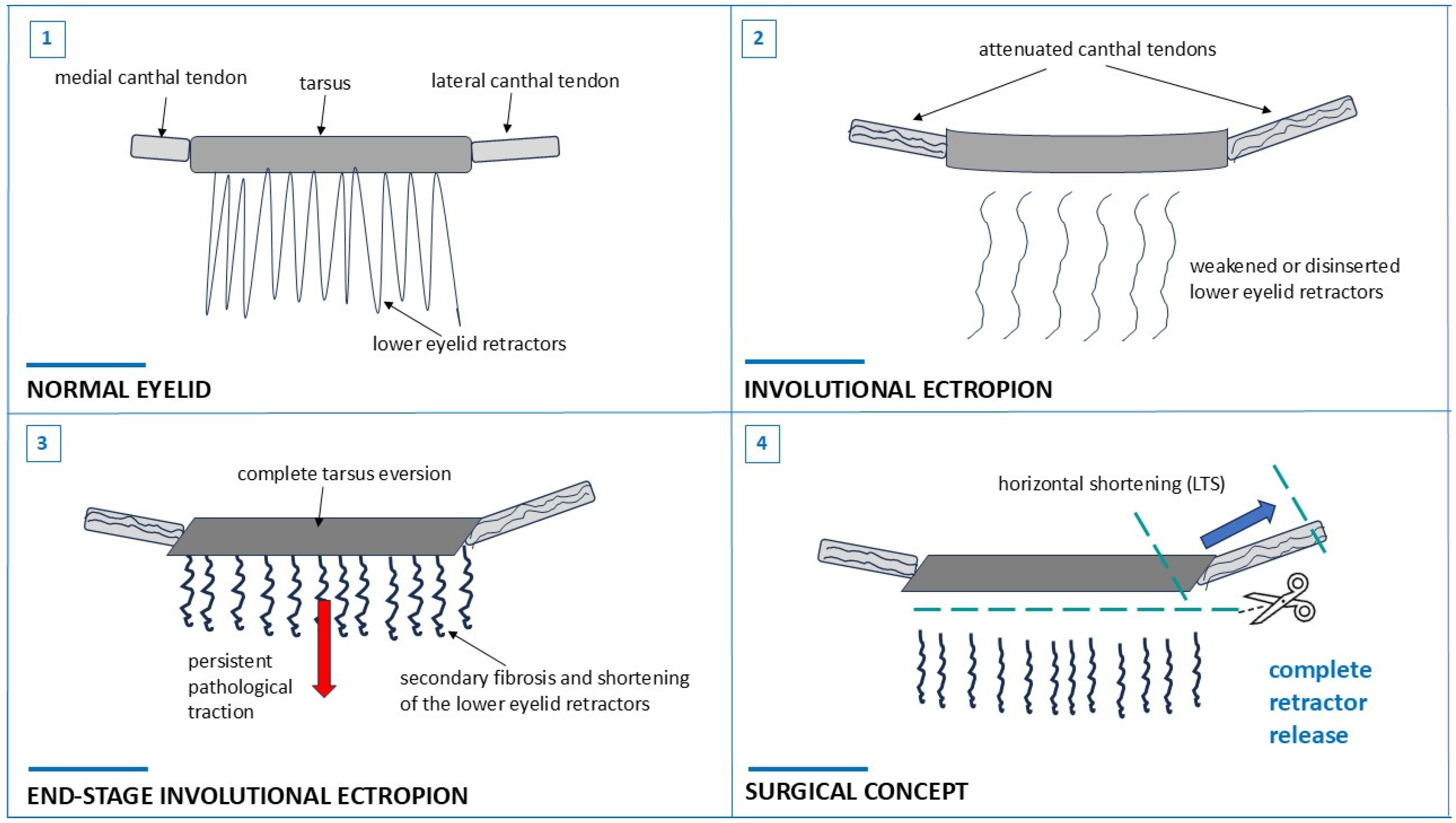
22]. We speculate that prolonged disease duration may further promote fibrosis and shortening of the retractor complex, resulting in pathological inferior traction that actively maintains the everted eyelid position (
Figure 3). Although supported by our intraoperative observations, this mechanism remains hypothetical, as no histopathological or biomechanical analyses were performed.
A particularly important observation in this series was the evolution of our surgical strategy during the study period. Initially, surgical management followed conventional principles, combining horizontal eyelid tightening with retractor reattachment or tightening when appropriate [
15,
16,
17,
18,
20,
23]. However, five of the six patients treated with this approach developed recurrent ectropion and subsequently required, or were considered candidates for, further surgical intervention. The only patient who remained free of recurrence was also the youngest in the series (73 years) and had the shortest duration of symptoms (3 months). Although no conclusions can be drawn from a single patient, it is plausible that the relatively short disease duration may have limited the development of secondary fibrotic changes within the posterior lamella. Consequently, restoration of normal retractor anatomy by reattachment or tightening may have been sufficient in this case.
In contrast, all remaining patients had a substantially longer history of ectropion, ranging from 8 to 22 months, and demonstrated marked intraoperative fibrosis, shortening, and reduced mobility of the lower eyelid retractors. During revision surgery, complete release of the fibrotic retractor complex eliminated the persistent inferior traction and allowed stable repositioning of the eyelid against the globe without recurrence during the available follow-up. These observations led to modification of our surgical approach. In the later patients included in this series, complete lower eyelid retractor release was therefore performed during the primary operation whenever advanced fibrosis and retractor shortening were identified intraoperatively.
Accordingly, the present study should not be interpreted as a comparison between retractor release and retractor reattachment or tightening. Rather, it describes the development of a modified surgical strategy based on progressively accumulated clinical experience in managing end-stage involutional ectropion. Within the limitations of this case series, no patient who underwent complete retractor release, either as a primary or revision procedure, developed recurrent ectropion during follow-up. Although encouraging, this observation cannot be interpreted as evidence of superiority over established techniques because treatment allocation was not standardized, the cohort was small, and several concomitant procedures were performed.
Interestingly, most published techniques addressing lower eyelid retractors aim to restore or reinforce their attachment to the inferior tarsal border by reattachment, tightening, or advancement [
15,
16,
17,
18,
23]. Our observations suggest that this concept may not be applicable to all patients. In carefully selected individuals with long-standing end-stage involutional ectropion and marked secondary fibrosis, reinforcement of an already shortened and fibrotic retractor complex may fail to eliminate the pathological traction maintaining eyelid eversion. We therefore believe that the indication for complete lower eyelid retractor release should be determined by the stage of the disease rather than by the diagnosis of involutional ectropion alone. In early disease, preservation and reattachment of the retractors may remain appropriate, whereas in advanced long-standing ectropion with fixed eyelid eversion and intraoperative evidence of fibrosis, complete release may be a more suitable approach for eliminating pathological traction. Whether this concept applies only to the most advanced stages of involutional ectropion or to a broader patient population remains to be determined.
The present study has several limitations. It represents a small retrospective case series from a single institution without a predefined treatment algorithm. The indication for complete retractor release evolved during the study period as our understanding of the underlying pathology developed. Furthermore, standardized functional and patient-reported outcome measures, as well as objective grading of retractor fibrosis, were unavailable. Preoperative and postoperative photographs were obtained retrospectively from routine clinical documentation and were not acquired using a standardized photographic protocol. Finally, follow-up was relatively limited. Accordingly, our findings should be regarded as hypothesis-generating rather than evidence supporting a change in standard surgical practice.
5. Conclusions
In this preliminary retrospective case series, our observations are consistent with the hypothesis that end-stage involutional ectropion should not be regarded simply as advanced horizontal eyelid laxity. Instead, prolonged disease may result in secondary cicatricial remodeling of the lower eyelid retractors, creating a distinct pathological stage in which complete retractor release may be considered in selected patients rather than conventional reattachment. Recognition of this advanced disease stage may help surgeons identify patients who could benefit from complete lower eyelid retractor release. Prospective studies with larger patient cohorts are required to determine the role of this proposed surgical approach and to compare its outcomes with those of established techniques.
Author Contributions
Conceptualization, B.K.E. and D.M. (Daliborka Miletić); methodology, B.K.E., D.M. (Daliborka Miletić) and I.B.; validation, B.K.E., D.M. (Daliborka Miletić), I.B. and M.B.; formal analysis, B.K.E., D.M. (Daliborka Miletić), I.B. and D.M. (Dora Marinčić); investigation, resources, data curation, B.K.E., D.M. (Daliborka Miletić) and I.B.; writing—original draft preparation, D.M. (Daliborka Miletić); writing—review and editing, B.K.E., D.M. (Daliborka Miletić), I.B. and D.M. (Dora Marinčić); supervision, B.K.E., D.M. (Daliborka Miletić) and M.B. All authors have read and agreed to the published version of the manuscript.
Funding
This research received no external funding.
Institutional Review Board Statement
The study was conducted in accordance with the Declaration of Helsinki and was approved by the Institutional Review Board of Sveti Duh University Hospital (approval number 01-839, dated 17 April 2026).
Informed Consent Statement
Due to the retrospective design and the use of anonymized data collected during routine clinical care, the requirement for informed consent was waived.
Data Availability Statement
The original contributions presented in this study are included in the article. Further inquiries can be directed to the corresponding author.
Acknowledgments
An artificial intelligence tool (Grammarly) was used only for language editing, including grammar, sentence structure, and clarity. The AI tool was not used to generate scientific content, interpret data, draw conclusions, or write the manuscript independently. The authors reviewed and approved the final text and take full responsibility for the manuscript’s content.
Conflicts of Interest
The authors declare no conflicts of interest.
Abbreviation
The following abbreviations are used in this manuscript:
References
- Rubino, C.; Trignano, E.; Dore, S.; Pinna, A.; Tsatsaris, N.; Ziani, F.; Sangalli, L.; Tettamanzi, M.; Rampazzo, S. 3-Step Combined Technique for Correction of Involutional Lower Eyelid Ectropion: A Case Series. J. Clin. Med. 2024, 14, 128. [Google Scholar] [CrossRef] [Scilit]
- Damasceno, R.W.; Osaki, M.H.; Dantas, P.E.C.; Belfort, R. Involutional Entropion and Ectropion of the Lower Eyelid: Prevalence and Associated Risk Factors in the Elderly Population. Ophthal. Plast. Reconstr. Surg. 2011, 27, 317–320. [Google Scholar] [CrossRef] [Scilit] [PubMed]
- Schellini, S.A.; Hoyama, E.; Shiratori, C.A.; Marques, M.E.A.; Padovani, C.R. Eyelid Alterations in Involutional Ectropion. J. Clin. Exp. Ophthalmol. 2011, 2, 135. [Google Scholar]
- Kocaoglu, F.A.; Katircioglu, Y.A.; Tok, O.Y.; Pulat, H.; Ornek, F. The Histopathology of Involutional Ectropion and Entropion. Can. J. Ophthalmol. 2009, 44, 677–679. [Google Scholar] [CrossRef] [Scilit] [PubMed]
- Bashour, M.; Harvey, J. Cause of involutional ectropion and entropion—Age-related tarsal changes are key. Ophthal Plast. Reconstr. Surg. 2000, 16, 131–141. [Google Scholar] [CrossRef] [Scilit] [PubMed]
- AlHarthi, A.S. Involutional Ectropion: Etiological Factors and Therapeutic Management. Int. Ophthalmol. 2022, 43, 1013–1026. [Google Scholar] [CrossRef] [Scilit] [PubMed]
- Lin, P.; Kitaguchi, Y.; Mupas-Uy, J.; Sabundayo, M.S.; Takahashi, Y.; Kakizaki, H. Involutional lower eyelid entropion: Causative factors and therapeutic management. Int. Ophthalmol. 2019, 39, 1895–1907. [Google Scholar] [CrossRef] [Scilit] [PubMed]
- Imbert, P. A Theory of Unified Pathogenesis for Tarsal Ectropion and Involutional Entropion. J. Fr. Ophtalmol. 2020, 43, 611–617. [Google Scholar] [CrossRef] [Scilit] [PubMed]
- Damasceno, R.W.; Avgitidou, G.; Belfort, R., Jr.; Dantas, P.E.C.; Holbach, L.M.; Heindl, L.M. Eyelid Aging: Pathophysiology and Clinical Management. Arq. Bras. Oftalmol. 2015, 78, 328–331. [Google Scholar] [CrossRef] [Scilit] [PubMed]
- Mcveigh, K.A.; Harrison, R.; Ford, R. Entropion and ectropion repair: A snapshot of surgical practice in the United Kingdom. Orbit 2018, 37, 105–109. [Google Scholar] [CrossRef] [Scilit] [PubMed]
- Collin, J.R.O. A Manual of Systematic Eyelid Surgery, 3rd ed.; Butterworth-Heinemann: Oxford, UK, 2006. [Google Scholar]
- Hou, X.; Guo, Y.; Li, S.; Lin, M.; Jia, R.; Rokohl, A.; Heindl, L.M. Lateral Tarsal Strip Procedure for Involutional Ectropion: A Retrospective Analysis of 85 Cases and a Comprehensive Literature Review. Adv. Ophthalmol. Pract. Res. 2021, 1, 100004. [Google Scholar] [CrossRef] [Scilit] [PubMed]
- Pascali, M.; Corsi, A.; Brinci, L.; Corsi, I.; Cervelli, V. The Tarsal Belt Procedure for the Correction of Ectropion: Description and Outcome in 42 Cases. Br. J. Ophthalmol. 2014, 98, 1691–1696. [Google Scholar] [CrossRef] [Scilit] [PubMed]
- Kam, K.Y.R.; Cole, C.J.; Bunce, C.; Watson, M.P.; Kamal, D.; Olver, J.M. The Lateral Tarsal Strip in Ectropion Surgery: Is It Effective When Performed in Isolation? Eye 2012, 26, 827–832. [Google Scholar] [CrossRef] [Scilit] [PubMed]
- Tse, D.T. Surgical Correction of Lower-Eyelid Tarsal Ectropion by Reinsertion of the Retractors. Arch. Ophthalmol. 1991, 109, 427. [Google Scholar] [CrossRef] [Scilit] [PubMed]
- Ghafouri, R.H.; Allard, F.D.; Migliori, M.E.; Freitag, S.K. Lower Eyelid Involutional Ectropion Repair With Lateral Tarsal Strip and Internal Retractor Reattachment With Full-Thickness Eyelid Sutures. Ophthal Plast. Reconstr. Surg. 2014, 30, 424–426. [Google Scholar] [CrossRef] [Scilit] [PubMed]
- Fong, K.C.S.; Mavrikakis, I.; Sagili, S.; Malhotra, R. Correction of Involutional Lower Eyelid Medial Ectropion with Transconjunctival Approach Retractor Plication and Lateral Tarsal Strip. Acta Ophthalmol. Scand. 2006, 84, 246–249. [Google Scholar] [CrossRef] [Scilit] [PubMed]
- Sagili, S.; Ha, J. Lower eyelid retractor (white-line) advancement to correct tarsal ectropion. Can. J. Ophthalmol. 2019, 54, 98–101. [Google Scholar] [CrossRef] [Scilit] [PubMed]
- Stefanyszyn, M.A.; Hidayat, A.A.; Flanagan, J.C. The histopathology of involutional ectropion. Ophthalmology 1985, 92, 120–127. [Google Scholar] [CrossRef] [Scilit] [PubMed]
- Gutoff, R.; Katowitz, J.A. (Eds.) Oculoplastics and Orbit (Essentials in Ophthalmology); Springer: Berlin/Heidelberg, Germany, 2007. [Google Scholar]
- Damasceno, R.W.; Osaki, M.H.; Dantas, P.E.; Belfort, R., Jr. Involutional ectropion and entropion: Clinicopathologic correlation between horizontal eyelid laxity and eyelid extracellular matrix. Ophthalmic Plast. Reconstr. Surg. 2011, 27, 321–326. [Google Scholar] [CrossRef] [Scilit] [PubMed]
- Kakizaki, H.; Malhotra, R.; Madge, S.N.; Selva, D. Lower eyelid anatomy: An update. Ann. Plast. Surg. 2009, 63, 344–351. [Google Scholar] [CrossRef] [Scilit] [PubMed]
- Shah-Desai, S.; Collin, R. Role of the lower lid retractors in involutional ectropion repair. Orbit 2001, 20, 81–86. [Google Scholar] [CrossRef] [Scilit] [PubMed]
| Disclaimer/Publisher’s Note: The statements, opinions and data contained in all publications are solely those of the individual author(s) and contributor(s) and not of MDPI and/or the editor(s). MDPI and/or the editor(s) disclaim responsibility for any injury to people or property resulting from any ideas, methods, instructions or products referred to in the content. |