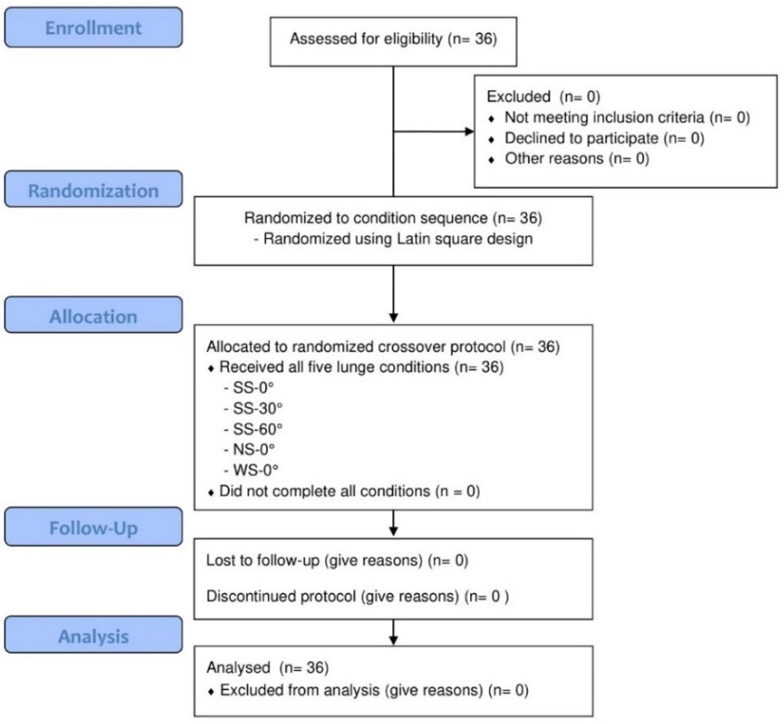
Effects of Foot Progression Angle and Stance Width on Lower-Limb Muscle Activation During the Holding Phase of Forward Lunge Exercises in Healthy Adults: A Randomized Within-Participant Crossover Study


Abstract
1. Introduction
2. Materials and Methods
2.1. Study Design and Participants
2.2. Experimental Procedure
2.3. Lunge Exercise Conditions
2.4. Surface Electromyography Measurement
2.5. Statistical Analysis
3. Results
3.1. General Characteristics of Participants
3.2. Statistical Assumption Checks
3.3. Muscle Activation According to Foot Progression Angle
3.4. Muscle Activation According to Stance Width
4. Discussion
5. Conclusions
Supplementary Materials
Author Contributions
Funding
Institutional Review Board Statement
Informed Consent Statement
Data Availability Statement
Conflicts of Interest
Abbreviations
| ANOVA | Analysis of variance |
| BF | Biceps femoris |
| GM | Gluteus medius |
| MSIT | Ministry of Science and ICT |
| MVC | Maximal voluntary contraction |
| NRF | National Research Foundation of Korea |
| NS-0° | Narrow stance with a 0° foot progression angle |
| RF | Rectus femoris |
| sEMG | Surface electromyography |
| SENIAM | Surface Electromyography for the Non-Invasive Assessment of Muscles |
| SS-0° | Standard stance with a 0° foot progression angle |
| SS-30° | Standard stance with a 30° foot progression angle |
| SS-60° | Standard stance with a 60° foot progression angle |
| ST | Semitendinosus |
| VL | Vastus lateralis |
| VM | Vastus medialis |
| WS-0° | Wide stance with a 0° foot progression angle |
| η2p | Partial eta squared |
References
- Ramari, C.; Hvid, L.G.; David, A.C.; Dalgas, U. The importance of lower-extremity muscle strength for lower-limb functional capacity in multiple sclerosis: A systematic review. Ann. Phys. Rehabil. Med. 2020, 63, 123–137. [Google Scholar] [CrossRef] [PubMed]
- Rostron, Z.P.; Green, R.A.; Kingsley, M.; Zacharias, A. Associations between measures of physical activity and muscle size and strength: A systematic review. Arch. Rehabil. Res. Clin. Transl. 2021, 3, 100124. [Google Scholar] [CrossRef] [PubMed]
- Kim, Y.S. Effects of the muscular strength training of lower extremities on the gait patterns and isokinetic strength in individuals with hemiplegia. Korean J. Adapt. Phys. Act. 2005, 13, 67–82. [Google Scholar]
- Yeo, S.J.; Yoon, S.D.; Park, G.D. A comparison of lower limb muscle activity in men in their 20s during lunge motion and kettlebell swing. Korean J. Sports Sci. 2016, 25, 1219–1226. [Google Scholar]
- Tayfur, B.; Johnson, A.K.; Palmieri-Smith, R. Changes in quadriceps rate of torque development after anterior cruciate ligament reconstruction and association to single-leg hop distance. Sports Health 2024, 16, 808–816. [Google Scholar] [CrossRef] [PubMed]
- Bordoni, B.; Varacallo, M.A. Anatomy, bony pelvis and lower limb: Thigh quadriceps muscle. In StatPearls [Internet]; StatPearls Publishing: Treasure Island, FL, USA, 2023. [Google Scholar]
- Schoenfeld, B.J. Squatting kinematics and kinetics and their application to exercise performance. J. Strength Cond. Res. 2010, 24, 3497–3506. [Google Scholar] [CrossRef] [PubMed]
- Clark, D.R.; Lambert, M.I.; Hunter, A.M. Muscle activation in the loaded free barbell squat: A brief review. J. Strength Cond. Res. 2012, 26, 1169–1178. [Google Scholar] [CrossRef] [PubMed]
- Eliassen, W.; Saeterbakken, A.H.; van den Tillaar, R. Comparison of bilateral and unilateral squat exercises on barbell kinematics and muscle activation. Int. J. Sports Phys. Ther. 2018, 13, 871–881. [Google Scholar] [CrossRef] [PubMed]
- Koderi, K.L.; Tan, K.; Azzfar, M.S.; Abd Malek, N.F.; Mohamad, N.I.; Nadzalan, A.M. The effects of stance width on muscle activation and performance during Romanian deadlift exercise. J. Phys. Conf. Ser. 2020, 1529, 022026. [Google Scholar] [CrossRef]
- Fischer, S.C.; Calley, D.Q.; Hollman, J.H. Effect of an exercise program that includes deadlifts on low back pain. J. Sport Rehabil. 2021, 30, 672–675. [Google Scholar] [CrossRef] [PubMed]
- Ada, L.; Dorsch, S.; Canning, C.G. Strengthening interventions increase strength and improve activity after stroke: A systematic review. Aust. J. Physiother. 2006, 52, 241–248. [Google Scholar] [CrossRef] [PubMed]
- Pietrosimone, B.; Lepley, A.S.; Harkey, M.S.; Luc-Harkey, B.A.; Blackburn, J.T.; Gribble, P.A.; Spang, J.T.; Sohn, D.H. Quadriceps strength predicts self-reported function post-ACL reconstruction. Med. Sci. Sports Exerc. 2016, 48, 1671–1677. [Google Scholar] [CrossRef] [PubMed]
- Bodkin, S.; Goetschius, J.; Hertel, J.; Hart, J. Relationships of muscle function and subjective knee function in patients after ACL reconstruction. Orthop. J. Sports Med. 2017, 5, 2325967117719041. [Google Scholar] [CrossRef] [PubMed]
- Cronin, J.; McNair, P.J.; Marshall, R.N. Lunge performance and its determinants. J. Sports Sci. 2003, 21, 49–57. [Google Scholar] [CrossRef] [PubMed]
- Turner, G.; Barker, K. Exercise selection to develop optimal explosive lunge movements for world-standard squash. Strength Cond. J. 2014, 36, 36–42. [Google Scholar] [CrossRef][Green Version]
- Goulette, D.; Griffith, P.; Schiller, M.; Rutherford, D.; Kernozek, T.W. Patellofemoral joint loading during the forward and backward lunge. Phys. Ther. Sport 2021, 47, 178–184. [Google Scholar] [CrossRef] [PubMed]
- Gao, L.; Lu, Z.; Liang, M.; Baker, J.S.; Gu, Y. Influence of different load conditions on lower extremity biomechanics during the lunge squat in novice men. Bioengineering 2022, 9, 272. [Google Scholar] [CrossRef] [PubMed]
- Wilson, D.J.; Gibson, K.; Masterson, G.L. Kinematics and kinetics of two styles of partial forward lunge. J. Sport Rehabil. 2008, 17, 387–398. [Google Scholar] [CrossRef] [PubMed]
- Jancová, J. Measuring the balance control system—Review. Acta Med. 2008, 51, 129–137. [Google Scholar]
- Choi, Y.; Lee, S. Changes in lower limb muscle activity based on angle of ankle abduction during lunge exercise. J. Phys. Ther. Sci. 2017, 29, 1947–1949. [Google Scholar] [CrossRef] [PubMed][Green Version]
- Park, H.Y.; Cha, Y.J.; Kim, N.H. Comparison of muscle activity in proximal muscle of lower extremities during lunge according to the anterior-posterior distance of foot position. J. Korean Soc. Phys. Med. 2018, 13, 131–138. [Google Scholar] [CrossRef]
- Asayama, A.; Tateuchi, H.; Yamagata, M.; Ichihashi, N. Influence of stance width and toe direction on medial knee contact force during bodyweight squats. J. Biomech. 2021, 129, 110824. [Google Scholar] [CrossRef] [PubMed]
- Ryu, H.J.; Kim, Y.T.; Park, H.J.; Kwon, J.W. Changes in lower limb muscle activity during lunge according to the different angle of ankle joint. J. Korean Phys. Ther. 2021, 33, 40–46. [Google Scholar] [CrossRef]
- Escamilla, R.F.; Zheng, N.; MacLeod, T.D.; Imamura, R.; Wang, S.; Wilk, K.E.; Yamashiro, K.; Fleisig, G.S. Patellofemoral joint loading in forward lunge with step length and height variations. J. Appl. Biomech. 2022, 38, 210–220. [Google Scholar] [CrossRef] [PubMed]
- Lim, C.Y.; In, J. Considerations for crossover design in clinical study. Korean J. Anesthesiol. 2021, 74, 293–299. [Google Scholar] [CrossRef] [PubMed]
- Kim, H.; Palmieri-Smith, R.M.; Kipp, K. Time-frequency analysis of muscle activation patterns in people with chronic ankle instability during landing and cutting tasks. Gait Posture 2020, 82, 203–208. [Google Scholar] [CrossRef] [PubMed]
- Pelliccia, A.; Sharma, S.; Gati, S.; Bäck, M.; Börjesson, M.; Caselli, S.; Collet, J.P.; Corrado, D.; Drezner, J.A.; Halle, M.; et al. 2020 ESC Guidelines on sports cardiology and exercise in patients with cardiovascular disease. Eur. Heart J. 2021, 42, 17–96. [Google Scholar] [CrossRef] [PubMed]
- Jeon, S.; Sontag, S.A.; Herda, T.J.; Trevino, M.A. Chronic training status affects muscle excitation of the vastus lateralis during repeated contractions. Sports Med. Health Sci. 2023, 5, 42–49. [Google Scholar] [CrossRef] [PubMed]
- Cohen, J. Statistical Power Analysis for the Behavioral Sciences, 2nd ed.; Lawrence Erlbaum Associates: Hillsdale, NJ, USA, 1988. [Google Scholar]
- Diong, J.; Butler, A.A.; Gandevia, S.C.; Héroux, M.E. Muscle electromyographic activity normalized to maximal muscle activity, not to Mmax, better represents voluntary activation. PLoS ONE 2022, 17, e0277947. [Google Scholar] [CrossRef] [PubMed]
- Moinuddin, A.; Goel, A.; Sethi, Y. The role of augmented feedback on motor learning: A systematic review. Cureus 2021, 13, e19695. [Google Scholar] [CrossRef] [PubMed]
- van Melick, N.; Meddeler, B.M.; Hoogeboom, T.J.; Nijhuis-van der Sanden, M.W.G.; van Cingel, R.E.H. How to determine leg dominance: The agreement between self-reported and observed performance in healthy adults. PLoS ONE 2017, 12, e0189876. [Google Scholar] [CrossRef] [PubMed]
- Bezerra, E.S.; Diefenthaeler, F.; Nunes, J.P.; Sakugawa, R.L.; Heberle, I.; Moura, B.M.; Moro, A.R.P.; Marcolin, G.; Paoli, A. Influence of trunk position during three lunge exercises on muscular activation in trained women. Int. J. Exerc. Sci. 2021, 14, 202–210. [Google Scholar] [CrossRef] [PubMed]
- Besomi, M.; Hodges, P.W.; Clancy, E.A.; Van Dieën, J.; Hug, F.; Lowery, M.; Merletti, R.; Søgaard, K.; Wrigley, T.; Besier, T.; et al. Consensus for experimental design in electromyography (CEDE) project: Amplitude normalization matrix. J. Electromyogr. Kinesiol. 2020, 53, 102438. [Google Scholar] [CrossRef] [PubMed]
- Hermens, H.J.; Freriks, B.; Disselhorst-Klug, C.; Rau, G. Development of recommendations for SEMG sensors and sensor placement procedures. J. Electromyogr. Kinesiol. 2000, 10, 361–374. [Google Scholar] [CrossRef] [PubMed]
- Yeo, S.S.; Kwon, J.W.; Kim, C.S. The effects of ankle joint angle on knee extensor electromyographic activity. J. Korean Soc. Phys. Med. 2009, 4, 15–21. [Google Scholar]
- Lee, M.M.; Park, D.S. A comparison of muscle activation and mechanical loading according to the degree of ankle joint motion during a sit-to-stand task. J. Korean Soc. Phys. Med. 2017, 12, 113–122. [Google Scholar] [CrossRef]
- Qiao, Y.J.; Kim, K.R.; Kim, M.K. Effects of altering foot position on quadriceps femoris activation during wall squat exercises. J. Korean Soc. Phys. Med. 2021, 16, 23–31. [Google Scholar] [CrossRef]
- Moore, D.; Semciw, A.I.; Pizzari, T. A systematic review and meta-analysis of common therapeutic exercises that generate highest muscle activity in the gluteus medius and gluteus minimus segments. Int. J. Sports Phys. Ther. 2020, 15, 856–881. [Google Scholar] [CrossRef] [PubMed]
- Marchetti, P.H.; Guiselini, M.A.; da Silva, J.J.; Tucker, R.; Behm, D.G.; Brown, L.E. Balance and lower limb muscle activation between in-line and traditional lunge exercises. J. Hum. Kinet. 2018, 62, 15–22. [Google Scholar] [CrossRef] [PubMed]
- Muyor, J.M.; Martín-Fuentes, I.; Rodríguez-Ridao, D.; Antequera-Vique, J.A. Electromyographic activity in the gluteus medius, gluteus maximus, biceps femoris, vastus lateralis, vastus medialis and rectus femoris during the monopodal squat, forward lunge and lateral step-up exercises. PLoS ONE 2020, 15, e0230841. [Google Scholar] [CrossRef] [PubMed]



| Condition | Definition |
|---|---|
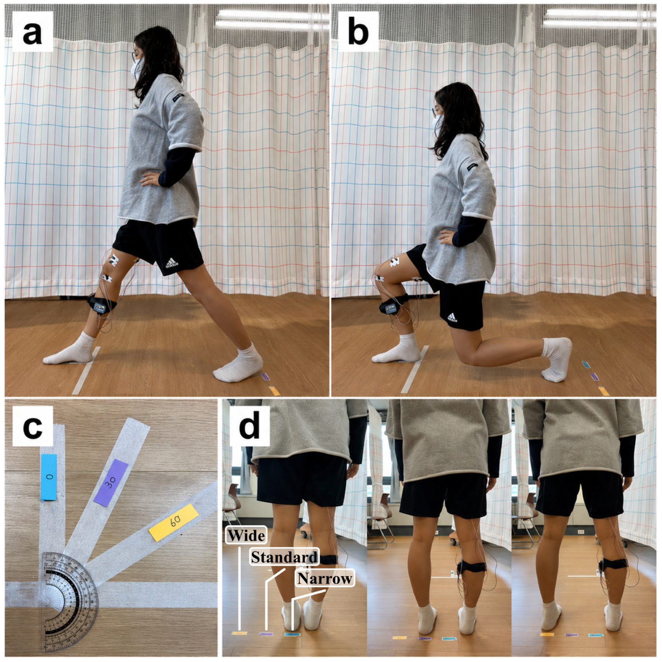
| SS-0° | Standard stance with a 0° foot progression angle |
| NS-0° | Narrow stance with a 0° foot progression angle |
| WS-0° | Wide stance with a 0° foot progression angle |
| SS-30° | Standard stance with a 30° foot progression angle |
| SS-60° | Standard stance with a 60° foot progression angle |
| Muscle | Electrode Placement |
|---|---|
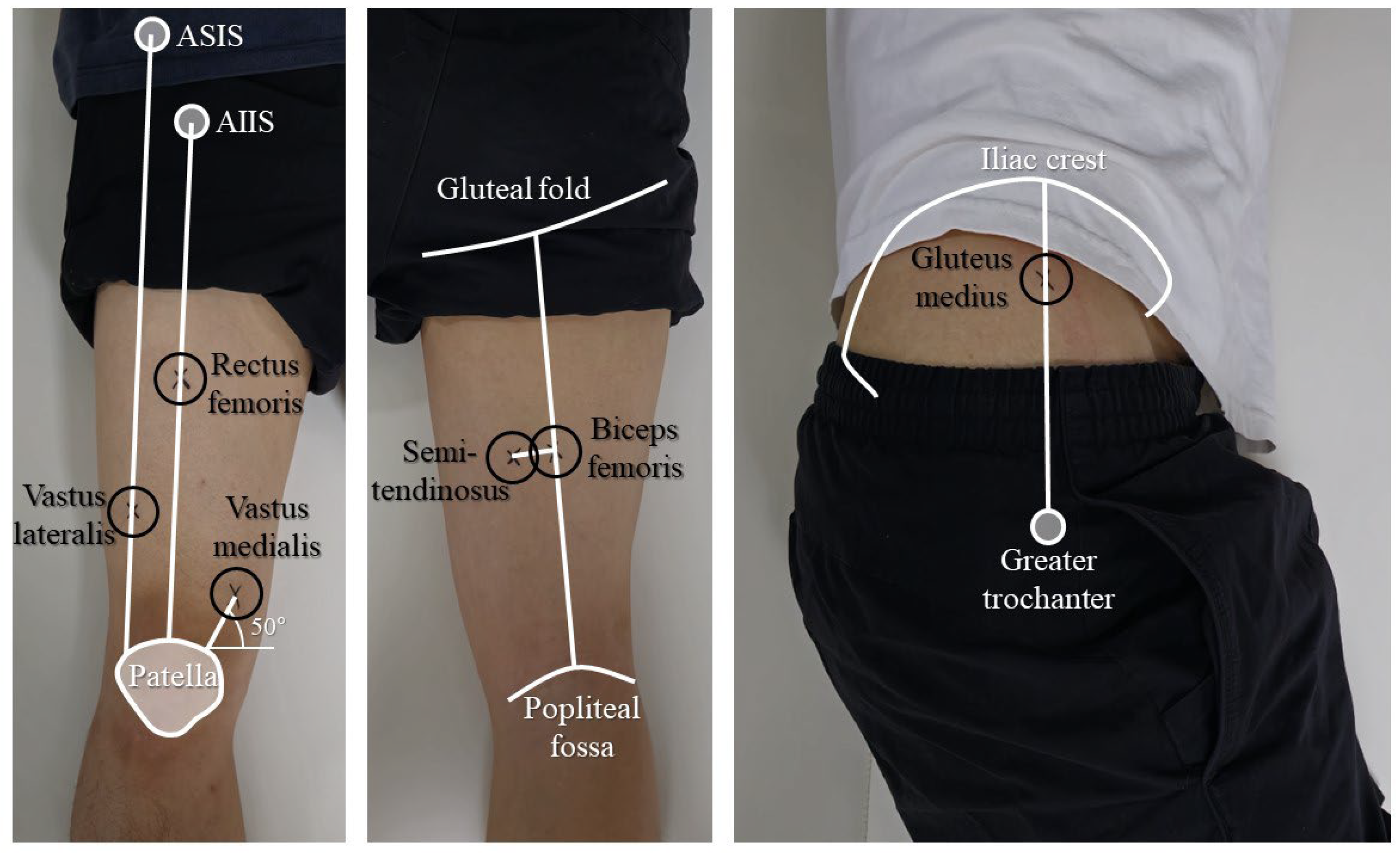
| Rectus femoris | At the midpoint of the anterior thigh along the line connecting the anterior inferior iliac spine and proximal patellar border |
| Gluteus medius | At one-third of the distance from the highest point of the iliac crest toward the greater trochanter |
| Vastus lateralis | Along the line toward the anterior superior iliac spine, 10 cm proximal to the superolateral patellar border |
| Vastus medialis | At a site 4 cm proximal to the superomedial patellar border, with an orientation of approximately 50° |
| Biceps femoris | On the posterior thigh, halfway between the gluteal fold and the popliteal fossa |
| Semitendinosus | 3 cm medial to the biceps femoris electrode placement site |
| Variable | Total Participants (n = 36) |
|---|---|
| Sex (male/female) | 17/19 |
| Age (years) | 28.39 ± 3.07 |
| Height (cm) | 165.00 ± 8.33 |
| Weight (kg) | 61.94 ± 14.98 |
| Body mass index (kg/m2) | 22.75 ± 1.85 |
| Dominant-side leg length (cm) | 77.11 ± 3.31 |
| Muscle | SS-0° | SS-30° | SS-60° | F(df) | ANOVA | η2p | 0–30° | 0–60° | 30–60° |
|---|---|---|---|---|---|---|---|---|---|
| RF | 18.99 ± 6.80 | 19.09 ± 7.12 | 20.35 ± 8.01 | 6.18 (2,70) | 0.003 | 0.150 | 1.000 | 0.003 | 0.026 |
| GM | 12.00 ± 3.01 | 10.01 ± 2.33 | 7.66 ± 2.30 | 118.93 (2,70) | <0.001 | 0.773 | <0.001 | <0.001 | <0.001 |
| VL | 35.63 ± 12.55 | 35.43 ± 10.50 | 37.54 ± 11.43 | 7.13 (2,70) | 0.002 | 0.169 | 1.000 | 0.022 | 0.001 |
| VM | 36.09 ± 15.99 | 34.78 ± 14.19 | 37.50 ± 15.99 | 4.03 (2,70) | 0.022 | 0.103 | 0.484 | 0.506 | 0.022 |
| BF | 4.91 ± 1.56 | 4.61 ± 2.38 | 4.91 ± 1.33 | 0.33 (1.68,58.93) | 0.682 | 0.009 | 1.000 | 1.000 | 1.000 |
| ST | 8.98 ± 3.17 | 8.68 ± 2.36 | 9.61 ± 2.77 | 0.98 (2,70) | 0.382 | 0.027 | 1.000 | 1.000 | 0.418 |
| Muscle | NS-0° | SS-0° | WS-0° | F(df) | ANOVA | η2p | NS–SS | NS–WS | SS–WS |
|---|---|---|---|---|---|---|---|---|---|
| RF | 17.36 ± 5.69 | 18.99 ± 6.80 | 23.05 ± 9.09 | 58.33 (1.44,50.45) | <0.001 | 0.625 | <0.001 | <0.001 | <0.001 |
| GM | 13.23 ± 3.54 | 12.00 ± 3.01 | 11.80 ± 3.60 | 9.60 (2,70) | <0.001 | 0.215 | 0.004 | <0.001 | 1.000 |
| VL | 33.16 ± 9.71 | 35.63 ± 12.55 | 36.44 ± 13.25 | 9.80 (2,70) | <0.001 | 0.219 | 0.009 | 0.001 | 0.767 |
| VM | 32.59 ± 12.52 | 36.09 ± 15.99 | 37.20 ± 16.71 | 10.79 (2,70) | <0.001 | 0.236 | 0.005 | <0.001 | 0.800 |
| BF | 5.06 ± 1.32 | 4.91 ± 1.56 | 5.62 ± 2.43 | 1.58 (2,70) | 0.214 | 0.043 | 1.000 | 0.720 | 0.369 |
| ST | 9.15 ± 3.17 | 8.98 ± 3.17 | 8.80 ± 2.63 | 0.14 (2,70) | 0.872 | 0.004 | 1.000 | 1.000 | 1.000 |
Disclaimer/Publisher’s Note: The statements, opinions and data contained in all publications are solely those of the individual author(s) and contributor(s) and not of MDPI and/or the editor(s). MDPI and/or the editor(s) disclaim responsibility for any injury to people or property resulting from any ideas, methods, instructions or products referred to in the content. |
© 2026 by the authors. Licensee MDPI, Basel, Switzerland. This article is an open access article distributed under the terms and conditions of the Creative Commons Attribution (CC BY) license.
Share and Cite
Shin, H.-J.; Kim, H.-M.; Cho, H.-Y.; Kim, S.-H. Effects of Foot Progression Angle and Stance Width on Lower-Limb Muscle Activation During the Holding Phase of Forward Lunge Exercises in Healthy Adults: A Randomized Within-Participant Crossover Study. J. Clin. Med. 2026, 15, 5567. https://doi.org/10.3390/jcm15145567
Shin H-J, Kim H-M, Cho H-Y, Kim S-H. Effects of Foot Progression Angle and Stance Width on Lower-Limb Muscle Activation During the Holding Phase of Forward Lunge Exercises in Healthy Adults: A Randomized Within-Participant Crossover Study. Journal of Clinical Medicine. 2026; 15(14):5567. https://doi.org/10.3390/jcm15145567
Chicago/Turabian StyleShin, Ho-Jin, Hong-Min Kim, Hwi-Young Cho, and Sung-Hyeon Kim. 2026. "Effects of Foot Progression Angle and Stance Width on Lower-Limb Muscle Activation During the Holding Phase of Forward Lunge Exercises in Healthy Adults: A Randomized Within-Participant Crossover Study" Journal of Clinical Medicine 15, no. 14: 5567. https://doi.org/10.3390/jcm15145567
APA StyleShin, H.-J., Kim, H.-M., Cho, H.-Y., & Kim, S.-H. (2026). Effects of Foot Progression Angle and Stance Width on Lower-Limb Muscle Activation During the Holding Phase of Forward Lunge Exercises in Healthy Adults: A Randomized Within-Participant Crossover Study. Journal of Clinical Medicine, 15(14), 5567. https://doi.org/10.3390/jcm15145567