Use of Artificial Intelligence in Rheumatoid Arthritis: Advancements and Novel Perspectives

 ,
,  ,
, 
 and
and 
Highlights
- RA is a complex disease with many unmet needs.
- AI may improve the management of RA patients by enabling early identification of the disease and stratifying patients based on prognostic factors or treatment response.
- ML models have proven accurate at combining large volumes of clinical and omics data to address some of these aspects.
- DL techniques may be more effective for image interpretation.
- The main limitations of AI in clinical practice are small cohorts, poor data generalizability, and the lack of external validation.
- GenAI, Agentic AI, and FL may represent future frontiers for addressing current AI gaps in clinical practice.
Abstract
1. Introduction
2. Materials and Methods
3. Results
3.1. Artificial Intelligence: From Conceptualization to Practical Application
3.2. Artificial Intelligence in Healthcare
3.3. Artificial Intelligence in Rheumatic Diseases

| Rheumatic Disorder | Study Design | Patients | Endpoint | Results | Author, Date, Reference |
|---|---|---|---|---|---|
| PsA | Multicenter, prospective, noninterventional study | 1278 eligible PsA patients treated with or scheduled to be treated with SEC from the AQUILA dataset | - Prediction of LDA and high HRQOL response at 16 weeks using binary ML; - Identification of predictors and quantification of their impact for individual patients using an XAI model | - Patient global assessment, physician global assessment, previous use of bDMARDs, TJC, and age emerging as the main LDA predictors; - Impact of disease, BDI, height, TJC, and BMI predicted to be associated with high HRQOL | Vodenčarević et al., 2025 [39] |
| SpA | Multicenter, prospective study collecting data from the BIOBADASER registry | 969 axial SpA patients who started treatment with a TNFi | - Identification of predictors of response to TNFi using a combination of statistical and AI methods (Anaxomics AI Data Science software) to extract relevant features from large datasets | - Female sex, age at diagnosis and at treatment initiation, and comorbidities predicted to be associated with an unfavorable response to TNFi | Fernández-Carballido et al., 2023 [40] |
| Multicenter, prospective, noninterventional study | 683 eligible axSpA patients treated with or scheduled to be treated with SEC from the AQUILA dataset | - Prediction of LDA and high HRQOL response at 16 weeks using binary ML; - Identification of predictors and quantification of their impact for individual patients using an XAI model | - BASDAI, previous use of bDMARDs, CRP values, ASAS HI, and height identified as predictors of LDA; - ASAS HI, BDI, BMI, height, and age predicted to be associated with high HRQOL | Vodenčarević et al., 2025 [39] | |
| Prospective multicenter study extrapolating MRI data from the DESIR and ASAS cohorts | 256 patients from the DESIR cohort with inflammatory back pain lasting more than 3 months and less than 3 years, of whom 27% met the ASAS criteria for SpA; 47 patients from the ASAS cohort, with 19% meeting the ASAS criteria for active sacroiliitis, used for external validation | Comparison of sensitivity, specificity, accuracy, Matthews correlation coefficient, and AUC of a DL model (Mask-RCNN) trained to detect active sacroiliitis on MRI according to the ASAS definition, compared to expert majority opinion | Similar performance of the DL model and experts in identifying BMO in sacroiliac joints and detecting sacroiliitis according to the ASAS definition | Bordner et al., 2023 [41] | |
| Retrospective multicenter study using MRI scan datasets from the Genodisc, Oxford Whole Spine, PREVENT, and MEASURE-1 studies | 686 axSpA patients enrolled in the MEASURE-1 and PREVENT trials | - Assessment of the accuracy of ML models (SpineNet software), with and without manual segmentation, for automated scoring of vertebral BMO on MRI | - Both models showing performance comparable to expert readers in detecting BMO; - Applying ML models without manual segmentation performed better than using an intermediate manual segmentation step | Jamaludin et al., 2025 [42] | |
| Retrospective multicenter study collecting conventional pelvis X-ray images from the PROOF, GESPIC, OptiRef, and DAMACT datasets | 2170 adult axSpA patients from the PROOF dataset, with 1483 X-rays used as training data; 525 axSpA patients from the GESPIC study and 361 axSpA patients from the OptiRef study, with 436 and 340 radiographs, respectively, used for inference analysis; plus 178 patients (89 with axSpA and 74 without SpA) from the DAMACT dataset used for inference | Comparison of AUC, accuracy, sensitivity, and specificity for detection and progression prediction of sacroiliitis between two neural networks: one anatomy-centered on the sacroiliac joints and the other on the full X-ray image | - Similar learning performance for both the standard and anatomy-centered models; - The anatomy-centered model shows higher consistency across datasets, as well as higher AUC scores, accuracy, sensitivity, and specificity compared to the comparator; - Ability of anatomy-centered models to identify patients progressing to radiographic sacroiliitis within 2 years | Dorfner et al., 2024, [43] | |
| SLE | Multicenter study | 446 cases, of which 382 allocated to the training and validation cohort (36 patients with cutaneous manifestations of SLE or other cutaneous diseases and 18 healthy controls) and 64 allocated to the external test cohort | Development and testing of an MMDLS for predicting cutaneous lupus subtypes | - Better performance of the MMDLS in diagnosing 13 skin conditions compared with single or dual models; - Increased diagnostic accuracy of junior dermatologists when the MMDLS is consulted | Li et al., 2024 [44] |
| Multicenter study and AI-enhanced meta-analysis | 12,850 patients with SLE from registry cohorts in Asia, Europe, North America, and Africa | Evaluation of the diagnostic and prognostic significance of NLR and PLR in SLE using an XGBoost ML model, guided by the Transparent Reporting of a Multivariable Prediction Model for Individual Prognosis or Diagnosis-AI, with SHAP, integrating traditional clinical biomarkers and multi-omics data | - Superior diagnostic accuracy of NLR compared with PLR for predicting active SLE, although with significant ethnic variation; - Higher risk of LN, CV events, or mortality associated with high NLR and PLR; - Improved accuracy in predicting active SLE when NLR and PLR are integrated into the AI model | Ali et al., 2025 [45] | |
| Observational study | 395 SLE patients requiring first hospitalization from the MIMIC-IV database and 100 critically ill patients with SLE allocated to the validation cohort | Development and validation of two predictive models: a traditional model based on logistic regression and an ML model using a stacking ensemble approach for assessing mortality risk in critically ill SLE patients | - AUC above 0.8 reported for both models; - The ML model outperformed logistic regression in terms of precision and specificity; - SHAP analysis made it possible to show the contribution of individual variables | Chen et al., 2025 [46] | |
| Systematic review | Over 800 patients, including NPSLE, SLE without neuropsychiatric involvement and healthy individuals | Evaluation of the performance of AI-based methods (ML or DL) in detecting NPSLE by integrating neuroimaging, CSF, or serum biomarkers | - Most studies relying on neuroimaging rather than biomarkers; - High performance of AI-based methods (pooled AUC 0.86; accuracy 0.87), but significant between-study heterogeneity that influenced sensitivity analyses; - No significant differences between ML and DL algorithms; - Lack of standardization, external validation, and explainable methods in most studies | Nouroozi et al., 2026 [47] | |
| LN | Monocenter retrospective study | 58 GN patients | Prediction of LN outcome and remission achievement by testing an AI-based neural network integrating laboratory and histopathological data | - Best performance of a multilayer perceptron with 40 neurons in the first hidden layer and 45 neurons in the second hidden layer (accuracy of 91.67%) with 100% precision for predicting complete LN remission | Stojanowski et al., 2022 [48] |
| Multicenter cohort study | 31,670 patients with chronic kidney disease across 6 medical centers | Evaluation of the performance of AI software in diagnosing glomerular disease by analysis of kidney TEM images | - Accuracy of TEM-AID in identifying glomerulonephritis subtypes, demonstrating high internal diagnostic performance and consistent external validation across five test sets; - Superior diagnostic performance of TEM-AID compared with pathologists | Ma et al., 2025 [49] | |
| SS | Multicenter cohort study | 100 patients, of whom 36 were used for training and internal validation datasets and 64 for the external test dataset | Assessment of the accuracy of CTG-PAM, based on graph theory, in scoring and diagnosing SS by analyzing cells and tissues in salivary gland biopsies | - High accuracy and better performance of CTG-PAM compared to traditional DL methods in diagnosing SS; - CTG-PAM’s diagnostic accuracy similar to that of expert pathologists and higher than that of junior pathologists | Wu et al., 2024 [50] |
| CTDs | Case series | 59 Japanese patients | Evaluation of the accuracy of diagnosing a CTD by the AI/RHEUM tool compared to the diagnosis made by a rheumatologist | - Full or partial agreement between the AI tool and traditional methods of diagnosing CTDs (92%), with a sensitivity of 90% and specificity of 96% | Porter et al., 1988 [36] |
| Cross-sectional study | 67 patients with CTD and early ILD development, including 21 with SSc, 23 with IIM, 9 with SS, 7 with SLE, 5 with MCTD, and 2 with RA | Accuracy of AI-based methods (AlqpHRTC) in diagnosing early lung involvement in CTDs (asymptomatic versus symptomatic) by analyzing HRCT datasets and combining these data with clinical and functional information | - Increased detection of high-attenuation lung volume and reticulations in symptomatic patients by AlqpHRTC, but not of ground-glass opacities or honeycombing; - High reliability of AlqpHRTC in detecting ILD features in asymptomatic patients | Hoffmann et al., 2025 [51] | |
| Vasculitis | Retrospective multicenter study | 137 GCA patients | Detection of the halo sign in CDU of temporal arteries using a DL approach | - Excellent accuracy in standardized images; - Identification of pathological signs in 90% of cases from non-standardized images; - Thrombus may increase the risk of bias | Roncato et al., 2020 [52] |
| Retrospective monocenter study | 1474 KD patients subdivided into training and validation cohorts at an 80/20 ratio | Development and validation of a DL AI model to detect coronary artery lesions | - Best performance achieved with a decision tree model based on 24 demographic, laboratory, and clinical features | Yang et al., 2025 [53] | |
| Systematic review and meta-analysis | 103,882 participants, of whom 12,541 were diagnosed with KD | Evaluation of the accuracy of ML methods to distinguish KD from other febrile conditions | - Twenty-nine studies included, of which 20 used ML; - The ability to identify KD showed a sensitivity of 0.91 and a specificity of 0.86 | Zhu et al., 2024 [61] | |
| Gout | Real-world, retrospective, prospective, multicenter cohort study | 6526 hospitalized gout patients, of whom 4074 were used to develop the prediction model, while 1746, 360, and 346 patients were allocated to groups for internal validation, external validation, and the prospective set, respectively | Prediction of gout recurrence through the construction and validation of a multidimensional AI model that takes into account diverse data categories, including comorbidities | - 3744 models screened, of which the IterImp_MM_FS_GB model was found to be the most effective for predicting gout recurrence; - 20 key predictors identified by SHAP analysis, including serum urate levels, neutrophil count, and tophi in patients with multiple comorbidities such as neoplasm and cardiovascular diseases | Li et al., 2025 [54] |
| Observational study | 6648 gout patients allocated to GIMS and 1651 gout patients allocated to EMRS | Comparison between the GIMS and EMRS cohorts in terms of kidney function preservation and maintenance of the serum urate target | - Reduced incidence of chronic kidney disease stage ≥ 3 and higher achievement of the target serum urate level in the GIMS group compared to the EMRS group | Qi et al., 2025 [62] | |
| OA | Single-blinded RCT | 82 knee OA patients | Development of an interactive mobile application that uses AI to provide personalized physical exercise programs to patients based on knee OA severity | - Higher accuracy in performing three prescribed exercises in patients using the mobile application than in controls; - Improvements in quality of life, function, and satisfaction in patients assigned to the mobile application group compared to controls | Thiengwittayaporn et al. 2023 [55] |
| Monocenter validation study | 5849 Thailand patients of whom 3455 had knee OA | Validation of DL models to predict early diagnosis of knee OA by processing textual data such as clinicians’ notes on patient symptoms | - Best performance achieved by the BiLSTM model, which improved further after applying a WOMAC-based processing approach; - Better performance of this model compared to other methods that use imaging or laboratory data | Thanyakunsajja et al., 2025 [56] | |
| Phase 2, double-blinded, observational study | 50 patients with knee OA | Comparison between an AI-generated (GPT-4) self-management guide for OA and recommendations created by clinicians | Better performance of GPT-4 compared to clinicians in content generation speed, accuracy, personalization, safety, and comprehensiveness | Du et al., 2025 [57] | |
| Cross-sectional study | 40 patients with knee OA | Comparing personalized rehabilitation programs generated by AI (ChatGPT-4.0 and Gemini Advanced) with physiotherapists’ consensus programs | - Greater agreement between ChatGPT-4.0 and physiotherapists’ consensus programs compared with Gemini Advanced; - Comparable or better performance of ChatGPT-4.0 than Gemini Advanced in most phases; - Limitations in exercise specificity, including frequency, sets, and progression criteria | Gürses et al., 2025 [58] | |
| Retrospective analysis study | 5966 knee radiographic images from the Osteoarthritis Initiative dataset used for model development, and 3392 knee radiographic images from the Multicenter Osteoarthritis Study dataset used for validation | Development of an ML-assisted method to predict KLG progression over 4–5 years in patients starting from a KLG score of 0, 1, or 2, integrating additional predictors such as demographics, comorbidities, history of meniscectomy, gait speed, WOMAC scores, and X-ray findings | - The model demonstrated good accuracy in predicting the progression of knee OA over 4–5 years | Lee et al., 2025 [63] | |
| Retrospective analysis study | 600 patients with knee OA enrolled in the Foundation for the National Institutes of Health Osteoarthritis Biomarkers Project, 297 of whom were classified as pain progressors according to the WOMAC scale during a 24- to 48-month follow-up period | Development of a nomogram model based on radiomics data (Neusoft Discovery) and clinical characteristics to predict pain progression | - Excellent predictive capability and accuracy in predicting pain progression using X-ray radiomics-based nomograms | Sun et al., 2026 [64] | |
| OP | RCT | 40,658 participants aged 40 years or older who underwent chest radiography without a history of DXA examination | Identification of individuals at high risk of OP using an AI model applied to chest radiographs | - 4912 participants classified as at high risk of OP by the AI model; - high proportion of patients with OP detected by DXA in this selected population; - increased odds ratio of OP in screened subjects who did not meet the formal criteria for DXA compared with those who did | Lin et al., 2024 [59] |
| Miscellanea of inflammatory rheumatic diseases | Prospective, multicenter, open-label crossover RCT | 600 patients from 3 rheumatology centers | Assessment and comparison of the diagnostic accuracy of the AI-based tools Ada and Rheport versus a diagnosis of inflammatory rheumatic disease made by rheumatologists | - Overall diagnostic accuracies of 52%, 63%, and 58%, respectively, for Rheport and Ada’s top 1 and top 5 disease suggestions; - Heterogeneous accuracy of Ada in making individual diagnoses, with its best performance in detecting RA compared with other diseases; - Poor agreement between Rheport and Ada’s top 1 and top 5 disease suggestions | Knitza et al., 2024 [65] |
3.4. Artificial Intelligence and Rheumatoid Arthritis: The State of the Art
3.4.1. Imaging
3.4.2. Early Diagnosis
3.4.3. Identification of Clinical Phenotypes
3.4.4. Treatment Response and Personalized Therapies
3.4.5. Follow-Up
| Domain | Study Design | Patients | Intervention | Results | Author, Date, Reference |
|---|---|---|---|---|---|
| Imaging | Retrospective observational study | 1694 US images obtained from 40 RA patients with long-standing (n.20) and untreated (n.20) disease | Application of two CNNs with different basic architectures (VGG-16 and Inception-v3) to classify hand and wrist joints as either healthy or diseased and to score images according to the OESS | - Good accuracy in distinguishing healthy from diseased scores compared with an expert rheumatologist (86.4% and 86.9%, respectively); - Accuracy of 75.0% for the Inception-v3 architecture model in four-class OESS | Andersen et al., 2019 [68] |
| Retrospective study | Dataset of musculoskeletal US images of the wrist from patients with suspected or known joint pathologies | Development of a Self-Attention U-Net-based DL model for synovitis segmentation and identification | - High accuracy in detecting synovial hypertrophy and effusion; - Improved performance compared with traditional U-Net models; - Greater ability to capture spatial patterns | Chang et al., 2024 [69] | |
| Retrospective study | 1244 US hand and wrist images obtained from 156 RA patients | Validation of four DL models based on a ResNet-type architecture to detect and score synovitis according to the OESS in static grayscale, dynamic grayscale, static PD, and dynamic PD, and comparison with a team of radiologists with varying levels of experience | - Dynamic PD, static grayscale, dynamic grayscale and static PD models emerged as the best-performing models for scores of 0/1/2/3, respectively; - Comparable results between DL models and experienced radiologists on a per-image basis; - Better performance of dynamic DL models than static models in most scoring processes and higher accuracy compared to radiologists | He et al., 2024 [70] | |
| Retrospective study | 216 hand radiographs of 108 patients with RA | Validation of a deep CNN for radiographic evaluation of joint space narrowing and bone erosion according to the Sharp/van der Heijde method | - Accuracy of 49.3–65.4% for joint space narrowing and 70.6–74.1% for erosion; - Discrete agreement between AI scores and clinicians’ judgment (correlation coefficient = 0.72–0.88 for joint space narrowing and 0.54–0.75 for erosion) | Hirano et al., 2019 [73] | |
| Retrospective study | 216 patients with RA undergoing hand and wrist radiographs | Development of a DL system that integrates contextual information from multiple joints to assess bone damage | - High accuracy in erosion assessment; - Improved accuracy compared with context-free models; - Ability to reproduce standard radiographic scores; - Reduced interobserver variability | Miyama et al., 2022 [72] | |
| Retrospective study | 326 RA patients who underwent cervical radiographs | Development of a DL model for automatic detection and classification of atlantoaxial subluxation on radiographic imaging in patients with RA | - High accuracy in identifying subluxation; - Good concordance with specialist assessment; -Potential support for screening and monitoring of RA cervical complications | Okita et al., 2023 [20] | |
| Retrospective study | Dataset of MRI images obtained from 26 RA patients | Development of a DL algorithm for automatic segmentation of synovitis on MRI | - Good accuracy in segmenting synovitis; - Reduced analysis time compared to manual assessment; - Potential improvement in reproducibility and standardization | Gaj S et al., 2020 [76] | |
| Imaging and early diagnosis | Retrospective feasibility study | 30 patients with early arthritis who underwent wrist MRI | Development of an automated algorithm for quantifying tenosynovitis on MRI in patients with early arthritis | - Good correlation with standard semiquantitative scoring, high reproducibility, and potential for objective assessment of early periarticular inflammation | Aizenberg et al., 2019 [74] |
| Retrospective feasibility study | 30 patients with early arthritis who underwent wrist MRI | Development of an automated algorithm for quantifying bone marrow edema on wrist MRI in patients with early arthritis | - Good feasibility and correlation with standard semiquantitative assessments; - Potential for objective and reproducible quantification of subclinical inflammation | Aizenberg et al., 2018 [75] | |
| Early diagnosis | Retrospective observational study | 2151 participants with recent-onset arthritis or clinically suspected arthritis (including healthy individuals) | Development of five ML models using symptoms, demographic data, and laboratory markers as input | - Moderate ability to predict the development of RA; - Ability to detect early signs of disease by MRI | Li et al., 2024 [82] |
| Retrospective multicenter cohort study with external validation | 350 patients with seronegative undifferentiated arthritis from 2 independent cohorts (210 from the KURAMA cohort used for training and 140 from the ANSWER cohort used for validation) | Development and external validation of a DL FNN integrating clinical and laboratory variables to predict progression to RA; model interpretability assessed using SHAP analysis. | - Excellent predictive performance of the FNN model (AUC of 0.924 in the training cohort and 0.777 in the external validation cohort); - MMP-3 emerged as the most influential predictor, followed by inflammatory and clinical variables | Fujii et al., 2025 [79] | |
| Retrospective, single-center, observational, analytical study | 377 participants, divided into diagnosed RA patients (54%) and symptomatic non-RA controls (46%) | Development of five ML models using symptoms, demographic data, and laboratory markers as inputs | - Strong ability to identify the most predictive features for early RA and potential support for early referral to primary care | Rahimi et al., 2026 [77] | |
| Multicenter cross-sectional, case–control study | 2863 subjects, including patients with RA (with clinical subgroups such as seronegative RA), OA, and healthy controls, recruited from seven independent cohorts and five clinical centers | - Development and validation of ML models based on targeted metabolomics analysis of plasma and serum samples to identify metabolic biomarkers; - Application of logistic regression, LASSO, Random Forest, SVM, and XGBoost to classify RA versus controls and RA versus OA | - Identification of 6 key-metabolites with diagnostic value, including L-phenylalanine, L-tryptophan, L-tyrosine, arachidonic acid, linoleic acid, and lactic acid; - Good discriminatory performance of ML models | Tang et al., 2025 [80] | |
| Multicenter retrospective observational study | 7771 patients from two independent cohorts including RA and non-RA subjects | Development of an ML model using NLP techniques, applied to structured and unstructured data from EMRs to identify patients with RA | - High accuracy in identifying patients with RA and good generalizability, with the model transferable between different centers | Maarseveen et al., 2020 [81] | |
| Clinical phenotype identification | Multicenter retrospective observational study | 5555 primary care patient records, including both RA patients and non-RA controls | Development of a case definition for RA using ML on EHRs, integrating structured information and free text with variable selection and supervised model training (Decision Tree, Random Forest, and XGBoost) | - High accuracy in identifying patients with RA and supporting the definition of computational clinical phenotypes | Pham et al., 2024 [86] |
| Retrospective, population-based study | Inpatient and outpatient medical records of 1643 participants with and without RA from 8 Minnesota counties | Clustering comorbidities in RA patients using unsupervised ML methods (hierarchical clustering, factor analysis, k-means clustering, and network analysis) | - Significant associations among mental and behavioral comorbidities, as well as among cardiovascular risk factors and diseases, in both RA patients and controls; - Associations between mental and behavioral comorbidities and younger age, and between dementia and older age; - Gender-specific comorbidities clustered together; - Numerous differences in comorbidity clustering between RA and non-RA cohorts, but these differences were minimized when considering comorbidities clustered together within each cohort | Crowson et al., [87] | |
| Multicenter observational case–control, retrospective study | 3,176,165 patients from 9 hospitals, of whom 3958 patients had RA and 5.1% of RA patients additionally had ILD | Study of the prevalence of ILD among RA patients and the characteristics associated with this phenotype versus non-ILD RA patients through the application of NLP to unstructured clinical information from EHRs and its standardization into SNOMED CT terminology | - Precision of the NLP model of 79.4% and 76.4% for ILD and RA, respectively; - Advanced age, infections, malignancies, higher inflammatory burden, pharmacological prescription, and cardiovascular disease among risk factors of RA-associated ILD; - High in-hospital mortality and death in RA patients with ILD | Román Ivorra et al., 2024 [89] | |
| Retrospective transcriptomic analysis study | - Four datasets from the GEO database, encompassing gene expression profiles from 25 synovial membrane samples from RA patients and 19 from healthy subjects, and 215 samples of renal fibrosis versus 124 controls | Evaluation of transcriptomic signatures in RA and renal fibrosis using a combination of ML algorithms (LASSO and Random Forest) and bioinformatics analysis | - BIRC3 and PSMB9 identified as hub differentially expressed genes in RA and renal fibrosis; - AUC of 0.829 through 10-fold cross-validation of the ML model | Qiu et al., 2025 [90] | |
| Retrospective transcriptomic analysis study | GSE3698 dataset from the GEO database including 18 RA samples and 11 PVNS samples | Evaluation of transcriptomic signatures and potential therapeutic targets of common genes between RA and PVNS, using a combination of ML methods (LASSO and Random Forest) | - 107 differentially expressed genes in RA and PVNS, with identification of 3 hub genes (PLIN, PPAP2A, and TYROBP); - Association of the 3 hub genes with 28 infiltrating immune cell types; - Good diagnostic performance of the model | Heng et al., 2023 [91] | |
| Translational observational study | 129 synovial tissue samples from 123 RA patients and 6 OA controls | Development of an ML model integrating synovial histology, RNA sequencing, and cellular profiling to identify molecular disease subtypes | - Identification of three distinct synovial molecular endotypes characterized by different inflammatory pathways and cellular composition; - Association of a high-inflammatory subtype with three plasma cell features; - Association of a low-inflammatory subtype with expression of fibroid genes and neuronal genes | Orange et al., 2018 [92] | |
| Treatment response prediction | Retrospective, observational study | 349 patients treated with MTX, classified as responders or non-responders according to DAS28 | Development of an ML model based on the integration of genetic (whole exome sequencing) and clinical data with biologically guided feature selection | - Good predictive performance and potential for application in therapy personalization | Lim et al., 2022 [96] |
| Multicenter observational study | 870 observations from three independent cohorts (ESPOIR, Leiden EAC, tREACH) of patients treated with MTX | Development of ML models (logistic regression, Random Forest, gradient boosting—LightGBM/CatBoost) using routine clinical and biological data to predict response to MTX at 9 months; Automatic variable selection and interpretation with SHAP used to identify predictive biomarkers | - Moderate predictive ability for response to MTX; - Identification of a clinically interpretable biomarker (lymphocytosis) associated with non-response | Duquesne et al., 2023 [97] | |
| Retrospective observational study | 1223 RA patients from German and Austrian cohorts treated with various bDMARDs, for whom 6- and 12-month follow-up data were available | Development of supervised ML models (XGBoost, AdaBoost, Random Forest, SVM, KNN) using baseline clinical data to predict initial and sustained response to bDMARDs; Nested cross-validation and SHAP used to identify the most relevant variables | - Moderate predictive ability for response to bDMARDs (AUC approximately 0.70–0.85); - Identification of relevant clinical variables, such as age, sex, disease duration, DAS28 scores, ESR, CRP, RF, and ACPA, associated with therapeutic efficacy | Salehi et al., 2024 [98] | |
| Multicenter observational study | 1166 patients with RA treated with anti-TNF agents | Development of supervised ML models to predict response to TNFi using clinical, demographic, and biological variables | - Moderate ability to predict response to TNFi; - Identification of specific clinical and biological variables associated with therapeutic outcomes; - Model robustness confirmed by validation in an independent cohort | Bouget et al., 2022 [95] | |
| Retrospective observational study | 425 RA patients | Development of a Stacked-Ensemble ML model using EHRs data to predict TNFi treatment efficacy | - Good accuracy of the ML framework model in identifying responders versus non-responders according to the EULAR criteria | Chen et al., 2022 [99] | |
| Retrospective observational study | 2700 RA patients treated with TNFi | Development of ML models, including Random Forest, LASSO, and other linear and nonlinear approaches, to predict response to TNFi by integrating clinical and genetic (SNP) data. The model evaluated both continuous outcomes (DAS28 variation) and responder versus non-responder classification | - Improvement in predictive performance with the addition of genetic data (increase in AUC from approximately 0.63 to 0.67); - Superior performance of nonlinear models, such as Random Forest; - Moderate overall predictive ability | Guan et al., 2019 [100] | |
| Prospective observational study | 80 patients with RA who were candidates for TNFi (adalimumab or etanercept) | Development and validation of a Random Forest ML model integrating multi-omics data, including gene expression and DNA methylation from PBMCs, monocytes, and CD4+ T cells, to predict response to TNFi before therapy initiation, with validation in a follow-up study involving a therapeutic switch | - High accuracy of multi-omics models in predicting response to TNFi before treatment initiation; - Identification of distinct molecular signatures between responders and non-responders to either adalimumab or etanercept | Tao et al., 2021 [101] | |
| Retrospective study | 452 RA patients from the Corrona RA registry treated with TCZ monotherapy and 853 matched patients from 4 RCTs | Comparison of the performance of logistic regression and Random Forest models in predicting remission rates under both controlled and real-life conditions | - Remission reported in consistent percentages of RA patients in both RCTs and real-world evidence; - Better discriminatory performance with the application of ML algorithms | Johansson et al., 2021 [102] | |
| Retrospective multicenter study | 264 patients with moderate-to-severe RA from the KOBIO registry and Asan Medical Centers | Training and validation of an XGBoost model to predict response to a 6-month treatment with JAKi | - Remission observed in 65% of patients on tofacitinib and 70% on baricitinib; - High accuracy of the ML model for predicting response to either tofacitinib or baricitinib (80% and 88%, respectively); - Lipid profile, inflammatory markers, and inflamed joint patterns identified as key predictive factors for the response to tofacitinib; - Patient global assessment, joint swelling, and concomitant hydroxychloroquine treatment emerging as key predictive factors for the response to baricitinib | Lee et al., 2025 [103] | |
| Retrospective multicenter study | 8404 RA patients treated with tofacitinib from 19 clinical trials | Prediction of serious infections through the application of statistical and ML methods (logistic regression, support vector machines with linear kernel, Random Forest, XGBoost, and boosted trees) | - Failure of the model to meet the threshold for accurate prediction (AUROC < 0.85); - Older age, glucocorticoid co-treatment, and previous infections identified as risk factors | Hetland et al., 2024 [104] | |
| Follow-up | Retrospective study | 210 RA patients from the KURAMA cohort followed up for 2 years | Prediction of RA relapse through the comparative use of three ML classifiers (Logistic Regression, Random Forest, and XGBoost) examining 73 US, laboratory, and clinical features | - Best performance of the XGBoost classifier compared with the other two (AUC = 0.747); - Identification of 10 features that may predict relapse, including wrist or metatarsophalangeal superb microvascular imaging scores | Matsuo et al., 2022 [109] |
| Retrospective single-center study | 5481 RA patients followed for 5 years | Application of GBoost decision tree models to predict risk of surgery, identify risk factors, and determine type of procedure | - AUC of 0.90 for the model predicting use of surgery and 0.58 for the model predicting type of surgery; - No significant demographic or comorbidity difference between patients who did and did not have surgery; - Prescription of NSAIDs more common among patients who did have surgery; - csDMARD and corticosteroid use having the greatest influence on the model predicting the use of surgery | Baxter et al., 2024 [110] | |
| Prospective cohort study | 48 patients subdivided into early RA, established RA, resolved RA, or uninflamed, undergoing synovial biopsy and an 18-month follow-up | Application of GMLVQ to discriminate among 117 cytokines and related molecule expressions | Significantly increased expression of CXCL4 and CXCL7 in patients with early RA compared with those with resolving arthritis or established disease | Yeo et al., 2015 [111] | |
| Prospective observational study | 30 patients with moderate-to-severe RA and 30 matched healthy controls | Evaluation of the performance of digital technology integrating ML, PROs, and digital health data (iPhone-guided tests and sensor data passively recorded from an Apple smartwatch) in remote disease activity monitoring | - Improved detection of RA severity using sensor-based data compared with PROs alone; - Good reliability in continuously assessing RA status with a combination of the two modalities | Creagh et al., 2024 [112] | |
| Single-center, multicampus, real-world observational study | 341 RA patients with a 6-month follow-up allocated to the intervention or control group | Development of an AI-assisted platform for recording joint symptoms, fatigue, medication adherence, laboratory tests, and emotional status to enhance treatment compliance and remote patient monitoring | - Significant decrease in DAS28 and disability scores in the intervention group compared with the control group; - Higher medication adherence in the intervention group; - Better satisfaction with the AI-based technology platform compared with standard care | Zhang et al., 2026 [113] |
4. Discussion
5. Future Perspectives
- (1)
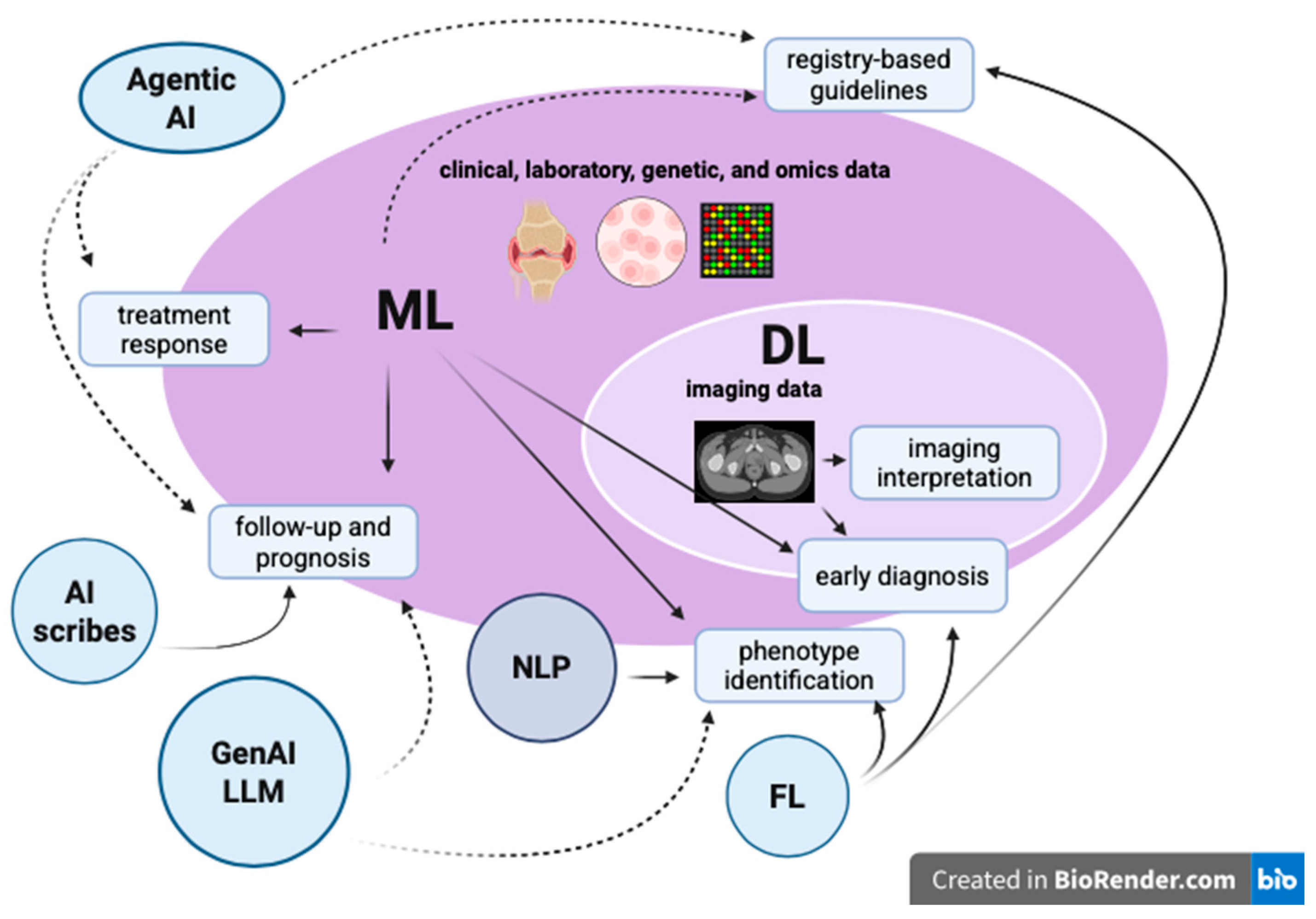
- AI may assist expert panels in formulating and continuously updating guidelines on crucial aspects of RA, ranging from early diagnosis to personalized treatments. In this context, it is important to note that applying AI models to the process of guideline creation—which mostly relies on the Consensus Development and GRADE methods—may result in more detailed and robust data review, drawing from registries, EHRs, and wearable devices. This approach can enhance objectivity, reduce bias, lower costs and time requirements, and enable continuous updates [124]. The growing availability of national real-world registries could promote the application of AI in drawing RA management guidelines from generalized real-world clinical data. Detailed analysis of registry data on recently introduced therapeutic molecules, such as JAKi, could better characterize their efficacy and safety profiles across multiple RA patient cohorts. For instance, data from the GISEA and ToRaRI studies may be analyzed to longitudinally assess efficacy, tolerability, and retention rates, which are essential for the development and validation of future predictive models [125,126]. However, AI models cannot completely replace human judgment, as expert supervision is required to draw final conclusions in the GRADE algorithm.
- (2)
- Another area of great interest is the use of GenAI and large language models (LLMs) [33]. This category includes computational methodologies that do not fall within conventional ML paradigms, such as simulation-based inference, graph-based modeling, and network analysis. Compared to conventional predictive AI models, GenAI not only analyzes existing data or produces classifications but also generates new outputs based on learned patterns [127]. In biomedical research, GenAI can generate new hypotheses, simulate biological scenarios, identify synthetic combinations of biomarkers, and design de novo molecular structures with predefined therapeutic properties [127]. Therefore, these approaches are mainly employed to investigate biological mechanisms, disease pathways, or complex interactions that are difficult to capture using traditional predictive models. Although clinical applications in rheumatology are still in their early stages, GenAI could offer promising perspectives in the management of several diseases, including RA. Specifically, these technologies could potentially aid in identifying novel biomolecular signatures, simulating disease trajectories, integrating multimodal datasets, and generating hypotheses regarding therapeutic response or disease progression [33]. Furthermore, by leveraging omics and imaging data, generative models could support precision medicine approaches by identifying hidden biological patterns and informing the design of future therapeutic strategies. However, the ability to generate biologically plausible outputs does not necessarily ensure clinical validity, and issues such as hallucinations, data dependence, bias, and real-world validation remain significant limitations [127].
- (3)
- An even more advanced and emerging field is Agentic AI. Agentic AI differs from conventional ML and static generative models because it operates as an autonomous decision-making agent, capable not only of making predictions but also of reasoning, planning, using external tools, maintaining contextual memory, and iteratively adjusting its decision-making process [127]. Although not yet validated for use in RA, Agentic AI could theoretically integrate real-time clinical, serological, imaging, genomic, and longitudinal data, continuously reassess disease activity, monitor therapeutic response, and support adaptive treat-to-target strategies through an iterative clinical decision-making process [127].
- (4)
- The development of more robust, multicenter, and generalizable AI models, while addressing the ethical, regulatory, and logistical challenges of sharing sensitive data, could be enabled by Federated Learning (FL). FL is a validated algorithm that combines medical data from multiple centers to improve predictive accuracy and reduce the risk of bias associated with small or highly selected cohorts [128]. As such, FL is emerging as a relevant strategy in fields such as oncology, immunotherapy, and precision medicine [128]. In rheumatology, FL could address challenges associated with relatively small, single-center, and poorly heterogeneous datasets and enhance the robustness of predictive models for early diagnosis, prognostic stratification, and therapeutic response prediction while maintaining patient confidentiality. Looking ahead, FL could also enable the creation of international collaborative AI networks in rheumatology [128]. Data heterogeneity across centers, the need for protocol standardization, computational complexity, risks of distributed bias, and the establishment of appropriate regulatory and governance frameworks represent major obstacles to this approach.
- (5)
- Finally, incorporating ambient AI scribes, which rely on LLMs, into daily clinical routines may reduce physician burnout and work exhaustion, which are largely due to the time spent on EHR documentation [129]. Ambient AI scribes enable the instantaneous transcription of conversations between physicians and patients to create a structured draft clinical note [130]. AI scribes are actively validated and increasingly adopted in rheumatology. The main limitations of this innovative approach are its costs and potential inaccuracies, which require constant human supervision.
6. Conclusions
Author Contributions
Funding
Institutional Review Board Statement
Informed Consent Statement
Data Availability Statement
Conflicts of Interest
Abbreviations
| ACPAs | anti-citrullinated peptide antibodies |
| AI | artificial intelligence |
| ANAs | antinuclear antibodies |
| APC | antigen-presenting cells |
| ASAS HI | assessment of spondyloarthritis international society health index |
| ASAS | assessment of spondyloarthritis international society |
| AUC | area under the ROC curve |
| AUROC | area under the receiver operating characteristic curve |
| axSpA | axial spondyloarthritis |
| BASDAI | Bath ankylosing spondylitis disease activity index |
| BDI | Beck depression inventory |
| bDMARDs | biologic disease-modifying anti-rheumatic drugs |
| BiLSTM | bidirectional long short-term memory |
| BMI | body mass index |
| BMO | bone marrow edema |
| CDU | color Doppler ultrasound |
| CNNs | Convolutional Neural Networks |
| CRP | C-reactive protein |
| csDMARDs | conventional synthetic disease-modifying anti-rheumatic drugs |
| CSF | cerebrospinal fluid |
| CTDs | connective tissue diseases |
| CTG-PAM | cell-tissue-graph-based pathological image analysis model |
| CV | cardiovascular; DL: deep learning |
| DAS28 | disease activity score on 28 joints |
| DL | deep learning |
| DXA | dual-energy X-ray absorptiometry |
| EHR | electronic health record |
| EMR | electronic medical record |
| EMRS | electronic medical records system |
| ePROS | electronic patient-reported outcomes |
| ESR | erythrocyte sedimentation rate |
| EULAR | European League Against Rheumatism |
| FL | Federate Learning |
| FLSs | fibroblast-like synoviocytes |
| FNN | Feed-forward neural network |
| GCA | giant cell arteritis |
| GenAI | generative artificial intelligence |
| GEO | Gene Expression Omnibus |
| GIMS | gout intelligent management system |
| GMLVQ | generalized matrix relevance |
| HLA | allele human leukocyte antigen |
| HR-pQCT | High-Resolution peripheral Quantitative Computed Tomography |
| HRQOL | health-related quality of life; IIM: idiopathic inflammatory myopathy |
| IL | interleukin-1 |
| ILD | interstitial lung disease |
| KD | Kawasaki disease; KLG: Kellgren–Lawrence grade |
| JAKi | Janus kinase inhibitors |
| LASSO | least absolute shrinkage and selection operator |
| LDA | low disease activity |
| LLMs | large language models |
| LN | lupus nephritis |
| MCTD | mixed connective tissue disease |
| ML | machine learning |
| MLS | macrophage-like synoviocytes |
| MMDLS | multimodal DL system |
| MMPs | matrix metalloproteinases |
| MRI | magnetic resonance imaging |
| NLP | natural language processing |
| NLR | neutrophil-to-lymphocyte ratio |
| NPSLE | neuropsychiatric systemic lupus erythematosus |
| NSAIDs | non-steroidal anti-inflammatory drugs |
| OA | osteoarthritis |
| OESS | OMERACT–EULAR Synovitis Scoring system |
| OP | osteoporosis |
| PBMCs | peripheral blood mononuclear cells |
| PD | power Doppler |
| PLR | platelet-to-lymphocyte ratio |
| PROs | patient-reported outcomes |
| PSA | psoriatic arthritis |
| PVNS | pigmented villonodular synovitis |
| RA | rheumatoid arthritis |
| RAPID-3 | Routine Assessment of Patient Index Data 3 |
| RCT | randomized controlled trial |
| RF | rheumatoid factor |
| RMD | rheumatic musculoskeletal disease |
| SEC | secukinumab |
| SHAP | Shapley additive explanation |
| SLE | systemic lupus erythematosus |
| SNP | single nucleotide polymorphism |
| SpA | spondyloarthritis |
| SS | Sjögren’s syndrome |
| SSc | systemic sclerosis |
| TEM | transmission electron microscopy |
| TCZ | tocilizumab |
| Th | T helper |
| TJC | tender joint count |
| TNFi | tumor necrosis factor inhibitors |
| TNF-α | tumor necrosis factor α |
| tsDMARDs | targeted synthetic disease-modifying anti-rheumatic drugs |
| U-Net | U-shaped convolutional neural networks |
| US | ultrasound |
| WOMAC | Western Ontario and McMaster Universities Arthritis Index |
| XAI | explainable artificial intelligence |
| XGBoost | extreme gradient boosting |
References
- Finckh, A.; Gilbert, B.; Hodkinson, B.; Bae, S.C.; Thomas, R.; Deane, K.D.; Alpizar-Rodriguez, D.; Lauper, K. Global Epidemiology of Rheumatoid Arthritis. Nat. Rev. Rheumatol. 2022, 18, 591–602. [Google Scholar] [CrossRef] [Scilit] [PubMed]
- Roudier, J. Association of MHC and Rheumatoid Arthritis Association of RA with HLA-DR4: The Role of Repertoire Selection. Arthritis Res. 2000, 2, 217–220. [Google Scholar] [PubMed]
- Sokolova, M.V.; Schett, G.; Steffen, U. Autoantibodies in Rheumatoid Arthritis: Historical Background and Novel Findings. Clin. Rev. Allergy Immunol. 2022, 63, 138–151. [Google Scholar] [PubMed]
- Jang, S.; Kwon, E.J.; Lee, J.J. Rheumatoid Arthritis: Pathogenic Roles of Diverse Immune Cells. Int. J. Mol. Sci. 2022, 23, 905. [Google Scholar] [CrossRef] [Scilit] [PubMed]
- Misra, D.P. Clinical Manifestations of Rheumatoid Arthritis, Including Comorbidities, Complications, and Long-Term Follow-Up. Best Pract. Res. Clin. Rheumatol. 2025, 39, 102020. [Google Scholar] [CrossRef] [Scilit] [PubMed]
- Wu, D.; Luo, Y.; Li, T.; Zhao, X.; Lv, T.; Fang, G.; Ou, P.; Li, H.; Luo, X.; Huang, A.; et al. Systemic Complications of Rheumatoid Arthritis: Focus on Pathogenesis and Treatment. Front. Immunol. 2022, 13, 1051082. [Google Scholar] [CrossRef] [Scilit] [PubMed]
- Anyfanti, P.; Ainatzoglou, A.; Angeloudi, E.; Michailou, O.; Defteraiou, K.; Bekiari, E.; Kitas, G.D.; Dimitroulas, T. Cardiovascular Risk in Rheumatoid Arthritis: Considerations on Assessment and Management. Mediterr. J. Rheumatol. 2024, 35, 402–410. [Google Scholar] [CrossRef] [Scilit] [PubMed]
- Kay, J.; Upchurch, K.S. ACR/EULAR 2010 Rheumatoid Arthritis Classification Criteria. Rheumatology 2012, 51, vi5–vi9. [Google Scholar] [CrossRef] [Scilit] [PubMed]
- van Steenbergen, H.W.; Doornkamp, F.; Alivernini, S.; Backlund, J.; Codreanu, C.; Cohen, S.B.; Combe, B.; Cope, A.P.; Deane, K.D.; England, B.R.; et al. EULAR/ACR Risk Stratification Criteria for Development of Rheumatoid Arthritis in the Risk Stage of Arthralgia. Ann. Rheum. Dis. 2025, 84, 1445–1457. [Google Scholar] [CrossRef] [Scilit] [PubMed]
- Burmester, G.R.; Pope, J.E. Novel Treatment Strategies in Rheumatoid Arthritis. Lancet 2017, 389, 2338–2348. [Google Scholar] [CrossRef] [Scilit] [PubMed]
- Burgers, L.E.; Raza, K.; Van Der Helm-Van Mil, A.H. Window of Opportunity in Rheumatoid Arthritis-Definitions and Supporting Evidence: From Old to New Perspectives. RMD Open 2019, 5, e000870. [Google Scholar] [CrossRef] [Scilit] [PubMed]
- Smolen, J.S.; Landewé, R.B.M.; Bergstra, S.A.; Kerschbaumer, A.; Sepriano, A.; Aletaha, D.; Caporali, R.; Edwards, C.J.; Hyrich, K.L.; Pope, J.E.; et al. EULAR Recommendations for the Management of Rheumatoid Arthritis with Synthetic and Biological Disease-Modifying Antirheumatic Drugs: 2022 Update. Ann. Rheum. Dis. 2022, 82, 3–18. [Google Scholar] [CrossRef] [Scilit] [PubMed]
- Nagy, G.; Roodenrijs, N.M.T.; Welsing, P.M.J.; Kedves, M.; Hamar, A.; Van Der Goes, M.C.; Kent, A.; Bakkers, M.; Blaas, E.; Senolt, L.; et al. EULAR Definition of Difficult-To-Treat Rheumatoid Arthritis. Ann. Rheum. Dis. 2021, 80, 31–35. [Google Scholar] [CrossRef] [Scilit] [PubMed]
- Van Vollenhoven, R.F. Unresolved Issues in Biologic Therapy for Rheumatoid Arthritis. Nat. Rev. Rheumatol. 2011, 7, 205–215. [Google Scholar] [CrossRef] [Scilit] [PubMed]
- Bécède, M.; Alasti, F.; Gessl, I.; Haupt, L.; Kerschbaumer, A.; Landesmann, U.; Loiskandl, M.; Supp, G.M.; Smolen, J.S.; Aletaha, D. Risk Profiling for a Refractory Course of Rheumatoid Arthritis. Semin. Arthritis Rheum. 2019, 49, 211–217. [Google Scholar] [CrossRef] [Scilit] [PubMed]
- Amisha; Malik, P.; Pathania, M.; Rathaur, V.K. Overview of Artificial Intelligence in Medicine. J. Fam. Med. Prim. Care 2019, 8, 2328–2331. [Google Scholar] [CrossRef] [Scilit] [PubMed]
- Khan, W.H.; Shayan Khan, M.; Khan, N.; Ahmad, A.; Siddiqui, Z.I.; Brojen Singh, R.K.; Zubbair Malik, M. Artificial Intelligence, Machine Learning and Deep Learning in Biomedical Fields: A Prospect in Improvising Medical Healthcare Systems. In Artificial Intelligence in Biomedical and Modern Healthcare Informatics; Elsevier: Amsterdam, The Netherlands, 2024; pp. 55–68. [Google Scholar]
- Mondillo, G.; Colosimo, S.; Perrotta, A.; Frattolillo, V.; Gicchino, M.F. Unveiling Artificial Intelligence’s Power: Precision, Personalization, and Progress in Rheumatology. J. Clin. Med. 2024, 13, 6559. [Google Scholar] [CrossRef] [Scilit] [PubMed]
- Sun, Y.; Lin, J.; Chen, W. Artificial Intelligence in Rheumatoid Arthritis. Rheumatol. Autoimmun. 2025, 5, 88–100. [Google Scholar] [CrossRef] [Scilit]
- Okita, Y.; Hirano, T.; Wang, B.; Nakashima, Y.; Minoda, S.; Nagahara, H.; Kumanogoh, A. Automatic Evaluation of Atlantoaxial Subluxation in Rheumatoid Arthritis by a Deep Learning Model. Arthritis Res. Ther. 2023, 25, 181. [Google Scholar] [CrossRef] [Scilit] [PubMed]
- Choi, R.Y.; Coyner, A.S.; Kalpathy-Cramer, J.; Chiang, M.F.; Peter Campbell, J. Introduction to Machine Learning, Neural Networks, and Deep Learning. Transl. Vis. Sci. Technol. 2020, 9, 14. [Google Scholar] [CrossRef] [Scilit] [PubMed]
- Janiesch, C.; Zschech, P.; Heinrich, K. Machine Learning and Deep Learning. Electron. Mark. 2021, 31, 685–695. [Google Scholar] [CrossRef] [Scilit]
- Kühl, N.; Schemmer, M.; Goutier, M.; Satzger, G. Artificial Intelligence and Machine Learning. Electron. Mark. 2022, 32, 2235–2244. [Google Scholar] [CrossRef] [Scilit]
- Sarker, I.H. Deep Learning: A Comprehensive Overview on Techniques, Taxonomy, Applications and Research Directions. SN Comput. Sci. 2021, 2, 420. [Google Scholar] [CrossRef] [Scilit] [PubMed]
- Zhang, Q.; Lu, J.; Jin, Y. Artificial Intelligence in Recommender Systems. Complex Intell. Syst. 2021, 7, 439–457. [Google Scholar] [CrossRef] [Scilit]
- Morooka, F.E.; Junior, A.M.; Sigahi, T.F.A.C.; Pinto, J.d.S.; Rampasso, I.S.; Anholon, R. Deep Learning and Autonomous Vehicles: Strategic Themes, Applications, and Research Agenda Using SciMAT and Content-Centric Analysis, a Systematic Review. Mach. Learn. Knowl. Extr. 2023, 5, 763–781. [Google Scholar] [CrossRef] [Scilit]
- Hinton, G.; Deng, L.; Yu, D.; Dahl, G.E.; Mohamed, A.; Jaitly, N.; Senior, A.; Vanhoucke, V.; Nguyen, P.; Sainath, T.N.; et al. Deep Neural Networks for Acoustic Modeling in Speech Recognition. IEEE Signal Process. Mag. 2012, 29, 82–97. [Google Scholar] [CrossRef] [Scilit]
- Plested, J.; Phiri, M.; Gedeon, T. Deep Transfer Learning for Image Classification: A Survey. Artif. Intell. Rev. 2026, 59, 100. [Google Scholar] [CrossRef] [Scilit]
- Shrestha, Y.R.; Ben-Menahem, S.M.; von Krogh, G. Organizational Decision-Making Structures in the Age of Artificial Intelligence. Calif. Manag. Rev. 2019, 61, 66–83. [Google Scholar] [CrossRef] [Scilit]
- Alhejaily, A.M.G. Artificial Intelligence in Healthcare (Review). Biomed. Rep. 2025, 22, 11. [Google Scholar] [PubMed]
- Mizna, S.; Arora, S.; Saluja, P.; Das, G.; Alanesi, W.A. An Analytic Research and Review of the Literature on Practice of Artificial Intelligence in Healthcare. Eur. J. Med. Res. 2025, 30, 382. [Google Scholar] [CrossRef] [Scilit] [PubMed]
- Ramos-Ruperto, L.; Mora-Delgado, J.; Rodríguez-González, A.; Sicilia, M.Á.; Pardilla, M.J.; Sempere, J.M.; Puchades, R. Machine Learning and Deep Learning in Internal Medicine: Demystifying Concepts. Rev. Clínica Esp. (Engl. Ed.) 2026, 226, 502412. [Google Scholar] [CrossRef] [Scilit] [PubMed]
- Rabbani, S.A.; El-Tanani, M.; Sharma, S.; Rabbani, S.S.; El-Tanani, Y.; Kumar, R.; Saini, M. Generative Artificial Intelligence in Healthcare: Applications, Implementation Challenges, and Future Directions. BioMedInformatics 2025, 5, 37. [Google Scholar] [CrossRef] [Scilit]
- Takita, H.; Kabata, D.; Walston, S.L.; Tatekawa, H.; Saito, K.; Tsujimoto, Y.; Miki, Y.; Ueda, D. A Systematic Review and Meta-Analysis of Diagnostic Performance Comparison between Generative AI and Physicians. npj Digit. Med. 2025, 8, 175. [Google Scholar] [CrossRef] [Scilit] [PubMed]
- Kingsland, L.C.; Lindberg, D.A.B.; Sharp, G.C. AI/RHEUM—A Consultant System for Rheumatology. J. Med. Syst. 1983, 7, 221–227. [Google Scholar] [CrossRef] [Scilit] [PubMed]
- Porter, J.F.; Kingsland, L.C.; Lindberg, D.A.B.; Shah, I.; Benge, J.M.; Hazelwood, S.E.; Kay, D.R.; Homma, M.; Akizuki, M.; Takano, M.; et al. The Ai/Rheum Knowledge-based Computer Consultant System in Rheumatology. Performance in the Diagnosis of 59 Connective Tissue Disease Patients from Japan. Arthritis Rheum. 1988, 31, 219–226. [Google Scholar] [CrossRef] [Scilit] [PubMed]
- Gossec, L.; Kedra, J.; Servy, H.; Pandit, A.; Stones, S.; Berenbaum, F.; Finckh, A.; Baraliakos, X.; Stamm, T.A.; Gomez-Cabrero, D.; et al. EULAR Points to Consider for the Use of Big Data in Rheumatic and Musculoskeletal Diseases. Ann. Rheum. Dis. 2020, 79, 69–76. [Google Scholar] [CrossRef] [Scilit] [PubMed]
- Kedra, J.; Radstake, T.; Pandit, A.; Baraliakos, X.; Berenbaum, F.; Finckh, A.; Fautrel, B.; Stamm, T.A.; Gomez-Cabrero, D.; Pristipino, C.; et al. Current Status of Use of Big Data and Artificial Intelligence in RMDs: A Systematic Literature Review Informing EULAR Recommendations. RMD Open 2019, 5, e001004. [Google Scholar] [CrossRef] [Scilit] [PubMed]
- Vodenčarević, A.; Brandt-Juergens, J.; Bär, S.; Kästner, P.; Köhm, M.; Simon, D.; Behrens, F.; Glassen, T.; Gmeiner, B.; Peterlik, D.; et al. Predicting Treatment Outcomes in Patients With Psoriatic Arthritis or Axial Spondyloarthritis: An Artificial Intelligence–Driven Approach. J. Rheumatol. 2025, 53, 152–161. [Google Scholar] [CrossRef] [Scilit] [PubMed]
- Fernández-Carballido, C.; Sanchez-Piedra, C.; Valls, R.; Garg, K.; Sánchez-Alonso, F.; Artigas, L.; Mas, J.M.; Jovaní, V.; Manrique, S.; Campos, C.; et al. Female Sex, Age, and Unfavorable Response to Tumor Necrosis Factor Inhibitors in Patients With Axial Spondyloarthritis: Results of Statistical and Artificial Intelligence–Based Data Analyses of a National Multicenter Prospective Registry. Arthritis Care Res. 2023, 75, 115–124. [Google Scholar] [CrossRef] [Scilit] [PubMed]
- Bordner, A.; Aouad, T.; Medina, C.L.; Yang, S.; Molto, A.; Talbot, H.; Dougados, M.; Feydy, A. A Deep Learning Model for the Diagnosis of Sacroiliitis According to Assessment of SpondyloArthritis International Society Classification Criteria with Magnetic Resonance Imaging. Diagn. Interv. Imaging 2023, 104, 373–383. [Google Scholar] [CrossRef] [Scilit] [PubMed]
- Jamaludin, A.; Windsor, R.; Ather, S.; Kadir, T.; Zisserman, A.; Braun, J.; Gensler, L.S.; Østergaard, M.; Poddubnyy, D.; Coroller, T.; et al. Automated Detection of Spinal Bone Marrow Oedema in Axial Spondyloarthritis: Training and Validation Using Two Large Phase 3 Trial Datasets. Rheumatology 2025, 64, 5446–5454. [Google Scholar] [CrossRef] [Scilit] [PubMed]
- Dorfner, F.J.; Vahldiek, J.L.; Donle, L.; Zhukov, A.; Xu, L.; Häntze, H.; Makowski, M.R.; Aerts, H.J.W.L.; Proft, F.; Rodriguez, V.R.; et al. Anatomy-Centred Deep Learning Improves Generalisability and Progression Prediction in Radiographic Sacroiliitis Detection. RMD Open 2024, 10, e004628. [Google Scholar] [CrossRef] [Scilit] [PubMed]
- Li, Q.; Yang, Z.; Chen, K.; Zhao, M.; Long, H.; Deng, Y.; Hu, H.; Jia, C.; Wu, M.; Zhao, Z.; et al. Human-Multimodal Deep Learning Collaboration in ‘Precise’ Diagnosis of Lupus Erythematosus Subtypes and Similar Skin Diseases. J. Eur. Acad. Dermatol. Venereol. 2024, 38, 2268–2279. [Google Scholar] [CrossRef] [Scilit] [PubMed]
- Ali, N.T.; Ali, G.S.; Mohsen Ali, H. NLR Outperforms PLR in SLE Diagnosis and Prognosis: An AI-Enhanced Meta-Analysis of 12 850 Patients with Ethnicity-Specific Cut-Offs. Lupus Sci. Med. 2025, 12, e001696. [Google Scholar] [CrossRef] [Scilit] [PubMed]
- Chen, Z.; Dai, Y.; Chen, Y.; Chen, H.; Wu, H.; Zhang, L. Prediction of Mortality Risk in Critically Ill Patients with Systemic Lupus Erythematosus: A Machine Learning Approach Using the MIMIC-IV Database. Lupus Sci. Med. 2025, 12, e001397. [Google Scholar] [CrossRef] [Scilit] [PubMed]
- Nouroozi, F.; Kazemi, H.S.; Alinezhad, A.; Goudarzi, N.; Khosravi, M.K.; Narimani, Z.; Asouri, Z.A.; Ahari, S.G.; Mehrjerdi, R.S.; Saeidi, R.; et al. Artificial Intelligence–Based Detection of Neuropsychiatric Lupus: An Exploratory Meta-Analysis of Neuroimaging and Multimodal Biomarker Models. Clin. Exp. Med. 2026, 26, 125. [Google Scholar] [CrossRef] [Scilit] [PubMed]
- Stojanowski, J.; Konieczny, A.; Rydzyńska, K.; Kasenberg, I.; Mikołajczak, A.; Gołębiowski, T.; Krajewska, M.; Kusztal, M. Artificial Neural Network—An Effective Tool for Predicting the Lupus Nephritis Outcome. BMC Nephrol. 2022, 23, 381. [Google Scholar] [CrossRef] [Scilit] [PubMed]
- Ma, P.; Li, J.; Zhang, Z.; Qiu, W.; Li, D.; Wang, J.; Li, B.; Guo, S.; Zhang, J.; Cen, Z.; et al. AI-Based System for Analysis of Electron Microscope Images in Glomerular Disease. JAMA Netw. Open 2025, 8, e2534985. [Google Scholar] [CrossRef] [Scilit] [PubMed]
- Wu, R.; Chen, Z.; Yu, J.; Lai, P.; Chen, X.; Han, A.; Xu, M.; Fan, Z.; Cheng, B.; Jiang, Y.; et al. A Graph-Learning Based Model for Automatic Diagnosis of Sjögren’s Syndrome on Digital Pathological Images: A Multicentre Cohort Study. J. Transl. Med. 2024, 22, 748. [Google Scholar] [CrossRef] [Scilit] [PubMed]
- Hoffmann, T.; Teichgräber, U.; Brüheim, L.B.; Lassen-Schmidt, B.; Renz, D.; Weise, T.; Krämer, M.; Oelzner, P.; Böttcher, J.; Güttler, F.; et al. The Association of Symptoms, Pulmonary Function Test and Computed Tomography in Interstitial Lung Disease at the Onset of Connective Tissue Disease: An Observational Study with Artificial Intelligence Analysis of High-Resolution Computed Tomography. Rheumatol. Int. 2025, 45, 194. [Google Scholar] [CrossRef] [Scilit] [PubMed]
- Roncato, C.; Perez, L.; Brochet-Guégan, A.; Allix-Béguec, C.; Raimbeau, A.; Gautier, G.; Agard, C.; Ploton, G.; Moisselin, S.; Lorcerie, F.; et al. Colour Doppler Ultrasound of Temporal Arteries for the Diagnosis of Giant Cell Arteritis: A Multicentre Deep Learning Study. Clin. Exp. Rheumatol. 2020, 38, 120–125. [Google Scholar] [PubMed]
- Yang, L.; Shen, X.; Liu, Y.; Chen, J.; Zou, Y.; Xu, L.; Ji, W.; Zhang, Y.; Liu, T.; Cao, Q. Development and Validation of KCPREDICT: A Deep Learning Model for Early Detection of Coronary Artery Lesions in Kawasaki Disease Patients. Pediatr. Cardiol. 2025, 47, 266–275. [Google Scholar] [CrossRef] [Scilit] [PubMed]
- Li, M.; Zhang, H.; Chen, S.; Zhong, F.; Liu, J.; Wu, J.; Lin, R.; Li, R.; Wu, Y.; Xie, D.; et al. Development and Validation of a Multidimensional and Interpretable Artificial Intelligence Model to Predict Gout Recurrence in Hospitalised Patients: A Real-World, Ambispective Multicentre Cohort Study in China. BMC Med. 2025, 23, 609. [Google Scholar] [CrossRef] [Scilit] [PubMed]
- Thiengwittayaporn, S.; Wattanapreechanon, P.; Sakon, P.; Peethong, A.; Ratisoontorn, N.; Charoenphandhu, N.; Charoensiriwath, S. Development of a Mobile Application to Improve Exercise Accuracy and Quality of Life in Knee Osteoarthritis Patients: A Randomized Controlled Trial. Arch. Orthop. Trauma Surg. 2023, 143, 729–738. [Google Scholar] [CrossRef] [Scilit] [PubMed]
- Thanyakunsajja, N.; Jitkajornwanich, K.; Xu, S.; Shin, D.; Charoenporn, P. Early Diagnosis of Knee Osteoarthritis With a Natural Language Processing–Driven Approach Based on Clinician Notes: Development and Validation Study. JMIR Form. Res. 2025, 9, e64536. [Google Scholar] [CrossRef] [Scilit] [PubMed]
- Du, K.; Li, A.; Zuo, Q.H.; Zhang, C.Y.; Guo, R.; Chen, P.; Du, W.S.; Li, S.M. Comparing Artificial Intelligence–Generated and Clinician-Created Personalized Self-Management Guidance for Patients With Knee Osteoarthritis: Blinded Observational Study. J. Med. Internet Res. 2025, 27, e67830. [Google Scholar] [CrossRef] [Scilit] [PubMed]
- Gürses, Ö.A.; Özüdoğru, A.; Tuncay, F.; Kararti, C. The Role of Artificial Intelligence Large Language Models in Personalized Rehabilitation Programs for Knee Osteoarthritis: An Observational Study. J. Med. Syst. 2025, 49, 73. [Google Scholar] [CrossRef] [Scilit] [PubMed]
- Lin, C.; Tsai, D.J.; Wang, C.C.; Chao, Y.P.; Huang, J.W.; Lin, C.S.; Fang, W.H. Osteoporotic Precise Screening Using Chest Radiography and Artificial Neural Network: The OPSCAN Randomized Controlled Trial. Radiology 2024, 311, e231937. [Google Scholar] [CrossRef] [Scilit] [PubMed]
- Doussiere, M.; Aboud, A.; Dequen, G.; Goëb, V. Artificial Intelligence in Rheumatology: From Algorithms to Clinical Impact in Osteoporosis and Chronic Inflammatory Rheumatic Diseases. J. Clin. Med. 2026, 15, 491. [Google Scholar] [CrossRef] [Scilit] [PubMed]
- Zhu, J.; Yang, F.; Wang, Y.; Wang, Z.; Xiao, Y.; Wang, L.; Sun, L. Accuracy of Machine Learning in Discriminating Kawasaki Disease and Other Febrile Illnesses: Systematic Review and Meta-Analysis. J. Med. Internet Res. 2024, 26, e57641. [Google Scholar] [CrossRef] [Scilit] [PubMed]
- Qi, H.; Lu, J.; Dalbeth, N.; Sun, M.; Liu, Z.; Ji, X.; Ji, A.; Wang, C.; Sun, W.; Li, X.; et al. An Artificial Intelligence-Based Gout Management System Reduced Chronic Kidney Disease Incident and Improved Target Serum Urate Achievement. Rheumatology 2025, 64, 3048–3056. [Google Scholar] [CrossRef] [Scilit] [PubMed]
- Lee, D.W.; Han, H.S.; Ro, D.H.; Lee, Y.S. Development of the Machine Learning Model That Is Highly Validated and Easily Applicable to Predict Radiographic Knee Osteoarthritis Progression. J. Orthop. Res. 2025, 43, 128–138. [Google Scholar] [CrossRef] [Scilit] [PubMed]
- Sun, Y.; Liu, J.; Deng, C.; Peng, C.; Pan, S.; Liu, X. Nomograms Based on X-Ray Radiomics for Predicting Pain Progression in Knee Osteoarthritis Using Data From the Foundation for the National Institutes of Health: Development and Validation Study. JMIR Med. Inform. 2026, 14, e78338. [Google Scholar] [CrossRef] [Scilit] [PubMed]
- Knitza, J.; Tascilar, K.; Fuchs, F.; Mohn, J.; Kuhn, S.; Bohr, D.; Muehlensiepen, F.; Bergmann, C.; Labinsky, H.; Morf, H.; et al. Diagnostic Accuracy of a Mobile AI-Based Symptom Checker and a Web-Based Self-Referral Tool in Rheumatology: Multicenter Randomized Controlled Trial. J. Med. Internet Res. 2024, 26, e55542. [Google Scholar] [CrossRef] [Scilit] [PubMed]
- Moon, J.; Jadhav, P.; Choi, S. Deep Learning Analysis for Rheumatologic Imaging: Current Trends, Future Directions, and the Role of Human. J. Rheum. Dis. 2025, 32, 73–88. [Google Scholar] [CrossRef] [Scilit] [PubMed]
- Ruta, S.; Reginato, A.M.; Pineda, C.; Gutierrez, M. General Applications of Ultrasound in Rheumatology: Why We Need It in Our Daily Practice. J. Clin. Rheumatol. 2015, 21, 133–143. [Google Scholar] [CrossRef] [Scilit] [PubMed]
- Andersen, J.K.H.; Pedersen, J.S.; Laursen, M.S.; Holtz, K.; Grauslund, J.; Savarimuthu, T.R.; Just, S.A. Neural Networks for Automatic Scoring of Arthritis Disease Activity on Ultrasound Images. RMD Open 2019, 5, e000891. [Google Scholar] [CrossRef] [Scilit] [PubMed]
- Chang, C.W.; Chang, C.Y.; Zhu, Y.X.; Wang, S.T. Wrist Joint Synovial Hypertrophy and Effusion Detection in Musculoskeletal Ultrasound Images Using Self-Attention U-Net. Multimed. Tools Appl. 2024, 83, 89317–89334. [Google Scholar] [CrossRef] [Scilit]
- He, X.; Wang, M.; Zhao, C.; Wang, Q.; Zhang, R.; Liu, J.; Zhang, Y.; Qi, Z.; Su, N.; Wei, Y.; et al. Deep Learning-Based Automatic Scoring Models for the Disease Activity of Rheumatoid Arthritis Based on Multimodal Ultrasound Images. Rheumatology 2024, 63, 866–873. [Google Scholar] [CrossRef] [Scilit] [PubMed]
- Wang, Q.; Yao, M.; Song, X.; Liu, Y.; Xing, X.; Chen, Y.; Zhao, F.; Liu, K.; Cheng, X.; Jiang, S.; et al. Automated Segmentation and Classification of Knee Synovitis Based on MRI Using Deep Learning. Acad. Radiol. 2024, 31, 1518–1527. [Google Scholar] [CrossRef] [Scilit] [PubMed]
- Miyama, K.; Bise, R.; Ikemura, S.; Kai, K.; Kanahori, M.; Arisumi, S.; Uchida, T.; Nakashima, Y.; Uchida, S. Deep Learning-Based Automatic-Bone-Destruction-Evaluation System Using Contextual Information from Other Joints. Arthritis Res. Ther. 2022, 24, 227. [Google Scholar] [CrossRef] [Scilit] [PubMed]
- Hirano, T.; Nishide, M.; Nonaka, N.; Seita, J.; Ebina, K.; Sakurada, K.; Kumanogoh, A. Development and Validation of a Deep-Learning Model for Scoring of Radiographic Finger Joint Destruction in Rheumatoid Arthritis. Rheumatol. Adv. Pract. 2019, 3, rkz047. [Google Scholar] [CrossRef] [Scilit] [PubMed]
- Aizenberg, E.; Shamonin, D.P.; Reijnierse, M.; van der Helm-van Mil, A.H.M.; Stoel, B.C. Automatic Quantification of Tenosynovitis on MRI of the Wrist in Patients with Early Arthritis: A Feasibility Study. Eur. Radiol. 2019, 29, 4477–4484. [Google Scholar] [CrossRef] [Scilit] [PubMed]
- Aizenberg, E.; Roex, E.A.H.; Nieuwenhuis, W.P.; Mangnus, L.; van der Helm-van Mil, A.H.M.; Reijnierse, M.; Bloem, J.L.; Lelieveldt, B.P.F.; Stoel, B.C. Automatic Quantification of Bone Marrow Edema on MRI of the Wrist in Patients with Early Arthritis: A Feasibility Study. Magn. Reson. Med. 2018, 79, 1127–1134. [Google Scholar] [CrossRef] [Scilit] [PubMed]
- Gaj Sibaji, C.C.Y.M.N.K.L.X. Automated Synovitis Segmentation in Patients with Rheumatoid Arthritis. In Proceedings of the International Society for Magnetic Resonance in Medicine, Virtual Conference & Exhibition, 8–14 August 2020; p. 28. [Google Scholar]
- Rahimi, F.; Rajaei, E.; Movafagh, N.; Hadianfard, A.M. Identification of Key Factors for Early Detection of Rheumatoid Arthritis in Primary Care Using Machine Learning. Sci. Rep. 2026, 16, 4036. [Google Scholar] [CrossRef] [Scilit] [PubMed]
- Gilvaz, V.J.; Sudheer, A.; Reginato, A.M. Emerging Artificial Intelligence Innovations in Rheumatoid Arthritis and Challenges to Clinical Adoption. Curr. Rheumatol. Rep. 2025, 27, 28. [Google Scholar] [CrossRef] [Scilit] [PubMed]
- Fujii, T.; Murata, K.; Kohjitani, H.; Onishi, A.; Murakami, K.; Tanaka, M.; Yamamoto, W.; Nagai, K.; Yoshikawa, A.; Etani, Y.; et al. Predicting Rheumatoid Arthritis Progression from Seronegative Undifferentiated Arthritis Using Machine Learning: A Deep Learning Model Trained on the KURAMA Cohort and Externally Validated with the ANSWER Cohort. Arthritis Res. Ther. 2025, 27, 65. [Google Scholar] [CrossRef] [Scilit] [PubMed]
- Tang, J.; Jiang, R.; Gao, H.; Xia, J.; Ma, Y.; Han, Z.; Yu, H.; Zhang, Y.; Xie, F.; Sheng, H.; et al. Development and Multi-Center Validation of Machine Learning Models Based on Targeted Metabolomics for Rheumatoid Arthritis. J. Transl. Med. 2025, 23, 1257. [Google Scholar] [CrossRef] [Scilit] [PubMed]
- Maarseveen, T.D.; Meinderink, T.; Reinders, M.J.T.; Knitza, J.; Huizinga, T.W.J.; Kleyer, A.; Simon, D.; van den Akker, E.B.; Knevel, R. Machine Learning Electronic Health Record Identification of Patients with Rheumatoid Arthritis: Algorithm Pipeline Development and Validation Study. JMIR Med. Inform. 2020, 8, e23930. [Google Scholar] [CrossRef] [Scilit] [PubMed]
- Li, Y.; Hassanzadeh, T.; Shamonin, D.P.; Reijnierse, M.; van der Helm-van Mil, A.H.M.; Stoel, B.C. Rheumatoid Arthritis Classification and Prediction by Consistency-Based Deep Learning Using Extremity MRI Scans. Biomed. Signal Process. Control 2024, 91, 105990. [Google Scholar] [CrossRef] [Scilit]
- Manzoor, M.F. Machine Learning for Early Disease Diagnosis: A Review of Techniques in Healthcare Applications. Prem. J. Sci. 2024, 5, 100043. [Google Scholar]
- Perera, J.; Delrosso, C.A.; Nerviani, A.; Pitzalis, C. Clinical Phenotypes, Serological Biomarkers, and Synovial Features Defining Seropositive and Seronegative Rheumatoid Arthritis: A Literature Review. Cells 2024, 13, 743. [Google Scholar] [CrossRef] [Scilit] [PubMed]
- Tang, M.; Guo, Y.; Lü, P.; Wang, B.; Gong, X. Decoding Rheumatoid Arthritis Comorbidities: Molecular Mechanisms and Computational Advances. Curr. Rheumatol. Rev. 2026, 22, e15733971435940. [Google Scholar] [CrossRef] [Scilit] [PubMed]
- Pham, A.N.Q.; Barber, C.E.H.; Drummond, N.; Jasper, L.; Klein, D.; Lindeman, C.; Widdifield, J.; Williamson, T.; Jones, C.A. Development and Validation of a Rheumatoid Arthritis Case Definition: A Machine Learning Approach Using Data from Primary Care Electronic Medical Records. BMC Med. Inform. Decis. Mak. 2024, 24, 360. [Google Scholar] [CrossRef] [Scilit] [PubMed]
- Crowson, C.S.; Gunderson, T.M.; Davis, J.M.; Myasoedova, E.; Kronzer, V.L.; Coffey, C.M.; Atkinson, E.J. Using Unsupervised Machine Learning Methods to Cluster Comorbidities in a Population-Based Cohort of Patients With Rheumatoid Arthritis. Arthritis Care Res. 2023, 75, 210–219. [Google Scholar] [CrossRef] [Scilit] [PubMed]
- Huang, Y.; Agarwal, S.K. Natural Language Processing to Enhance Rheumatoid Arthritis Care in Clinical Studies: A Scoping Review of Applications, Data, Approaches, Challenges and Future Directions. Rheumatol. Int. 2026, 46, 164. [Google Scholar] [CrossRef] [Scilit] [PubMed]
- Román Ivorra, J.A.; Trallero-Araguas, E.; Lopez Lasanta, M.; Cebrián, L.; Lojo, L.; López-Muñíz, B.; Fernández-Melon, J.; Núñez, B.; Silva-Fernández, L.; Veiga Cabello, R.; et al. Prevalence and Clinical Characteristics of Patients with Rheumatoid Arthritis with Interstitial Lung Disease Using Unstructured Healthcare Data and Machine Learning. RMD Open 2024, 10, e003353. [Google Scholar] [CrossRef] [Scilit] [PubMed]
- Qiu, J.; Xu, Y.; Tong, L.; Yang, X.; Wu, X. Identification of Potential Pathogenic Genes Associated with the Comorbidity of Rheumatoid Arthritis and Renal Fibrosis Using Bioinformatics and Machine Learning. Sci. Rep. 2025, 15, 21686. [Google Scholar] [CrossRef] [Scilit] [PubMed]
- Heng, H.; Li, D.; Su, W.; Liu, X.; Yu, D.; Bian, Z.; Li, J. Exploration of Comorbidity Mechanisms and Potential Therapeutic Targets of Rheumatoid Arthritis and Pigmented Villonodular Synovitis Using Machine Learning and Bioinformatics Analysis. Front. Genet. 2023, 13, 1095058. [Google Scholar] [CrossRef] [Scilit] [PubMed]
- Orange, D.E.; Agius, P.; DiCarlo, E.F.; Robine, N.; Geiger, H.; Szymonifka, J.; McNamara, M.; Cummings, R.; Andersen, K.M.; Mirza, S.; et al. Identification of Three Rheumatoid Arthritis Disease Subtypes by Machine Learning Integration of Synovial Histologic Features and RNA Sequencing Data. Arthritis Rheumatol. 2018, 70, 690–701. [Google Scholar] [CrossRef] [Scilit] [PubMed]
- Konzett, V.; Laskou, F.; Smolen, J.S.; Edwards, C.J.; Aletaha, D.; van der Heijde, D.; Winthrop, K.L.; Takeuchi, T.; Caporali, R.; Verschueren, P.; et al. Efficacy of Synthetic and Biological DMARDs: A Systematic Literature Review Informing the 2025 Update of the EULAR Recommendations for the Management of Rheumatoid Arthritis. Ann. Rheum. Dis. 2026, 85, 1039–1054. [Google Scholar] [CrossRef] [Scilit] [PubMed]
- Taylor, P.C.; Matucci Cerinic, M.; Alten, R.; Avouac, J.; Westhovens, R. Managing Inadequate Response to Initial Anti-TNF Therapy in Rheumatoid Arthritis: Optimising Treatment Outcomes. Ther. Adv. Musculoskelet. Dis. 2022, 14, 1–14. [Google Scholar] [CrossRef] [Scilit]
- Bouget, V.; Duquesne, J.; Hassler, S.; Cournède, P.H.; Fautrel, B.; Guillemin, F.; Pallardy, M.; Broët, P.; Mariette, X.; Bitoun, S. Machine Learning Predicts Response to TNF Inhibitors in Rheumatoid Arthritis: Results on the ESPOIR and ABIRISK Cohorts. RMD Open 2022, 8, e002442. [Google Scholar] [CrossRef] [Scilit] [PubMed]
- Lim, L.J.; Lim, A.J.W.; Ooi, B.N.S.; Tan, J.W.L.; Koh, E.T.; Chong, S.S.; Khor, C.C.; Tucker-Kellogg, L.; Lee, C.G.; Leong, K.P. Machine Learning Using Genetic and Clinical Data Identifies a Signature That Robustly Predicts Methotrexate Response in Rheumatoid Arthritis. Rheumatology 2022, 61, 4175–4186. [Google Scholar] [CrossRef] [Scilit] [PubMed]
- Duquesne, J.; Bouget, V.; Cournède, P.H.; Fautrel, B.; Guillemin, F.; De Jong, P.H.P.; Heutz, J.W.; Verstappen, M.; Van Der Helm-Van Mil, A.H.M.; Mariette, X.; et al. Machine Learning Identifies a Profile of Inadequate Responder to Methotrexate in Rheumatoid Arthritis. Rheumatology 2023, 62, 2402–2409. [Google Scholar] [CrossRef] [Scilit] [PubMed]
- Salehi, F.; Lopera Gonzalez, L.I.; Bayat, S.; Kleyer, A.; Zanca, D.; Brost, A.; Schett, G.; Eskofier, B.M. Machine Learning Prediction of Treatment Response to Biological Disease-Modifying Antirheumatic Drugs in Rheumatoid Arthritis. J. Clin. Med. 2024, 13, 3890. [Google Scholar] [CrossRef] [Scilit] [PubMed]
- Chen, S.; Gupta, N.; Galbraith, W.B.; Shah, V.; Cirrone, J. Prediction of Drug Effectiveness in Rheumatoid Arthritis Patients Based on Machine Learning Algorithms. In Proceedings of the ACM International Conference Proceeding Series; Association for Computing Machinery: New York, NY, USA, 2022; pp. 147–154. [Google Scholar]
- Guan, Y.; Zhang, H.; Quang, D.; Wang, Z.; Parker, S.C.J.; Pappas, D.A.; Kremer, J.M.; Zhu, F. Machine Learning to Predict Anti–Tumor Necrosis Factor Drug Responses of Rheumatoid Arthritis Patients by Integrating Clinical and Genetic Markers. Arthritis Rheumatol. 2019, 71, 1987–1996. [Google Scholar] [CrossRef] [Scilit] [PubMed]
- Tao, W.; Concepcion, A.N.; Vianen, M.; Marijnissen, A.C.A.; Lafeber, F.P.G.J.; Radstake, T.R.D.J.; Pandit, A. Multiomics and Machine Learning Accurately Predict Clinical Response to Adalimumab and Etanercept Therapy in Patients With Rheumatoid Arthritis. Arthritis Rheumatol. 2021, 73, 212–222. [Google Scholar] [CrossRef] [Scilit] [PubMed]
- Johansson, F.D.; Collins, J.E.; Yau, V.; Guan, H.; Kim, S.C.; Losina, E.; Sontag, D.; Stratton, J.; Trinh, H.; Greenberg, J.; et al. Predicting Response to Tocilizumab Monotherapy in Rheumatoid Arthritis: A Real-World Data Analysis Using Machine Learning. J. Rheumatol. 2021, 48, 1364–1370. [Google Scholar] [CrossRef] [Scilit] [PubMed]
- Lee, Y.J.; Choi, G.; Yeo, J.; Baek, J.; Choi, H.; Kim, M.; Kim, Y.G.; Kim, B.Y.; Koo, J. Machine Learning-Based Prediction of Response to Janus Kinase Inhibitors in Patients with Rheumatoid Arthritis Using Clinical Data. Front. Immunol. 2025, 16, 1689144. [Google Scholar] [CrossRef] [Scilit] [PubMed]
- Hetland, M.L.; Strangfeld, A.; Bonfanti, G.; Soudis, D.; Deuring, J.J.; Edwards, R.A. Machine Learning Prediction and Explanatory Models of Serious Infections in Patients with Rheumatoid Arthritis Treated with Tofacitinib. Arthritis Res. Ther. 2024, 26, 153. [Google Scholar] [CrossRef] [Scilit] [PubMed]
- Huang, Y.; Bazzazzadehgan, S.; Maharjan, S.; Lin, Y.; Bentley, J.P.; Agarwal, S.K.; Yang, Y. Predicting the Risk of Venous Thromboembolism Events in Older Adults with Rheumatoid Arthritis after Initiating Targeted Disease-Modifying Antirheumatic Drugs: A Comparison of the Random Survival Forest and Regularized Cox Regression Models. Curr. Med. Res. Opin. 2026, 42, 615–630. [Google Scholar] [CrossRef] [Scilit] [PubMed]
- Eriakha, E.B.; Han, Y.; Li, M.; Li, J.; Huang, Y. Machine Learning for Predicting Treatment Response to Biologic and Targeted Synthetic Disease-Modifying Antirheumatic Drugs in Rheumatoid Arthritis: A Scoping Review. BMC Rheumatol. 2025, 9, 132. [Google Scholar] [CrossRef] [Scilit] [PubMed]
- Benavent, D.; Carmona, L.; García Llorente, J.F.; Montoro, M.; Ramirez, S.; Otón, T.; Loza, E.; Gómez-Centeno, A. Artificial Intelligence to Predict Treatment Response in Rheumatoid Arthritis and Spondyloarthritis: A Scoping Review. Rheumatol. Int. 2025, 45, 91. [Google Scholar] [CrossRef] [Scilit] [PubMed]
- Maldonado-Cañón, K.; Coral-Alvarado, P.; Méndez-Patarroyo, P.; Bautista-Molano, W.; Quintana-López, G. ANAs and Triple Positivity Effect on Disease Activity and Sustained Remission in Rheumatoid Arthritis: A Retrospective Real-World Machine Learning Approach. Clin. Rheumatol. 2025, 44, 4895–4907. [Google Scholar] [CrossRef] [Scilit] [PubMed]
- Matsuo, H.; Kamada, M.; Imamura, A.; Shimizu, M.; Inagaki, M.; Tsuji, Y.; Hashimoto, M.; Tanaka, M.; Ito, H.; Fujii, Y. Machine Learning-Based Prediction of Relapse in Rheumatoid Arthritis Patients Using Data on Ultrasound Examination and Blood Test. Sci. Rep. 2022, 12, 7224. [Google Scholar] [CrossRef] [Scilit] [PubMed]
- Baxter, N.B.; Lin, C.; Wallace, B.I.; Chen, J.; Kuo, C.; Chung, K.C. Development of a Machine Learning Model to Predict the Use of Surgery in Patients With Rheumatoid Arthritis. Arthritis Care Res. 2024, 76, 636–643. [Google Scholar] [CrossRef] [Scilit] [PubMed]
- Yeo, L.; Adlard, N.; Biehl, M.; Juarez, M.; Smallie, T.; Snow, M.; Buckley, C.D.; Raza, K.; Filer, A.; Scheel-Toellner, D. Expression of Chemokines CXCL4 and CXCL7 by Synovial Macrophages Defines an Early Stage of Rheumatoid Arthritis. Ann. Rheum. Dis. 2016, 75, 763–771. [Google Scholar] [CrossRef] [Scilit] [PubMed]
- Creagh, A.P.; Hamy, V.; Yuan, H.; Mertes, G.; Tomlinson, R.; Chen, W.H.; Williams, R.; Llop, C.; Yee, C.; Duh, M.S.; et al. Digital Health Technologies and Machine Learning Augment Patient Reported Outcomes to Remotely Characterise Rheumatoid Arthritis. npj Digit. Med. 2024, 7, 33. [Google Scholar] [CrossRef] [Scilit] [PubMed]
- Zhang, Z.; Zhang, X.; Zhang, L.; Du, M.; Zhang, L.; Yang, S.; Cai, X.; Hou, S. An Intelligent Interactive Management Platform for Rheumatoid Arthritis Care: Real-World Observational Study. JMIR Med. Inform. 2026, 14, e90784. [Google Scholar] [CrossRef] [Scilit] [PubMed]
- Perronne, L.; Binvignat, M.; Foulquier, N.; Saraux, A.; Laredo, J.D.; de Margerie-Mellon, C.; Fournier, L.; Sellam, J. Algorithmic Approaches in Hand Imaging for Rheumatic Musculoskeletal Diseases: A Systematic Literature Review. Semin. Arthritis Rheum. 2025, 73, 152750. [Google Scholar] [CrossRef] [Scilit] [PubMed]
- Danieli, M.G.; Brunetto, S.; Gammeri, L.; Palmeri, D.; Claudi, I.; Shoenfeld, Y.; Gangemi, S. Machine Learning Application in Autoimmune Diseases: State of Art and Future Prospectives. Autoimmun. Rev. 2024, 23, 103496. [Google Scholar] [CrossRef] [Scilit] [PubMed]
- Rajkomar, A.; Dean, J.; Kohane, I. Machine Learning in Medicine. N. Engl. J. Med. 2019, 380, 1347–1358. [Google Scholar] [CrossRef] [Scilit] [PubMed]
- Kreimeyer, K.; Foster, M.; Pandey, A.; Arya, N.; Halford, G.; Jones, S.F.; Forshee, R.; Walderhaug, M.; Botsis, T. Natural Language Processing Systems for Capturing and Standardizing Unstructured Clinical Information: A Systematic Review. J. Biomed. Inform. 2017, 73, 14–29. [Google Scholar] [CrossRef] [Scilit] [PubMed]
- Topol, E.J. High-Performance Medicine: The Convergence of Human and Artificial Intelligence. Nat. Med. 2019, 25, 44–56. [Google Scholar] [CrossRef] [Scilit] [PubMed]
- Shen, D.; Wu, G.; Suk, H.-I. Deep Learning in Medical Image Analysis. Annu. Rev. Biomed. Eng. 2017, 19, 221–248. [Google Scholar] [CrossRef] [Scilit] [PubMed]
- Litjens, G.; Kooi, T.; Bejnordi, B.E.; Setio, A.A.A.; Ciompi, F.; Ghafoorian, M.; van der Laak, J.A.W.M.; van Ginneken, B.; Sánchez, C.I. A Survey on Deep Learning in Medical Image Analysis. Med. Image Anal. 2017, 42, 60–88. [Google Scholar] [CrossRef] [Scilit] [PubMed]
- Ma, J.; Yu, J.; Xie, A.; Huang, T.; Liu, W.; Ma, M.; Tao, Y.; Zang, F.; Zheng, Q.; Zhu, W.; et al. Large Language Model Evaluation in Autoimmune Disease Clinical Questions Comparing ChatGPT 4o, Claude 3.5 Sonnet and Gemini 1.5 Pro. Sci. Rep. 2025, 15, 17635. [Google Scholar] [CrossRef] [Scilit] [PubMed]
- Roemer, A.; Schlicker, N.; Kernder, A.; Albe, B.; Hack, J.; Hirsch, M.; Mayr, A.; Kuhn, S.; Knitza, J. Large Language Models Enhance Diagnostic Reasoning of Medical Students in Rheumatology: A Randomized Controlled Trial. BMC Med. Educ. 2026, 26, 579. [Google Scholar] [CrossRef] [Scilit] [PubMed]
- Nagendran, M.; Chen, Y.; Lovejoy, C.A.; Gordon, A.C.; Komorowski, M.; Harvey, H.; Topol, E.J.; Ioannidis, J.P.A.; Collins, G.S.; Maruthappu, M. Artificial Intelligence versus Clinicians: Systematic Review of Design, Reporting Standards, and Claims of Deep Learning Studies in Medical Imaging. BMJ 2020, 368, m689. [Google Scholar] [CrossRef] [Scilit] [PubMed]
- Gangemi, S.; Allegra, A.; Di Gioacchino, M.; Gammeri, L.; Cacciola, I.; Canonica, G.W. The Innovative Potential of Artificial Intelligence Applied to Patient Registries to Implement Clinical Guidelines. Mach. Learn. Knowl. Extr. 2026, 8, 38. [Google Scholar] [CrossRef] [Scilit]
- D’Alessandro, F.; Cazzato, M.; Laurino, E.; Morganti, R.; Bardelli, M.; Frediani, B.; Buongarzone, C.; Moroncini, G.; Guiducci, S.; Cometi, L.; et al. ToRaRI (Tofacitinib in Rheumatoid Arthritis a Real-Life Experience in Italy): Effectiveness, Safety Profile of Tofacitinib and Concordance between Patient-Reported Outcomes and Physician’s Global Assessment of Disease Activity in a Retrospective Study in Central-Italy. Clin. Rheumatol. 2024, 43, 657–665. [Google Scholar] [CrossRef] [Scilit] [PubMed]
- Fornaro, M.; Caporali, R.; Biggioggero, M.; Bugatti, S.; De Stefano, L.; Cauli, A.; Congia, M.; Conti, F.; Chimenti, M.S.; Bazzani, C.; et al. Effectiveness and Safety of Filgotinib in Rheumatoid Arthritis Patients: Data from the GISEA Registry. Clin. Exp. Rheumatol. 2024, 42, 1043–1050. [Google Scholar] [CrossRef] [Scilit] [PubMed]
- Niazi, S.K. Artificial Intelligence in Small-Molecule Drug Discovery: A Critical Review of Methods, Applications, and Real-World Outcomes. Pharmaceuticals 2025, 18, 1271. [Google Scholar] [CrossRef] [Scilit]
- Alshorman, J.; Mehran, M.J.; Bahrami, Y.; Mohammadzadeh, S.; Barzigar, R.; Morshedi, M.; Haider, K.H.; Tembo, K.M.; Rong, S.-J.; Jadgal, N.; et al. Artificial Intelligence in Immunotherapy: Revolutionizing Diagnostic and Therapeutic Applications in Cancer and Autoimmune Diseases. Clin. Exp. Med. 2026, 26, 185. [Google Scholar] [CrossRef] [Scilit] [PubMed]
- Lukac, P.J.; Turner, W.; Vangala, S.; Chin, A.T.; Khalili, J.; Shih, Y.-C.T.; Sarkisian, C.; Cheng, E.M.; Mafi, J.N. Ambient AI Scribes in Clinical Practice: A Randomized Trial. NEJM AI 2025, 2, AIoa2501000. [Google Scholar] [CrossRef] [Scilit] [PubMed]
- Shah, S.J.; Crowell, T.; Jeong, Y.; Devon-Sand, A.; Smith, M.; Yang, B.; Ma, S.P.; Liang, A.S.; Delahaie, C.; Hsia, C.; et al. Physician Perspectives on Ambient AI Scribes. JAMA Netw. Open 2025, 8, e251904. [Google Scholar] [CrossRef] [Scilit] [PubMed]


| Domain | Category | Description |
|---|---|---|
| Data type | Clinical | Studies primarily based on clinical, laboratory, anamnestic, genetic, biomarker, questionnaire, EHR/EMR, or patient-reported outcome data, without direct use of medical imaging. |
| Instrumental | Studies mainly using imaging or instrumental diagnostic data, including radiographs, MRI, ultrasound, thermography, HR-pQCT, scintigraphy, smartphone imaging, or other digital sensor-based modalities. | |
| Clinical + Instrumental | Studies integrating both clinical and instrumental/imaging data within the same analytical workflow or predictive model. | |
| Task | Diagnostic | Studies focused on disease identification, classification, screening, early diagnosis, or severity assessment of RA and related rheumatic diseases using computational models. |
| Predictive | Studies aimed at predicting disease risk, clinical outcomes, progression, remission, flare occurrence, or therapeutic response. | |
| Clinical Decision Support and Monitoring | Studies dedicated to clinical decision support, patient monitoring, workflow optimization, healthcare data management, or digital rehabilitation systems. | |
| Drug Discovery and Molecular Modeling | Studies involving biomarker discovery, therapeutic target identification, molecular mechanism analysis, drug repurposing, pharmacological screening, or computational biological simulations. | |
| Healthcare System and Infrastructure | Studies related to digital health platforms, datasets, telemedicine systems, healthcare infrastructures, methodological frameworks, or healthcare organization. | |
| AI algorithm | Traditional Machine Learning | Conventional machine learning models, including Random Forest, support vector machine, XGBoost, logistic regression, k-nearest neighbors, Decision Trees, and classical ensemble methods. |
| Deep Learning | Deep neural network architectures, including CNNs, DenseNet, ResNet, U-Net, transformer-based vision models, and other deep learning approaches. | |
| NLP/Generative AI | Natural language processing and generative AI methods, including text mining, knowledge extraction, BERT-based models, LLMs, and text-generative systems. | |
| Other Computational Approaches | Advanced computational methods not directly classifiable into previous categories, including graph-based modeling, simulation-based inference, fuzzy systems, optimization algorithms, and complex statistical-computational frameworks. |
Disclaimer/Publisher’s Note: The statements, opinions and data contained in all publications are solely those of the individual author(s) and contributor(s) and not of MDPI and/or the editor(s). MDPI and/or the editor(s) disclaim responsibility for any injury to people or property resulting from any ideas, methods, instructions or products referred to in the content. |
© 2026 by the authors. Licensee MDPI, Basel, Switzerland. This article is an open access article distributed under the terms and conditions of the Creative Commons Attribution (CC BY) license.
Share and Cite
Talotta, R.; Sfravara, F.; Fiorentino, F.; Barberi, E.; Gangemi, S. Use of Artificial Intelligence in Rheumatoid Arthritis: Advancements and Novel Perspectives. J. Clin. Med. 2026, 15, 5482. https://doi.org/10.3390/jcm15145482
Talotta R, Sfravara F, Fiorentino F, Barberi E, Gangemi S. Use of Artificial Intelligence in Rheumatoid Arthritis: Advancements and Novel Perspectives. Journal of Clinical Medicine. 2026; 15(14):5482. https://doi.org/10.3390/jcm15145482
Chicago/Turabian StyleTalotta, Rossella, Felice Sfravara, Filippo Fiorentino, Emmanuele Barberi, and Sebastiano Gangemi. 2026. "Use of Artificial Intelligence in Rheumatoid Arthritis: Advancements and Novel Perspectives" Journal of Clinical Medicine 15, no. 14: 5482. https://doi.org/10.3390/jcm15145482
APA StyleTalotta, R., Sfravara, F., Fiorentino, F., Barberi, E., & Gangemi, S. (2026). Use of Artificial Intelligence in Rheumatoid Arthritis: Advancements and Novel Perspectives. Journal of Clinical Medicine, 15(14), 5482. https://doi.org/10.3390/jcm15145482