1. Introduction
Why we have a nasal septal deviation is not clear. Acquired and congenital factors that contribute to the development of nasal septal deviation, such as trauma and craniofacial anomalies, are well-studied [
1,
2,
3,
4,
5,
6,
7,
8,
9,
10]. However, these factors alone cannot explain the high incidence of nasal septal deviations. This is important because nasal septal deviation is among the leading causes of nasal obstruction, along with enlarged adenoids, concha bullosa, and inferior turbinate hypertrophy, all of which may lead to mouth breathing. Chronic mouth breathing over an extended period can increase the constricting forces on the maxilla, thereby affecting its transverse and vertical growth [
11,
12,
13]. Further maxillary constriction can cause occlusal disharmony, narrowing of the pharyngeal airway, alterations in tongue posture resulting in retroglossal airway narrowing, and potentially contribute to obstructive sleep apnea [
14,
15,
16,
17,
18,
19].
Previous studies have shown that nasal septal deviation is rare among long-snouted mammals and is more common in mammals with shorter faces [
3,
20]. Similarly, from an evolutionary point of view, it has been suggested that humans may have developed a high incidence of nasal septal deviation as a result of the continued diminution of our face [
20,
21]. It has also been reported that individuals with a more retrognathic nasomaxillary complex exhibit a greater magnitude of septal deviation compared to individuals with a more mesognathic midface [
9]. In addition, nasal septal deviation is more common in individuals with reduced facial height [
22]. Similarly, experimental reduction in the maxillary length through surgery restricts normal septal growth and induces septal deviation [
23]. These observations suggest a possible developmental etiology for nasal septal deviation, possibly due to a constricted maxilla. Based on this theory, another factor that may cause deviations is when cartilage volume exceeds the available space for the cartilage due to a decrease in the maxilla’s transverse dimension. This suggests reciprocal effects between the nasal septum and maxilla; a constricted maxilla produces nasal septal deviation, and in turn, nasal septal deviation through mouth breathing worsens the maxillary constriction.
However, studies investigating maxillary transverse deficiency as a developmental factor in nasal septal deviation are rare and indirect, focusing mainly on associations between nasal septal deviation and maxillary constriction [
24,
25,
26,
27].
Here, we further investigate the effect of maxillary constriction on the development of nasal septum deviation in growing rats, and the mechanism by which maxillary constriction may contribute to nasal septal deviation. Considering that the incidence of maxillary constriction is increasing in humans [
3,
28,
29,
30,
31,
32,
33,
34,
35,
36,
37], establishing a relationship between maxillary constriction and nasal septal deviation development may have significant health implications.
2. Materials and Methods
2.1. Animal Study
Growing Wistar rats (n = 60, average weight 47 g, 21 days old) were studied at two centers: Constriction experiments were completed at Universidad Nacional Autónoma de México (UNAM), while the Expansion experiments were completed at New York University, All experiments were approved by the Institutional Animal Care and Research Advisory Committee (CICUAL, ID 10389, approved 9 August 2024), from Universidad Nacional Autónoma de México (UNAM), and the National Research Council’s Guide for the Care and Use of Laboratory Animals from New York University (IACUC Protocol # 151003, approved 9 September 2018). Although using 2 centers was not ideal, to avoid introducing variability, the same investigator fabricated, calibrated, and delivered the constriction and expansion appliances at both centers. Collected specimens were fixed and then transferred to the same facility for analysis consistency. Therefore, all specimen analyses were conducted at the same imaging facility, using the same microCT equipment (µCT; Skyscan1172; Bruker microCT, Kontich, Belgium), and measurements were completed by the same investigators.
Rats at this stage are in a growing phase, and their molars are fully erupted. All animals were housed in polycarbonate cages in a 12 h light/dark environment at a controlled temperature of 21–23 °C and relative humidity of 40–60%. Animals were housed in an individually ventilated rack system and fed a specially prepared soft diet with tap water ad libitum. The mixture consisted of 200 g of standard chow (Teklad, Envigo, Indianapolis, Indiana, IN, USA), 600 mL of water, and 45 g of gelatin, which was homogenized and subsequently molded into bars to facilitate consumption by the rats. This diet was used to ensure adequate nutritional intake while minimizing the possibility of dislodging the appliance.
For randomization, the animals were numbered 1 to 60, and 15 random numbers were subsequently generated for each group, using the standard = RAND() function in Microsoft Excel. The four groups were: (1) an untreated Control, (2) a Sham Group, (3) Experimental Group 1 (Constriction), and (4) Experimental Group 2 (Expansion). Allocation was not concealed, as the effects of constriction or expansion could be observed during daily assessment of appliance integrity.
2.2. Transverse Force Application
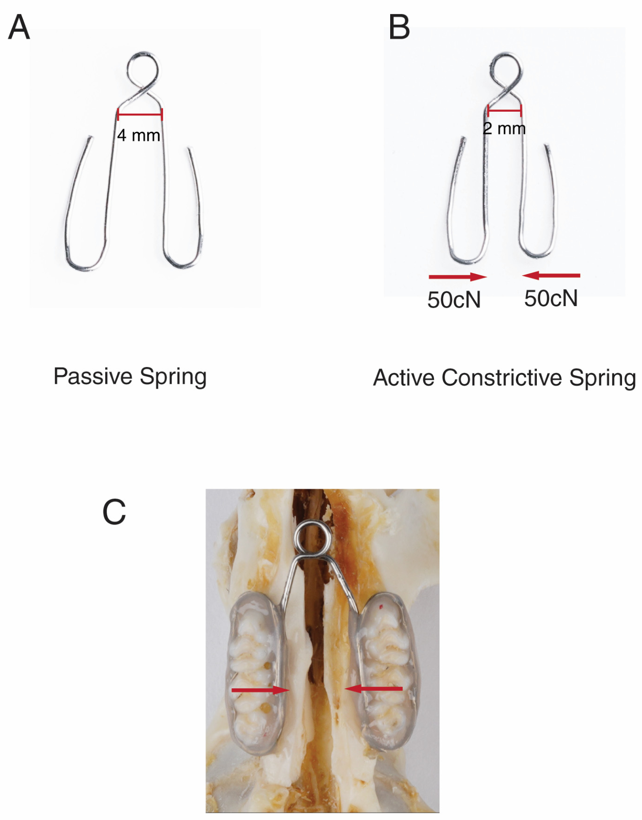
On day 0, animals from the Sham and Experimental Groups were sedated with isoflurane delivered through the SomnoSuite anesthesia system (Kent Scientific, Torrington, CT, USA). Isoflurane was administered at 1% during the initial sedation phase, increased to 3% to achieve deep anesthesia, and subsequently reduced to 0.5% for maintenance. Anesthesia was verified by lack of response to toe-pinch. Springs were fabricated from 0.016″ stainless steel wires (3M Unitek, Monrovia, CA, USA). The Experimental Groups received a calibrated, custom-designed constriction or expansion spring that delivered compression or tensile transverse force (100cN) to the molars (
Figure 1). This force was selected based on previous studies demonstrating that high cellular activity was observed in the suture at 100cN [
38]. Considering that rats would have a normal vertical chewing force, averaging between 54 and 75 N, this force is not regarded as excessive [
39]. The springs were fabricated, calibrated, and delivered by the same investigator at both centers. While rats were sedated, the springs were cemented with composite resin on the interproximal and occlusal surfaces of the molars by the same investigator. The Sham group received a similar spring, but it was not activated and produced no forces. Compressive or tensile forces were applied for 28 days. Animals were monitored daily under inhalational anesthesia with isoflurane–nitrous oxide as the standard method of general anesthesia to ensure the integrity of the appliance. No appliances were dislodged or needed to be reinstalled. Therefore, all animals were included in the analysis and final results.
2.3. Micro-CT Imaging
Animals were euthanized by CO2 narcosis on day 28, and samples were collected for micro-CT analysis (μCT). After euthanasia, the whole skull was dissected and fixed for over 72 h with 4% (w/v) paraformaldehyde in 0.1 M phosphate buffer, pH 7.4, followed by storage in 70% ethanol.
The whole maxilla was scanned by micro-computed tomography (µCT; Skyscan1172; Bruker microCT, Kontich, Belgium). The specimens were scanned at 13.55 µm voxel size, 100 KV, 0.300 degrees rotation step (192.30 degrees angular range), and a 1910 ms exposure per view in 70% ETOH. Results were analyzed utilizing the NRecon (1.6.9.16) software on the HP open platform (OpenVMS Alpha Version 1.3-1 session manager) for 3D reconstruction and viewing of images. Superimpositions were performed using Amira 6.0.0.
2.4. Intra-Oral and Intra-Nasal Measurements
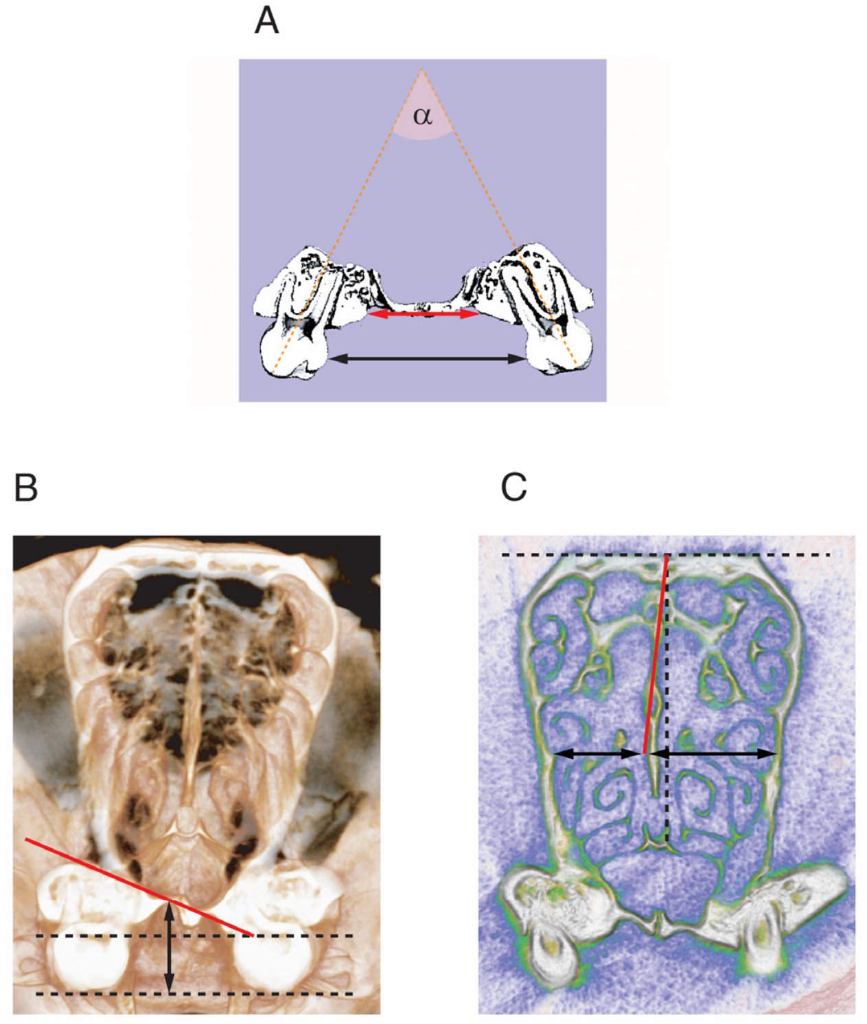
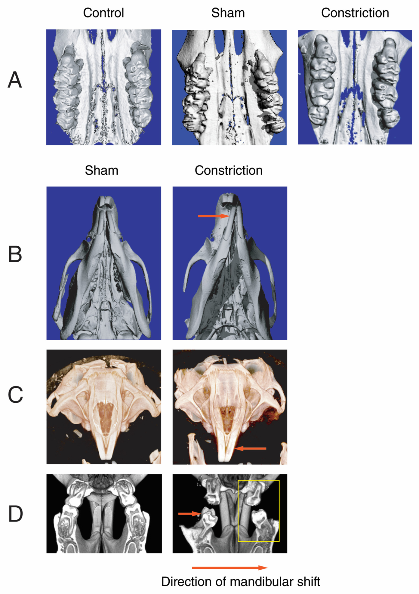
Palatal width (at the level of intersection between alveolar bone and palatal walls) and interdental width (the distance between the height of contour of the first molars) were analyzed in the mid-coronal plane at the level of the upper first molar from the three-dimensional images of µCT (
Figure 2A). Changes in the upper molar inclination were studied by measuring the angle (a) formed by the long axes of the upper right and left first molars in the same plane.
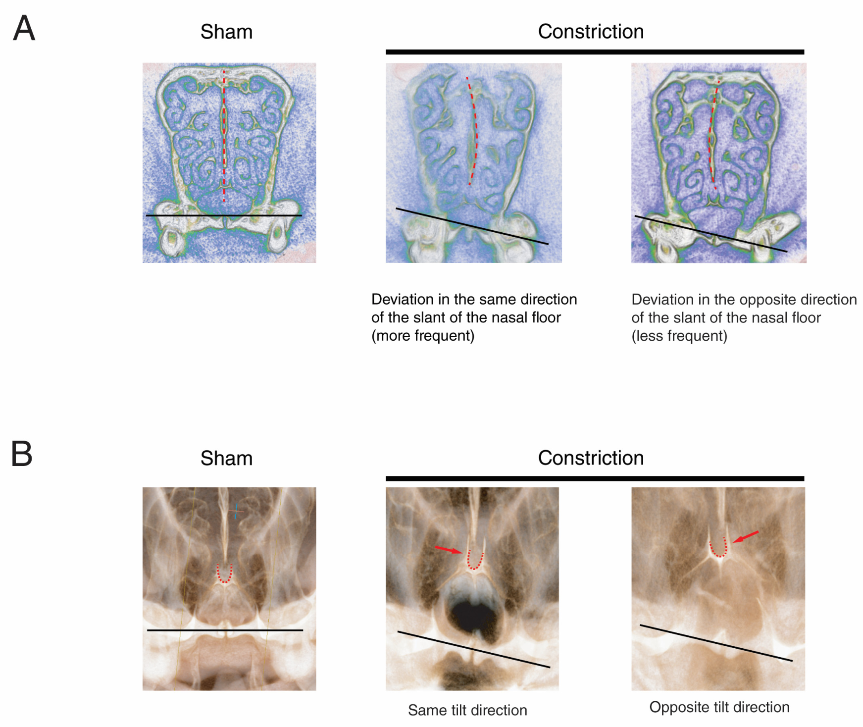
The slant of the nasal floor was measured on the coronal plane of the µCT image, specifically in the area where the slant was most pronounced. One line was drawn tangent to the nasal floor and parallel to the orbits, and another line was drawn along the steepest slant of the nasal floor. The angle between these lines was measured and recorded as the slant of the nasal floor (
Figure 2B). On the same µCT image, the palatal height was measured as the perpendicular distance from the highest point on the palate to the occlusal plane (
Figure 2B).
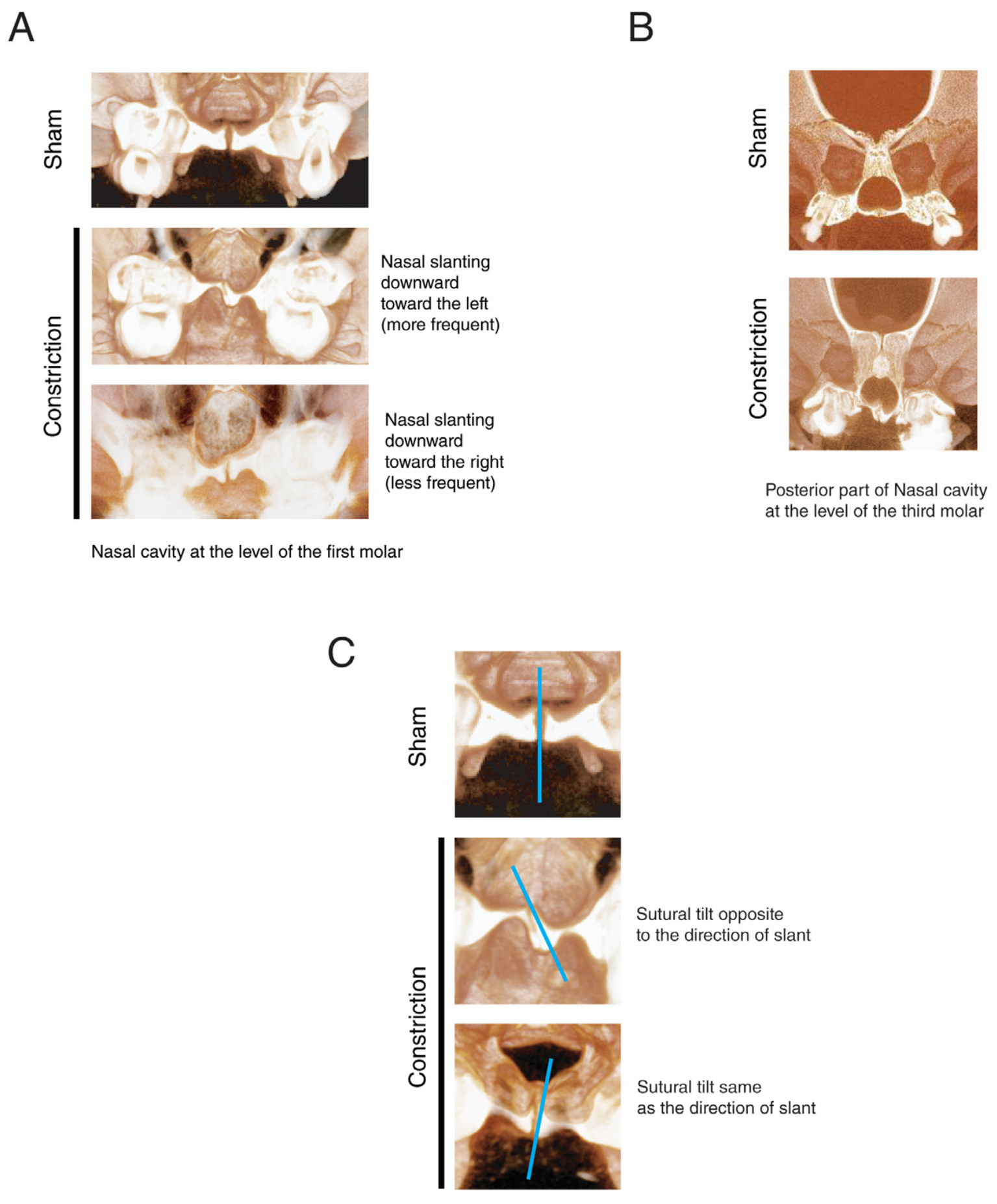
The degree of the nasal septum deviation was assessed by measuring the angle between the tangent line to the upper part of the nasal septum and a perpendicular line drawn from the horizontal line connecting the center of the two orbits. (
Figure 2C). The width of the left and right nasal cavities was measured on the area of maximum deviation on a horizontal line from the septum to the lateral walls of the nasal cavity (
Figure 2C). Measurements were performed on soft tissue view of the coronal section of microCT images, where the deviation was most significant. Investigators completing these morphometric measurements were blinded to group allocation to avoid bias.
2.5. Extra-Oral Measurements
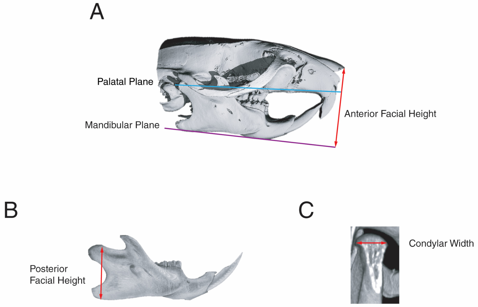
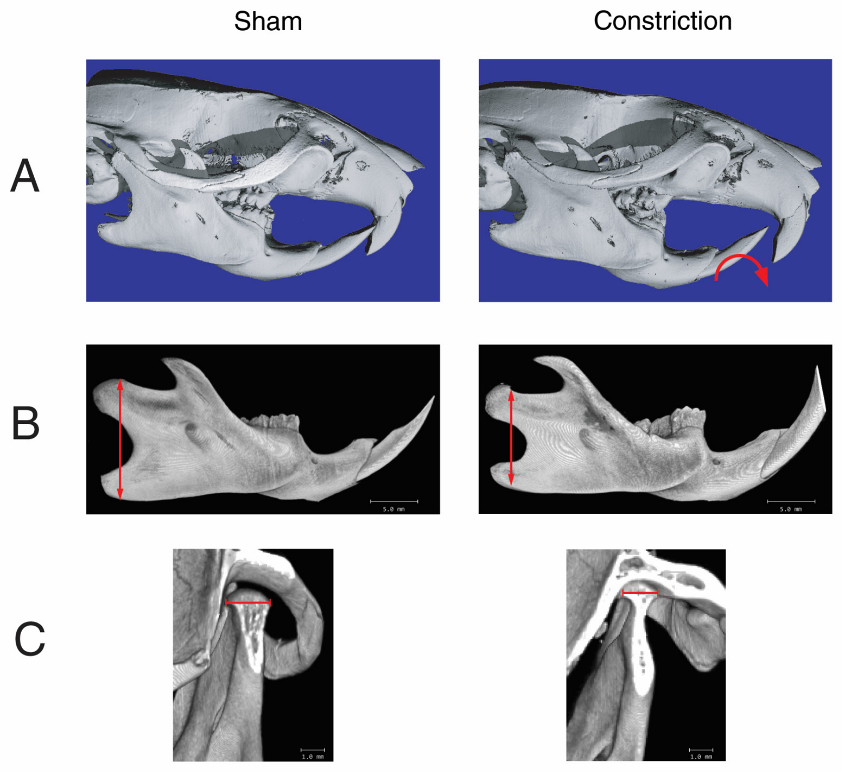
Skull changes in response to constriction were studied by measuring the angle between the mandibular plane (the line tangent to the lower border of the mandible) and the palatal plane (the line tangent to the palate). The anterior facial height was measured on the perpendicular line to the mandibular plane from the most anterior point of the nasal bone (
Figure 3A). The posterior facial height was measured from the highest point in the condylar process to the lowest point on the gonial angle (
Figure 3B). Maximum condylar width was measured in the coronal view from the medial to the lateral heights of contour (
Figure 3C).
2.6. Statistical Analysis
Two examiners completed all morphological quantifications. The random and systematic errors were calculated using the formula described by Dahlberg and Houston [
40,
41]. The random and systematic errors were found to be small both intra-observer (0.015 and 0.017 mm, respectively) and inter-observer (0.018 and 0.02 mm, respectively).
Significant differences between the Experimental Groups, Shams, and Controls were assessed using analysis of variance (ANOVA). A pairwise multiple-comparison analysis was performed using Tukey’s post hoc test. Two-tailed p-values were calculated; p < 0.05 was set as the level of statistical significance.
Table 1,
Table 2,
Table 3 and
Table 4 report 11 continuous morphometric outcomes across four anatomical domains (dental arch,
Table 1; craniofacial skeleton,
Table 2; nasal floor,
Table 3; nasal septum,
Table 4), measured on an overlapping set of specimens. To control the family-wise error rate across this full set of comparisons, a Holm–Bonferroni step-down correction was applied to the 11 outcome-level tests (Constriction vs. Control and sham for
Table 1; Constriction vs. Sham for
Table 2,
Table 3 and
Table 4), with α = 0.05. Holm’s method was chosen over a fixed Bonferroni correction for its greater statistical power, and over a false-discovery-rate approach because these are pre-specified, confirmatory comparisons rather than an exploratory screen.
Posterior crossbite, anterior crossbite, and mandibular shift (
Table 5) were treated as present when the measured displacement exceeded the intra- and inter-observer measurement error established above (≥0.1 mm for posterior crossbite; ≥0.5 mm for anterior crossbite and mandibular shift), and compared between groups with Fisher’s exact test (two-tailed). Two examiners independently classified each animal, and inter-examiner agreement was assessed using Cohen’s κ (0.91). A Holm–Bonferroni correction was applied across this family of 3 comparisons (α = 0.05).
Directional laterality of mandibular shift, nasal floor slanting, and nasal septal deviation within the Constriction group (
Table 6) used the same mandibular shift threshold (≥0.5 mm) and an angular threshold of ≥5° for nasal floor slanting and nasal septal deviation, chosen because it falls within the non-overlapping range separating Sham from Constriction on these measures (
Table 3 and
Table 4). For each phenomenon, the observed left/right split was tested against a 50:50 chance expectation with an exact binomial test (two-tailed). The same binomial approach was used to test cross-phenomenon concordance—whether the direction of one phenomenon matched another within the same animal—against a null concordance of 0.50. Sham animals were excluded from these analyses, since none showed a shift, slant, or deviation in either direction (
Table 5). The resulting six comparisons (three own-direction tests, and three concordance tests) were treated as one Holm–Bonferroni family (α = 0.05).
A multivariate model (MANOVA or ANCOVA) was considered for both outcome families but not pursued. With n = 15/group, the sample size does not support stable multivariate covariance estimation for the largest domain in either family (4 correlated continuous outcomes,
Table 2; 6 categorical/directional outcomes,
Table 6), and the distributional assumptions these models require could not be confidently verified at this sample size. More importantly, every univariate comparison in this study showed a large effect (
p ≤ 0.017 for all comparisons;
p < 0.0001 for all continuous outcomes and 5 of 6 categorical outcomes), so a multivariate approach would not alter any conclusions. Holm–Bonferroni correction of the univariate tests was therefore retained as the more conservative and defensible approach throughout.
4. Discussion
Various factors can contribute to nasal septal deviation, including increased intrauterine pressure, trauma during birth, trauma from injuries, genetic factors, and nasal floor asymmetry [
1,
2,
3,
4,
5,
6,
7,
8,
9,
10]. Here, we explore whether constriction of the maxilla can also contribute to the development of nasal septal deviation.
Our experiments demonstrate that maxilla constriction affects nasal septum shape. However, it does not establish if this effect is direct or indirect. This agrees with previous findings that found a correlation between maxillary width and nasal septal deviation in humans [
42,
43]. It should be emphasized that this observation does not claim that maxillary constriction is the only etiological factor in nasal septal deviation, but it increases clinicians’ awareness of the need to explore maxillary constriction as a contributing factor.
The causes of the constriction can be due to a number of environmental factors, including ecological allergens/nasal obstruction, dietary changes, and decreased rates of breastfeeding [
28,
29,
30,
31,
32,
33,
34,
35,
36,
37]. However, how the maxillary constriction contributes to the development of nasal septum deviation is not clear. Three possibilities exist.
Hypothesis 1: One possible way maxillary constriction may affect the growth of the nasal septum is by creating a growth mismatch between the septum and maxilla. This mismatch may cause the nasal septum (cartilage and bone) to bulge away from the midsagittal plane. This hypothesis aligns with previous studies in humans, which have shown that when normal midline nasal septal growth is disrupted, the frequency of septal deviation increases [
3,
9,
20,
44,
45].
For this hypothesis to be correct, we need to demonstrate that the nasal septum exhibits robust interstitial growth that persists even in the absence of the space required for growth of the septum due to maxillary constriction. But which part of the nasal septum has the interstitial growth?
The nasal septum is composed of three parts: the cartilaginous septum and the bony septum, which is composed of the perpendicular plate of ethmoid and the vomer. The ossification of the vomer via the intramembranous pathway typically is completed by birth, and the ossification of the perpendicular plate accelerates until age 10, then slows significantly afterward [
46,
47]. It has been established that the bony part of the septum does not have interstitial growth and primarily functions as a growth site [
48,
49]. On the other hand, since the nasal cartilage is considered the anterior extension of the chondrocranium, it is regarded as a primary cartilage and, therefore, has interstitial growth. It has been suggested that this interstitial growth can produce enough mechanical forces that may help in the maxilla’s displacement and suture activation and, therefore, cause maxillary growth [
50,
51]. Thus, the nasal septum has been proposed as the growth center for the maxillary complex [
8,
52,
53,
54,
55,
56,
57,
58,
59]. While this claim is evident primarily in long-snouted animal models [
60], it is controversial for the human facial skeleton [
61]. One group believes that the nasal septum in humans is similar to that of many long-snouted animals and acts as a growth center for the maxilla [
6,
7,
8,
9,
53,
57,
61], and nasal septal deviation can produce a force that can cause asymmetry in the skull [
61,
62,
63,
64,
65]. On the other hand, many others could not find the evidence that the mechanical force produced by nasal septum interstitial growth was strong enough to displace the maxilla. These scientists believe that the soft tissue (periosteal matrix) and oronasal spaces (capsular matrix) are responsible for maxillary growth [
48,
66,
67], and nasal septum growth is secondary to midfacial growth. They believe that the nasal septum is more important for transferring occlusal forces to the skull, rather than for the growth of the maxillary complex [
61,
68,
69]. The opponents to the role of the nasal septum as a growth center argue that if the mechanical force produced by the nasal septum were large enough, it would prevent the development of nasal septum deviation in the first place by pushing the maxilla downward and forward.
While the interstitial growth of the nasal septum is undeniable, the magnitude of its contribution to nasal septal deviation is questionable. This is because the majority of the nasal septum’s growth decreases significantly by the age of two, while maxillary growth continues by age 14–16 [
70]. Based on this observation, nasal septal growth can only partially explain the development of septal deviation.
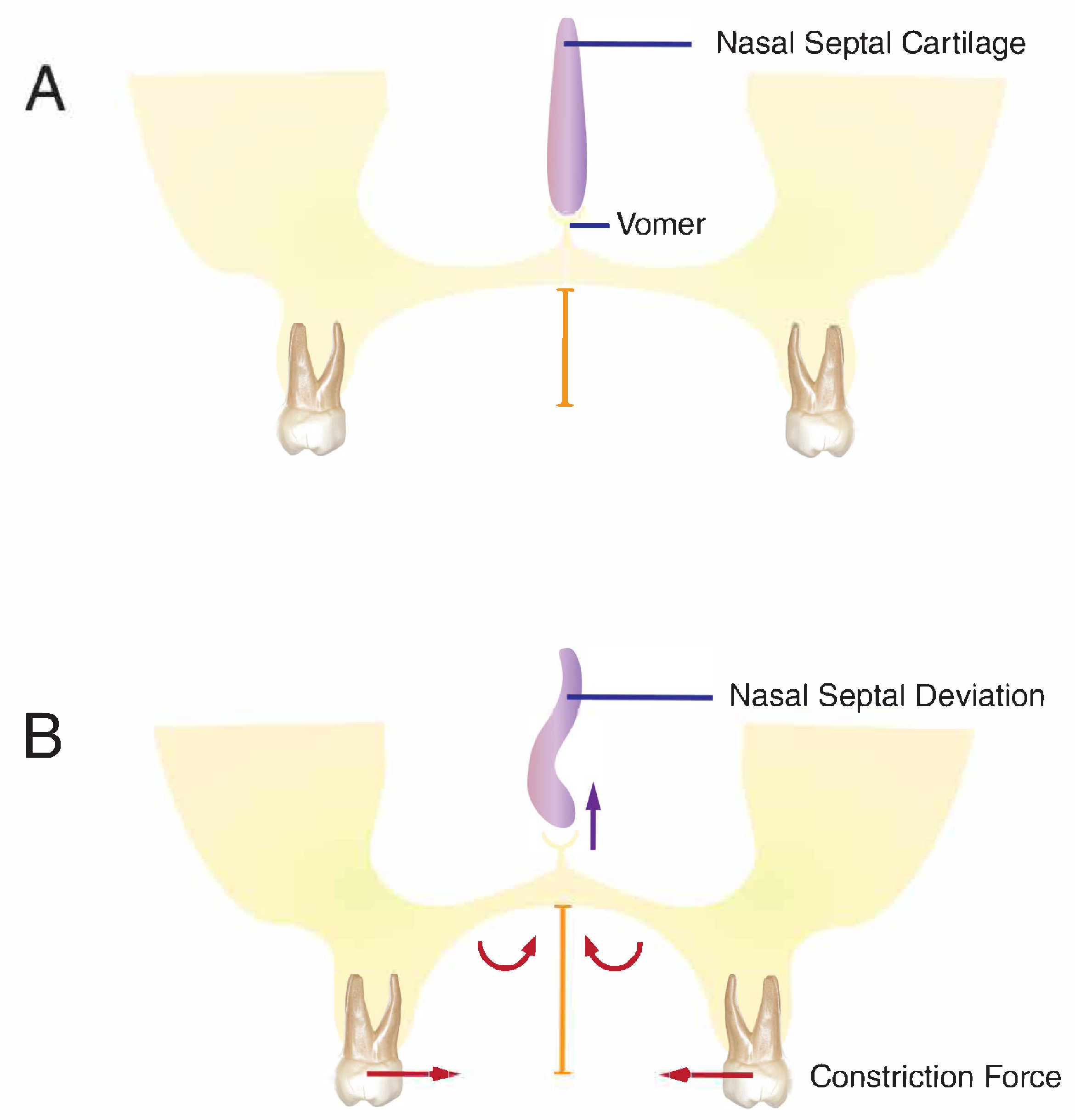
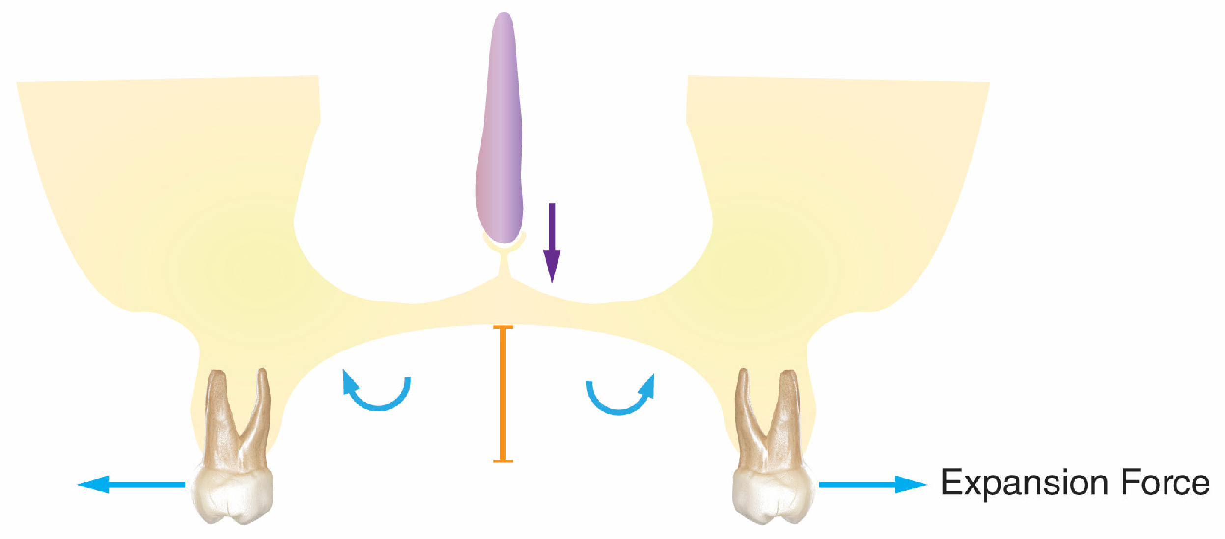
Hypothesis 2: The second possibility that may explain why maxillary constriction affects the nasal septum deviation is that constriction pushes the palatal shelves upward toward the space occupied by the nasal septum, causing a deviation in the septum. This bulging in response to constriction can be explained mechanically by the appearance of significant moments in the system (
Figure 9). We argue that the constriction forces applied far from the center of resistance of each hemimaxilla can produce considerable moments in the system that push the maxilla up in the midline toward the nasal cavity and decrease the space for the septum (
Figure 9B). If this hypothesis is correct, one would expect the application of tensile forces to produce opposite moments and decrease bulging in the middle of the nasal floor, as observed in our experiment (
Figure 10). This analysis is in agreement with previous observations that nasal septal deviation is associated with an increase in palatal arch height [
3,
4,
11,
71]. Our current data does not examine this hypothesis since, in our model, maxillary constriction was always associated with mandibular shift. However, this hypothesis remains a strong possibility biomechanically.
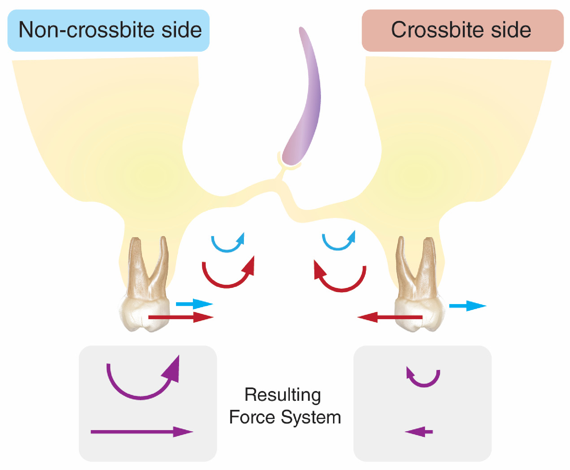
Symmetrical moments produced by constricting forces should cause bulging in the middle of the nasal floor, but no slanting should occur. In other words, mechanical constriction cannot explain the nasal septal floor slanting, since it produces symmetrical forces and moments. However, in our experiment, a significant nasal floor slant was observed, indicating that mechanical factors were unequal and disrupted the balance between the right and left nasal floors. Therefore, constricting forces alone cannot explain the slant in the nasal floor, suggesting the possibility of other factor(s). Here, we propose that the mandibular shift, occurring in response to the maxillary constriction, can play a role in the development of nasal floor slanting. It should be emphasized that mandibular shift is always secondary to maxillary constriction or expansion, as we observed here, and does not occur separately, especially in animals in the absence of any trauma.
Maxillary constriction prevents proper contact between the posterior teeth and forces the mandible to shift to one side to establish a posterior occlusion, resulting in a crossbite, as observed in our experiments. This produces large horizontal forces toward the medial side of the non-crossbite side. These forces produce an additional moment that can push the palate even further up on that side (
Figure 11). On the other hand, on the crossbite side, the horizontal forces remain small and directed laterally, producing a lateral moment that rotates the palate in the opposite direction of the moment created by constricting forces, thereby reducing the magnitude of the rotation of the palatal shelf on that side. This differential moment between the left and right contributes to the slanting of the nasal floor.
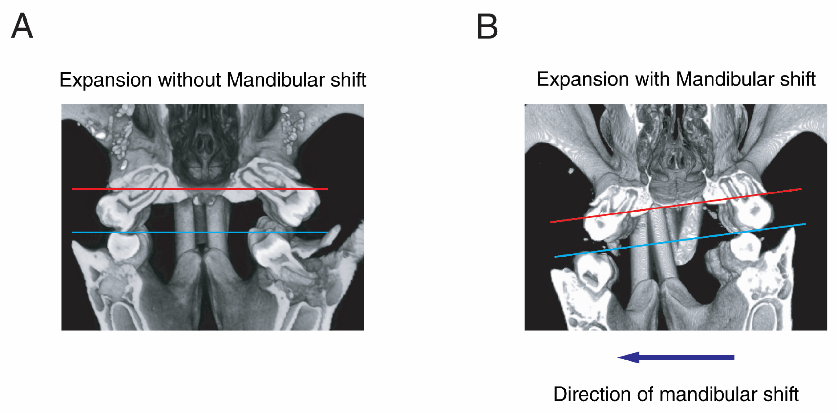
In our study, no specific trend was observed in the shift in the mandible toward one side. However, the direction of shift in the mandible and nasal floor cant was always the same. To further test the hypothesis that mandibular shift contributes to the slanting of the nasal floor, we studied animals subjected to expansion (tensile forces). Here, we expected no nasal floor slanting. However, similar in animals subjected to constriction forces, the bite in these animals changed, and the majority of the animals demonstrated a shift in their mandibles toward a normal bite on one side and a scissor bite on the other (
Figure 8). However, some animals were able to continue biting in the center. In none of the animals did the expander cause bulging of the nasal floor as was expected. In the absence of mandibular shift, no slanting of the nasal floor was observed. But in the presence of mandibular shift, mild to moderate nasal floor slanting was observed. In this scenario, similar to a constriction group, the direction of nasal floor slanting and mandibular shift was the same. These experiments together demonstrate that the slanting was caused by mandibular shift and could be exaggerated by constricting forces. It should be emphasized that the shift itself did not cause the large horizontal forces and moments. The shift is associated with a change in the biomechanics of occlusion, such as a change in the inclination of the lower molars, that will continue even as the mandible gradually shifts, leading to adaptation of form and the development of permanent asymmetry. It should also be noted that since the changes in nasal septal deviation occur throughout life [
9,
46,
72,
73], one can expect the dynamic changes in occlusion that can occur throughout life to contribute to changes in nasal deviation.
In our experiment, we did not establish whether the effect of constriction on nasal septal deviation is direct (Hypothesis 1 or 2) or indirect through mandibular shift (Hypothesis 3). This is due to a study limitation, which shows that constriction is always associated with mandibular shift, and separating the effect of one from the other was not possible. However, we are currently developing a model to induce mandibular shift in the absence of constriction or expansion to study the isolated effect of mandibular shift on nasal cavity architecture. Based on current data, we proposed the third hypothesis, that constriction-induced mandibular shift may indirectly affect nasal septal deviation. We know that the nasal floor is part of the palatal process of the maxilla, and its growth is a combination of bony resorption on the nasal side, along with bony apposition over the oral surface, which is considered part of natural cortical drift [
49]. The change in loading of the maxilla and the appearance of moments and horizontal forces due to constriction forces and mandibular shift produce cortical drifting in the opposite direction, which causes changes in the floor of the nose and the height of the palate [
74,
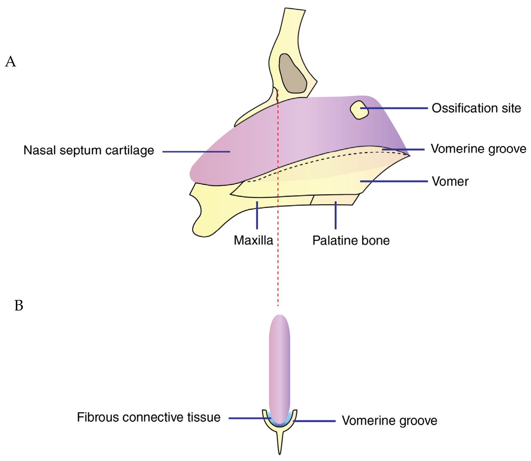
75]. We believe that this change in occlusal forces and the appearance of new moments that caused nasal floor slanting can be the third source for the development of nasal deviation. Nasal floor slanting is accompanied by a change in the direction of the vomer, as was observed in these experiments. This is important, since the vomer holds the base of the nasal septal cartilage (
Figure 12). Nasal septal cartilage lies in the vomerine groove, a shallow channel at the superior border of the vomer bone. Therefore, a change in the direction of the vomer could easily cause nasal septal deviation. Our observation that nasal floor slanting and vomer inclination could be considered as a third factor in the development of nasal septal deviation is in agreement with previous studies, which show a high incidence of nasal septal deviation associated with nasal floor slanting [
65,
70,
76].
It should be emphasized that hypothesis three that mandibular shift can cause nasal floor slanting (always in the same direction) and may affect nasal septal deviation by changing the direction of vomer is in agreement with the observation that in our experiment all mandibular shifts were associated with change in direction of vomer. However, while vomer and nasal septal deviation were always in the same direction, the nasal floor slanting and the vomer inclination were not always in the same direction. This differs from previous studies, which noted that nasal septum deviation is more pronounced on the lower side of the nasal floor [
3,
43,
76,
77]. This difference may be due to the limited sample size in our experiment. These observations suggest that there is a complex mechanical environment in the oral and nasal cavities, and that we are far from understanding the full aspect of these interactions.
In our experiments, constriction of the maxilla was accompanied by C-shape deformation of the nasal septum, in accordance with the classification system proposed by Guyuron [
78]. However, we did not observe any other type of deformation. This may be due to a short study duration. Our observation is in agreement with previous reports that have shown the most frequent nasal septal deviation associated with nasal floor slanting is the C-shape deformity [
43]. Perhaps that can explain why the application of expansion forces can be more successful in the treatment of C-shape deviation, as has been reported before [
79].
This study sheds light on nasal floor slanting as the third source of nasal septal deviation. However, in addition to nasal septal deviation, nasal slanting has other side effects. Studies demonstrated that severe slanting of the nasal floor may also contribute to nasal obstruction [
80], which emphasizes the importance of clinical diagnosis of constricted maxilla that can cause mandibular shift and indirectly contribute to nasal floor slanting.
Rats in this experiment demonstrated clockwise rotation of the mandible, increased lower facial height, and decreased posterior facial height. These changes were not due to a change in the animal’s breathing pattern and were induced mostly by constriction of the upper arch and uprighting of the upper posterior teeth. However, they produced similar skeletal effects to those observed in mouth-breathing patients [
81,
82]. This similarity can be due to the effect that mouth breathing has on the constriction of the upper arch [
71]. Chronic mouth breathing can increase the constricting forces on the maxilla, which will affect the transverse and vertical growth of the maxilla [
11,
12,
13].
It should be emphasized that our study neither tested nor denied the possible effect of septal growth on the maxilla [
51,
54,
55,
56]. However, we demonstrated that even in the presence of the mechanical forces from the septum, maxillary growth can be substantially altered by numerous other factors. In fact, the maxilla itself can change the direction of septal growth, which can cause chronic blockage of the nasal cavity and mouth breathing [
36,
83,
84,
85]. Based on this reciprocal theory, deformity of craniofacial structures worsens nasal obstruction and chronic mouth breathing, which, in turn, leads to craniofacial maldevelopment. This negative feedback loop highlights the need to interrupt the cycle as early as possible through proper diagnosis and early interceptive treatment. While current data indicate a potential negative feedback loop between the oral and nasal cavities, these findings should not be over-interpreted or generalized to humans without further validation. Further research in this area, especially in humans, is required.




 ,
,