1. Introduction
Nonalcoholic fatty liver disease (NAFLD) is increasingly recognized not only as an isolated liver disease, but as a complex metabolic disorder with systemic implications [
1]. The pathogenesis of NAFLD is closely linked to lipotoxicity, insulin resistance, and chronic low-grade inflammation [
2,
3]. Emerging evidence suggests that the excessive accumulation of hepatic lipids triggers the activation of potent pro-inflammatory pathways, most notably the sphingomyelinase–ceramide axis [
4]. This activation releases a cascade of inflammatory mediators into the bloodstream, which can eventually compromise the microvascular and mucosal integrity of distant organs.
In the field of otolaryngology and audiology, the detrimental effects of metabolic syndromes, obesity, and diabetes on the auditory system have been widely investigated, though research has mostly focused on the inner ear [
5,
6]. Furthermore, a broader paradigm shift in hearing research suggests that audiologic evaluations should not be limited solely to local ear-related issues; systemically, vascular risk factors and serum electrolyte levels also play critical roles in the pathogenesis of hearing loss [
7,
8]. Numerous studies link metabolic disturbances to sensorineural hearing loss, generally pointing to cochlear microvascular damage and subsequent hair cell apoptosis [
9]. However, the impact of systemic low-grade inflammation on the middle ear and Eustachian tube mucosa has received very little attention. It is highly possible that the circulating inflammatory mediators present in NAFLD could lead to subclinical mucosal edema and microvascular congestion in the tympanic cavity, altering its biomechanics.
Detecting these minor, subclinical changes is difficult with standard hearing tests. Conventional pure-tone audiometry (conducted within the frequency range of 125 to 8000 Hz) and classical 226 Hz tympanometry often lack the sensitivity needed to catch early biomechanical shifts before they manifest as overt conductive hearing loss. Wideband Tympanometry (WBT), on the other hand, offers a more advanced and sensitive approach [
10]. Tympanometry remains a fundamental, objective, and non-invasive tool in otorhinolaryngological and audiological diagnostics, crucial for evaluating middle ear pathologies. However, while conventional tympanometry is limited to a single 226 Hz probe tone providing static compliance data, wideband tympanometry (WBT) offers a comprehensive analysis across a broad frequency spectrum. Furthermore, acoustic reflex measurements performed during immittance testing provide vital clinical insights into the stapedius muscle contraction, middle ear compliance dynamics, and neural pathway integrity. By measuring the acoustic immittance of the middle ear across a wide range of frequencies (typically 226 to 8000 Hz), WBT can track dynamic parameters like wideband absorbance and Resonance Frequency (RF). This allows clinicians to detect very subtle changes in the mass and stiffness of the middle ear system.
In this study, we hypothesized that the systemic lipotoxicity and inflammation associated with NAFLD might cause subclinical micro-edema in the middle ear mucosa, leading to early biomechanical alterations even in the absence of subjective hearing complaints. We aimed to use WBT to investigate subclinical middle ear biomechanical changes in normal-hearing patients with ultrasonographically confirmed hepatosteatosis, and to compare the findings with a healthy control group.
2. Materials and Methods
2.1. Study Design and Participants
This prospective observational clinical study was conducted at the Department of Otorhinolaryngology. The study protocol was approved by the Biruni University Clinical Research Ethics Committee (Approval No: 2026-07; Date: 1 July 2026), and written informed consent was obtained from all participants in accordance with the Declaration of Helsinki.
The study cohort consisted of 58 adults. Participants were consecutively recruited from the internal medicine outpatient clinic. Patients who underwent abdominal ultrasonography and were diagnosed with hepatic steatosis were referred to the otorhinolaryngology department for further evaluation. Following a detailed medical history and criteria screening, eligible patients were assigned to either the NAFLD group (n = 28) or the healthy control group (n = 30). For the healthy control group, the absence of hepatic steatosis was objectively confirmed via abdominal ultrasonography, alongside normal liver function tests and no history of chronic metabolic or systemic diseases.
2.2. Inclusion and Exclusion Criteria
The study included participants aged between 18 and 45 years. All participants were required to have normal otoscopic examination findings, an intact tympanic membrane, a Type A tympanogram on standard 226 Hz tympanometry, and normal pure-tone hearing thresholds (defined as ≤20 dB HL across the 250–8000 Hz frequency range). Patients were excluded from the study if they had a history of otologic surgery, chronic otitis media, tympanic membrane perforation, otosclerosis, active upper respiratory tract infections, or any degree of conductive or sensorineural hearing loss (air-bone gap > 10 dB). Participants in the healthy control group were not taking any regular medications, including systemic anti-inflammatory drugs, ototoxic medications, or treatments for metabolic diseases, ensuring an unconfounded baseline for wideband tympanometry measurements. Furthermore, individuals with a history of active smoking, a history of regular alcohol consumption, allergic rhinitis, chronic sinusitis, or nasal polyposis were excluded. Finally, patients with coexisting systemic conditions, including diabetes mellitus, essential hypertension, autoimmune disorders, or chronic kidney disease, were also excluded from the study cohort.
2.3. Radiological and Biochemical Evaluations
In the study group, the diagnosis and grading of hepatosteatosis (Grades 1 to 3) were confirmed via abdominal ultrasonography (USG) performed by an experienced radiologist according to well-established clinical criteria [
11]. Fasting venous blood samples were collected from all participants in both groups between 08:00 and 10:00 AM following an overnight fast of at least 8 h. The biochemical parameters analyzed using standard automated laboratory assays included aspartate aminotransferase (AST), alanine aminotransferase (ALT), gamma-glutamyl transferase (GGT), total cholesterol, high-density lipoprotein (HDL), low-density lipoprotein (LDL), triglycerides, and fasting glucose.
2.4. Audiological and Wideband Tympanometry (WBT) Measurements
Following the otorhinolaryngological examination, pure-tone audiometry was performed using a clinical audiometer (AC40; Interacoustics, Middelfart, Denmark) in a sound-attenuated booth. Air conduction thresholds were measured across the frequency range of 125 to 8000 Hz, while bone conduction thresholds were evaluated between 250 and 4000 Hz. Standard clinical masking procedures, including masked-bone conduction assessments, were systematically employed whenever a significant air–bone gap or interaural attenuation threshold was detected. Subsequent wideband acoustic immittance measurements were conducted using a wideband tympanometry system (Titan; Interacoustics, Middelfart, Denmark). Additionally, acoustic reflex testing (including both ipsilateral and contralateral pathways) was systematically conducted for each individual case in both the NAFLD and control groups, using the same system to ensure comprehensive middle ear and neural pathway evaluation. Wideband absorbance (ranging from 0 to 1) was measured across a frequency spectrum of 226 to 8000 Hz. The primary parameters extracted for statistical analysis included Resonance Frequency (RF) in Hertz (Hz), wideband absorbance values at specific frequencies (250, 500, 1000, 2000, 4000, and 8000 Hz), Peak Absorbance, Peak Absorbance Frequency, and middle ear pressure (daPa) at 226 Hz. Measurements were recorded separately for the right and left ears.
2.5. Statistical Analysis
Statistical analyses were performed using SPSS software, version 26.0 (IBM Corp., Armonk, NY, USA). The normality of the data distribution was evaluated using the Shapiro–Wilk test. Normally distributed continuous variables were expressed as mean ± standard deviation (SD), and categorical variables were reported as frequencies and percentages. The independent samples t-test was utilized to compare continuous variables, including age and wideband tympanometry parameters, between the study and control groups. The Chi-square test was employed to compare sex distribution. For the subgroup analysis within the NAFLD cohort (Grade 1 versus Grades 2–3), the independent samples t-test was applied. A two-sided p-value of <0.05 was considered statistically significant.
4. Discussion
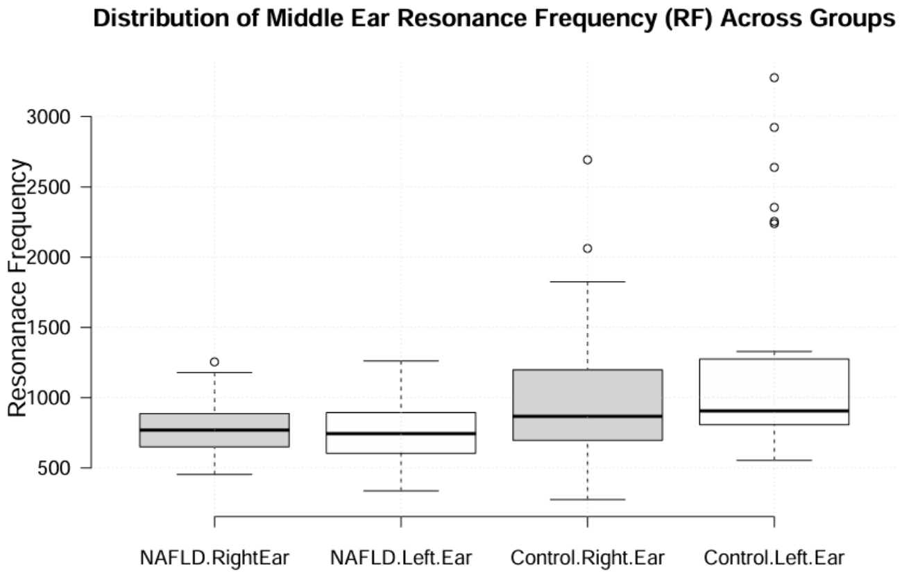
Our primary findings indicated that normal-hearing non-alcoholic fatty liver disease (NAFLD) patients exhibited a significant decrease in middle ear Resonance Frequency (RF) and a simultaneous increase in wideband absorbance at lower frequencies (250 Hz and 500 Hz) compared to healthy controls. Currently, there are very few studies in the literature investigating the relationship between NAFLD and the middle ear mechanics. The present study is one of the few to specifically examine this association. Although studies specifically investigating middle ear biomechanics via wideband tympanometry in NAFLD are exceptionally scarce, our findings align conceptually with earlier reports demonstrating the negative impacts of metabolic dysfunction and abdominal fat accumulation on the auditory system. The previous literature has indicated that chronic inflammation, insulin resistance, and systemic metabolic stress—which form the core pathophysiology of hepatic steatosis—can lead to subclinical neural and cochlear impairments, often manifested as elevated pure-tone thresholds or sensorineural hearing loss [
12,
13]. While those earlier reports primarily focused on the sensory and neural components of hearing, our study expands this paradigm by demonstrating that the metabolic burden of NAFLD also alters the peripheral conductive mechanics of the middle ear, as objectively shown by WBT. The simultaneous presence of subclinical hearing deficits reported in the past literature and the low-frequency acoustic absorbance elevations and reduced resonance frequencies observed in our cohort further substantiate the hypothesis that NAFLD-associated lipotoxicity and systemic inflammation exert a widespread impact on the entire auditory system, ranging from middle ear compliance to cochlear and neural function. These findings collectively suggest that the middle ear mucosa, like the cochlea, is vulnerable to the systemic inflammatory burden of metabolic diseases, positioning WBT as a promising non-invasive screening tool for early otologic manifestations of systemic metabolic disorders. In our study, we also conducted subgroup analysis which suggested that mass-dominated biomechanical shifts might occur at the early stages of hepatic fat accumulation (Grade 1) and may not necessarily progress in a linear fashion with advancing ultrasonographic severity (Grades 2–3). However, it is important to approach this subgroup result with caution due to the unequal distribution of participants between the disease grades. These outcomes support the notion that the systemic inflammatory burden and lipotoxicity associated with NAFLD could exert a subclinical “mass effect” on the middle ear and Eustachian tube mucosa.
In wideband acoustic immittance evaluations, the Resonance Frequency represents the point where the mass and stiffness components of the middle ear system cancel each other out [
10]. Pathologies that increase the stiffness of the tympano-ossicular system characteristically shift the RF to higher frequencies. Conversely, conditions that increase the mass of the middle ear—such as early-stage congestion or mucosal edema—tend to shift the RF toward lower frequencies and predominantly increase absorbance in the low-frequency spectrum [
14]. The reduction in RF and the elevated absorbance at 250–500 Hz observed in our NAFLD cohort appear to be consistent with the acoustic profile of a mass-dominated middle ear system. Given our exclusion criteria, which minimized confounding otologic factors and other major systemic inflammatory diseases, it is plausible to consider that this mass effect might be related to subclinical micro-edema within the middle ear mucosa, potentially driven by NAFLD pathophysiology.
The underlying mechanism linking a hepatic metabolic disorder to potential middle ear mucosal alterations might be explored through the lens of lipotoxicity and chronic, low-grade systemic inflammation. NAFLD is increasingly recognized as a systemic condition that can trigger vascular and endothelial dysfunction [
15]. In this context, the sphingomyelinase–ceramide pathway could play a contributory role. As extensively described in the recent literature, excessive accumulation of hepatic lipids activates the sphingomyelinase pathway, leading to the systemic circulation of toxic ceramides [
16]. Elevated circulating ceramides have been reported to directly disrupt endothelial tight junctions, increase microvascular permeability, and promote the release of pro-inflammatory cytokines [
17]. Since the middle ear cavity and the Eustachian tube are lined with a vascularized respiratory mucosa, they are likely susceptible to such circulating inflammatory mediators. One plausible mechanistic explanation for our findings could involve the sphingomyelinase–ceramide pathway, which has been implicated in the systemic lipotoxicity and endothelial dysfunction associated with NAFLD. However, it is important to emphasize that this remains a speculative hypothesis at this stage, as we did not directly measure circulating ceramide levels or serum inflammatory markers (such as CRP), or perform histological analysis of the middle ear mucosa to confirm micro-edema. Future studies incorporating these biomarkers are required to validate this proposed mechanism.
Moreover, our subgroup analysis comparing early-stage (Grade 1) and advanced-stage (Grades 2–3) hepatosteatosis revealed similar biomechanical alterations in both subgroups, suggesting that the mucosal response may be triggered during the initial phases of lipotoxicity rather than exhibiting a dose-dependent progression. Several factors may explain this lack of linearity. First, ultrasonographic grading primarily reflects hepatic fat infiltration and does not reliably distinguish between simple steatosis and steatohepatitis (NASH), as it does not assess inflammation [
18]. Since our study used USG-based grading rather than histopathological assessment or inflammatory biomarkers, patients with higher USG grades may have had varying degrees of inflammation. Second, the small sample size in the advanced-stage subgroup (
n = 13) limits statistical power to detect subtle differences. Third, the cross-sectional design captures a single time point; longitudinal studies are needed to evaluate true progression. Therefore, while our findings suggest an early, non-linear mucosal response, they should be interpreted with caution and validated in larger, histologically or biomarker-stratified cohorts.
From a practical clinical perspective, our findings introduce a novel paradigm for the early screening and interdisciplinary management of NAFLD patients. Traditionally, otologic evaluations are not routine in the clinical pathway of patients with metabolic fatty liver diseases. However, our objective WBT results demonstrate that subclinical middle ear biomechanical alterations occur even in the earliest stages (Grade 1) of hepatic fat accumulation, long before noticeable hearing loss develops. Clinically, this positions wideband tympanometry as a highly sensitive, rapid, and non-invasive screening tool that can detect early metabolic microvascular and mucosal changes in the auditory system. Identifying these subclinical “mass effects” allows clinicians to recognize early otologic risks, implement closer audiological monitoring, and counsel patients on stricter lifestyle modifications to mitigate systemic lipotoxicity before permanent sensory or conductive damage ensues. Furthermore, these results emphasize the need for a collaborative, interdisciplinary approach between gastroenterologists and otolaryngologists for a more comprehensive assessment of metabolic syndrome manifestations.
Limitations of the Study
While the present study provides preliminary insights into the potential subclinical otologic manifestations of NAFLD, several limitations should be acknowledged. First, the relatively small overall sample size may limit the generalizability of the findings to the broader NAFLD population. More specifically, regarding our subgroup analysis, the limited number of patients in the Grade 1 (n = 15) and Grade 2–3 (n = 13) categories inherently reduces the statistical power of this comparison. Furthermore, the grouping and potential clinical overlap between Grade 2 and Grade 3 hepatosteatosis led to an unequal distribution of participants among severity grades, which serves as a structural limitation in analyzing advanced disease stages. Consequently, the lack of a statistically significant dose-dependent deterioration should be interpreted with caution, as a larger cohort might be required to detect subtle, progressive biomechanical differences between advancing disease stages. Therefore, larger, multicenter cohorts would be beneficial to further validate these initial observations. Second, the diagnosis and grading of hepatosteatosis were based primarily on abdominal ultrasonography (USG). Although USG is widely used as a first-line imaging modality, it is important to acknowledge its inherent limitations: conventional gray-scale USG cannot reliably differentiate between simple steatosis and steatohepatitis as it does not directly assess the key histologic features of non-alcoholic steatohepatitis (NASH), namely lobular inflammation and hepatocyte ballooning. Therefore, our study reflects an association with the degree of hepatic fat accumulation rather than with the severity of necroinflammation or fibrosis. Third, the healthy control group was significantly younger than the NAFLD cohort. Given that chronological aging can independently influence middle ear compliance, tissue elasticity, and acoustic immittance parameters, this age discrepancy stands as a potential confounding factor. Although we adjusted for age as a covariate in our statistical models to isolate the independent impact of hepatic steatosis, future age-matched cohorts are necessary to completely eliminate the confounding effects of age on wideband tympanometry outcomes. Finally, while we have proposed a pathophysiological mechanism involving systemic lipotoxicity and potential mucosal micro-edema, we did not directly measure circulating pro-inflammatory cytokines, serum ceramide levels, or histological changes in the middle ear mucosa. Therefore, our mechanistic interpretation remains speculative and requires validation in future molecular and histological studies.