1. Introduction
Brachial plexus injuries (BPIs) are among the most complex and disabling conditions in trauma and peripheral nerve surgery, producing both short- and long-term disability [
1,
2,
3]. Their annual incidence is estimated at 0.8–2.4 per 100,000 inhabitants, although rates vary with geography and trauma mechanism [
1,
2,
3,
4]. Most cases involve young men exposed to high-energy events such as road-traffic collisions, motorcycle accidents, and contact sports; BPIs can also complicate traumatic shoulder dislocation. Violent traction or direct compression disrupts the plexus and may cause anything from transient neurapraxia to complete cervical root avulsion [
5,
6,
7].
Lesions are usually classified according to Seddon or Sunderland, which grade the depth of axonal and connective-tissue damage [
1,
3,
7]. Prognosis depends on the lesion level and timely intervention, as delays often lead to permanent neuromuscular dysfunction [
1,
7]. Magnetic resonance neurography, myelography, electromyography, and nerve-conduction studies guide surgical planning and help predict spontaneous recovery [
1,
7]. Clinically, BPIs present with severe motor and sensory deficits of the upper limb, neuropathic pain, progressive muscle atrophy and joint instability, all of which can severely diminish quality of life and prevent return to work [
5,
6,
7]. Loss of dynamic stabilisation by the rotator cuff and periscapular muscles predisposes to glenohumeral dislocation and progressive arthropathy, further deepening disability [
8,
9,
10]. Despite advances in trauma care and microsurgical techniques, delayed or inadequate management still leaves many patients with profound deficits, underscoring the need for better surgical solutions [
7,
11].
Current repairs rely on nerve grafting, direct transfers and neurotisation, which can restore useful movement in upper-plexus lesions (C5-C6) when performed within 3–6 months [
1,
7,
8]. Targeted muscle reinnervation and distal nerve transfers expand options, but outcomes remain variable and time-dependent [
12,
13,
14]. Denervation of the supraspinatus, infraspinatus, deltoid, and serratus anterior abolishes shoulder abduction, flexion, and rotation, leaving the joint unstable and nonfunctional [
14]. When irreversible damage persists, shoulder arthrodesis offers pain relief and a stable position, but it is invasive, restricts compensatory motion, and limits future myoelectric–prosthetic control [
12,
13,
14]. Lower-plexus injuries (C7-T1) further hamper rehabilitation because hand and wrist paralysis curtails functional gain despite tendon transfers and reinnervation techniques [
1,
7]. These challenges create an urgent need for alternatives that stabilise the shoulder and preserve signals suitable for bionic prostheses [
1,
7,
14].
The reconstructive algorithm for BPI depends critically on the level of lesion (preganglionic vs. postganglionic), the timing of injury, and the availability of donor nerves. In preganglionic avulsions, particularly when involving C5–T1, donor options are limited, and even early reconstruction yields modest functional recovery. In late-presenting or globally denervated limbs, the likelihood of achieving useful hand or elbow function declines sharply, often leaving patients with a painful, flail limb despite extensive reconstruction.
This study describes a combined surgical approach comprising transhumeral amputation, a passive biceps brachii traction sling, regenerative peripheral nerve interfaces (RPNIs) performed systematically at the time of amputation, and planned prosthetic rehabilitation in selected patients with irreversible brachial plexus injury and painful flail shoulder. Amputation alone provides inconsistent analgesia, and phantom limb pain (PLP) may persist. Contemporary strategies often combine amputation with neuromodulatory or biologic interfaces such as spinal cord stimulation, peripheral nerve stimulation, targeted muscle reinnervation (TMR), and RPNI. In this series, RPNIs were performed systematically. TMR was not indicated due to the absence of reliable donor input. The sling component was designed as a passive mechanical construct intended to resist inferior glenohumeral traction and to optimise the residual limb for prosthetic fitting while preserving channels for myoelectric control [
12,
13,
14]; it was not designed to restore active shoulder motion or replace rotator cuff or periscapular function.
The aim of this report is to describe the technical details of this combined approach and to assess its feasibility and perioperative safety descriptively. Because the series was retrospective, uncontrolled, and involved five patients receiving a complex, multi-component intervention, no causal inference regarding the specific contribution of the biceps sling to any observed clinical changes can be drawn. The hypothesis that the biceps sling provides meaningful passive resistance to inferior glenohumeral traction and improves residual limb stability remains unproven and requires objective biomechanical and radiographic evaluation in future prospective studies.
2. Materials and Methods
This retrospective, uncontrolled five-case series was designed as a technical note to assess the feasibility, perioperative safety, descriptive clinical outcomes, and early prosthetic integration of modified transhumeral amputation with a passive biceps brachii traction sling. The procedure was intended to provide passive resistance to inferior glenohumeral traction and to optimise a prosthesis-ready stump, not to restore active shoulder stability or replace rotator cuff, deltoid, or periscapular function. Given the small uncontrolled cohort, the study was not designed or powered to assess comparative efficacy or superiority over conventional transhumeral amputation or other reconstructive pathways.
The study was conducted at Brixsana Private Clinic and Bolzano General Hospital and included all consecutive patients with irreversible brachial plexus palsy who underwent modified transhumeral amputation incorporating a biceps brachii traction sling between January 2022 and July 2023. According to local institutional policy, formal Ethics Committee approval was waived because the study involved retrospective analysis of anonymized clinical data, in accordance with the Declaration of Helsinki. Written informed consent was obtained from all participants, including specific consent for the acquisition and publication of clinical photographs and video recordings for scientific, educational, and conference purposes.
Eligible cases showed complete loss of upper-limb function after failed reconstructive surgery for severe brachial plexus injury, disabling pain from inferior glenohumeral subluxation, no realistic further reconstructive options, and a sustained amputation request maintained for at least twelve months after detailed counselling. Preoperative assessment included routine laboratory investigations, imaging of the upper arm and glenohumeral joint, and psychological screening to exclude uncontrolled psychiatric disorders. In addition, preoperative electromyographic (EMG) mapping was performed to identify and characterise potential detection sites in the proximal upper arm and shoulder region for future control of a myoelectric bionic prosthesis (
Supplementary Video S1).
For each patient, the extent and level of the brachial plexus lesion, involved nerve roots, and preoperative biceps brachii function were documented using the Medical Research Council grading system. Detailed patient characteristics are presented in Table 2.
Surgery Technique
The biceps brachii traction sling was designed as a passive stabilising construct to resist inferior glenohumeral translation and optimise the residual limb for prosthetic fitting. It was not intended to restore active shoulder stability or replace rotator cuff, deltoid, or periscapular function.
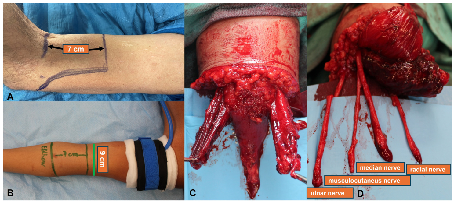
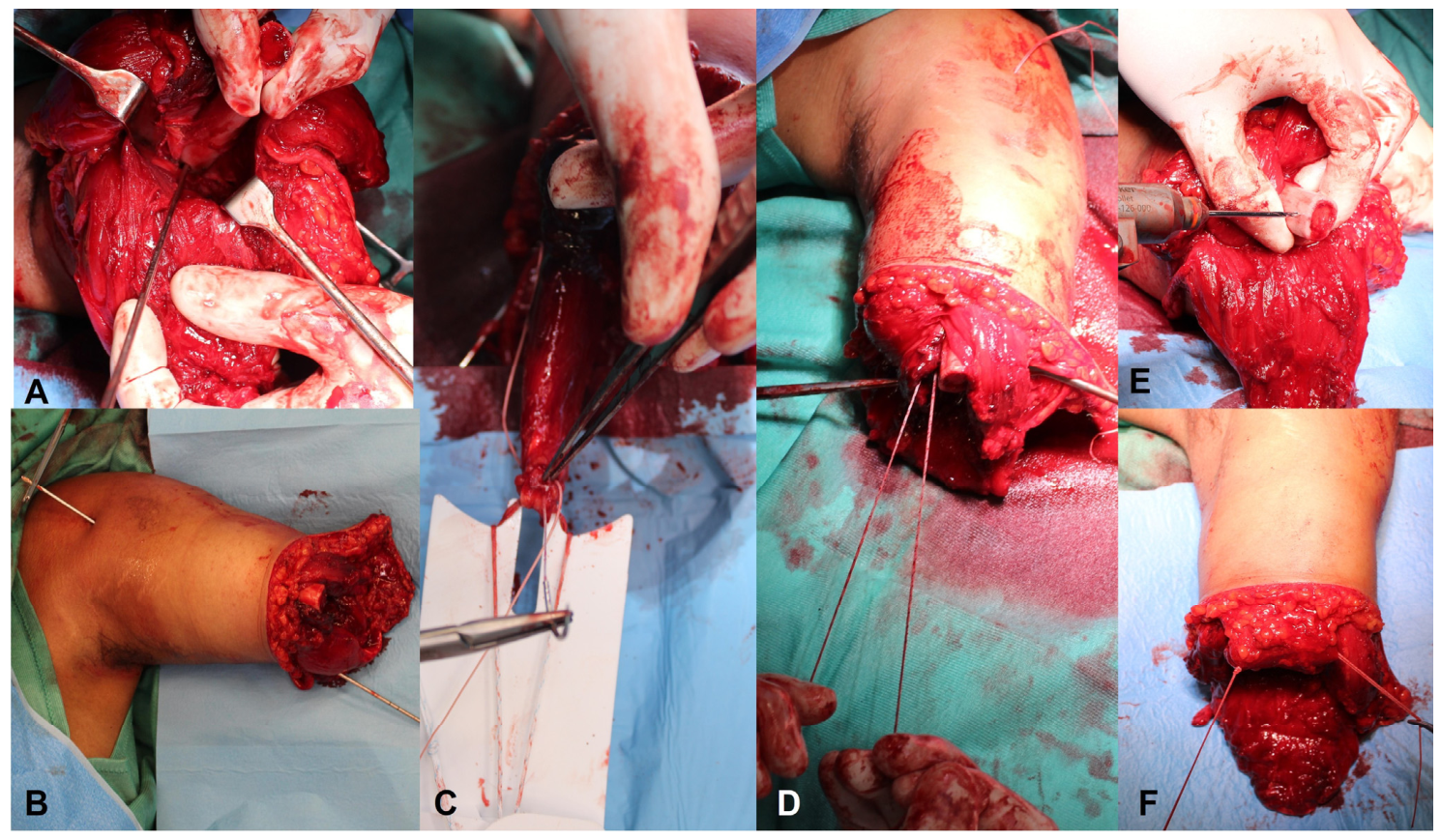
The surgical procedures were performed by board-certified plastic surgeons with extensive expertise in microsurgery, peripheral nerve surgery, and bionic reconstructive surgery, assisted by junior trainees. All procedures were performed under general anaesthesia with upper-arm tourniquet control and additional axillary plexus blockade. The patient was positioned supine, and the amputation level was planned approximately 7 cm proximal to the antecubital crease (
Figure 1A,B). Following skin incision and elevation of a posterior skin flap, the distal insertion of the biceps brachii tendon was identified and detached from the radial tuberosity together with a small periosteal sleeve. The brachialis muscle was then split longitudinally over approximately 5 cm, along its fibre orientation, to facilitate subsequent stump coverage and improve surgical exposure (
Figure 1C). To provide additional soft tissue coverage and enhance the stability of the residual limb, the triceps brachii tendon was detached distally, and the muscle was mobilised as a muscle flap. The ulnar, musculocutaneous, median, and radial nerves were identified, mobilised, and transected (
Figure 1D). The major vascular structures of the arm were ligated and divided. The humerus was circumferentially exposed over a length of approximately 9 cm, transected using an oscillating saw, and the cut surface was smoothed and rounded.
Passive mechanical stabilisation was achieved using a traction sling, followed by dorsal tension band fixation of the biceps brachii tendon to the humerus. For this purpose, approximately 8 cm of the humerus was exposed (
Figure 2A), and the biceps brachii muscle was bluntly passed around the humerus. After determination of the optimal muscle tension by controlled traction, an eyelet guide wire was inserted obliquely from the distal dorsal aspect of the humerus and advanced proximal ventrally through the bone, exiting through the skin (
Figure 2B). The guide-wire tract was subsequently enlarged with a cannulated 4.2-mm drill bit through both cortices of the humerus. Thereafter, the dorsal cortex was selectively enlarged using a cannulated 8-mm drill bit. The tendon was secured to a ToggleLoc™ with ZipLoop
® fixation device (13-mm ToggleLoc button with standard #7 ZipLoop) using two MaxBraid
® 2-0 simple interrupted sutures (Zimmer Biomet, 1800 West Center St., Warsaw, IN 46581-0587, USA) (
Figure 2C). The suture limbs were shuttled through the humeral tunnel to the ventral aspect over the guide wire. Final fixation of the biceps brachii tendon was achieved within the dorsal humeral tunnel using the 13-mm ToggleLoc button while maintaining maximal tension on the musculotendinous unit (
Figure 2D). At this stage, accurate repositioning of the biceps brachii muscle around the distal humeral stump is critical. To prevent displacement, the muscle was additionally secured to the distal humerus through two lateral drill holes using two MaxBraid
® 2-0 simple interrupted sutures (
Figure 2E,F). The previously created brachialis muscle flaps, which had been split longitudinally along the direction of their muscle fibres, were subsequently wrapped around the biceps brachii muscle and apposed ventrally along the median plane using interrupted sutures.
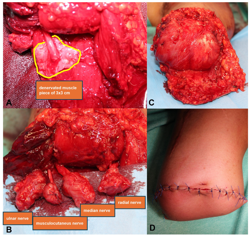
For neuroma prophylaxis, the ulnar, musculocutaneous, median, and radial nerve stumps were each capped and implanted into denervated muscle targets to create RPNIs (
Figure 3A,B). This was intended to reduce the risk of symptomatic neuroma-related pain and preserve the potential for future interface-based prosthetic control. The triceps brachii muscle flap was then transposed ventrally to augment soft tissue coverage and optimise contouring of the residual limb (
Figure 3C). Following meticulous haemostasis, a closed-suction drain was placed. Definitive stump closure was completed with inset of the posterior skin flap and tension-free layered wound closure (
Figure 3D). TMR was not performed because no reliable donor motor input was available. Spinal cord stimulation and peripheral nerve stimulation were not part of the treatment protocol.
Follow-up data were available for all patients, with a maximum follow-up of 30 months. Patient-reported outcomes (DASH, VAS, SF-36) were recorded preoperatively and at the most recent follow-up visit, which occurred no earlier than 6 months postoperatively, with final outcome evaluation completed at a maximum follow-up of 30 months (Table 3). Individual follow-up durations, timing of postoperative outcome assessment, timing of prosthetic fitting, rehabilitation protocol, occupational therapy duration, prosthesis use at last follow-up, revision surgery status, and complications per patient are reported in Table 4. Postoperative rehabilitation followed a three-phase protocol: (1) wound healing and stump maturation (weeks 0–8), supervised by the treating plastic surgeon; (2) prosthetic fitting, socket fabrication, and myoelectric control training (months 3–6) (
Supplementary Video S2), supervised by a certified prosthetist and occupational therapist; and (3) functional and occupational integration (months 6–30), with structured occupational therapy sessions of 45 min twice weekly for a minimum of 10–12 weeks. Patients fitted with a myoelectric prosthesis underwent EMG-guided biofeedback training before definitive socket fitting. Rehabilitation content, prosthesis type, and daily wear duration at last follow-up are reported individually in Table 4.
No objective measures of shoulder position, glenohumeral subluxation grade, stump stability, socket load distribution, radiographic alignment, or motion analysis were obtained in this series. The claim that the biceps sling provides passive resistance to inferior glenohumeral traction, therefore, remains a design hypothesis based on anatomical and biomechanical rationale rather than a demonstrated finding. The
Supplementary Videos (S3 and S4) are provided as illustrative material to document the clinical appearance and prosthetic function achieved; they do not constitute objective evidence of sling-mediated shoulder or stump stabilisation and should not be interpreted as such.
3. Results
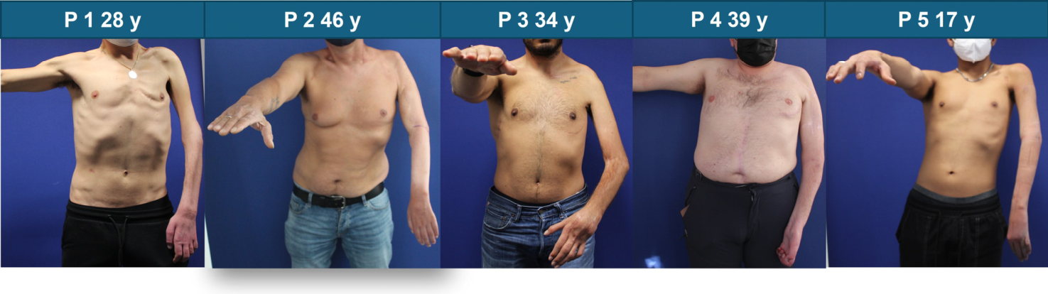
The cohort included five male patients aged 17 to 46 years, all with injuries involving the same upper extremity. Demographic and injury-related data are reported in
Table 1 and
Figure 4.
All patients had severe brachial plexus injury with complete loss of useful upper-limb function after previous unsuccessful reconstructive procedures. Detailed lesion characteristics, preoperative biceps function, previous treatments, main preoperative complaints, pain management, indication for transhumeral amputation, injury year, date of surgery, and injury-to-amputation interval are reported in
Table 2. All patients who reported pain and distress related it to the perception of the flail limb as a nonfunctional appendage. Four of the five patients had complete preganglionic brachial plexus injury with no residual biceps function, while one patient retained partial biceps strength (MRC 3/5) following an intercostal-to-musculocutaneous nerve transfer.
Preoperative and postoperative patient-reported outcomes are summarised in
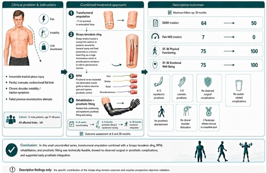
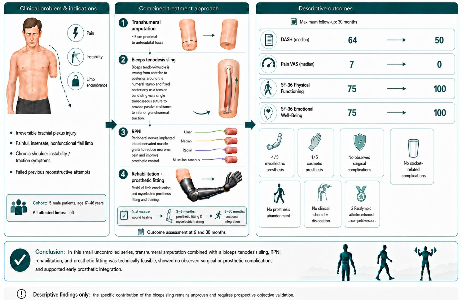
Table 3. After combined transhumeral amputation, biceps sling construction, RPNI, rehabilitation, and prosthetic fitting, patient-reported scores changed as follows in this small uncontrolled series. These changes cannot be attributed to the biceps sling specifically; they may reflect amputation itself, removal of the painful and burdensome flail limb, RPNI, rehabilitation, prosthetic fitting, psychological adaptation, regression to the mean, or a combination of these factors. Median DASH score changed from 64 (IQR, 62–67) preoperatively to 50 (IQR, 47–53) postoperatively. In this setting, DASH score changes should not be interpreted as evidence of restored upper-limb function. DASH improvement in this cohort may reflect pain relief, removal of a burdensome limb, altered functional expectations, prosthetic compensation, and psychosocial adaptation rather than any improvement in native limb performance. Median VAS pain score changed from 7 (IQR, 6–7) to 0 (IQR, 0–1). VAS captured overall pain intensity only and did not separately measure phantom limb pain, mechanical traction-related pain, neuropathic pain, or deafferentation pain. The observed change in VAS, therefore, cannot be interpreted as evidence of improvement in any specific pain subtype, including phantom limb pain or traction-related pain. SF-36 Physical Functioning changed from 75 (IQR, 75–80) to 100, and SF-36 Emotional Well-Being changed from 75 (IQR, 75–80) to 100 (IQR, 95–100). These data are reported descriptively and without hypothesis testing.
All five patients proceeded to prosthetic evaluation within 3–6 months postoperatively. Four patients were fitted with a myoelectric prosthesis and reported daily wear times of 4–8 h at 30 months of follow-up. One patient elected to use a cosmetic prosthesis only because of limited occupational need. No postoperative surgical complications, socket-related complications, prosthesis abandonment, or clinical failure of the construct were observed during follow-up. Individual patient-level outcomes, including follow-up duration, timing of postoperative outcome assessment, time to prosthetic fitting, prosthesis type at last follow-up, daily wear duration, occupational therapy duration, revision surgery status, and complications, are summarised in
Table 4.
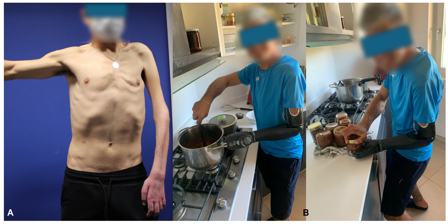
Representative preoperative appearance and postoperative functional use of the prosthesis in patient 1 are shown in
Figure 5. The
Supplementary Videos S3 and S4 further demonstrate the functionality of the prosthesis and the stability of the shoulder joint during a training exercise performed by patient 1; these videos are illustrative only and do not constitute objective evidence of sling-mediated stabilisation. No objective radiographic, biomechanical, or socket-load measurements of shoulder position, glenohumeral subluxation, or stump stability were obtained preoperatively or postoperatively. The absence of clinical dislocation and socket-related complications during follow-up is a descriptive safety observation only and cannot be interpreted as evidence that the sling provided effective passive glenohumeral stabilisation.
Of note, two patients (P1 and P3) were Paralympic athletes and returned to competitive sport after amputation, prosthetic fitting, and rehabilitation, subsequently achieving competitive success in multiple events. Given the small sample size and descriptive design, no inferential statistical analysis was performed. Outcomes are therefore presented descriptively at the individual-patient level.
4. Discussion
This retrospective five-case series provides preliminary data on a combined surgical approach, comprising elective transhumeral amputation, a passive biceps brachii traction sling, RPNI, and planned prosthetic rehabilitation. In this highly selected cohort, all patients showed improvement in pain, patient-reported disability, and selected SF-36 domains. Notably, all patients reported preoperative overall pain, comprising PLP, mechanical traction-related pain, neuropathic pain, deafferentation pain, and distress associated with perceiving the flail limb as a nonfunctional and burdensome appendage. In this context, the observed improvements should be interpreted not merely as changes in questionnaire scores, but also as clinically meaningful relief from the physical and psychological burden of a painful, insensate, and nonfunctional upper limb [
1,
7,
14]. These benefits were maintained during the available follow-up period, which extended up to 30 months, with no clinical dislocation, socket-related complication, prosthesis abandonment, or obvious failure of the construct observed. Rather, they likely reflect the combined effects of the complex intervention, including amputation itself, removal of the painful and burdensome flail limb, RPNI, rehabilitation, prosthetic fitting, psychological adaptation, regression to the mean, and other contextual factors inherent to this treatment pathway.
These benefits were maintained during the available follow-up period, which extended up to 30 months, with no clinical dislocation, socket-related complications, prosthesis abandonment, or obvious failure of the construct observed. However, these changes cannot be attributed to the biceps sling specifically. Rather, they likely reflect the combined effects of the complex intervention, including amputation itself, removal of the painful and burdensome flail limb, RPNI, rehabilitation, prosthetic fitting, psychological adaptation, regression to the mean, and other contextual factors inherent to this treatment pathway.
The main contribution of this report is not the general concept of myoplasty or myodesis, which is well established in transhumeral amputation surgery [
11]. Rather, it describes a structured biceps brachii-based passive traction sling applied to the specific and challenging setting of flail shoulder after irreversible BPI. In these patients, loss of active glenohumeral and periscapular control exposes the shoulder and residual limb to inferior traction, limb encumbrance, mechanical discomfort, and difficulty with prosthetic fitting [
1,
7,
14]. The described construct was designed to redirect the residual biceps muscle-tendon unit around the distal humeral stump, creating a passive suspension vector intended to improve stump contour, reduce inferior traction, and facilitate prosthetic rehabilitation. Therefore, the technique should be interpreted as a modified passive myodesis/sling configuration rather than as a dynamic stabilising procedure or as a proven alternative superior to conventional transhumeral amputation techniques [
11,
14].
The biomechanical rationale of the sling remains hypothetical. The central hypothesis is that wrapping the biceps muscle-tendon unit around the distal humeral stump creates a passive suspension vector that resists inferior glenohumeral traction and improves residual limb contour and prosthetic socket stability in patients with total or near-total brachial plexus palsy. In the present cohort, four of five patients had complete preganglionic brachial plexus injury with absent biceps function, and one patient retained partial biceps strength after previous intercostal-to-musculocutaneous nerve transfer. Because the biceps was denervated in the majority of cases, the sling was not expected to provide active shoulder motion; it was intended as a passive structural suspension. Denervated muscle undergoes progressive atrophy and cannot provide dynamic glenohumeral control. No objective measures of shoulder position, subluxation grade, stump stability, socket load, radiographic alignment, or biomechanical performance were obtained. The
Supplementary Videos are illustrative clinical documentation only and do not constitute evidence of sling efficacy. The absence of clinical dislocation and socket-related complications during follow-up is a descriptive safety observation that is consistent with, but does not confirm, the hypothesised mechanism.
Pain reduction was among the most relevant clinical findings. Median VAS pain score changed from 7 to 0, suggesting substantial relief of the symptoms associated with the painful flail limb. However, VAS captured overall pain and did not distinguish between mechanical pain, neuropathic pain, deafferentation pain, and PLP. These pain components are biologically and clinically distinct, and amputation does not necessarily address centrally mediated neuropathic pain [
1,
7]. Because PLP, traction-related pain, mechanical pain, and neuropathic pain were not measured separately using validated instruments, the observed VAS change cannot be interpreted as evidence of improvement in any of these specific pain subtypes. The observed change in overall VAS may partly reflect the removal of the weight and encumbrance of the flail limb, reduction in traction-related symptoms, improved residual limb stability, rehabilitation, and prosthetic fitting. Therefore, the observed improvement should not be interpreted as direct evidence that the biceps sling specifically reduced neuropathic pain or PLP. Rather, it supports the clinical relevance of a combined strategy addressing mechanical instability, neuroma prophylaxis, and prosthetic readiness [
1,
7,
14].
RPNIs were systematically performed in this cohort as part of the reconstructive strategy. This was intended to reduce the risk of symptomatic neuroma-related pain and to preserve the possibility of future interface-based prosthetic control strategies [
3,
14]. In contrast, TMR was not performed because no reliable donor motor input was available. TMR requires dependable proximal motor signals, and the predominance of pan-plexus avulsion without residual biceps function made this approach unsuitable in the present series. Neuromodulatory approaches such as spinal cord stimulation or peripheral nerve stimulation may be considered in selected refractory cases and may act synergistically with biologic interfaces, although these modalities were not used in the present cohort [
1,
7,
14].
The improvement in SF-36 Physical Functioning and Emotional Well-Being suggests that the procedure may have effects beyond pain reduction; however, these changes occurred in the context of a complex, multi-component intervention and cannot be attributed to the sling specifically. In total BPI, a flail and nonfunctional limb may contribute to distress, altered body image, social limitation, and reduced autonomy [
1,
7,
14]. In this setting, removal of the painful and insensate limb, reduction in limb weight, and creation of a prosthesis-ready stump may support psychosocial adaptation and improve perceived quality of life [
7,
8,
9,
10,
11,
14]. Similarly, DASH score improvement in this setting should not be interpreted as evidence of restored upper-limb function. In patients with a previously nonfunctional and burdensome flail limb, DASH changes may primarily reflect pain relief, removal of the limb’s encumbrance, altered functional expectations following amputation, prosthetic compensation, and psychosocial adaptation rather than any change in native upper-limb performance.
Four patients were fitted with myoelectric prostheses and achieved daily use, while one patient elected to use a cosmetic prosthesis because of limited occupational need. Of particular clinical relevance, two patients who were Paralympic athletes returned to competitive sport after surgery, prosthetic fitting, and rehabilitation, subsequently achieving important competitive results. This observation highlights the potential impact of the procedure not only on pain and prosthetic readiness, but also on social reintegration, participation, and patient-perceived autonomy.
Elective amputation for irreversible BPI remains uncommon in Europe and is generally reserved for carefully selected patients after failed reconstructive attempts [
2,
7,
11]. In contrast, several Asian centres, particularly in Japan, have traditionally prioritised limb-preserving strategies such as nerve transfers and free functional muscle transplantation [
15,
16]. These approaches can achieve partial functional restoration and social reintegration without amputation, but they often require multiple staged procedures and prolonged rehabilitation and may provide variable return of meaningful function [
17]. The present series reflects a complementary strategy in which, for selected patients with non-reconstructable lesions, persistent disability, and a sustained request for amputation after counselling, TA combined with passive stump stabilisation, RPNI-based nerve management, and planned prosthetic rehabilitation may represent a realistic reconstructive endpoint rather than a failure of treatment.
The decision to proceed with elective amputation should remain cautious and multidisciplinary, involving surgeons, physiatrists, occupational therapists, psychologists, pain specialists, and the patient [
1,
7,
14]. Preoperative counselling is essential to clarify realistic functional expectations, address the possibility of persistent neuropathic or PLP, and reduce the risk of prosthesis rejection, which remains a relevant issue in upper-limb amputation [
1,
7,
14]. The present findings may be of interest to centres treating complex irreversible brachial plexus injuries, particularly when conventional nerve reconstruction, tendon transfer, free functional muscle transplantation, or arthrodesis are unlikely to provide acceptable functional recovery or symptom control.
Several limitations must be acknowledged. This was a retrospective uncontrolled series of five patients, and the findings should be considered preliminary and hypothesis-generating. First, the multi-component nature of the intervention, comprising elective amputation, biceps sling construction, RPNI, rehabilitation, and prosthetic fitting, means that the specific contribution of any single element, including the sling, cannot be isolated. The observed changes in DASH, VAS, and SF-36 may reflect amputation itself, removal of a painful and burdensome limb, RPNI, rehabilitation, prosthetic fitting, psychological adaptation, regression to the mean, or other contextual factors, rather than any specific effect of the biceps sling. Second, no objective biomechanical, radiographic, socket-load, motion-analysis, or EMG-based assessment of shoulder position, glenohumeral subluxation grade, or stump stability was performed preoperatively or postoperatively. The mechanistic claim that the sling provides passive resistance to inferior glenohumeral traction, therefore, remains an unproven design hypothesis that requires objective investigation. Third, pain was assessed using VAS, which did not distinguish between mechanical pain, neuropathic pain, deafferentation pain, traction-related pain, and PLP. PLP and traction-related pain were not measured with validated instruments, and the VAS change cannot be attributed to improvement in any specific pain subtype. Fourth, DASH should be interpreted as a measure of global perceived disability rather than proof of restored native upper-limb function. In this cohort, DASH changes may primarily reflect psychosocial adaptation, altered expectations, and removal of a burdensome limb rather than functional improvement. Fifth, no comparison with conventional transhumeral amputation without a sling was made, and no control group was available. Future prospective multicentre studies with larger cohorts, standardised rehabilitation protocols, objective assessment of glenohumeral and stump stability with radiographic and biomechanical methods, control groups, longer follow-up, and validated neuropathic and PLP instruments will be necessary to confirm these preliminary observations and refine patient-selection criteria.




 ,
,