Current and Future Perspectives in Mohs Micrographic Surgery for Non-Melanoma Skin Cancers: A Narrative Review


Abstract
1. Introduction
2. History
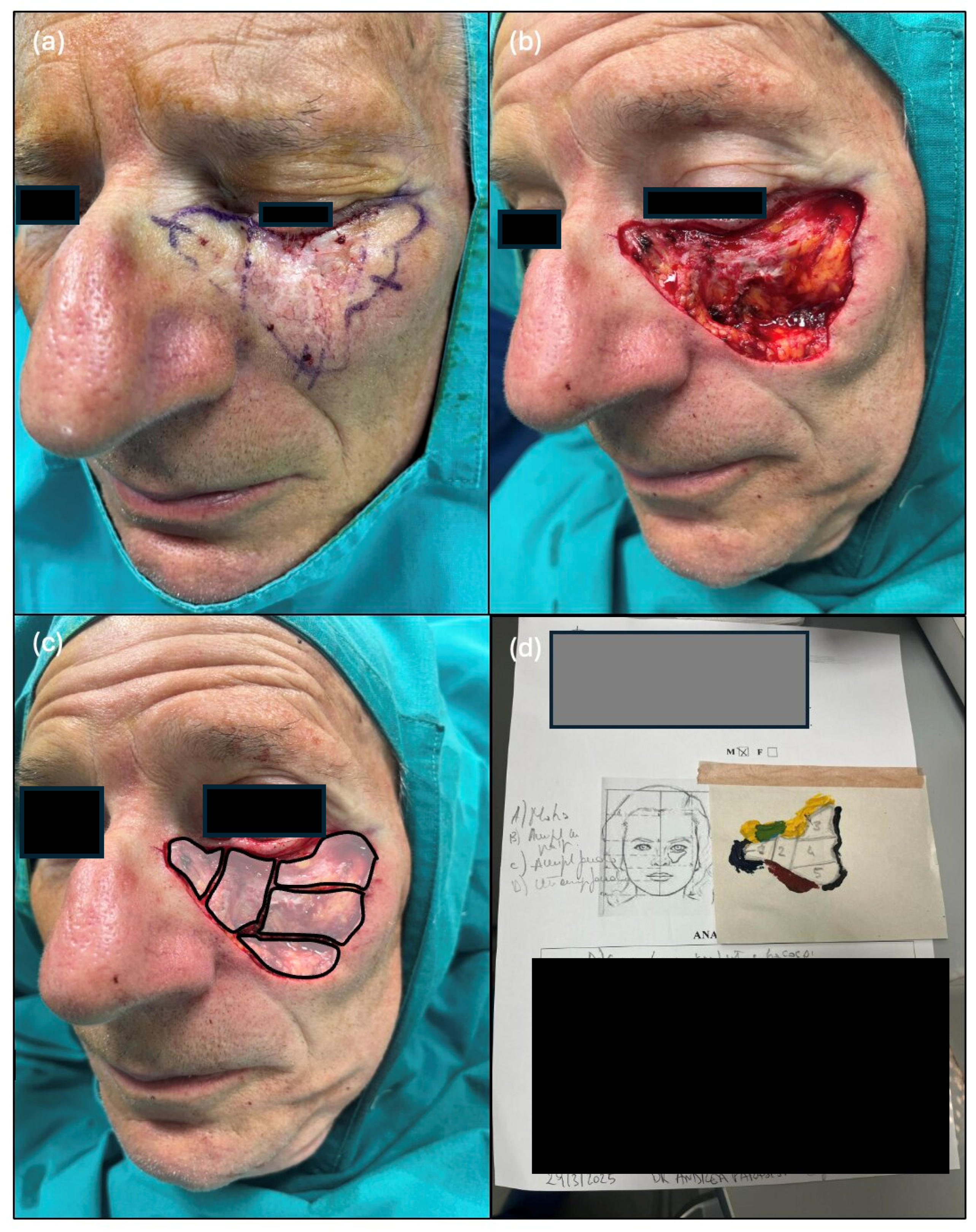
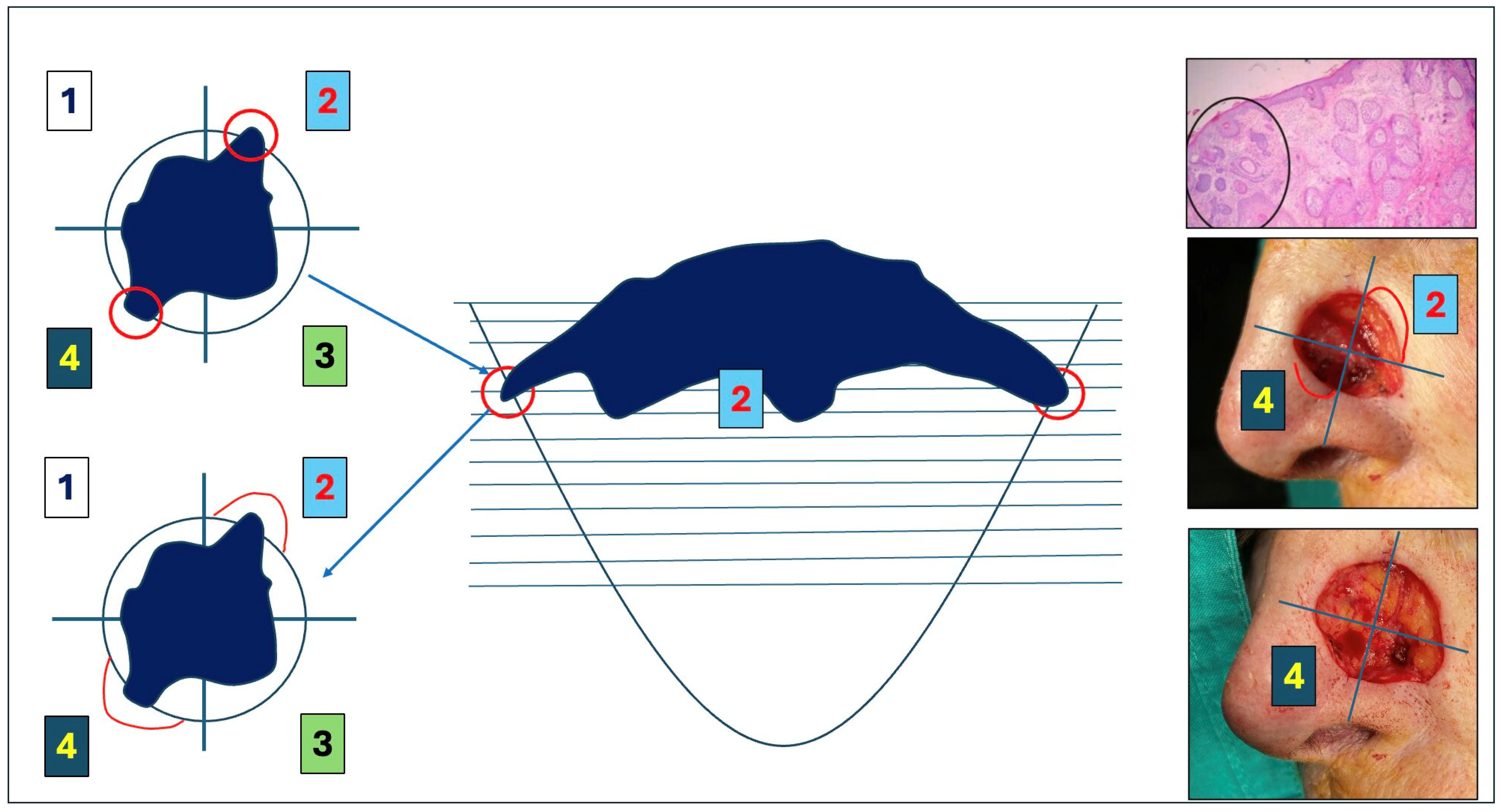
3. Surgical Technique
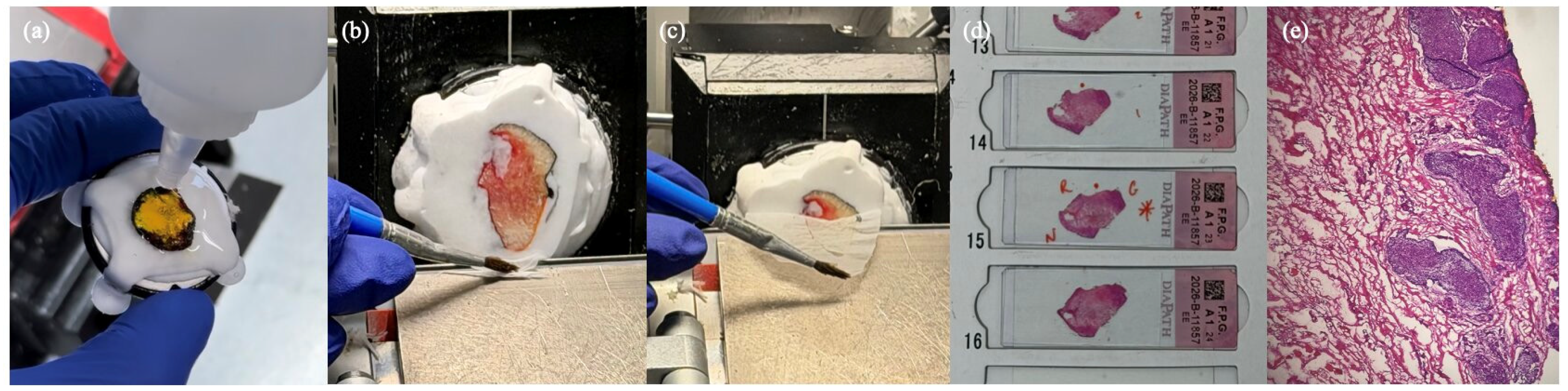
4. Histologic Preparation of the Specimen
5. Preoperative Consideration
5.1. Selecting the Right Local Anesthetic for MMS
5.2. Discontinuation of Anticoagulant and/or Anti-Platelet Therapy Before MMS
5.3. Prophylactic Antibiotics
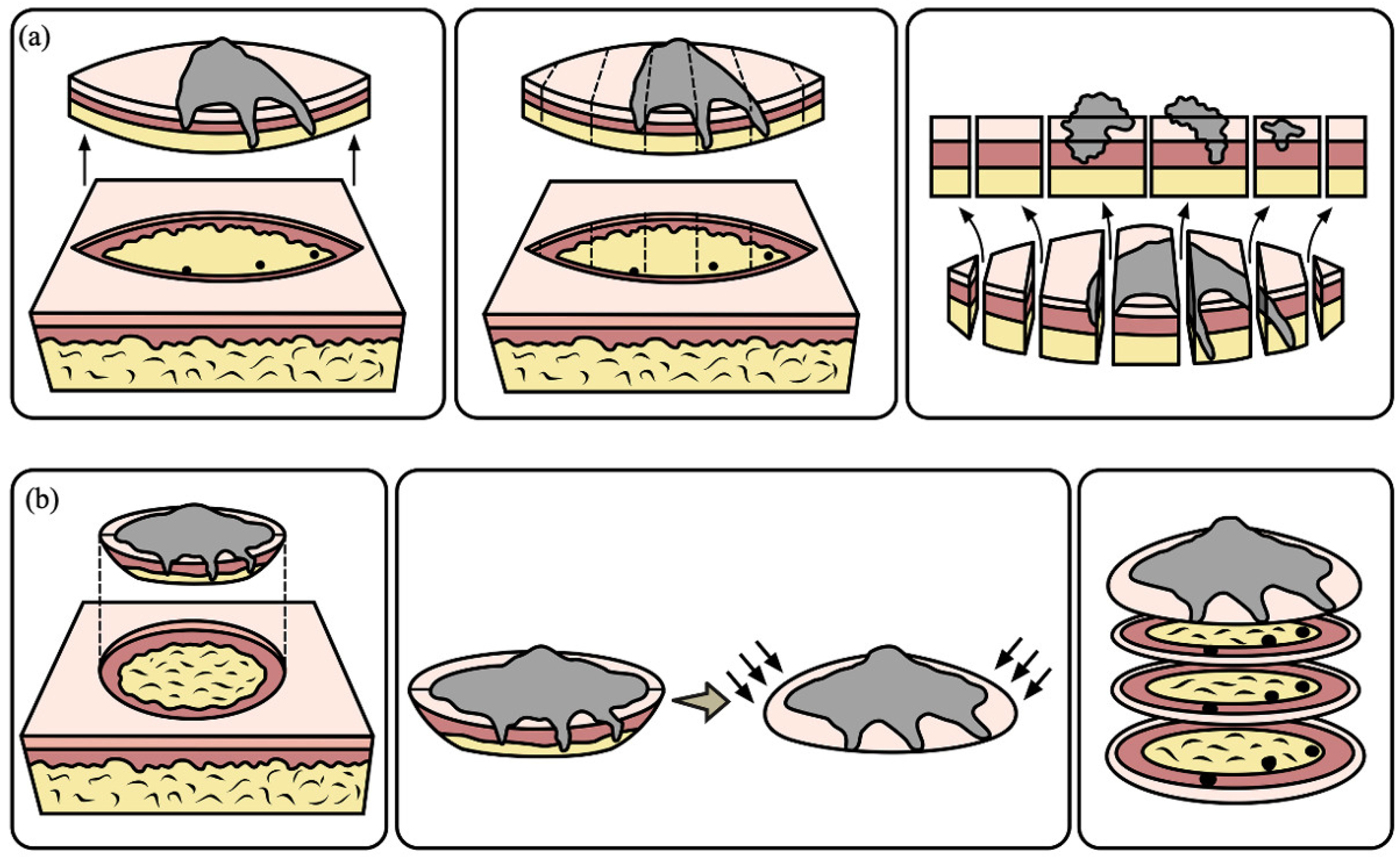
6. Different Surgical Techniques in MMS
7. Limitations of MMS
8. Mohs Micrographic Surgery for Basal Cell Carcinoma
8.1. Epidemiology
8.2. Staging
8.3. Indications for Mohs Micrographic Surgery for Basal Cell Carcinoma
9. Preoperative Evaluation in MMS
9.1. Dermoscopy
9.2. Reflectance Confocal Microscopy (RCM)
9.3. Optical Coherence Tomography (OCT)
9.4. Line-Field Confocal Optical Coherence Tomography (LC-OCT)
10. Future Perspectives
11. MMS Outcomes
12. Mohs Micrographic Surgery for Cutaneous Squamous Cell Carcinoma
12.1. Epidemiology and Clinical Significance
12.2. Prognostic Staging and Risk Factors
12.3. Indications for Mohs Micrographic Surgery for Squamous Cell Carcinoma
12.4. Advantages of Mohs Micrographic Surgery for Cutaneous Squamous Cell Carcinoma
12.5. Outcomes
13. Mohs Micrographic Surgery for Other Rare Non-Melanoma Skin Cancer
Author Contributions
Funding
Institutional Review Board Statement
Informed Consent Statement
Data Availability Statement
Acknowledgments
Conflicts of Interest
References
- Bittner, G.C.; Cerci, F.B.; Kubo, E.M.; Tolkachjov, S.N. Mohs micrographic surgery: A review of indications, technique, outcomes, and considerations. An. Bras. Dermatol. 2021, 96, 263–277. [Google Scholar] [CrossRef] [Scilit] [PubMed]
- Wang, R.; Chen, Y.; Shao, X.; Chen, T.; Zhong, J.; Ou, Y.; Chen, J. Burden of Skin Cancer in Older Adults from 1990 to 2021 and Modelled Projection to 2050. JAMA Dermatol. 2025, 161, 715. [Google Scholar] [CrossRef] [Scilit] [PubMed]
- Bray, F.; Laversanne, M.; Sung, H.; Ferlay, J.; Siegel, R.L.; Soerjomataram, I.; Jemal, A. Global cancer statistics 2022: GLOBOCAN estimates of incidence and mortality worldwide for 36 cancers in 185 countries. CA Cancer J. Clin. 2024, 74, 229–263. [Google Scholar] [CrossRef] [Scilit] [PubMed]
- Stratigos, A.J.; Garbe, C.; Dessinioti, C.; Lebbe, C.; van Akkooi, A.; Bataille, V.; Bastholt, L.; Dreno, B.; Dummer, R.; Fargnoli, M.C.; et al. European consensus-based interdisciplinary guideline for invasive cutaneous squamous cell carcinoma: Part 2. Treatment—Update 2023. Eur. J. Cancer 2023, 193, 113252. [Google Scholar] [PubMed]
- Peris, K.; Fargnoli, M.C.; Kaufmann, R.; Arenberger, P.; Bastholt, L.; Seguin, N.B.; Bataille, V.; Brochez, L.; del Marmol, V.; Dummer, R.; et al. European consensus-based interdisciplinary guideline for diagnosis and treatment of basal cell carcinoma—Update 2023. Eur. J. Cancer 2023, 192, 113254. [Google Scholar] [PubMed]
- Mohs, F.E. Chemosurgery: A microscopically controlled method of cancer excision. Arch. Surg. 1941, 42, 279. [Google Scholar]
- Tromovitch, T.A.; Stegeman, S.J. Microscopically Controlled Excision of Skin Tumors: Chemosurgery (Mohs): Fresh Tissue Technique. Arch. Dermatol. 1974, 110, 231–232. [Google Scholar] [PubMed]
- Litaiem, N.; Hayder, F.; Benlagha, I.; Karray, M.; Dziri, C.; Zeglaoui, F. The Use of Dermoscopy in the Delineation of Basal Cell Carcinoma for Mohs Micrographic Surgery: A Systematic Review with Meta-Analysis. Dermatol. Pract. Concept. 2022, 12, e2022176. [Google Scholar] [PubMed]
- Kouba, D.J.; LoPiccolo, M.C.; Alam, M.; Bordeaux, J.S.; Cohen, B.; Hanke, C.W.; Jellinek, N.; Maibach, H.I.; Tanner, J.W.; Vashi, N.; et al. Guidelines for the use of local anesthesia in office-based dermatologic surgery. J. Am. Acad. Dermatol. 2016, 74, 1201–1219. [Google Scholar] [CrossRef] [Scilit] [PubMed]
- Dokic, Y.; Mireles, N.S.; Hu, A.Y.; Joshi, T.P.; Shimizu, I.; Ranario, J.S. Efficacy of 0.25% Lidocaine Versus 0.5% Lidocaine in Dermatologic Surgery: A Double-Blind, Randomized Controlled Trial. Dermatol. Surg. 2025, 51, 30–33. [Google Scholar] [PubMed]
- Alam, M.; Schaeffer, M.R.; Geisler, A.; Poon, E.; Fosko, S.W.; Srivastava, D. Safety of Local Intracutaneous Lidocaine Anesthesia Used by Dermatologic Surgeons for Skin Cancer Excision and Postcancer Reconstruction: Quantification of Standard Injection Volumes and Adverse Event Rates. Dermatol. Surg. 2016, 42, 1320–1324. [Google Scholar] [CrossRef] [Scilit] [PubMed]
- Wang, A.; Grushchak, S.; Kaul, S.; Lee, P.; Feldman, J. Toxicity of Infiltrative Lidocaine in Dermatologic Surgery: Are Current Limits Valid? Dermatol. Pract. Concept. 2021, 11, e2021120. [Google Scholar] [CrossRef] [Scilit] [PubMed]
- Morganroth, P.A.; Gelfand, J.M.; Jambusaria, A.; Margolis, D.J.; Miller, C.J. A randomized, double-blind comparison of the total dose of 1.0% lidocaine with 1:100,000 epinephrine versus 0.5% lidocaine with 1:200,000 epinephrine required for effective local anesthesia during Mohs micrographic surgery for skin cancers. J. Am. Acad. Dermatol. 2009, 60, 444–452. [Google Scholar] [CrossRef] [Scilit] [PubMed]
- Chen, P.; Smith, H.; Vinciullo, C. Bupivacaine as an Adjunct to Lidocaine in Mohs Micrographic Surgery: A Prospective Randomized Controlled Trial. Dermatol. Surg. 2018, 44, 607–610. [Google Scholar] [CrossRef] [Scilit] [PubMed]
- Jeha, G.M.; Malinosky, H.R.; Taylor, L.; Greenway, H.T.; Kelley, B. Liposomal and Nonliposomal Bupivacaine for Mohs Surgery: A Systematic Review. Dermatol. Surg. 2024, 50, 16–20. [Google Scholar] [PubMed]
- Zuberi, H.Z.; Alkul, M.; Alkul, S.; Seervai, R.N.H.; Jacox, E.; Orengo, I.F. Anticipation, perception, and recollection of acute pain from local anesthesia injection prior to Mohs surgery. Arch. Dermatol. Res. 2025, 317, 490. [Google Scholar] [CrossRef] [Scilit] [PubMed]
- Chen, A.; Albertini, J.G.; Bordeaux, J.S.; Chen, M.W.; Cook, J.L.; Davidson, C.; Donnelly, K.C.; Galaria, I.I.; Hannan, C.M.; Kantor, J.; et al. Evidence-based clinical practice guideline: Reconstruction after skin cancer resection. J. Am. Acad. Dermatol. 2021, 85, 423–441. [Google Scholar] [CrossRef] [Scilit] [PubMed]
- Nast, A.; Ernst, H.; Rosumeck, S.; Erdmann, R.; Jacobs, A.; Sporbeck, B. Risk of complications due to anticoagulation during dermatosurgical procedures: A systematic review and meta-analysis. J. Eur. Acad. Dermatol. Venereol. 2014, 28, 1603–1609. [Google Scholar] [PubMed]
- Fahmy, L.M.; Dowd, M.L.; Loesch, E.; Denson, E.M.; Pisano, C.E.; Samie, F.H. Postoperative Bleeding Complications Associated With Novel Oral Anticoagulants in Mohs Micrographic Surgery. Dermatol. Surg. 2024, 50, 1–4. [Google Scholar] [PubMed]
- Maragh, S.L.H.; Brown, M.D. Prospective evaluation of surgical site infection rate among patients with Mohs micrographic surgery without the use of prophylactic antibiotics. J. Am. Acad. Dermatol. 2008, 59, 275–278. [Google Scholar] [CrossRef] [Scilit] [PubMed]
- Barbieri, J.S.; Etzkorn, J.R.; Margolis, D.J. Use of Antibiotics for Dermatologic Procedures From 2008 to 2016. JAMA Dermatol. 2019, 155, 465–470. [Google Scholar] [CrossRef] [Scilit] [PubMed]
- Campbell, E.; Virk, A.; Lessard, S.; Lohman, M.E.; Otley, C. Antibiotic Prophylaxis in Dermatologic Surgery: Advisory Statement 2025 (APDSAS 2025). Dermatol. Surg. 2026. [Google Scholar] [CrossRef] [Scilit] [PubMed]
- Patel, J.R.; Gimenez, M.C.; Shamloul, N.; Brown, M.D.; Smith, F.L. Retrospective Review of the Infection Rate of Lower Extremities Treated with Mohs Micrographic Surgery with and Without Prophylactic Antibiotics. Dermatol. Surg. 2025, 51, 749–751. [Google Scholar] [CrossRef] [Scilit] [PubMed]
- Cammarata, E.; Esposto, E.; Veronese, F.; Airoldi, C.; Zavattaro, E.; Boggio, P.; Savoia, P. Safety margins for dermatofibrosarcoma protuberans: A comparison between wide local excision and Mohs Tubingen technique. Eur. J. Dermatol. 2020, 30, 289–293. [Google Scholar] [CrossRef] [Scilit] [PubMed]
- Paradisi, A.; Abeni, D.; Rusciani, A.; Cigna, E.; Wolter, M.; Scuderi, N.; Rusciani, L.; Kaufmann, R.; Podda, M. Dermatofibrosarcoma protuberans: Wide local excision vs. Mohs micrographic surgery. Cancer Treat. Rev. 2008, 34, 728–736. [Google Scholar] [CrossRef] [Scilit] [PubMed]
- Surmanowicz, P.; Sivanand, A.; Du, A.X.; Mahmood, M.N.; Gniadecki, R. Muffin Technique Micrographic Surgery for Non-melanoma Skin Cancer. Front. Med. 2021, 7, 637223. [Google Scholar] [CrossRef] [Scilit] [PubMed]
- Gualdi, G.; La Rosa, G.; Di Buduo, A.; Paradisi, A.; Soglia, S.; Calzavara-Pinton, P.; Amerio, P. Conventional surgery compared with formalin-fixed tissue Mohs surgery (slow Mohs) for DFSP: A comparative analysis of 83 cases. J. Eur. Acad. Dermatol. Venereol. 2023, 37, e1393–e1395. [Google Scholar] [PubMed]
- van Loo, E.; Mosterd, K.; Krekels, G.A.M.; Roozeboom, M.H.; Ostertag, J.U.; Dirksen, C.D.; Steijlen, P.M.; Neumann, H.A.M.; Nelemans, P.J.; Kelleners-Smeets, N.W.J. Surgical excision versus Mohs’ micrographic surgery for basal cell carcinoma of the face: A randomised clinical trial with 10year follow-up. Eur. J. Cancer 2014, 50, 3011–3020. [Google Scholar] [CrossRef] [Scilit] [PubMed]
- Lomas, A.; Leonardi-Bee, J.; Bath-Hextall, F. A systematic review of worldwide incidence of nonmelanoma skin cancer. Br. J. Dermatol. 2012, 166, 1069–1080. [Google Scholar] [CrossRef] [Scilit] [PubMed]
- Wehner, M.R. Keratinocyte Carcinoma: A Review. JAMA 2026, 335, 70–81. [Google Scholar] [CrossRef] [Scilit] [PubMed]
- Cameron, M.C.; Lee, E.; Hibler, B.P.; Barker, C.A.; Mori, S.; Cordova, M.; Nehal, K.S.; Rossi, A.M. Basal cell carcinoma. J. Am. Acad. Dermatol. 2019, 80, 303–317. [Google Scholar] [CrossRef] [Scilit] [PubMed]
- Bichakjian, C.; Armstrong, A.; Baum, C.; Bordeaux, J.S.; Brown, M.; Busam, K.J.; Eisen, D.B.; Iyengar, V.; Lober, C.; Margolis, D.J.; et al. Guidelines of care for the management of basal cell carcinoma. J. Am. Acad. Dermatol. 2018, 78, 540–559. [Google Scholar] [CrossRef] [Scilit] [PubMed]
- Connolly, S.M.; Baker, D.R.; Coldiron, B.M.; Fazio, M.J.; Storrs, P.A.; Vidimos, A.T.; Zalla, M.J.; Brewer, J.D.; Begolka, W.S.; Berger, T.G.; et al. AAD/ACMS/ASDSA/ASMS 2012 appropriate use criteria for Mohs micrographic surgery: A report of the American Academy of Dermatology, American College of Mohs Surgery, American Society for Dermatologic Surgery Association, and the American Society for Mohs Surgery. J. Am. Acad. Dermatol. 2012, 67, 531–550. [Google Scholar] [CrossRef] [Scilit] [PubMed]
- Boostani, M.; Bozsányi, S.; Suppa, M.; Cantisani, C.; Lőrincz, K.; Bánvölgyi, A.; Holló, P.; Wikonkál, N.M.; Huss, W.J.; Brady, K.L.; et al. Novel imaging techniques for tumor margin detection in basal cell carcinoma: A systematic scoping review of FDA and EMA-approved imaging modalities. Int. J. Dermatol. 2025, 64, 287–301. [Google Scholar] [PubMed]
- Yeom, S.D.; Lee, S.H.; Ko, H.S.; Chung, K.; Shin, J.; Choi, G.S.; Byun, J.W. Effectiveness of dermoscopy in Mohs micrographic surgery (MMS) for nonmelanoma skin cancer (NMSC). Int. J. Dermatol. 2017, 56, e136–e139. [Google Scholar] [CrossRef] [Scilit] [PubMed]
- Litaiem, N.; Karray, M.; Jones, M.; Rammeh, S.; Zeglaoui, F. Effectiveness of dermoscopy in the demarcation of surgical margins in slow Mohs surgery. Dermatol. Ther. 2020, 33, e14196. [Google Scholar] [CrossRef] [Scilit] [PubMed]
- Dika, E.; Fanti, P.A.; Christman, H.; Ravaioli, G.M.; Patrizi, A. Videodermoscopy and Curettage: The Value of Simple Procedures During Mohs Surgery. Dermatol. Surg. 2017, 43, 1411–1417. [Google Scholar] [CrossRef] [Scilit] [PubMed]
- Suzuki, H.S.; Serafini, S.Z.; Sato, M.S. Utility of dermoscopy for demarcation of surgical margins in Mohs micrographic surgery. An. Bras. Dermatol. 2014, 89, 38–43. [Google Scholar] [CrossRef] [Scilit] [PubMed]
- Jayasekera, P.S.A.; Dodd, J.; Oliphant, T.; Langtry, J.A.A.; Lawrence, C.M. Dermoscopy prior to Mohs micrographic surgery does not improve tumour margin assessment and leads to fewer Mohs stages. Br. J. Dermatol. 2018, 178, 565–566. [Google Scholar] [PubMed]
- Asilian, A.; Momeni, I. Comparison between examination with naked eye, curretage and dermoscopy in determining tumor extension before Mohs micrographic surgery. Adv. Biomed. Res. 2013, 2, 2. [Google Scholar] [CrossRef] [Scilit] [PubMed]
- Pan, Z.Y.; Lin, J.R.; Cheng, T.T.; Wu, J.Q.; Wu, W.Y. In Vivo Reflectance Confocal Microscopy of Basal Cell Carcinoma: Feasibility of Preoperative Mapping of Cancer Margins. Dermatol. Surg. 2012, 38, 1945–1950. [Google Scholar] [CrossRef] [Scilit] [PubMed]
- Venturini, M.; Gualdi, G.; Zanca, A.; Lorenzi, L.; Pellacani, G.; Calzavara-Pinton, P.G. A new approach for presurgical margin assessment by reflectance confocal microscopy of basal cell carcinoma. Br. J. Dermatol. 2016, 174, 380–385. [Google Scholar] [PubMed]
- Lupu, M.; Voiculescu, V.M.; Caruntu, A.; Tebeica, T.; Caruntu, C. Preoperative Evaluation through Dermoscopy and Reflectance Confocal Microscopy of the Lateral Excision Margins for Primary Basal Cell Carcinoma. Diagnostics 2021, 11, 120. [Google Scholar] [CrossRef] [Scilit] [PubMed]
- Alawi, S.A.; Kuck, M.; Wahrlich, C.; Batz, S.; McKenzie, G.; Fluhr, J.W.; Lademann, J.; Ulrich, M. Optical coherence tomography for presurgical margin assessment of non-melanoma skin cancer—A practical approach. Exp. Dermatol. 2013, 22, 547–551. [Google Scholar] [CrossRef] [Scilit] [PubMed]
- Wang, K.X.; Meekings, A.; Fluhr, J.W.; McKenzie, G.; Lee, D.A.; Fisher, J.; Markowitz, O.; Siegel, D.M. Optical Coherence Tomography–Based Optimization of Mohs Micrographic Surgery of Basal Cell Carcinoma: A Pilot Study. Dermatol. Surg. 2013, 39, 627–633. [Google Scholar] [CrossRef] [Scilit] [PubMed]
- Paradisi, A.; Cornacchia, L.; Cappilli, S.; Abeni, D.; Federico, F.; Di Stefani, A.; Mannino, M.; Peris, K. Preoperative evaluation of high-risk basal cell carcinoma with line-field confocal optical coherence tomography (LC-OCT) reduces Mohs micrographic surgery stage number: A case-control study. EJC Skin Cancer 2024, 2, 100015. [Google Scholar] [CrossRef] [Scilit]
- Mirza, F.N.; Haq, Z.; Abdi, P.; Diaz, M.J.; Libby, T.J. Artificial Intelligence for Mohs and Dermatologic Surgery: A Systematic Review and Meta-Analysis. Dermatol. Surg. 2024, 50, 799–806. [Google Scholar] [CrossRef] [Scilit] [PubMed]
- Jeha, G.M.; Qiblawi, S.; Jairath, N.; Sable, K.; LeBlanc, K.; Aylward, J.; Xu, Y.G. ChatGPT and Generative Artificial Intelligence in Mohs Surgery: A New Frontier of Innovation. J. Investig. Dermatol. 2023, 143, 2105–2107. [Google Scholar] [CrossRef] [Scilit] [PubMed]
- Fardos, M.; Miller, P.; Ward, J.; Miller, R. Applications, Ethical Considerations, and Patient Perspectives on Artificial Intelligence in Dermatologic Surgery: Focus on Mohs Surgery. Dermatol. Surg. 2025, 51, 930–934. [Google Scholar] [CrossRef] [Scilit] [PubMed]
- Lacerda, P.N.; Lange, E.P.; Luna, N.M.; Miot, H.A.; Abbade, L.P.F. Efficacy of micrographic surgery versus conventional excision in reducing recurrence for basal cell carcinoma and squamous cell carcinoma: A systematic review and meta-analysis. J. Eur. Acad. Dermatol. Venereol. 2024, 38, 1058–1069. [Google Scholar] [CrossRef] [Scilit] [PubMed]
- Mosterd, K.; Krekels, G.A.M.; Nieman, F.H.M.; Ostertag, J.U.; AB Essers, B.; Dirksen, C.D.; Steijlen, P.M.; Vermeulen, A.; Neumann, H.; Kelleners-Smeets, N.W. Surgical excision versus Mohs’ micrographic surgery for primary and recurrent basal-cell carcinoma of the face: A prospective randomised controlled trial with 5-years’ follow-up. Lancet Oncol. 2008, 9, 1149–1156. [Google Scholar] [CrossRef] [Scilit] [PubMed]
- Islam, R.; Jones, C.; Stewart, T.; Kok, C.; Frew, J.; Elhindi, J.; Lee, S.; Bonilla, G.M. Ten Years of Scalp Keratinocyte Carcinomas (Basal Cell Carcinomas and Squamous Cell Carcinomas) Treated by Mohs Micrographic Surgery at a Large Australian Centre, a Descriptive Study. Australas. J. Dermatol. 2025, 67, 18–23. [Google Scholar] [CrossRef] [Scilit] [PubMed]
- Andersen, Y.M.F.; Karmisholt, K.; Høgsberg, T.; Nielsen, M.-L.; Brosbøl, A.; Stave, S.; Schøsler, L.; Omland, S.; Daugaard, C.A.; Glud, M. 10 Years of Mohs Micrographic Surgery in Denmark: Results from a Nationwide Cohort. Acta Derm. Venereol. 2025, 105, adv41118. [Google Scholar] [CrossRef] [Scilit] [PubMed]
- Smeets, N.W.J.; Krekels, G.A.M.; Ostertag, J.U.; AB Essers, B.; Dirksen, C.D.; Nieman, F.H.; Neumann, H.M. Surgical excision vs Mohs’ micrographic surgery for basal-cell carcinoma of the face: Randomised controlled trial. Lancet 2004, 364, 1766–1772. [Google Scholar] [CrossRef] [Scilit] [PubMed]
- Grošelj, A.; Jamšek, Č.; Kranjc Brezar, S.; Čemažar, M.; Omerzel, M.; Pušnik, L.; Serša, G. Long-term outcomes of reduced-dose bleomycin in electrochemotherapy for basal cell carcinoma in elderly patients. Sci. Rep. 2025, 15, 13688. [Google Scholar] [CrossRef] [Scilit] [PubMed]
- Sung, H.; Ferlay, J.; Siegel, R.L.; Laversanne, M.; Soerjomataram, I.; Jemal, A.; Bray, F. Global Cancer Statistics 2020: GLOBOCAN Estimates of Incidence and Mortality Worldwide for 36 Cancers in 185 Countries. CA Cancer J. Clin. 2021, 71, 209–249. [Google Scholar] [CrossRef] [Scilit] [PubMed]
- Apalla, Z.; Nashan, D.; Weller, R.B.; Castellsagué, X. Skin Cancer: Epidemiology, Disease Burden, Pathophysiology, Diagnosis, and Therapeutic Approaches. Dermatol. Ther. 2017, 7, 5–19. [Google Scholar] [CrossRef] [Scilit] [PubMed]
- Roky, A.H.; Islam, M.M.; Ahasan, A.M.F.; Mostaq, S.; Mahmud, Z.; Amin, M.N.; Mahmud, A. Overview of skin cancer types and prevalence rates across continents. Cancer Pathog. Ther. 2025, 3, 89–100. [Google Scholar] [CrossRef] [Scilit] [PubMed]
- Naik, P.P.; Desai, M.B. Basal Cell Carcinoma: A Narrative Review on Contemporary Diagnosis and Management. Oncol. Ther. 2022, 10, 317–335. [Google Scholar] [CrossRef] [Scilit] [PubMed]
- Leiter, U.; Eigentler, T.; Garbe, C. Epidemiology of Skin Cancer. In Sunlight, Vitamin D and Skin Cancer; Springer: New York, NY, USA, 2014; pp. 120–140. [Google Scholar]
- Caudill, J.; Thomas, J.E.; Burkhart, C.G. The risk of metastases from squamous cell carcinoma of the skin. Int. J. Dermatol. 2023, 62, 483–486. [Google Scholar] [PubMed]
- Klein, J.C.; Shahwan, K.T.; Petric, U.B.; Mallela, T.; Voller, L.; Ruiz, E.; Ran, N.; Granger, E.E.; Koyfman, S.; Vidimos, A.; et al. Impact of immunosuppression on cutaneous squamous cell carcinoma outcomes. J. Am. Acad. Dermatol. 2025, 94, 151–160. [Google Scholar] [CrossRef] [Scilit] [PubMed]
- Rosenthal, A.; Conde, G.; Dodson, J.; Juhasz, M.; Gharavi, N. Immunosuppression as an Independent Risk Factor for Poor Outcomes in Cutaneous Squamous Cell Carcinoma: A Prospective Study. Dermatol. Surg. 2025, 51, 852–858. [Google Scholar] [CrossRef] [Scilit] [PubMed]
- Thompson, A.K.; Kelley, B.F.; Prokop, L.J.; Murad, M.H.; Baum, C.L. Risk Factors for Cutaneous Squamous Cell Carcinoma Recurrence, Metastasis, and Disease-Specific Death. JAMA Dermatol. 2016, 152, 419. [Google Scholar] [CrossRef] [Scilit] [PubMed]
- Knuutila, J.S.; Kaijala, O.; Lehto, S.; Vahlberg, T.; Nissinen, L.; Kähäri, V.-M.; Riihilä, P. Clinical Risk Factors for Cutaneous Squamous Cell Carcinoma in Patients with Actinic Keratosis or Cutaneous Squamous Cell Carcinoma in Situ: A Retrospective Double-cohort Study. Acta Derm. Venereol. 2024, 104, adv40990. [Google Scholar] [CrossRef] [Scilit] [PubMed]
- Sandoval-Clavijo, A.; Martí-Martí, I.; Ferrándiz-Pulido, C.; Verdaguer-Faja, J.; Jaka, A.; Toll, A. Human Papillomavirus-Related Cutaneous Squamous Cell Carcinoma. Cancers 2025, 17, 897. [Google Scholar] [CrossRef] [Scilit] [PubMed]
- Zheng, S.; Yu, H.; Zhang, J.; Lau, W.C.; Chen, M.; Cheng, H.; Xian, H.; Ming, W.-K.; Cheng, L.N.; He, Y.; et al. Prediction of relative survival trends in patients with cutaneous squamous cell carcinoma using a model-based period analysis: A retrospective analysis of the surveillance, epidemiology, and end results database. BMJ Open 2024, 14, e086488. [Google Scholar] [CrossRef] [Scilit] [PubMed]
- Tan, B.; Seth, I.; Fischer, O.; Hewitt, L.; Melville, G.; Bulloch, G.; Ashford, B. Sex Disparity for Patients with Cutaneous Squamous Cell Carcinoma of the Head and Neck: A Systematic Review. Cancers 2022, 14, 5830. [Google Scholar] [CrossRef] [Scilit] [PubMed]
- Samarasinghe, V.; Madan, V.; Lear, J.T. Management of high-risk squamous cell carcinoma of the skin. Expert. Rev. Anticancer. Ther. 2011, 11, 763–769. [Google Scholar] [CrossRef] [Scilit] [PubMed]
- Marrazzo, G.; Zitelli, J.A.; Brodland, D. Clinical outcomes in high-risk squamous cell carcinoma patients treated with Mohs micrographic surgery alone. J. Am. Acad. Dermatol. 2019, 80, 633–638. [Google Scholar] [CrossRef] [Scilit] [PubMed]
- Goldenberg, A.; Ortiz, A.; Kim, S.S.; Jiang, S.B. Squamous cell carcinoma with aggressive subclinical extension: 5-year retrospective review of diagnostic predictors. J. Am. Acad. Dermatol. 2015, 73, 120–126. [Google Scholar] [CrossRef] [Scilit] [PubMed]
- Ruiz, E.S.; Karia, P.S.; Besaw, R.; Schmults, C.D. Performance of the American Joint Committee on Cancer Staging Manual, 8th Edition vs the Brigham and Women’s Hospital Tumor Classification System for Cutaneous Squamous Cell Carcinoma. JAMA Dermatol. 2019, 155, 819. [Google Scholar] [CrossRef] [Scilit] [PubMed]
- Girardi, F.M.; Wagner, V.P.; Machado, C.D.C.; Wysong, A.; Ran, N.A.; Granger, E.E.; Koyfman, S.A.; Vidimos, A.T.; Carr, D.R.; Shahwan, K.T.; et al. Validation of Current Staging Systems in HNCSCC: A Multinational Cohort Study. Head. Neck 2025, 47, 3385–3393. [Google Scholar] [CrossRef] [Scilit] [PubMed]
- Karia, P.S.; Morgan, F.C.; Califano, J.A.; Schmults, C.D. Comparison of Tumor Classifications for Cutaneous Squamous Cell Carcinoma of the Head and Neck in the 7th vs 8th Edition of the AJCC Cancer Staging Manual. JAMA Dermatol. 2018, 154, 175. [Google Scholar] [CrossRef] [Scilit] [PubMed]
- Shahwan, K.T.; Walker, T.D.; Tan, A.; Ruiz, E.; Ran, N.; Granger, E.E.; Koyfman, S.; Vidimos, A.; Wysong, A.; Hirotsu, K.E.; et al. Identifying the impact of minor risk factors in Brigham and Women’s Hospital stage T1 cutaneous squamous cell carcinomas on risk of poor outcomes: A retrospective cohort study. J. Am. Acad. Dermatol. 2025, 92, 1319–1326. [Google Scholar] [CrossRef] [Scilit] [PubMed]
- Ran, N.A.; Granger, E.E.; Brodland, D.G.; Cañueto, J.; Carr, D.R.; Carter, J.B.; Carucci, J.A.; Hirotsu, K.E.; Koyfman, S.A.; Mangold, A.R.; et al. Risk Factor Number and Recurrence, Metastasis, and Disease-Related Death in Cutaneous Squamous Cell Carcinoma. JAMA Dermatol. 2025, 161, 597. [Google Scholar] [CrossRef] [Scilit] [PubMed]
- Pahalyants, V.; Jairath, N.K.; Maas, D.E.; Cheraghlou, S.; Mandal, S.; Friedman, S.; Criscito, M.C.; Lee, N.; Doudican, N.A.; Ruiz, E.S.; et al. Satellitosis or in-transit metastasis in cutaneous squamous cell carcinoma: Risk factors and the prognostic significance. J. Am. Acad. Dermatol. 2025, 93, 1432–1440. [Google Scholar] [CrossRef] [Scilit] [PubMed]
- Hirotsu, K.E.; Aasi, S.Z.; Samson, K.K.; Zheng, C.; Nazaroff, J.R.; Voller, L.M.; Ruiz, E.S.; Ran, N.A.; Granger, E.E.; Koyfman, S.A.; et al. Lymphovascular invasion is an independent predictor of metastasis and disease-specific death in cutaneous squamous cell carcinoma: A multicenter retrospective study. J. Am. Acad. Dermatol. 2025, 93, 368–377. [Google Scholar] [CrossRef] [Scilit] [PubMed]
- van Hof, K.S.; Wakkee, M.; Sewnaik, A.; Herkendaal, A.F.; Tans, L.; Mast, H.; Bos, R.R.v.D.; Mureau, M.A.M.; Offerman, M.P.J.; de Jong, R.J.B. Long-term outcomes, quality of life, and costs of treatment modalities for T1–T2 lip carcinomas. Oral Dis. 2024, 30, 2063–2074. [Google Scholar] [PubMed]
- Zürcher, S.; Martignoni, Z.; Hunger, R.E.; Benzaquen, M.; Seyed Jafari, S.M. Mohs Micrographic Surgery for Cutaneous Squamous Cell Carcinoma. Cancers 2024, 16, 2394. [Google Scholar] [CrossRef] [Scilit] [PubMed]
- Salmon, P.J.M.; Hussain, W.; Geisse, J.K.; Grekin, R.C.; Mortimer, N.J. Sclerosing Squamous Cell Carcinoma of the Skin, An Underemphasized Locally Aggressive Variant: A 20-Year Experience. Dermatol. Surg. 2011, 37, 664–670. [Google Scholar] [CrossRef] [Scilit] [PubMed]
- Abril-Pérez, C.; Mansilla-Polo, M.; Escutia-Muñoz, B.; Sanmartín, O.; Garcés, J.R.; Rodríguez-Prieto, M.A.; Ruiz-Salas, V.; de Eusebio-Murillo, E.; Miñano-Medrano, R.; González-Sixto, B.; et al. Mohs micrographic surgery in immunosuppressed vs immunocompetent patients: Results of a prospective nationwide cohort study (REGESMOHS, Spanish registry of Mohs surgery). J. Eur. Acad. Dermatol. Venereol. 2025, 39, 426–434. [Google Scholar] [PubMed]
- Li, K.L.; Schmalbach, C.E. Mohs Micrographic Surgery Efficacy for Solid Organ Transplant Head and Neck Cutaneous Squamous Cell Carcinoma. Otolaryngol.–Head Neck Surg. 2025, 172, 1286–1290. [Google Scholar] [CrossRef] [Scilit] [PubMed]
- Udkoff, J.; Beal, B.T.; Brodland, D.G.; Knackstedt, T. Cost effectiveness of intermediate-risk squamous cell carcinoma treated with Mohs micrographic surgery compared with wide local excision. J. Am. Acad. Dermatol. 2022, 86, 303–311. [Google Scholar] [CrossRef] [Scilit] [PubMed]
- Lee, C.B.; Roorda, B.M.; Wakkee, M.; Voorham, Q.; Mooyaart, A.; Vijlder, H.; Nijsten, T.; Bos, R.R.V.D. Recurrence rates of cutaneous squamous cell carcinoma of the head and neck after Mohs micrographic surgery vs. standard excision: A retrospective cohort study. Br. J. Dermatol. 2019, 181, 338–343. [Google Scholar] [CrossRef] [Scilit]
- Crum, O.M.; O’Hern, K.; Demer, A.M.; Brewer, J.D. Disease-Specific Mortality of Dermatofibrosarcoma Protuberans After Mohs Surgery Versus Wide Local Excision: A Systematic Review and Meta-Analysis. Dermatol. Surg. 2024, 50, 317–321. [Google Scholar] [CrossRef] [Scilit] [PubMed]
- Tolkachjov, S.N.; Hocker, T.L.; Camilleri, M.J.; Baum, C.L. Treatment of Porocarcinoma With Mohs Micrographic Surgery. Dermatol. Surg. 2016, 42, 745–750. [Google Scholar] [CrossRef] [Scilit] [PubMed]
- Xu, Y.G.; Aylward, J.; Longley, B.J.; Hinshaw, M.A.; Snow, S.N. Eccrine Porocarcinoma Treated by Mohs Micrographic Surgery. Dermatol. Surg. 2015, 41, 685–692. [Google Scholar] [CrossRef] [Scilit] [PubMed]
- Yadlapati, S.; Rosa-Nieves, P.M.; Mehta, N.; Merritt, B.G.; Carrasquillo, O.Y. Treatment of sebaceous carcinoma with Mohs micrographic surgery versus wide local excision: A systematic review. Int. J. Dermatol. 2024, 63, 1357–1362. [Google Scholar] [CrossRef] [Scilit] [PubMed]



| Feature | High-Risk BCC | Low-Risk BCC |
|---|---|---|
| Location/size | Area H (any size) Area M (most subtypes) Area L with specific criteria | Area L without high-risk features |
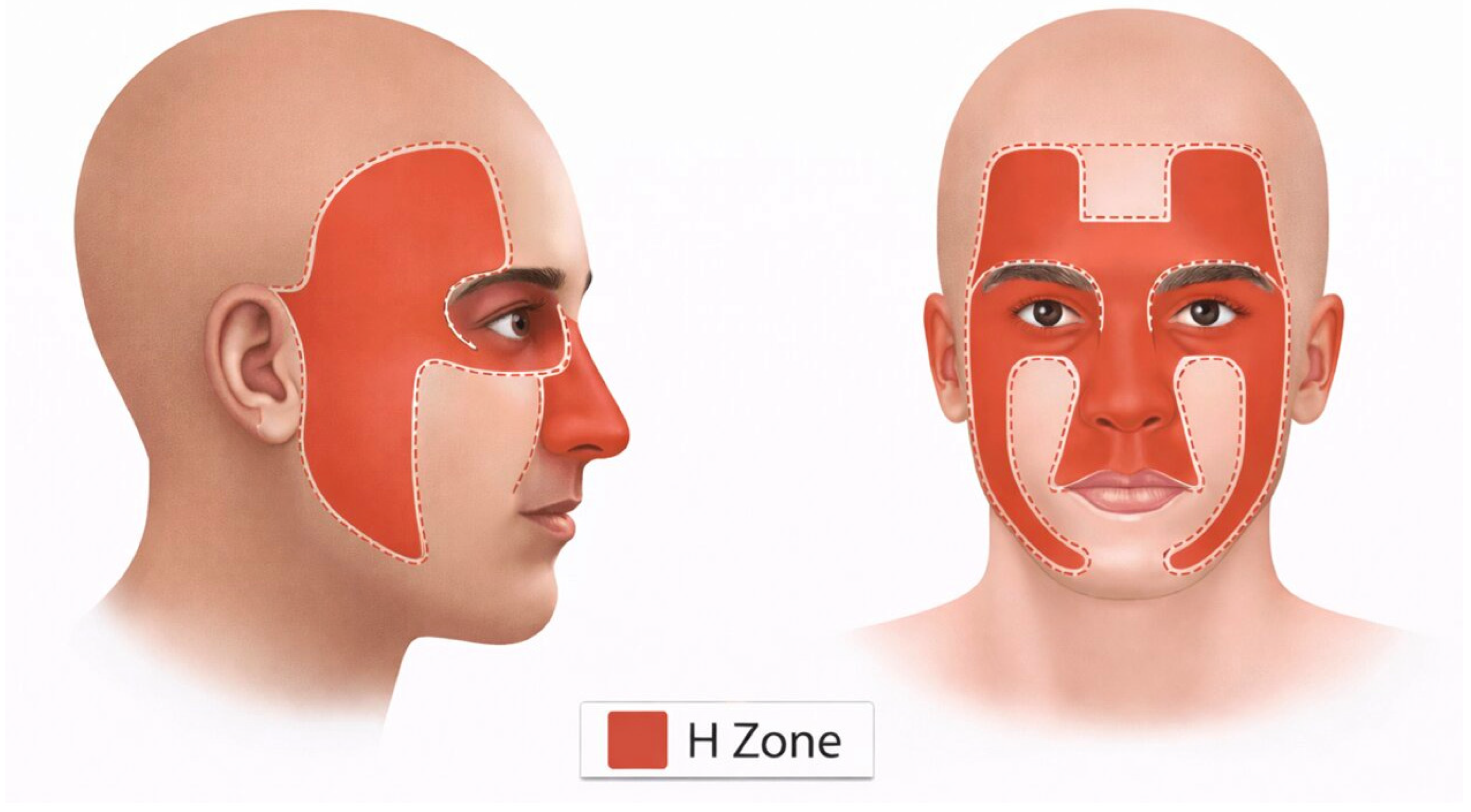
| Area H involvement | Eyelids, nose, lips, eyebrows, chin, temples, ears, preauricular skin; hands, feet, ankles, nail unit, nipple/areola complex, genitalia | – |
| Area M involvement | Cheeks, forehead, neck, jawline, scalp, pretibial surface (except superficial BCC < 0.6 cm in healthy patients) | Superficial BCC < 0.6 cm in healthy patients |
| Area L involvement | Recurrent aggressive or nodular BCC Primary aggressive BCC > 0.5 cm Primary nodular BCC > 2 cm (healthy) Primary nodular BCC > 1 cm (immunocompromised) | Primary superficial BCC |
| Borders | Poorly defined | Well defined |
| Primary vs. recurrent | Recurrent | Primary |
| Site of prior radiation therapy | Yes | No |
| Age at onset | <40 years | ≥40 years |
| Genetic syndromes | Gorlin syndrome, xeroderma pigmentosum | No |
| Histologic subtype | Morpheaform, infiltrative, micronodular, basosquamous | Superficial, nodular |
| Perineural invasion | Present | Absent |
| High-Risk cSCC | Low-Risk cSCC | |
|---|---|---|
| Clinical Characteristics | ||
| Location/size | Area H Area M ≥ 1 cm Area L ≥ 2 cm | Area M < 1 cm Area L < 2 cm |
| Borders | Poorly defined | Well defined |
| Primary vs. recurrent | Recurrent | Primary |
| Site of prior radiation therapy or chronic inflammatory process | Yes | No |
| Rapidly growing tumor | Yes | No |
| Neurologic symptoms | Yes | No |
| Pathologic Characteristics | ||
| High-risk subtype | Yes | No |
| Degree of differentiation | Poorly differentiated | Well or moderately differentiated |
| Depth | ≥2 mm or Clark level IV, V | <2 mm or Clark level I, II, III |
| Perineural or lymphovascular involvement | Yes | No |
Disclaimer/Publisher’s Note: The statements, opinions and data contained in all publications are solely those of the individual author(s) and contributor(s) and not of MDPI and/or the editor(s). MDPI and/or the editor(s) disclaim responsibility for any injury to people or property resulting from any ideas, methods, instructions or products referred to in the content. |
© 2026 by the authors. Licensee MDPI, Basel, Switzerland. This article is an open access article distributed under the terms and conditions of the Creative Commons Attribution (CC BY) license.
Share and Cite
Paradisi, A.; Brunetti, F.; Jeha, G.M.; Tolkachjov, S.N. Current and Future Perspectives in Mohs Micrographic Surgery for Non-Melanoma Skin Cancers: A Narrative Review. J. Clin. Med. 2026, 15, 4754. https://doi.org/10.3390/jcm15124754
Paradisi A, Brunetti F, Jeha GM, Tolkachjov SN. Current and Future Perspectives in Mohs Micrographic Surgery for Non-Melanoma Skin Cancers: A Narrative Review. Journal of Clinical Medicine. 2026; 15(12):4754. https://doi.org/10.3390/jcm15124754
Chicago/Turabian StyleParadisi, A., F. Brunetti, G. M. Jeha, and S. N. Tolkachjov. 2026. "Current and Future Perspectives in Mohs Micrographic Surgery for Non-Melanoma Skin Cancers: A Narrative Review" Journal of Clinical Medicine 15, no. 12: 4754. https://doi.org/10.3390/jcm15124754
APA StyleParadisi, A., Brunetti, F., Jeha, G. M., & Tolkachjov, S. N. (2026). Current and Future Perspectives in Mohs Micrographic Surgery for Non-Melanoma Skin Cancers: A Narrative Review. Journal of Clinical Medicine, 15(12), 4754. https://doi.org/10.3390/jcm15124754