Neurophysiology of Sleep-Deprivation Part 1: Effects of Sleep-Deprivation on Event-Related Potentials (ERPs)—Systematic and Mechanistic Review

Abstract
1. Introduction
2. Materials and Methods
2.1. Search Strategy and Information Sources
- sleep deprivation/total sleep deprivation/sleep loss/prolonged wakefulness
- event-related potentials/ERP/EEG evoked potentials/P300/N2/CNV/MMN/ERN/Pe
- Reference list screening of eligible articles, and
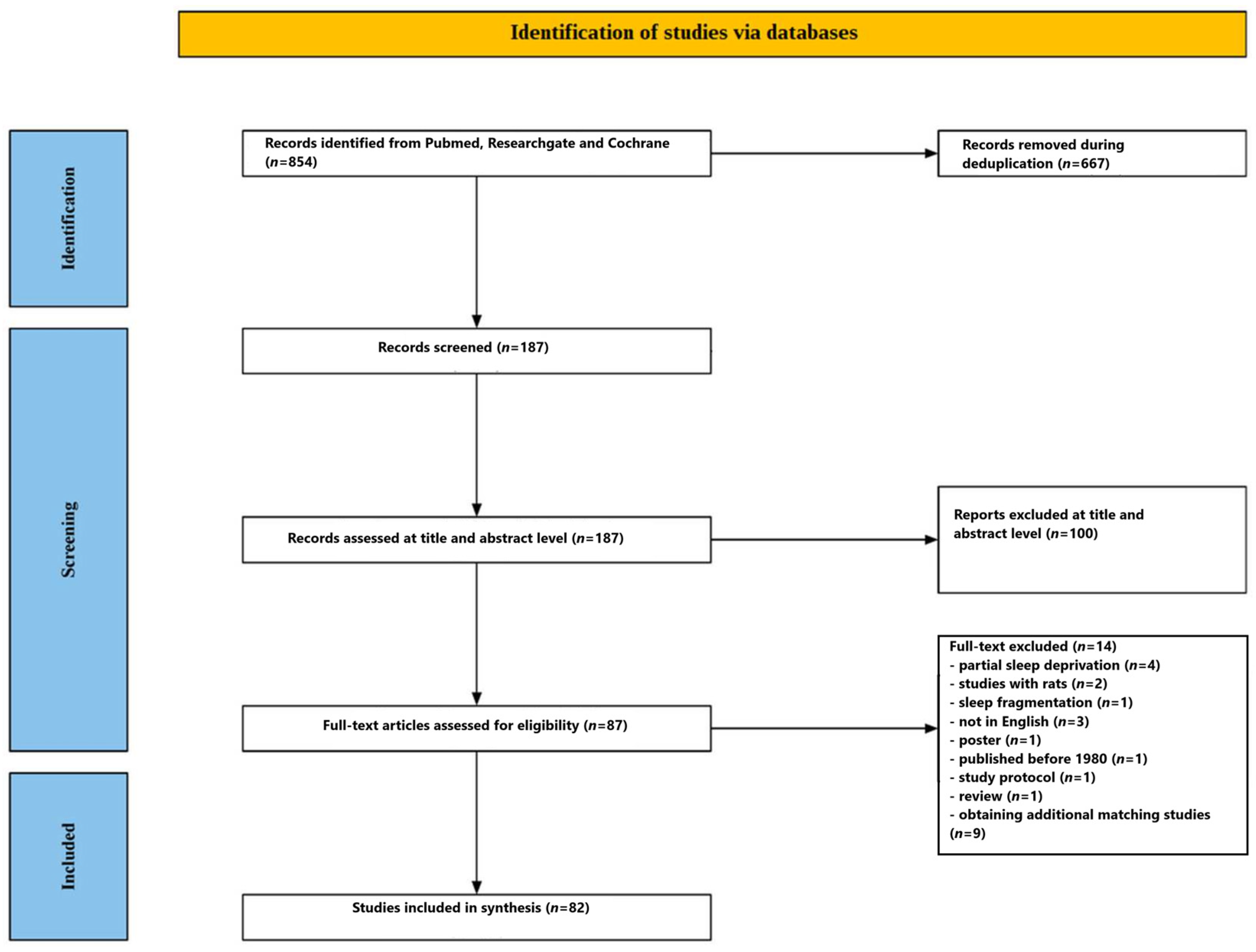
- Forward and backward citation searching (“similar,” cited, and citing articles), which yielded additional studies beyond the database search. This supplementary step is explicitly reflected in the review flow description.
2.2. Eligibility Criteria
2.2.1. Inclusion Criteria
- included human participants (healthy or clinical populations),
- examined sleep deprivation (primarily acute/total sleep deprivation),
- reported ERP outcomes derived from EEG (including standard ERP components such as P1/N1, P2/N2, P3/P300, CNV, MMN, ERN/Ne, Pe, LPP, etc.),
- used an experimental, quasi-experimental, or observational design with analysable ERP data,
- were published in English, and
- were available as full-text publications.
2.2.2. Exclusion Criteria
- examined partial sleep deprivation rather than the target exposure (total sleep deprivation),
- focused on sleep fragmentation instead of sleep deprivation,
- were animal studies (e.g., rat experiments),
- were non-English publications,
- were posters, study protocols, or reviews,
- or were published before 1980.
2.3. Study Selection Process
- Title and abstract screening, followed by
- Full-text review of potentially eligible records.
2.4. Data Extraction
- Number of study
- Study design (within-subject, between-group, crossover, intervention/countermeasure design)
- Participant characteristics (sample size, age, sex, population type, inclusion/exclusion criteria)
- Sleep deprivation protocol (duration of wakefulness, recovery sleep, naps, pharmacological countermeasures, control conditions)
- Task/paradigm (oddball, Go/NoGo, stop-signal, flanker, N-back, emotional tasks, vigilance tasks, etc.)
- ERP methodology (components analysed, scalp sites/regions, amplitude/latency windows, preprocessing/artifact handling when reported)
- Behavioural outcomes (accuracy, reaction time, errors, vigilance metrics)
- Main ERP findings (direction of effects, latency/amplitude changes, topographic or condition-specific differences)
- Recovery/countermeasure effects (if applicable)
2.5. Data Synthesis
- sleep deprivation duration and protocols,
- participant populations (healthy, clinical, specialised occupational/athlete cohorts),
- ERP tasks and cognitive domains,
- ERP preprocessing and measurement conventions,
- and outcomes reported (different components, latency vs. amplitude, stimulus-locked vs. response-locked analyses),
- Participant/sample characteristics,
- ERP paradigm/task family, and
- ERP component domain (e.g., P3/P300, early sensory/pre-attentive components, P2/N2, ERN/Pe, CNV, affective and memory-related components).
2.6. Risk of Bias Assessment
- RoB 2 for randomised studies, and
- ROBINS-I for non-randomised studies.
2.7. Registration of a Systematic Review
3. Results
3.1. Participants’ Characteristics
3.2. ERP Paradigms
- P2 was often interpreted as early selection or perceptual-attentional engagement,
- N2 as comparison/conflict monitoring or control, and
- P3 as updating/resource allocation/context revision.
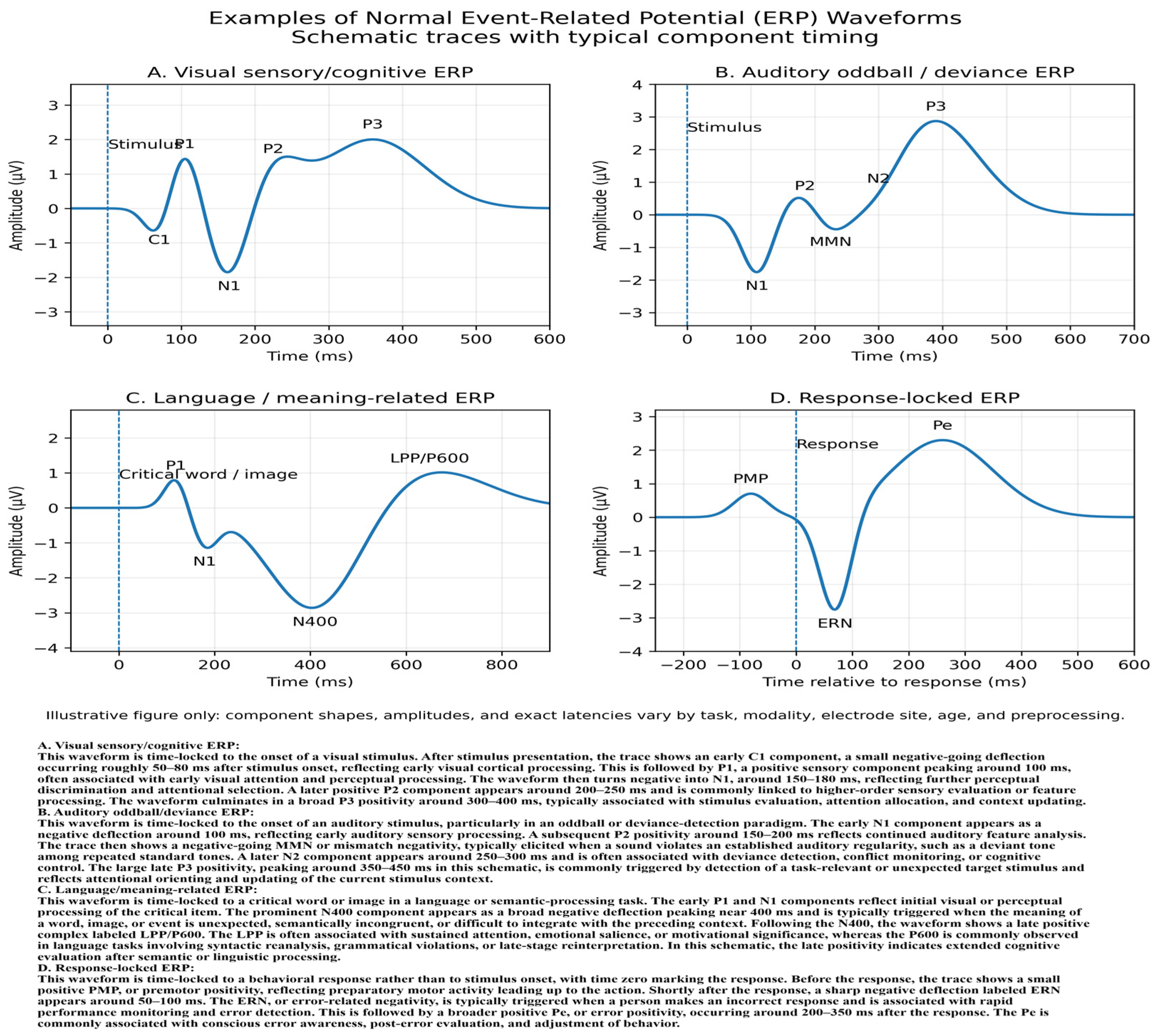
3.3. ERPs Results
3.3.1. Global Pattern: Late-Stage Vulnerability, Temporal Slowing, and Selective Compensation
3.3.2. P3/P300 Complex Across Paradigms: The Most Robust ERP Marker of Sleep Loss
3.3.3. Early Sensory and Perceptual Components (P1/N1/P2): Often Preserved, but Task-Dependent Vulnerabilities Are Common
3.3.4. MMN and Pre-Attentive Auditory Deviance Processing
3.3.5. N2 and Related Control-Oriented Negativities: Conflict Detection, Inhibition, and Switching Are Reliably Disrupted
3.3.6. Inhibitory Control and Executive Control: Dissociation of Early and Late Control Stages
3.3.7. Error Monitoring and Performance Monitoring (ERN/Ne, Pe): Weakened Frontal Monitoring and Reduced Error Differentiation
3.3.8. Working Memory ERPs: Staged Disruption of the P2–N2–P3 Complex
3.3.9. Selective Attention, Orienting, Visual Search, and Sensory–Motor Preparation
3.3.10. Auditory Processing, AEPs, and Sleep Inertia-Related ERP Changes
3.3.11. CNV and Preparatory Processing: Robust Vulnerability of Anticipatory Brain States
3.3.12. Novelty Processing, Involuntary Attention Capture, and Distraction
3.3.13. Emotion, Motivation, Empathy, Pain, and Semantic Processing: Late Positive and Domain-Specific ERP Alterations
3.3.14. Topography, Lateralisation, and Source/Network-Level Findings
3.3.15. Latency Effects as a General Signature of Slowed Neural Processing
3.3.16. Recovery Sleep, Naps, Pharmacological/Behavioural Countermeasures, and Reversibility
3.3.17. ERP–Behaviour Relationships, Individual Differences, and Translational Relevance
3.3.18. Risk of Bias Assessment
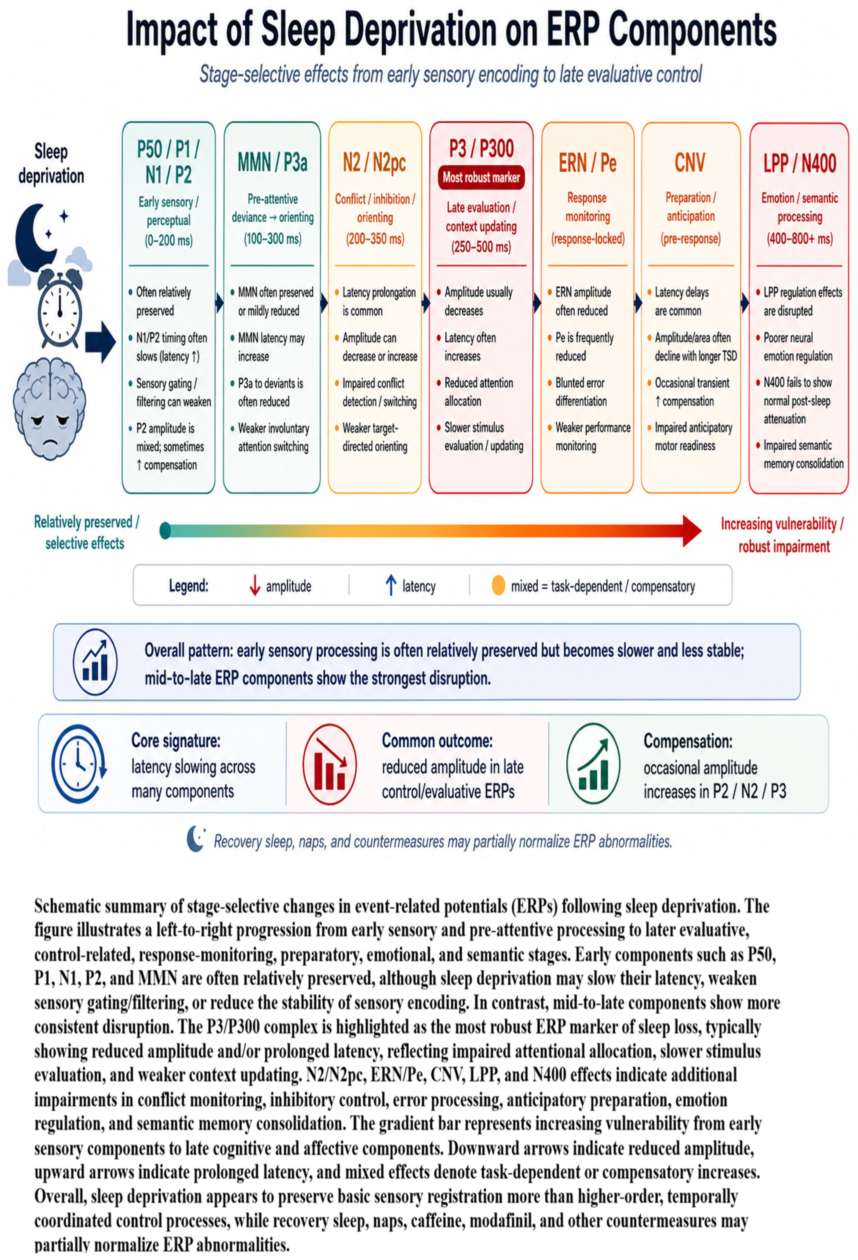
3.3.19. Synthetic Summary of the Evidence
4. Discussion
4.1. Overall Pattern of ERP Changes After Sleep Deprivation
4.2. P300/P3 as the Most Consistent Marker of Sleep Loss Effects
4.3. Early Sensory and Pre-Attentive Processing: Relative Preservation with Selective Vulnerability
4.4. P2 and N2: Intermediate Markers of Selection, Conflict, and Compensatory Control
4.5. Performance Monitoring After Sleep Deprivation: ERN/Ne–Pe Dissociation
4.6. Novelty Detection, Orienting, Selective Attention, and Reorienting
4.7. CNV and Anticipatory Preparation: Impaired Readiness Before Stimulus Onset
4.8. Semantic Processing, Recognition Memory, and Sleep-Dependent Representational Updating
4.9. Affective and Social Processing: Altered Emotional Salience, Regulation, and Face-Related ERPs
4.10. Sensory Gating, Salience Processing, and Modality-Specific ERP Effects
4.11. Oscillatory State, Phase Locking, and Temporal Instability of ERP Responses
4.12. Recovery, Naps, Countermeasures, and Component-Specific Restoration
5. Homeostatic vs. Circadian Influences: Two-Process Model Integration
6. Conclusions
Supplementary Materials
Author Contributions
Funding
Institutional Review Board Statement
Informed Consent Statement
Data Availability Statement
Conflicts of Interest
References
- Banks, S.; Dinges, D.F. Behavioral and physiological consequences of sleep restriction. J. Clin. Sleep Med. 2007, 3, 519–528. [Google Scholar] [CrossRef] [PubMed]
- Alhola, P.; Polo-Kantola, P. Sleep deprivation: Impact on cognitive performance. Neuropsychiatr. Dis. Treat. 2007, 3, 553–567. [Google Scholar]
- Durmer, J.S.; Dinges, D.F. Neurocognitive consequences of sleep deprivation. Semin. Neurol. 2005, 25, 117–129. [Google Scholar] [CrossRef] [PubMed]
- Dinges, D.; Lim, J. Sleep Deprivation and Vigilant Attention. Ann. N. Y. Acad. Sci. 2008, 1129, 305–322. [Google Scholar]
- Lowe, C.J.; Safati, A.; Hall, P.A. The neurocognitive consequences of sleep restriction: A meta-analytic review. Neurosci. Biobehav. Rev. 2017, 80, 586–604. [Google Scholar] [CrossRef]
- Belenky, G.; Wesensten, N.J.; Thorne, D.R.; Thomas, M.L.; Sing, H.C.; Redmond, D.P.; Russo, M.B.; Balkin, T.J. Patterns of performance degradation and restoration during sleep restriction and subsequent recovery: A sleep dose-response study. J. Sleep Res. 2003, 12, 1–12. [Google Scholar] [CrossRef]
- Van Dongen, H.P.; Maislin, G.; Mullington, J.M.; Dinges, D.F. The cumulative cost of additional wakefulness: Dose-response effects on neurobehavioral functions and sleep physiology from chronic sleep restriction and total sleep deprivation. Sleep 2003, 26, 117–126. [Google Scholar] [CrossRef]
- Killgore, W.D. Effects of sleep deprivation on cognition. Prog. Brain Res. 2010, 185, 105–129. [Google Scholar]
- Goel, N.; Rao, H.; Durmer, J.S.; Dinges, D.F. Neurocognitive consequences of sleep deprivation. Semin. Neurol. 2009, 29, 320–339. [Google Scholar] [CrossRef] [PubMed]
- Williamson, A.M.; Feyer, A.M. Moderate sleep deprivation produces impairments in cognitive and motor performance equivalent to legally prescribed levels of alcohol intoxication. Occup. Environ. Med. 2000, 57, 649–655. [Google Scholar] [CrossRef]
- Dawson, D.; Reid, K. Fatigue, alcohol and performance impairment. Nature 1997, 388, 235. [Google Scholar] [CrossRef]
- Leproult, R.; Van Cauter, E. Role of sleep and sleep loss in hormonal release and metabolism. Endocr. Dev. 2010, 17, 11–21. [Google Scholar]
- Spiegel, K.; Leproult, R.; Van Cauter, E. Impact of sleep debt on metabolic and endocrine function. Lancet 1999, 354, 1435–1439. [Google Scholar] [CrossRef] [PubMed]
- Taheri, S.; Lin, L.; Austin, D.; Young, T.; Mignot, E. Short sleep duration is associated with reduced leptin, elevated ghrelin, and increased body mass index. PLoS Med. 2004, 1, e62. [Google Scholar] [CrossRef]
- Aldabal, L.; Bahammam, A.S. Metabolic, endocrine, and immune consequences of sleep deprivation. Open Respir. Med. J. 2011, 5, 31–43. [Google Scholar] [CrossRef] [PubMed]
- Crispim, C.A.; Zalcman, I.; Dáttilo, M.; Padilha, H.G.; Edwards, B.; Waterhouse, J.; Tufik, S.; de Mello, M.T. The influence of sleep and sleep loss upon food intake and metabolism. Nutr. Res. Rev. 2007, 20, 195–212. [Google Scholar] [CrossRef]
- Itani, O.; Jike, M.; Watanabe, N.; Kaneita, Y. Short sleep duration and health outcomes: A systematic review, meta-analysis, and meta-regression. Sleep Med. 2017, 32, 246–256. [Google Scholar] [CrossRef] [PubMed]
- Cappuccio, F.P.; Cooper, D.; D’Elia, L.; Strazzullo, P.; Miller, M.A. Sleep duration predicts cardiovascular outcomes: A systematic review and meta-analysis of prospective studies. Eur. Heart J. 2011, 32, 1484–1492. [Google Scholar] [CrossRef]
- Irwin, M.R. Sleep and inflammation: Partners in sickness and in health. Nat. Rev. Immunol. 2019, 19, 702–715. [Google Scholar] [CrossRef]
- Besedovsky, L.; Lange, T.; Haack, M. The Sleep-Immune Crosstalk in Health and Disease. Physiol. Rev. 2019, 99, 1325–1380. [Google Scholar] [CrossRef]
- Irwin, M.R.; Olmstead, R.; Carroll, J.E. Sleep Disturbance, Sleep Duration, and Inflammation: A Systematic Review and Meta-Analysis of Cohort Studies and Experimental Sleep Deprivation. Biol. Psychiatry 2016, 80, 40–52. [Google Scholar] [CrossRef] [PubMed]
- Irwin, M.; Opp, M. Sleep Health: Reciprocal Regulation of Sleep and Innate Immunity. Neuropsychopharmacology 2017, 42, 129–155. [Google Scholar]
- Goldstein, A.N.; Walker, M.P. The role of sleep in emotional brain function. Annu. Rev. Clin. Psychol. 2014, 10, 679–708. [Google Scholar] [CrossRef]
- van der Helm, E.; Walker, M.P. Sleep and affective brain regulation. Soc. Personal. Psychol. Compass 2012, 6, 773–791. [Google Scholar] [CrossRef]
- Walker, M.P.; van der Helm, E. Overnight therapy? The role of sleep in emotional brain processing. Psychol. Bull. 2009, 135, 731–748. [Google Scholar] [CrossRef]
- Watson, N.F.; Badr, M.S.; Belenky, G.; Bliwise, D.L.; Buxton, O.M.; Buysse, D.; Dinges, D.F.; Gangwisch, J.; Grandner, M.A.; Kushida, C.; et al. Recommended Amount of Sleep for a Healthy Adult: A Joint Consensus Statement of the American Academy of Sleep Medicine and Sleep Research Society. Sleep 2015, 38, 843–844. [Google Scholar] [CrossRef]
- Luck, S.J. An Introduction to the Event-Related Potential Technique, 2nd ed.; MIT Press: Cambridge, MA, USA, 2014. [Google Scholar]
- Picton, T.W.; Bentin, S.; Berg, P.; Donchin, E.; Hillyard, S.A.; Johnson, R., Jr.; Miller, G.A.; Ritter, W.; Ruchkin, D.S.; Rugg, M.D.; et al. Guidelines for using human event-related potentials to study cognition: Recording standards and publication criteria. Psychophysiology 2000, 37, 127–152. [Google Scholar] [CrossRef]
- Woodman, G.F. A brief introduction to the use of event-related potentials in studies of perception and attention. Atten. Percept. Psychophys. 2010, 72, 2031–2046. [Google Scholar] [CrossRef] [PubMed]
- Polich, J. Updating P300: An integrative theory of P3a and P3b. Clin. Neurophysiol. 2007, 118, 2128–2148. [Google Scholar] [CrossRef]
- Kappenman, E.S.; Luck, S.J. Best Practices for Event-Related Potential Research in Clinical Populations. Biol. Psychiatry Cogn. Neurosci. Neuroimaging 2016, 1, 110–115. [Google Scholar] [PubMed]
- Gratton, G.; Coles, M.G.; Donchin, E. A new method for off-line removal of ocular artifact. Electroencephalogr. Clin. Neurophysiol. 1983, 55, 468–484. [Google Scholar] [CrossRef]
- Delorme, A.; Makeig, S. EEGLAB: An open source toolbox for analysis of single-trial EEG dynamics including independent component analysis. J. Neurosci. Methods 2004, 134, 9–21. [Google Scholar] [CrossRef] [PubMed]
- Panjwani, U.; Ray, K.; Chatterjee, A.; Bhaumik, S.; Kumar, S. Electrophysiological correlates of cognition improve with nap during sleep deprivation. Eur. J. Appl. Physiol. 2010, 108, 549–556. [Google Scholar] [CrossRef]
- Zhang, Q.; Liao, Y.; Qi, J.; Zhao, Y.; Zhu, T.; Liu, Z.; Liu, X. A visual ERP study of impulse inhibition following a zaleplon-induced nap after sleep deprivation. PLoS ONE 2014, 9, e95653. [Google Scholar] [CrossRef]
- Jackson, M.L.; Croft, R.J.; Owens, K.; Pierce, R.J.; Kennedy, G.A.; Crewther, D.; Howard, M.E. The effect of acute sleep deprivation on visual evoked potentials in professional drivers. Sleep 2008, 31, 1261–1269. [Google Scholar] [CrossRef][Green Version]
- Lee, H.J.; Kim, L.; Suh, K.Y. Cognitive deterioration and changes of P300 during total sleep deprivation. Psychiatry Clin. Neurosci. 2003, 57, 490–496. [Google Scholar] [CrossRef] [PubMed]
- Brauer, K.A. Sleep Deprivation and Recovery: The Effects of P300 Three and Six Hours Post Recovery. Ph.D. Thesis, Missouri State University, Springfield, MO, USA, 2016. Available online: https://bearworks.missouristate.edu/theses/2953 (accessed on 30 March 2026).
- Lee, H.J.; Kim, L.; Kim, Y.K.; Suh, K.Y.; Han, J.; Park, M.K.; Park, K.W.; Lee, D.H. Auditory event-related potentials and psychological changes during sleep deprivation. Neuropsychobiology 2004, 50, 1–5. [Google Scholar] [CrossRef]
- Jones, K.B.; Frizzell, T.; Fickling, S.; Pawlowski, G.; Brodie, S.M.; Lakhani, B.; Venter, J.; D’Arcy, R.C.N. Brain vital sign monitoring of sleep deprivation detects situational cognitive impairment. Front. Hum. Neurosci. 2024, 18, 1358551. [Google Scholar] [CrossRef] [PubMed]
- Zukerman, G.; Goldstein, A.; Babkoff, H. The effect of 24–40 hours of sleep deprivation on the P300 response to auditory target stimuli. Aviat. Space Environ. Med. 2007, 78, B216–B223. [Google Scholar]
- Ren, Z.; Mao, X.; Zhang, Z.; Wang, W. The impact of sleep deprivation on cognitive function in healthy adults: Insights from auditory P300 and reaction time analysis. Front. Neurosci. 2025, 19, 1559969. [Google Scholar] [CrossRef]
- Trujillo, L.T.; Kornguth, S.; Schnyer, D.M. An ERP examination of the different effects of sleep deprivation on exogenously cued and endogenously cued attention. Sleep 2009, 32, 1285–1297. [Google Scholar] [CrossRef]
- Zhang, L.; Shao, Y.; Liu, Z.; Li, C.; Chen, Y.; Zhou, Q. Decreased Information Replacement of Working Memory After Sleep Deprivation: Evidence from an Event-Related Potential Study. Front. Neurosci. 2019, 13, 408. [Google Scholar] [CrossRef]
- Kusztor, A.; Raud, L.; Juel, B.E.; Nilsen, A.S.; Storm, J.F.; Huster, R.J. Sleep deprivation differentially affects subcomponents of cognitive control. Sleep 2019, 42, zsz016. [Google Scholar] [CrossRef]
- Peng, Z.; Dai, C.; Ba, Y.; Zhang, L.; Shao, Y.; Tian, J. Effect of Sleep Deprivation on the Working Memory-Related N2-P3 Components of the Event-Related Potential Waveform. Front. Neurosci. 2020, 14, 469. [Google Scholar] [CrossRef] [PubMed]
- Song, T.; Yu, K.; Wang, L.; Xu, L.; Xu, M.; Peng, Z.; Dai, C.; Wang, H.; Yang, T.; Shao, Y.; et al. Total Sleep Deprivation Triggers Greater Activation in the Parietal Brain in the Visual Working Memory Updating Processes: An Event-Related Potentials Study. Front. Neurosci. 2022, 16, 736437. [Google Scholar] [CrossRef]
- Peng, Z.; Dai, C.; Cai, X.; Zeng, L.; Li, J.; Xie, S.; Wang, H.; Yang, T.; Shao, Y.; Wang, Y. Total Sleep Deprivation Impairs Lateralization of Spatial Working Memory in Young Men. Front. Neurosci. 2020, 14, 562035. [Google Scholar] [CrossRef] [PubMed]
- Yin, Y.; Chen, S.; Song, T.; Zhou, Q.; Shao, Y. Cognitive Load Moderates the Effects of Total Sleep Deprivation on Working Memory: Evidence from Event-Related Potentials. Brain Sci. 2023, 13, 898. [Google Scholar] [CrossRef]
- Izullah, F.R.; Koivisto, M.; Nieminen, V.; Luimula, M.; Hämäläinen, H. Aging and sleep deprivation affect different neurocognitive stages of spatial information processing during a virtual driving task—An ERP study. Transp. Res. Part F Traffic Psychol. Behav. 2022, 89, 399–406. [Google Scholar] [CrossRef]
- Renn, R.P.; Cote, K.A. Performance monitoring following total sleep deprivation: Effects of task type and error rate. Int. J. Psychophysiol. 2013, 88, 64–73. [Google Scholar] [CrossRef]
- Qi, J.-L.; Shao, Y.-C.; Miao, D.; Fan, M.; Bi, G.-H.; Yang, Z. The effects of 43 hours of sleep deprivation on executive control functions: Event-related potentials in a visual Go/No go task. Soc. Behav. Personal. Int. J. 2010, 38, 29–42. [Google Scholar] [CrossRef]
- Jin, X.; Ye, E.; Qi, J.; Wang, L.; Lei, Y.; Chen, P.; Mi, G.; Zou, F.; Shao, Y.; Yang, Z. Recovery Sleep Reverses Impaired Response Inhibition Due to Sleep Restriction: Evidence from a Visual Event Related Potentials Study. PLoS ONE 2015, 10, e0142361. [Google Scholar]
- Liu, Q.; Zhou, R.; Liu, L.; Zhao, X. Effects of 72hours total sleep deprivation on male astronauts’ executive functions and emotion. Compr. Psychiatry 2015, 61, 28–35. [Google Scholar] [CrossRef]
- Gosselin, A.; De Koninck, J.; Campbell, K.B. Disentangling specific inhibitory versus general decision-making processes during sleep deprivation using a Go/NoGo ERP paradigm. Int. J. Psychophysiol. 2019, 141, 18–27. [Google Scholar]
- Xu, L.; Song, T.; Peng, Z.; Dai, C.; Wang, L.; Shao, Y.; Wang, L.; Weng, X.; Han, M. Acute Sleep Deprivation Impairs Motor Inhibition in Table Tennis Athletes: An ERP Study. Brain Sci. 2022, 12, 746. [Google Scholar] [CrossRef]
- Peng, Z.; Hou, Y.; Xu, L.; Wang, H.; Wu, S.; Song, T.; Shao, Y.; Yang, Y. Recovery sleep attenuates impairments in working memory following total sleep deprivation. Front. Neurosci. 2023, 17, 1056788. [Google Scholar] [CrossRef]
- Gosselin, A.; De Koninck, J.; Campbell, K.B. Total sleep deprivation and novelty processing: Implications for frontal lobe functioning. Clin. Neurophysiol. 2005, 116, 211–222. [Google Scholar] [CrossRef] [PubMed]
- Muller-Gass, A.; Campbell, K. Sleep deprivation moderates neural processes associated with passive auditory capture. Brain Cogn. 2019, 132, 89–97. [Google Scholar] [CrossRef]
- Peszka, J.; Harsh, J. Effect of sleep deprivation on NREM sleep ERPs and related activity at sleep onset. Int. J. Psychophysiol. 2002, 46, 275–286. [Google Scholar] [CrossRef]
- Wu, S.; Yue, P.; Wu, L.; Wang, C.; Lin, X.; Li, X. Electrophysiological Mechanism of Attention of Sleep Deprivation: Evidence from Event-Related Potentials (ERP) Data. Cureus 2023, 15, e33464. [Google Scholar] [CrossRef]
- Chatterjee, A.; Ray, K.; Panjwani, U.; Thakur, L.; Anand, J.P. Meditation as an intervention for cognitive disturbances following total sleep deprivation. Indian J. Med. Res. 2012, 136, 1031–1038. [Google Scholar] [PubMed]
- Ray, K.; Chatterjee, A.; Panjwani, U.; Kumar, S.; Sahu, S.; Ghosh, S.; Thakur, L.; Anand, J.P. Modafinil improves event related potentials P300 and contingent negative variation after 24 h sleep deprivation. Life Sci. 2012, 91, 94–99. [Google Scholar] [CrossRef]
- Szelenberger, W.; Piotrowski, T.; Dabrowska, A.J. Increased prefrontal event-related current density after sleep deprivation. Acta Neurobiol. Exp. 2005, 65, 19–28. [Google Scholar] [CrossRef] [PubMed]
- López Zunini, R.; Muller-Gass, A.; Campbell, K. The effects of total sleep deprivation on semantic priming: Event-related potential evidence for automatic and controlled processing strategies. Brain Cogn. 2014, 84, 14–25. [Google Scholar] [CrossRef]
- Corsi-Cabrera, M.; Arce, C.; Del Río-Portilla, I.Y.; Pérez-Garci, E.; Guevara, M.A. Amplitude reduction in visual event-related potentials as a function of sleep deprivation. Sleep 1999, 22, 181–189. [Google Scholar] [CrossRef]
- Shao, Y.; Xu, L.; Peng, Z.; An, X.; Gong, J.; Han, M. Non-Linear Effects of Acute Sleep Deprivation on Spatial Working Memory: Cognitive Depletion and Neural Compensation. Brain Sci. 2025, 15, 18. [Google Scholar] [CrossRef] [PubMed]
- Lee, C.-S.; Hsieh, S.; Young, H.-Y.; Tsai, L.-L. Impairment of Error Monitoring Following Sleep Deprivation. Sleep 2005, 28, 707–713. [Google Scholar] [CrossRef]
- Lian, J.; Xu, L.; Song, T.; Peng, Z.; Gong, X.; Chen, J.; Zhong, X.; An, X.; Chen, S.; Shao, Y. Decreased Functional Connectivity of Brain Networks in the Alpha Band After Sleep Deprivation Is Associated with Decreased Inhibitory Control in Young Male Adults. Int. J. Environ. Res. Public Health 2023, 20, 4663. [Google Scholar] [CrossRef] [PubMed]
- Magnuson, J.R.; Kang, H.J.; Dalton, B.H.; McNeil, C.J. Neural effects of sleep deprivation on inhibitory control and emotion processing. Behav. Brain Res. 2022, 426, 113845. [Google Scholar] [CrossRef]
- Li, Y.; Ma, M.; Shao, Y.; Wang, W. Enhanced effective connectivity from the middle frontal gyrus to the parietal lobe is associated with impaired mental rotation after total sleep deprivation: An electroencephalogram study. Front. Neurosci. 2022, 16, 910618. [Google Scholar] [CrossRef]
- Lam, Y.C.; Li, C.; Hsiao, J.H.; Lau, E.Y.Y. A sleepless night disrupts the resolution of emotional conflicts: Behavioural and neural evidence. J. Sleep Res. 2024, 33, e14176. [Google Scholar] [CrossRef]
- Hsieh, S.; Li, T.H.; Tsai, L.L. Impact of monetary incentives on cognitive performance and error monitoring following sleep deprivation. Sleep 2010, 33, 499–507. [Google Scholar] [CrossRef] [PubMed]
- Alfarra, R.; Fins, A.I.; Chayo, I.; Tartar, J.L. Changes in attention to an emotional task after sleep deprivation: Neurophysiological and behavioral findings. Biol. Psychol. 2015, 104, 1–7. [Google Scholar] [CrossRef]
- Mograss, M.A.; Guillem, F.; Brazzini-Poisson, V.; Godbout, R. The effects of total sleep deprivation on recognition memory processes: A study of event-related potential. Neurobiol. Learn. Mem. 2009, 91, 343–352. [Google Scholar] [CrossRef] [PubMed]
- Hsieh, S.; Cheng, I.C.; Tsai, L.L. Immediate error correction process following sleep deprivation. J. Sleep Res. 2007, 16, 137–147. [Google Scholar] [CrossRef]
- Sun, L.; Liang, S.; Yu, S.; He, J. Effects of sleep deprivation and hazard types on the hazard perception of young novice drivers: An ERP study. Neurosci. Lett. 2024, 827, 137739. [Google Scholar] [CrossRef]
- Krull, K.R.; Smith, L.T.; Sinha, R.; Parsons, O.A. Simple reaction time event-related potentials: Effects of alcohol and sleep deprivation. Alcohol. Clin. Exp. Res. 1993, 17, 771–777. [Google Scholar] [CrossRef]
- Liu, L.; Zhou, R. Effect of 72 h of Sleep Deprivation on the Iowa Gambling Task. Noro Psikiyatr. Ars. 2016, 53, 357–360. [Google Scholar] [CrossRef]
- Wiggins, E.; Mottarella, M.; Good, K.; Eggleston, S.; Stevens, C. 24-h sleep deprivation impairs early attentional modulation of neural processing: An event-related brain potential study. Neurosci. Lett. 2018, 677, 32–36. [Google Scholar] [CrossRef] [PubMed]
- Tavakoli, P.; Murkar, A.; Porteous, M.; Carrier, J.; Robillard, R. The Effects of Total Sleep Deprivation on Attention Capture Processes in Young and Older Adults: An ERP Study. Exp. Aging Res. 2023, 49, 130–151. [Google Scholar] [CrossRef]
- Cote, K.A.; Mondloch, C.J.; Sergeeva, V.; Taylor, M.; Semplonius, T. Impact of total sleep deprivation on behavioural neural processing of emotionally expressive faces. Exp. Brain Res. 2014, 232, 1429–1442. [Google Scholar] [CrossRef]
- Danos, P.; Kasper, S.; Scholl, H.P.; Kaiser, J.; Ruhrmann, S.; Höflich, G.; Möller, H.J. Clinical response to sleep deprivation and auditory-evoked potentials--preliminary results. Pharmacopsychiatry 1994, 27, 70–71. [Google Scholar] [PubMed]
- Bortoletto, M.; Tona Gde, M.; Scozzari, S.; Sarasso, S.; Stegagno, L. Effects of sleep deprivation on auditory change detection: A N1-mismatch negativity study. Int. J. Psychophysiol. 2011, 81, 312–316. [Google Scholar] [CrossRef]
- Raz, A.; Deouell, L.Y.; Bentin, S. Is pre-attentive processing compromised by prolonged wakefulness? Effects of total sleep deprivation on the mismatch negativity. Psychophysiology 2001, 38, 787–795. [Google Scholar] [CrossRef]
- Murphy, T.I.; Richard, M.; Masaki, H.; Segalowitz, S.J. The effect of sleepiness on performance monitoring: I know what I am doing, but do I care? J. Sleep Res. 2006, 15, 15–21. [Google Scholar] [CrossRef] [PubMed]
- Boardman, J.M.; Cross, Z.R.; Bravo, M.M.; Andrillon, T.; Aidman, E.; Anderson, C.; Drummond, S.P.A. Awareness of errors is reduced by sleep loss. Psychophysiology 2024, 61, e14523. [Google Scholar] [CrossRef]
- Zhang, Y.; Miao, H.; Wang, C.; Wu, B.; Chen, X.; Chi, L. Effects of acute sleep deprivation on post-error adjustments and error processing. Int. J. Psychophysiol. 2025, 211, 112554. [Google Scholar] [CrossRef]
- Hoedlmoser, K.; Griessenberger, H.; Fellinger, R.; Freunberger, R.; Klimesch, W.; Gruber, W.; Schabus, M. Event-related activity and phase locking during a psychomotor vigilance task over the course of sleep deprivation. J. Sleep Res. 2011, 20, 377–385. [Google Scholar]
- Eidelman-Rothman, M.; Ben-Simon, E.; Freche, D.; Keil, A.; Hendler, T.; Levit-Binnun, N. Sleepless and desynchronized: Impaired inter trial phase coherence of steady-state potentials following sleep deprivation. Neuroimage 2019, 202, 116055. [Google Scholar] [CrossRef]
- Zhang, L.; Huang, Y.; Zhang, Y.; Xin, W.; Shao, Y.; Yang, Y. Enhanced high-frequency precuneus-cortical effective connectivity is associated with decreased sensory gating following total sleep deprivation. Neuroimage 2019, 197, 255–263. [Google Scholar]
- Ohga, T.; Miyao, M.; Sakakibara, H.; Yamada, S.; Terashima, M. The influence of sleep deprivation on the contingent negative variation. Nagoya J. Med. Sci. 1985, 47, 91–100. [Google Scholar] [PubMed]
- Zhang, X.; Feng, S.; Yang, X.; Peng, Y.; Du, M.; Zhang, R.; Sima, J.; Zou, F.; Wu, X.; Wang, Y.; et al. Neuroelectrophysiological alteration associated with cognitive flexibility after 24 h sleep deprivation in adolescents. Conscious. Cogn. 2024, 124, 103734. [Google Scholar] [CrossRef]
- Ferrara, M.; De Gennaro, L.; Ferlazzo, F.; Curcio, G.; Cristiani, R.; Bertini, M. Topographical changes in N1-P2 amplitude upon awakening from recovery sleep after slow-wave sleep deprivation. Clin. Neurophysiol. 2002, 113, 1183–1190. [Google Scholar] [CrossRef]
- Zhang, J.; Lau, E.Y.Y.; Hsiao, J.H.-W. Using emotion regulation strategies after sleep deprivation: ERP and behavioral findings. Cogn. Affect. Behav. Neurosci. 2019, 19, 283–295. [Google Scholar] [CrossRef] [PubMed]
- Song, T.; Xu, L.; Peng, Z.; Wang, L.; Dai, C.; Xu, M.; Shao, Y.; Wang, Y.; Li, S. Total sleep deprivation impairs visual selective attention and triggers a compensatory effect: Evidence from event-related potentials. Cogn. Neurodyn. 2023, 17, 621–631. [Google Scholar] [CrossRef]
- Ramdani, C.; Carbonnell, L.; Rabat, A.; Meckler, C.; Burle, B.; Hasbroucq, T.; Vidal, F. Sleep deprivation affects the sensitivity of proactive and reactive action monitoring: A behavioural and ERP analysis. Biol. Psychol. 2013, 93, 237–245. [Google Scholar] [CrossRef]
- Shao, Y.; Peng, Z.; Xu, L.; Lian, J.; An, X.; Cheng, M.-Y. Decrease in the P2 Amplitude of Object Working Memory after 8 h-Recovery Sleep Following 36 h-Total Sleep Deprivation: An ERP Study. Brain Sci. 2023, 13, 1470. [Google Scholar] [CrossRef] [PubMed]
- Díaz-Leines, S.; Gama-Moreno, O.; Poblano, A.; Flores-Avalos, B. Psychoacoustics and Neurophysiological Alterations after 30-36 Hours of Sleep Deprivation. Neuropsychobiology 2017, 75, 89–95. [Google Scholar] [CrossRef] [PubMed]
- Chen, S.; Song, T.; Peng, Z.; Xu, L.; Lian, J.; An, X.; Shao, Y. Total Sleep Deprivation Triggers a Compensatory Mechanism During Conflict Monitoring Process: Evidence from Event-Related Potentials. Arch. Clin. Neuropsychol. 2024, 39, 367–377. [Google Scholar] [CrossRef]
- Duan, H.; Wang, Y.J.; Lei, X. The effect of sleep deprivation on empathy for pain: An ERP study. Neuropsychologia 2021, 163, 108084. [Google Scholar] [CrossRef]
- Song, T.; Du, F.; Xu, L.; Peng, Z.; Wang, L.; Dai, C.; Xu, M.; Zhang, Y.; Shao, Y.; Weng, X.; et al. Total sleep deprivation selectively impairs motor preparation sub-stages in visual search task: Evidence from lateralized readiness potentials. Front. Neurosci. 2023, 17, 989512. [Google Scholar] [CrossRef]
- Zhang, L.; Shao, Y.; Jin, X.; Cai, X.; Du, F. Decreased effective connectivity between insula and anterior cingulate cortex during a working memory task after prolonged sleep deprivation. Behav. Brain Res. 2021, 409, 113263. [Google Scholar] [CrossRef]
- Schuh-Hofer, S.; Baumgärtner, U.; Treede, R.D. Effect of sleep deprivation on the electrophysiological signature of habituation to noxious laser stimuli. Eur. J. Pain 2015, 19, 1197–1209. [Google Scholar] [CrossRef]
- Xu, L.; Yang, X.; Peng, Z.; Song, T.; Wang, L.; Dai, C.; Xu, M.; Shao, Y.; Lv, J. Modafinil ameliorates the decline in pronunciation-related working memory caused by 36-h acute total sleep deprivation: An ERP study. Neurobiol. Learn. Mem. 2022, 192, 107625. [Google Scholar] [CrossRef]
- Peeke, S.C.; Callaway, E.; Jones, R.T.; Stone, G.C.; Doyle, J. Combined effects of alcohol and sleep deprivation in normal young adults. Psychopharmacology 1980, 67, 279–287. [Google Scholar] [CrossRef]
- Gauthier, P.; Gottesmann, C. Influence of total sleep deprivation on event-related potentials in man. Psychophysiology 1983, 20, 351–355. [Google Scholar] [CrossRef]
- Song, Y.; Huang, Y.; Gao, Y.; Zhang, M.; Shao, Y.; Zhou, G.; Sun, H.; Wang, G.; Jia, T.; Shi, J.; et al. Stress Management in Athletes: Predictive Effects of Sleep Deprivation-Induced Cognitive Control Changes on Competition Performance. J. Neurosci. 2025, 45, e1683242025. [Google Scholar] [CrossRef]
- Pressman, M.R.; Spielman, A.J.; Pollak, C.P.; Weitzman, E.D. Long-latency auditory evoked responses during sleep deprivation and in narcolepsy. Sleep 1982, 5, S147–S156. [Google Scholar] [CrossRef] [PubMed]
- Smith, M.E.; McEvoy, L.K.; Gevins, A. The impact of moderate sleep loss on neurophysiologic signals during working-memory task performance. Sleep 2002, 25, 784–794. [Google Scholar] [CrossRef]
- Lin, C.C.; Yang, C.M. Evidence of sleep-facilitating effect on formation of novel semantic associations: An event-related potential (ERP) study. Neurobiol. Learn. Mem. 2014, 116, 69–78. [Google Scholar] [CrossRef] [PubMed]
- Chen, X.; Zhang, L.; Yang, D.; Li, C.; An, G.; Wang, J.; Shao, Y.; Fan, R.; Ma, Q. Effects of Caffeine on Event-Related Potentials and Neuropsychological Indices After Sleep Deprivation. Front. Behav. Neurosci. 2020, 14, 108. [Google Scholar] [CrossRef] [PubMed]
- Zhang, N.; Wu, X.; Cui, N.; Shao, Y.; Zhang, Y. Short-term recovery sleep attenuates the impairment of response inhibition after total sleep deprivation: Evidence from an event-related potentials study. Int. J. Psychophysiol. 2025, 212, 112563. [Google Scholar] [CrossRef]
- Humphrey, D.G.; Kramer, A.F.; Stanny, R.R. Influence of extended wakefulness on automatic and nonautomatic processing. Hum. Factors 1994, 36, 652–669. [Google Scholar] [CrossRef]
- Peng, Z.; Wang, Z.; Xu, L.; Shao, Y.; Jiao, F.; Lv, J. Sleep deprivation impairs spatial cognitive processing and Alters brain connectivity in table tennis athletes. Neuroscience 2025, 564, 13–20. [Google Scholar] [CrossRef] [PubMed]
- Kutas, M.; McCarthy, G.; Donchin, E. Augmenting mental chronometry: The P300 as a measure of stimulus evaluation time. Science 1977, 197, 792–795. [Google Scholar] [CrossRef] [PubMed]
- Donchin, E.; Coles, M.G. Is the P300 component a manifestation of context updating? Behav. Brain Sci. 1988, 11, 357–427. [Google Scholar] [CrossRef]
- Huang, W.J.; Chen, W.W.; Zhang, X. The neurophysiology of P 300--an integrated review. Eur. Rev. Med. Pharmacol. Sci. 2015, 19, 1480–1488. [Google Scholar]
- Tagliabue, C.F.; Veniero, D.; Benwell, C.S.Y.; Cecere, R.; Savazzi, S.; Thut, G. The EEG signature of sensory evidence accumulation during decision formation closely tracks subjective perceptual experience. Sci. Rep. 2019, 9, 4949. [Google Scholar] [CrossRef]
- Twomey, D.M.; Murphy, P.R.; Kelly, S.P.; O’Connell, R.G. The classic P300 encodes a build-to-threshold decision variable. Eur. J. Neurosci. 2015, 42, 1636–1643. [Google Scholar] [CrossRef]
- O’Connell, R.G.; Dockree, P.M.; Kelly, S.P. A supramodal accumulation-to-bound signal that determines perceptual decisions in humans. Nat. Neurosci. 2012, 15, 1729–1735. [Google Scholar] [CrossRef]
- Bledowski, C.; Prvulovic, D.; Hoechstetter, K.; Scherg, M.; Wibral, M.; Goebel, R.; Linden, D.E. Localizing P300 generators in visual target and distractor processing: A combined event-related potential and functional magnetic resonance imaging study. J. Neurosci. 2004, 24, 9353–9360. [Google Scholar] [CrossRef]
- Mantini, D.; Corbetta, M.; Perrucci, M.G.; Romani, G.L.; Del Gratta, C. Large-scale brain networks account for sustained and transient activity during target detection. Neuroimage 2009, 44, 265–274. [Google Scholar] [CrossRef]
- Stevens, M.C.; Laurens, K.R.; Liddle, P.F.; Kiehl, K.A. The hemodynamics of oddball processing during single-tone and two-tone target detection tasks. Int. J. Psychophysiol. 2006, 60, 292–303. [Google Scholar] [CrossRef] [PubMed]
- Delfin, C.; Wallinius, M.; Björnsdotter, M.; Ruzich, E.; Andiné, P. Prolonged NoGo P3 latency as a possible neurobehavioral correlate of aggressive and antisocial behaviors: A Go/NoGo ERP study. Biol. Psychol. 2022, 168, 108245. [Google Scholar] [CrossRef] [PubMed]
- Luijten, M.; Machielsen, M.W.; Veltman, D.J.; Hester, R.; de Haan, L.; Franken, I.H. Systematic review of ERP and fMRI studies investigating inhibitory control and error processing in people with substance dependence and behavioural addictions. J. Psychiatry Neurosci. 2014, 39, 149–169. [Google Scholar] [CrossRef] [PubMed] [PubMed Central]
- Smith, J.L.; Johnstone, S.J.; Barry, R.J. Movement-related potentials in the Go/NoGo task: The P3 reflects both cognitive and motor inhibition. Clin. Neurophysiol. 2008, 119, 704–714. [Google Scholar] [CrossRef] [PubMed]
- Hillyard, S.A.; Anllo-Vento, L. Event-related brain potentials in the study of visual selective attention. Proc. Natl. Acad. Sci. USA 1998, 95, 781–787. [Google Scholar] [CrossRef] [PubMed] [PubMed Central]
- Luck, S.J.; Heinze, H.J.; Mangun, G.R.; Hillyard, S.A. Visual event-related potentials index focused attention within bilateral stimulus arrays. II. Functional dissociation of P1 and N1 components. Electroencephalogr. Clin. Neurophysiol. 1990, 75, 528–542. [Google Scholar] [CrossRef] [PubMed]
- Näätänen, R.; Michie, P.T. Early selective-attention effects on the evoked potential: A critical review and reinterpretation. Biol. Psychol. 1979, 8, 81–136. [Google Scholar] [CrossRef] [PubMed]
- Hansen, J.C.; Hillyard, S.A. Endogenous brain potentials associated with selective auditory attention. Electroencephalogr. Clin. Neurophysiol. 1980, 49, 277–290. [Google Scholar] [CrossRef] [PubMed]
- Woldorff, M.G.; Hillyard, S.A. Modulation of early auditory processing during selective listening to rapidly presented tones. Electroencephalogr. Clin. Neurophysiol. 1991, 79, 170–191. [Google Scholar] [CrossRef] [PubMed]
- van Diepen, R.M.; Mazaheri, A. The Caveats of Observing Inter-Trial Phase-Coherence in Cognitive Neuroscience. Sci. Rep. 2018, 8, 2990. [Google Scholar] [CrossRef]
- Näätänen, R.; Paavilainen, P.; Rinne, T.; Alho, K. The mismatch negativity (MMN) in basic research of central auditory processing: A review. Clin. Neurophysiol. 2007, 118, 2544–2590. [Google Scholar] [CrossRef] [PubMed]
- Garrido, M.I.; Kilner, J.M.; Stephan, K.E.; Friston, K.J. The mismatch negativity: A review of underlying mechanisms. Clin. Neurophysiol. 2009, 120, 453–463. [Google Scholar] [CrossRef] [PubMed] [PubMed Central]
- Paavilainen, P. The mismatch-negativity (MMN) component of the auditory event-related potential to violations of abstract regularities: A review. Int. J. Psychophysiol. 2013, 88, 109–123. [Google Scholar] [CrossRef] [PubMed]
- Freedman, R.; Olsen-Dufour, A.M.; Olincy, A.; Consortium on the Genetics of Schizophrenia. P50 inhibitory sensory gating in schizophrenia: Analysis of recent studies. Schizophr. Res. 2020, 218, 93–98. [Google Scholar] [CrossRef] [PubMed] [PubMed Central]
- Arciniegas, D.B.; Topkoff, J.L. Applications of the P50 evoked response to the evaluation of cognitive impairments after traumatic brain injury. Phys. Med. Rehabil. Clin. N. Am. 2004, 15, 177–203. [Google Scholar] [CrossRef] [PubMed]
- Vlcek, P.; Bob, P.; Raboch, J. Sensory disturbances, inhibitory deficits, and the P50 wave in schizophrenia. Neuropsychiatr. Dis. Treat. 2014, 10, 1309–1315. [Google Scholar] [CrossRef]
- Potter, D.; Summerfelt, A.; Gold, J.; Buchanan, R.W. Review of clinical correlates of P50 sensory gating abnormalities in patients with schizophrenia. Schizophr. Bull. 2006, 32, 692–700. [Google Scholar] [CrossRef] [PubMed] [PubMed Central]
- Eimer, M. Attending to quadrants and ring-shaped regions: ERP effects of visual attention in different spatial selection tasks. Psychophysiology 1999, 36, 491–503. [Google Scholar] [CrossRef] [PubMed]
- Hong, X.; Bo, K.; Meyyappan, S.; Tong, S.; Ding, M. Decoding attention control and selection in visual spatial attention. Hum. Brain Mapp. 2020, 41, 3900–3921. [Google Scholar] [CrossRef] [PubMed] [PubMed Central]
- Kunnath, A.J.; Gifford, R.H.; Wallace, M.T. Cholinergic modulation of sensory perception and plasticity. Neurosci. Biobehav. Rev. 2023, 152, 105323. [Google Scholar] [CrossRef] [PubMed] [PubMed Central]
- Potts, G.F. An ERP index of task relevance evaluation of visual stimuli. Brain Cogn. 2004, 56, 5–13. [Google Scholar] [CrossRef] [PubMed]
- Lenartowicz, A.; Escobedo-Quiroz, R.; Cohen, J.D. Updating of context in working memory: An event-related potential study. Cogn. Affect. Behav. Neurosci. 2010, 10, 298–315. [Google Scholar] [CrossRef] [PubMed] [PubMed Central]
- Crowley, K.E.; Colrain, I.M. A review of the evidence for P2 being an independent component process: Age, sleep and modality. Clin. Neurophysiol. 2004, 115, 732–744. [Google Scholar] [CrossRef] [PubMed]
- Tremblay, K.L.; Ross, B.; Inoue, K.; McClannahan, K.; Collet, G. Is the auditory evoked P2 response a biomarker of learning? Front. Syst. Neurosci. 2014, 8, 28. [Google Scholar] [CrossRef] [PubMed] [PubMed Central]
- Han, N.T.; Yan, T.; Zhuang, R.; Kokkinakis, A.V.; Cao, L. Sensory Attenuation of Auditory P2 Responses is Modulated by the Sense of Action Timing Control. Psychophysiology 2025, 62, e70134. [Google Scholar] [CrossRef] [PubMed] [PubMed Central]
- Folstein, J.R.; Van Petten, C. Influence of cognitive control and mismatch on the N2 component of the ERP: A review. Psychophysiology 2008, 45, 152–170. [Google Scholar] [CrossRef] [PubMed] [PubMed Central]
- Donkers, F.C.; van Boxtel, G.J. The N2 in go/no-go tasks reflects conflict monitoring not response inhibition. Brain Cogn. 2004, 56, 165–176. [Google Scholar] [CrossRef] [PubMed]
- van Veen, V.; Carter, C.S. The anterior cingulate as a conflict monitor: fMRI and ERP studies. Physiol. Behav. 2002, 77, 477–482. [Google Scholar] [CrossRef] [PubMed]
- Wang, C.; Ulbert, I.; Schomer, D.L.; Marinkovic, K.; Halgren, E. Responses of human anterior cingulate cortex microdomains to error detection, conflict monitoring, stimulus-response mapping, familiarity, and orienting. J. Neurosci. 2005, 25, 604–613. [Google Scholar] [CrossRef] [PubMed] [PubMed Central]
- Falkenstein, M.; Hoormann, J.; Hohnsbein, J. ERP components in Go/Nogo tasks and their relation to inhibition. Acta Psychol. 1999, 101, 267–291. [Google Scholar] [CrossRef] [PubMed]
- Huster, R.J.; Enriquez-Geppert, S.; Lavallee, C.F.; Falkenstein, M.; Herrmann, C.S. Electroencephalography of response inhibition tasks: Functional networks and cognitive contributions. Int. J. Psychophysiol. 2013, 87, 217–233. [Google Scholar] [CrossRef] [PubMed]
- Correa, A.; Rao, A.; Nobre, A.C. Anticipating conflict facilitates controlled stimulus-response selection. J. Cogn. Neurosci. 2009, 21, 1461–1472. [Google Scholar] [CrossRef] [PubMed] [PubMed Central]
- Hong, X.; Chen, Y.; Wang, J.; Shen, Y.; Li, Q.; Zhao, B.; Guo, X.; Feng, W.; Wu, W.; Li, C. Effects of multi-domain cognitive training on working memory retrieval in older adults: Behavioral and ERP evidence from a Chinese community study. Sci. Rep. 2021, 11, 1207, Erratum in Sci. Rep. 2021, 11, 9271. https://doi.org/10.1038/s41598-021-88990-2. [Google Scholar] [CrossRef] [PubMed] [PubMed Central]
- Cepeda-Freyre, H.A.; Garcia-Aguilar, G.; Eguibar, J.R.; Cortes, C. Brain Processing of Complex Geometric Forms in a Visual Memory Task Increases P2 Amplitude. Brain Sci. 2020, 10, 114. [Google Scholar] [CrossRef] [PubMed] [PubMed Central]
- Shalchy, M.A.; Pergher, V.; Pahor, A.; Van Hulle, M.M.; Seitz, A.R. N-Back Related ERPs Depend on Stimulus Type, Task Structure, Pre-processing, and Lab Factors. Front. Hum. Neurosci. 2020, 14, 549966. [Google Scholar] [CrossRef] [PubMed] [PubMed Central]
- Falkenstein, M.; Hoormann, J.; Christ, S.; Hohnsbein, J. ERP components on reaction errors and their functional significance: A tutorial. Biol. Psychol. 2000, 51, 87–107. [Google Scholar] [CrossRef] [PubMed]
- Falkenstein, M.; Hohnsbein, J.; Hoormann, J.; Blanke, L. Effects of crossmodal divided attention on late ERP components. II. Error processing in choice reaction tasks. Electroencephalogr. Clin. Neurophysiol. 1991, 78, 447–455. [Google Scholar] [CrossRef] [PubMed]
- Yeung, N.; Botvinick, M.M.; Cohen, J.D. The neural basis of error detection: Conflict monitoring and the error-related negativity. Psychol. Rev. 2004, 111, 931–959. [Google Scholar] [CrossRef] [PubMed]
- Holroyd, C.B.; Coles, M.G.H. The neural basis of human error processing: Reinforcement learning, dopamine, and the error-related negativity. Psychol. Rev. 2002, 109, 679–709. [Google Scholar] [CrossRef] [PubMed]
- Olvet, D.M.; Hajcak, G. The error-related negativity (ERN) and psychopathology: Toward an endophenotype. Clin. Psychol. Rev. 2008, 28, 1343–1354. [Google Scholar] [CrossRef] [PubMed] [PubMed Central]
- Nieuwenhuis, S.; Ridderinkhof, K.R.; Blom, J.; Band, G.P.; Kok, A. Error-related brain potentials are differentially related to awareness of response errors: Evidence from an antisaccade task. Psychophysiology 2001, 38, 752–760. [Google Scholar] [CrossRef] [PubMed]
- Endrass, T.; Reuter, B.; Kathmann, N. ERP correlates of conscious error recognition: Aware and unaware errors in an antisaccade task. Eur. J. Neurosci. 2007, 26, 1714–1720. [Google Scholar] [CrossRef] [PubMed]
- Wessel, J.R. Error awareness and the error-related negativity: Evaluating the first decade of evidence. Front. Hum. Neurosci. 2012, 6, 88. [Google Scholar] [CrossRef] [PubMed] [PubMed Central]
- Hewig, J.; Coles, M.G.; Trippe, R.H.; Hecht, H.; Miltner, W.H. Dissociation of Pe and ERN/Ne in the conscious recognition of an error. Psychophysiology 2011, 48, 1390–1396. [Google Scholar] [CrossRef] [PubMed]
- Godefroid, E.; Pourtois, G.; Wiersema, J.R. Joint effects of sensory feedback and interoceptive awareness on conscious error detection: Evidence from event related brain potentials. Biol. Psychol. 2016, 114, 49–60. [Google Scholar] [CrossRef] [PubMed]
- Ridderinkhof, K.R.; Ramautar, J.R.; Wijnen, J.G. To P(E) or not to P(E): A P3-like ERP component reflecting the processing of response errors. Psychophysiology 2009, 46, 531–538. [Google Scholar] [CrossRef] [PubMed]
- Hughes, G.; Yeung, N. Dissociable correlates of response conflict and error awareness in error-related brain activity. Neuropsychologia 2011, 49, 405–415. [Google Scholar] [CrossRef] [PubMed] [PubMed Central]
- Cavanagh, J.F.; Frank, M.J. Frontal theta as a mechanism for cognitive control. Trends Cogn. Sci. 2014, 18, 414–421. [Google Scholar] [CrossRef] [PubMed] [PubMed Central]
- Ullsperger, M. Beyond peaks and troughs: Multiplexed performance monitoring signals in the EEG. Psychophysiology 2024, 61, e14553. [Google Scholar] [CrossRef] [PubMed]
- Justo-Guillén, E.; Ricardo-Garcell, J.; Rodríguez-Camacho, M.; Rodríguez-Agudelo, Y.; Lelo de Larrea-Mancera, E.S.; Solís-Vivanco, R. Auditory mismatch detection, distraction, and attentional reorientation (MMN-P3a-RON) in neurological and psychiatric disorders: A review. Int. J. Psychophysiol. 2019, 146, 85–100. [Google Scholar] [CrossRef] [PubMed]
- Ungan, P.; Karsilar, H.; Yagcioglu, S. Pre-attentive Mismatch Response and Involuntary Attention Switching to a Deviance in an Earlier-Than-Usual Auditory Stimulus: An ERP Study. Front. Hum. Neurosci. 2019, 13, 58. [Google Scholar] [CrossRef] [PubMed] [PubMed Central]
- Escera, C.; Corral, M.J. Role of mismatch negativity and novelty-P3 in involuntary auditory attention. J. Psychophysiol. 2007, 21, 251–264. [Google Scholar] [CrossRef]
- Wronka, E.; Kaiser, J.; Coenen, A.M.L. Neural generators of the auditory evoked potential components P3a and P3b. Acta Neurobiol. Exp. 2012, 72, 51–64. [Google Scholar] [CrossRef] [PubMed]
- Løvstad, M.; Funderud, I.; Lindgren, M.; Endestad, T.; Due-Tønnessen, P.; Meling, T.; Voytek, B.; Knight, R.T.; Solbakk, A.K. Contribution of subregions of human frontal cortex to novelty processing. J. Cogn. Neurosci. 2012, 24, 378–395. [Google Scholar] [CrossRef] [PubMed] [PubMed Central][Green Version]
- Wienke, A.S.; Basar-Eroglu, C.; Schmiedt-Fehr, C.; Mathes, B. Novelty N2-P3a Complex and Theta Oscillations Reflect Improving Neural Coordination Within Frontal Brain Networks During Adolescence. Front. Behav. Neurosci. 2018, 12, 218. [Google Scholar] [CrossRef] [PubMed] [PubMed Central]
- Rangel-Gomez, M.; Meeter, M. Electrophysiological analysis of the role of novelty in the von Restorff effect. Brain Behav. 2013, 3, 159–170. [Google Scholar] [CrossRef] [PubMed] [PubMed Central]
- Wetzel, N.; Schröger, E. On the development of auditory distraction: A review. PsyCh J. 2014, 3, 72–91. [Google Scholar] [CrossRef] [PubMed]
- Hansen, J.C. Processing negativity: Comparison process or selective processing? Behav. Brain Sci. 1990, 13, 242–243. [Google Scholar] [CrossRef]
- Teder, W.; Alho, K.; Reinikainen, K.; Näätänen, R. Interstimulus interval and the selective-attention effect on auditory ERPs: “N1 enhancement” versus processing negativity. Psychophysiology 1993, 30, 71–81. [Google Scholar] [CrossRef] [PubMed]
- Strauss, D.J.; Corona-Strauss, F.I.; Mai, A.; Hillyard, S.A. Unraveling the effects of selective auditory attention in ERPs: From the brainstem to the cortex. Neuroimage 2025, 316, 121295. [Google Scholar] [CrossRef] [PubMed]
- Ross, B.; Hillyard, S.A.; Picton, T.W. Temporal dynamics of selective attention during dichotic listening. Cereb. Cortex 2010, 20, 1360–1371. [Google Scholar] [CrossRef] [PubMed]
- Correa-Jaraba, K.S.; Cid-Fernández, S.; Lindín, M.; Díaz, F. Involuntary Capture and Voluntary Reorienting of Attention Decline in Middle-Aged and Old Participants. Front. Hum. Neurosci. 2016, 10, 129. [Google Scholar] [CrossRef] [PubMed] [PubMed Central]
- Näätänen, R.; Alho, K.; Schröger, E. Electrophysiology of Attention. In Stevens’ Handbook of Experimental Psychology; Pashler, H., Ed.; John Wiley & Sons: Hoboken, NJ, USA, 2002. [Google Scholar] [CrossRef]
- Ruhnau, P.; Wetzel, N.; Widmann, A.; Schröger, E. The modulation of auditory novelty processing by working memory load in school age children and adults: A combined behavioral and event-related potential study. BMC Neurosci. 2010, 11, 126. [Google Scholar] [CrossRef]
- Linssen, A.M.; Vuurman, E.F.; Sambeth, A.; Nave, S.; Spooren, W.; Vargas, G.; Santarelli, L.; Riedel, W.J. Contingent negative variation as a dopaminergic biomarker: Evidence from dose-related effects of methylphenidate. Psychopharmacology 2011, 218, 533–542. [Google Scholar] [CrossRef] [PubMed] [PubMed Central]
- Mento, G. The passive CNV: Carving out the contribution of task-related processes to expectancy. Front. Hum. Neurosci. 2013, 7, 827. [Google Scholar] [CrossRef] [PubMed] [PubMed Central]
- van Rijn, H.; Kononowicz, T.W.; Meck, W.H.; Ng, K.K.; Penney, T.B. Contingent negative variation and its relation to time estimation: A theoretical evaluation. Front. Integr. Neurosci. 2011, 5, 91. [Google Scholar] [CrossRef] [PubMed] [PubMed Central]
- Fan, J.; Kolster, R.; Ghajar, J.; Suh, M.; Knight, R.T.; Sarkar, R.; McCandliss, B.D. Response anticipation and response conflict: An event-related potential and functional magnetic resonance imaging study. J. Neurosci. 2007, 27, 2272–2282. [Google Scholar] [CrossRef] [PubMed] [PubMed Central]
- Nagai, Y.; Critchley, H.D.; Featherstone, E.; Fenwick, P.B.; Trimble, M.R.; Dolan, R.J. Brain activity relating to the contingent negative variation: An fMRI investigation. Neuroimage 2004, 21, 1232–1241. [Google Scholar] [CrossRef] [PubMed]
- Funderud, I.; Løvstad, M.; Lindgren, M.; Endestad, T.; Due-Tønnessen, P.; Meling, T.R.; Knight, R.T.; Solbakk, A.K. Preparatory attention after lesions to the lateral or orbital prefrontal cortex--an event-related potentials study. Brain Res. 2013, 1527, 174–188. [Google Scholar] [CrossRef] [PubMed] [PubMed Central]
- Kutas, M.; Federmeier, K.D. Thirty years and counting: Finding meaning in the N400 component of the event-related brain potential (ERP). Annu. Rev. Psychol. 2011, 62, 621–647. [Google Scholar] [CrossRef] [PubMed] [PubMed Central]
- Lindborg, A.; Musiolek, L.; Ostwald, D.; Rabovsky, M. Semantic surprise predicts the N400 brain potential. Neuroimage Rep. 2023, 3, 100161. [Google Scholar] [CrossRef] [PubMed] [PubMed Central]
- Aurnhammer, C.; Delogu, F.; Schulz, M.; Brouwer, H.; Crocker, M.W. Retrieval (N400) and integration (P600) in expectation-based comprehension. PLoS ONE 2021, 16, e0257430. [Google Scholar] [CrossRef] [PubMed] [PubMed Central]
- Hill, H.; Strube, M.; Roesch-Ely, D.; Weisbrod, M. Automatic vs. controlled processes in semantic priming--differentiation by event-related potentials. Int. J. Psychophysiol. 2002, 44, 197–218. [Google Scholar] [CrossRef] [PubMed]
- Chwilla, D.J.; Brown, C.M.; Hagoort, P. The N400 as a function of the level of processing. Psychophysiology 1995, 32, 274–285. [Google Scholar] [CrossRef] [PubMed]
- Liang, X.; Xiao, F.; Lei, Y.; Li, H.; Chen, Q. N400/frontal negativity reveals the controlled processes of taxonomic and thematic relationships in semantic priming for artifacts. Psychophysiology 2020, 57, e13486. [Google Scholar] [CrossRef] [PubMed]
- Tsivilis, D.; Allan, K.; Roberts, J.; Williams, N.; Downes, J.J.; El-Deredy, W. Old-new ERP effects and remote memories: The late parietal effect is absent as recollection fails whereas the early mid-frontal effect persists as familiarity is retained. Front. Hum. Neurosci. 2015, 9, 532. [Google Scholar] [CrossRef] [PubMed] [PubMed Central]
- Rugg, M.D.; Curran, T. Event-related potentials and recognition memory. Trends Cogn. Sci. 2007, 11, 251–257. [Google Scholar] [CrossRef] [PubMed]
- Curran, T.; Hancock, J. The FN400 indexes familiarity-based recognition of faces. Neuroimage 2007, 36, 464–471. [Google Scholar] [CrossRef] [PubMed] [PubMed Central]
- Finnigan, S.; Humphreys, M.S.; Dennis, S.; Geffen, G. ERP ‘old/new’ effects: Memory strength and decisional factor(s). Neuropsychologia 2002, 40, 2288–2304. [Google Scholar] [CrossRef] [PubMed]
- Yang, H.; Laforge, G.; Stojanoski, B.; Nichols, E.S.; McRae, K.; Köhler, S. Late positive complex in event-related potentials tracks memory signals when they are decision relevant. Sci. Rep. 2019, 9, 9469. [Google Scholar] [CrossRef]
- Jaeger, A.; Parente, M.A.M.P. Event-related potentials and the study of memory retrieval: A critical review. Dement. Neuropsychol. 2008, 2, 248–255. [Google Scholar] [CrossRef] [PubMed] [PubMed Central]
- Friedman, D.; Johnson, R., Jr. Event-related potential (ERP) studies of memory encoding and retrieval: A selective review. Microsc. Res. Tech. 2000, 51, 6–28. [Google Scholar] [CrossRef] [PubMed]
- Borovsky, A.; Elman, J.L.; Kutas, M. Once is Enough: N400 Indexes Semantic Integration of Novel Word Meanings from a Single Exposure in Context. Lang. Learn. Dev. 2012, 8, 278–302. [Google Scholar] [CrossRef] [PubMed] [PubMed Central]
- Bakker, I.; Takashima, A.; van Hell, J.G.; Janzen, G.; McQueen, J.M. Tracking lexical consolidation with ERPs: Lexical and semantic-priming effects on N400 and LPC responses to newly-learned words. Neuropsychologia 2015, 79, 33–41. [Google Scholar] [CrossRef] [PubMed]
- Leckey, M.; Federmeier, K.D. The P3b and P600(s): Positive contributions to language comprehension. Psychophysiology 2020, 57, e13351. [Google Scholar] [CrossRef] [PubMed] [PubMed Central]
- Hajcak, G.; Foti, D. Significance?... Significance! Empirical, methodological, and theoretical connections between the late positive potential and P300 as neural responses to stimulus significance: An integrative review. Psychophysiology 2020, 57, e13570. [Google Scholar] [CrossRef] [PubMed]
- Olofsson, J.K.; Nordin, S.; Sequeira, H.; Polich, J. Affective picture processing: An integrative review of ERP findings. Biol. Psychol. 2008, 77, 247–265. [Google Scholar] [CrossRef] [PubMed] [PubMed Central]
- Hajcak, G.; Dunning, J.P.; Foti, D. Motivated and controlled attention to emotion: Time-course of the late positive potential. Clin. Neurophysiol. 2009, 120, 505–510. [Google Scholar] [CrossRef] [PubMed]
- Meynadasy, M.A.; Brush, C.J.; Sheffler, J.; Mach, R.; Carr, D.; Kiosses, D.; Hajcak, G.; Sachs-Ericsson, N. Emotion regulation and the late positive potential (LPP) in older adults. Int. J. Psychophysiol. 2022, 177, 202–212. [Google Scholar] [CrossRef] [PubMed] [PubMed Central]
- Babkirk, S.; Rios, V.; Dennis, T.A. The late positive potential predicts emotion regulation strategy use in school-aged children concurrently and two years later. Dev. Sci. 2015, 18, 832–841. [Google Scholar] [CrossRef] [PubMed] [PubMed Central]
- Thiruchselvam, R.; Blechert, J.; Sheppes, G.; Rydstrom, A.; Gross, J.J. The temporal dynamics of emotion regulation: An EEG study of distraction and reappraisal. Biol. Psychol. 2011, 87, 84–92. [Google Scholar] [CrossRef] [PubMed]
- Shen, Y.; Xue, S.; Wang, K.; Qiu, J. Neural time course of emotional conflict control: An ERP study. Neurosci. Lett. 2013, 541, 34–38. [Google Scholar] [CrossRef] [PubMed]
- Zhu, J.; Li, J.; Li, X.; Rao, J.; Hao, Y.; Ding, Z.; Wang, G. Neural Basis of the Emotional Conflict Processing in Major Depression: ERPs and Source Localization Analysis on the N450 and P300 Components. Front. Hum. Neurosci. 2018, 12, 214. [Google Scholar] [CrossRef] [PubMed] [PubMed Central]
- Clayson, P.E.; Larson, M.J. Adaptation to emotional conflict: Evidence from a novel face emotion paradigm. PLoS ONE 2013, 8, e75776. [Google Scholar] [CrossRef] [PubMed] [PubMed Central]
- Hinojosa, J.A.; Mercado, F.; Carretié, L. N170 sensitivity to facial expression: A meta-analysis. Neurosci. Biobehav. Rev. 2015, 55, 498–509. [Google Scholar] [CrossRef] [PubMed]
- Turano, M.T.; Lao, J.; Richoz, A.R.; Lissa, P.; Degosciu, S.B.A.; Viggiano, M.P.; Caldara, R. Fear boosts the early neural coding of faces. Soc. Cogn. Affect. Neurosci. 2017, 12, 1959–1971, Erratum in Soc. Cogn. Affect. Neurosci. 2017, 12, 1993. https://doi.org/10.1093/scan/nsx136. [Google Scholar] [CrossRef] [PubMed] [PubMed Central]
- Wieser, M.J.; Brosch, T. Faces in context: A review and systematization of contextual influences on affective face processing. Front. Psychol. 2012, 3, 471. [Google Scholar] [CrossRef] [PubMed] [PubMed Central]
- Leppänen, J.M.; Moulson, M.C.; Vogel-Farley, V.K.; Nelson, C.A. An ERP study of emotional face processing in the adult and infant brain. Child Dev. 2007, 78, 232–245. [Google Scholar] [CrossRef] [PubMed] [PubMed Central]
- Fan, Y.; Han, S. Temporal dynamic of neural mechanisms involved in empathy for pain: An event-related brain potential study. Neuropsychologia 2008, 46, 160–173. [Google Scholar] [CrossRef] [PubMed]
- Coll, M.P. Meta-analysis of ERP investigations of pain empathy underlines methodological issues in ERP research. Soc. Cogn. Affect. Neurosci. 2018, 13, 1003–1017. [Google Scholar] [CrossRef] [PubMed] [PubMed Central]
- Pereira, D.R.; Cardoso, S.; Ferreira-Santos, F.; Fernandes, C.; Cunha-Reis, C.; Paiva, T.O.; Almeida, P.R.; Silveira, C.; Barbosa, F.; Marques-Teixeira, J. Effects of inter-stimulus interval (ISI) duration on the N1 and P2 components of the auditory event-related potential. Int. J. Psychophysiol. 2014, 94, 311–318. [Google Scholar] [CrossRef] [PubMed]
- Lijffijt, M.; Lane, S.D.; Meier, S.L.; Boutros, N.N.; Burroughs, S.; Steinberg, J.L.; Moeller, F.G.; Swann, A.C. P50, N100, and P200 sensory gating: Relationships with behavioral inhibition, attention, and working memory. Psychophysiology 2009, 46, 1059–1068. [Google Scholar] [CrossRef] [PubMed] [PubMed Central]
- Gjini, K.; Burroughs, S.; Boutros, N.N. Relevance of attention in auditory sensory gating paradigms in schizophrenia A pilot study. J. Psychophysiol. 2011, 25, 60–66. [Google Scholar] [CrossRef] [PubMed] [PubMed Central]
- Wan, L.; Friedman, B.H.; Boutros, N.N.; Crawford, H.J. P50 sensory gating and attentional performance. Int. J. Psychophysiol. 2008, 67, 91–100. [Google Scholar] [CrossRef] [PubMed] [PubMed Central]
- Stone, J.L.; Calderon-Arnulphi, M.; Watson, K.S.; Patel, K.; Mander, N.S.; Suss, N.; Fino, J.; Hughes, J.R. Brainstem auditory evoked potentials—A review and modified studies in healthy subjects. J. Clin. Neurophysiol. 2009, 26, 167–175. [Google Scholar] [CrossRef] [PubMed]
- Kerneis, S.; Caillaud, E.; Bakhos, D. Auditory brainstem response: Key parameters for good-quality recording. Eur. Ann. Otorhinolaryngol. Head Neck Dis. 2023, 140, 181–185. [Google Scholar] [CrossRef] [PubMed]
- Iannetti, G.D.; Hughes, N.P.; Lee, M.C.; Mouraux, A. Determinants of laser-evoked EEG responses: Pain perception or stimulus saliency? J. Neurophysiol. 2008, 100, 815–828. [Google Scholar] [CrossRef] [PubMed] [PubMed Central]
- Ronga, I.; Valentini, E.; Mouraux, A.; Iannetti, G.D. Novelty is not enough: Laser-evoked potentials are determined by stimulus saliency, not absolute novelty. J. Neurophysiol. 2013, 109, 692–701. [Google Scholar] [CrossRef] [PubMed] [PubMed Central]
- Mouraux, A.; Iannetti, G.D. Nociceptive laser-evoked brain potentials do not reflect nociceptive-specific neural activity. J. Neurophysiol. 2009, 101, 3258–3269, Erratum in J. Neurophysiol. 2010, 103, 1145. [Google Scholar] [CrossRef] [PubMed]
- van der Miesen, M.M.; Joosten, E.A.; Kaas, A.L.; Linden, D.E.J.; Peters, J.C.; Vossen, C.J. Habituation to pain: Self-report, electroencephalography, and functional magnetic resonance imaging in healthy individuals. A scoping review and future recommendations. Pain 2024, 165, 500–522. [Google Scholar] [CrossRef] [PubMed] [PubMed Central]
- Gavin, W.J.; Lin, M.H.; Davies, P.L. Developmental trends of performance monitoring measures in 7- to 25-year-olds: Unraveling the complex nature of brain measures. Psychophysiology 2019, 56, e13365. [Google Scholar] [CrossRef] [PubMed] [PubMed Central]
- Rousselet, G.A.; Husk, J.S.; Bennett, P.J.; Sekuler, A.B. Single-trial EEG dynamics of object and face visual processing. Neuroimage 2007, 36, 843–862. [Google Scholar] [CrossRef] [PubMed]
- Fellinger, R.; Klimesch, W.; Gruber, W.; Freunberger, R.; Doppelmayr, M. Pre-stimulus alpha phase-alignment predicts P1-amplitude. Brain Res. Bull. 2011, 85, 417–423. [Google Scholar] [CrossRef] [PubMed] [PubMed Central]
- van den Berg, B.; Appelbaum, L.; Clark, K.; Lorist, M.M.; Woldorff, M.G. Visual search performance is predicted by both prestimulus and poststimulus electrical brain activity. Sci. Rep. 2016, 6, 37718. [Google Scholar] [CrossRef]
- Mathewson, K.E.; Gratton, G.; Fabiani, M.; Beck, D.M.; Ro, T. To see or not to see: Prestimulus alpha phase predicts visual awareness. J. Neurosci. 2009, 29, 2725–2732. [Google Scholar] [CrossRef] [PubMed] [PubMed Central]
- Benwell, C.S.Y.; Tagliabue, C.F.; Veniero, D.; Cecere, R.; Savazzi, S.; Thut, G. Prestimulus EEG Power Predicts Conscious Awareness But Not Objective Visual Performance. eNeuro 2017, 4, ENEURO.0182-17.2017. [Google Scholar] [CrossRef] [PubMed] [PubMed Central]
- Dou, W.; Morrow, A.; Iemi, L.; Samaha, J. Pre-stimulus alpha-band phase gates early visual cortex responses. Neuroimage 2022, 253, 119060. [Google Scholar] [CrossRef] [PubMed]
- Fakche, C.; VanRullen, R.; Marque, P.; Dugué, L. α Phase-Amplitude Tradeoffs Predict Visual Perception. eNeuro 2022, 9, ENEURO.0244-21.2022. [Google Scholar] [CrossRef] [PubMed] [PubMed Central]
- Himmelstoss, N.A.; Brötzner, C.P.; Zauner, A.; Kerschbaum, H.H.; Gruber, W.; Lechinger, J.; Klimesch, W. Prestimulus amplitudes modulate P1 latencies and evoked traveling alpha waves. Front. Hum. Neurosci. 2015, 9, 302. [Google Scholar] [CrossRef] [PubMed] [PubMed Central]
- Kashiwase, Y.; Matsumiya, K.; Kuriki, I.; Shioiri, S. Temporal dynamics of visual attention measured with event-related potentials. PLoS ONE 2013, 8, e70922. [Google Scholar] [CrossRef] [PubMed] [PubMed Central]
- Yang, H.; Paller, K.A.; van Vugt, M. The steady state visual evoked potential (SSVEP) tracks “sticky” thinking, but not more general mind-wandering. Front. Hum. Neurosci. 2022, 16, 892863. [Google Scholar] [CrossRef] [PubMed] [PubMed Central]
- Kritzman, L.; Eidelman-Rothman, M.; Keil, A.; Freche, D.; Sheppes, G.; Levit-Binnun, N. Steady-state visual evoked potentials differentiate between internally and externally directed attention. Neuroimage 2022, 254, 119133. [Google Scholar] [CrossRef] [PubMed]
- Mazaheri, A.; Jensen, O. Posterior alpha activity is not phase-reset by visual stimuli. Proc. Natl. Acad. Sci. USA 2006, 103, 2948–2952. [Google Scholar] [CrossRef] [PubMed] [PubMed Central]
- Min, B.K.; Busch, N.A.; Debener, S.; Kranczioch, C.; Hanslmayr, S.; Engel, A.K.; Herrmann, C.S. The best of both worlds: Phase-reset of human EEG alpha activity and additive power contribute to ERP generation. Int. J. Psychophysiol. 2007, 65, 58–68. [Google Scholar] [CrossRef] [PubMed]
- Fuentemilla, L.; Marco-Pallarés, J.; Grau, C. Modulation of spectral power and of phase resetting of EEG contributes differentially to the generation of auditory event-related potentials. Neuroimage 2006, 30, 909–916. [Google Scholar] [CrossRef] [PubMed]
- Hanslmayr, S.; Klimesch, W.; Sauseng, P.; Gruber, W.; Doppelmayr, M.; Freunberger, R.; Pecherstorfer, T.; Birbaumer, N. Alpha phase reset contributes to the generation of ERPs. Cereb. Cortex 2007, 17, 1–8. [Google Scholar] [CrossRef] [PubMed]
- David, O.; Harrison, L.; Friston, K.J. Modelling event-related responses in the brain. Neuroimage 2005, 25, 756–770. [Google Scholar] [CrossRef]
- Fell, J.; Dietl, T.; Grunwald, T.; Kurthen, M.; Klaver, P.; Trautner, P.; Schaller, C.; Elger, C.E.; Fernández, G. Neural bases of cognitive ERPs: More than phase reset. J. Cogn. Neurosci. 2004, 16, 1595–1604. [Google Scholar] [CrossRef]
- Cavanagh, J.F.; Shackman, A.J. Frontal midline theta reflects anxiety and cognitive control: Meta-analytic evidence. J. Physiol. Paris. 2015, 109, 3–15. [Google Scholar] [CrossRef]
- Morand-Beaulieu, S.; Perrault, M.-A.; Lavoie, M.E. Test-retest reliability of event-related potentials across three tasks. J. Psychophysiol. 2022, 36, 100–117. [Google Scholar] [CrossRef]
- Malcolm, B.R.; Foxe, J.J.; Butler, J.S.; Mowrey, W.B.; Molholm, S.; De Sanctis, P. Long-term test-retest reliability of event-related potential (ERP) recordings during treadmill walking using the mobile brain/body imaging (MoBI) approach. Brain Res. 2019, 1716, 62–69. [Google Scholar] [CrossRef] [PubMed]
- Hong, J.S.; Lee, J.H.; Yoon, Y.H.; Choi, J.H.; Shin, J.E.; Kim, S.M.; Park, Y.G. The assessment of reliability of cognitive evoked potential in normal person. Ann. Rehabil. Med. 2013, 37, 263–268. [Google Scholar] [CrossRef] [PubMed]
- Takahashi, M.; Arito, H. Maintenance of alertness and performance by a brief nap after lunch under prior sleep deficit. Sleep 2000, 23, 813–819. [Google Scholar] [CrossRef]
- Takahashi, M.; Arito, H. Sleep inertia and autonomic effects on post-nap P300 event-related potential. Ind. Health 1998, 36, 347–353. [Google Scholar] [CrossRef] [PubMed]
- Kononowicz, T.W.; Penney, T.B. The contingent negative variation (CNV): Timing isn’t everything. Curr. Opin. Behav. Sci. 2016, 8, 231–237. [Google Scholar] [CrossRef]
- Saletu, M.; Anderer, P.; Saletu-Zyhlarz, G.M.; Mandl, M.; Saletu, B.; Zeitlhofer, J. Modafinil improves information processing speed and increases energetic resources for orientation of attention in narcoleptics: Double-blind, placebo-controlled ERP studies with low-resolution brain electromagnetic tomography (LORETA). Sleep Med. 2009, 10, 850–858. [Google Scholar] [CrossRef]
- Barry, R.J.; De Blasio, F.M.; Cave, A.E. Caffeine Effects on ERP Components and Performance in an Equiprobable Auditory Go/NoGo Task. J. Caffeine Res. 2014, 4, 83–92. [Google Scholar] [CrossRef]
- Barry, R.J.; Fogarty, J.S.; De Blasio, F.M. Caffeine as a Tool to Explore Active Cognitive Processing Stages in Two-Choice Tasks. J. Caffeine Adenosine Res. 2020, 10, 71–83. [Google Scholar] [CrossRef]
- Barry, R.J.; De Blasio, F.M. Sequential processing in the equiprobable auditory Go/NoGo task: A temporal PCA study. Int. J. Psychophysiol. 2013, 89, 123–127. [Google Scholar] [CrossRef]
- Potts, G.F. Impact of reward and punishment motivation on behavior monitoring as indexed by the error-related negativity. Int. J. Psychophysiol. 2011, 81, 324–331. [Google Scholar] [CrossRef]
- Boksem, M.A.; Tops, M.; Wester, A.E.; Meijman, T.F.; Lorist, M.M. Error-related ERP components and individual differences in punishment and reward sensitivity. Brain Res. 2006, 1101, 92–101. [Google Scholar] [CrossRef]
- LoTemplio, S.B.; Lopes, C.L.; McDonnell, A.S.; Scott, E.E.; Payne, B.R.; Strayer, D.L. Updating the relationship of the Ne/ERN to task-related behavior: A brief review and suggestions for future research. Front. Hum. Neurosci. 2023, 17, 1150244. [Google Scholar] [CrossRef] [PubMed]
- Nuñez-Estupiñan, X.; Berticelli, L.Z.; de Almeida, R.M.M.; Gauer, G. Aversiveness of errors and the error-related negativity (ERN): A systematic review on the affective states’ manipulations findings. Cogn. Affect. Behav. Neurosci. 2022, 22, 754–776. [Google Scholar] [CrossRef]
- Montoya-Pedrón, A.; Ocaña Montoya, C.M.; Santos Toural, J.E.; Acosta Lee, T.; Sánchez-Hechavarría, M.E.; López-Galán, E.; Muñoz-Bustos, G.A. Contingent Negative Variation in the Evaluation of Neurocognitive Disorders Due to Possible Alzheimer’s Disease. Neurol. Int. 2024, 16, 126–138. [Google Scholar] [CrossRef]
- Ling, X.; Wang, S.; Zhang, S.; Li, W.; Zhang, Q.; Cai, W.; Li, H. Contingent negative variation as an evaluation indicator of neurocognitive disorder after traumatic brain injury. Front. Psychiatry 2023, 14, 1255608. [Google Scholar] [CrossRef]
- Borbély, A. The two-process model of sleep regulation: Beginnings and outlook. J. Sleep Res. 2022, 31, e13598. [Google Scholar] [CrossRef] [PubMed]
- Page, M.J.; McKenzie, J.E.; Bossuyt, P.M.; Boutron, I.; Hoffmann, T.C.; Mulrow, C.D.; Shamseer, L.; Tetzlaff, J.M.; Akl, E.A.; Brennan, S.E.; et al. The PRISMA 2020 statement: An updated guideline for reporting systematic reviews. BMJ 2021, 372, n71. [Google Scholar] [CrossRef] [PubMed]



| Recovery/Nap/Other Modifiers | Main ERP Effects | Behavioural/Subjective Effects | Task/ERP Focus | Sleep Manipulation | Sample | Study |
|---|---|---|---|---|---|---|
| 30 min nap mitigated cognitive slowing and ERP latency changes | TSD: P300 latency ↑; CNV M1 latency ↑. Nap: P300 latency and CNV M1 latency improved toward baseline | Sleepiness ↑; RT slowed after TSD; nap improved sleepiness and RT | Auditory oddball (P300); CNV task | 24 h TSD; +30 min nap (1–3 a.m.) condition | 9 healthy young men | [34] |
| Zaleplon: short-term cost, later ERP timing benefit | TSD and abrupt awakening: NoGo-P3 latency ↑, amplitude ↓. Zaleplon caused worse immediate NoGo-P3 latency (sleep inertia), but better later (4–6 h) latency vs. placebo | Inhibition performance worsened after TSD; nap helped over time; no strong behavioural zaleplon-placebo difference | Visual Go/NoGo (NoGo-N2, NoGo-P3) | 30 h TSD; 2 h nap + zaleplon vs. placebo | 16 male undergrads (crossover) | [35] |
| TSD mainly affected later cognitive processing, not selective peripheral vision | No “tunnel vision” ERP signature. P300 amplitude ↓ (latency unchanged). Early sensory ERPs mostly unchanged. Parvocellular P100 latency ↑ (specific effect) | RT slower and omissions ↑ globally (central + peripheral equally) | Tunnel-vision visual task; pattern-reversal VEP/ERP | ~27 h TSD vs. rested | 19 professional truck drivers | [36] |
| Shows selective vulnerability of vigilance/simple speed | P300 latency ↑, amplitude ↓ (both amplitude metrics); no major time-of-day interaction | Vigilance and simple RT worsened; complex cognitrone performance relatively preserved (practice effects noted) | Auditory oddball P300 + neurocognitive tests | 38 h TSD (4 repeated sessions) | 30 college students | [37] |
| Brief recovery sleep did not produce durable P300 recovery; later decline persisted | No reliable latency effects. Peak-to-peak P300 amplitude declined by 6 h post-recovery (Cz/Pz significant) | Not primary emphasis | Auditory oddball P300 at Fz/Cz/Pz | ~24 h TSD + 110 min recovery sleep; tested 30 min, 3 h, 6 h later | 14 analysed (17 enrolled) students | [38] |
| P300/N200 changes tracked sleepiness; P200 amplitude tracked mood/anxiety/fatigue | P300 latency ↑, amplitude ↓; N200 latency ↑, amplitude reduced; N100 unchanged; P200 amplitude ↑ | Sleepiness, fatigue, anxiety, negative mood ↑ | Auditory AERP (N100, P200, N200, P300) + mood/fatigue scales | 37 h TSD (morning/evening repeated sessions) | 24 college students | [39] |
| Portable EEG system detected TSD effects mainly in P300 amplitude | P300 amplitude decreased in TSD group (group × time); N100 unchanged; N400 time effect but interaction not significant; latencies largely unchanged | Behavioural data collected but reported separately | NeuroCatch “brain vital signs” (N100, P300, N400) | Overnight TSD vs. control | 30 healthy adults (15 TSD, 15 control) | [40] |
| TSD changed normal evening “speeding” pattern of P300 latency | P300 latency ↑ (~27 ms overall); amplitude unchanged; N100 unaffected. Circadian pattern of P300 latency altered by TSD | Sleepiness markedly ↑; RTs showed time-of-day effects; P300 latency correlated with RT only when sleep deprived | Auditory oddball P300 (Pz focus) | One-night TSD vs. normal sleep; 3 sessions/day | 18 students (15 ERP datasets) | [41] |
| Nightshift history moderated RT impact more than ERP latency impact | P300 latency ↑ (~18.8 ms overall); amplitude ↓ trend (ns) | RT slowed overall; acute group slowed much more than chronic nightshift group | Auditory oddball P300 + RT | 24 h TSD | 26 adults (13 acute/no nightshift, 13 chronic nightshift) | [42] |
| TSD selectively weakened voluntary (endogenous) attentional selection at early stage | Early P1 unaffected by day. Parietal N1 reduction for endogenous spatial cues only (not exogenous). P2 ↑, P3 ↓ generally with TSD | RT slowed; accuracy dropped modestly; timeouts increased sharply | Exogenous vs. endogenous ANT; visual ERPs (P1, N1, P2, P3) | 24 h TSD | 26 military personnel (14 cadets, 12 soldiers) | [43] |
| 8 h recovery sleep partially restored behaviour and ERPs, not fully | TSD caused latency prolongation + amplitude reductions across P200/N200/P300 (frontal prominence); P200 highlighted as “information replacement” marker | Accuracy ↓, misses ↑, RT slower under TSD; partial improvement after recovery | 2-back pronunciation WM; P200/N200/P300 | Baseline (TIB) → 36 h TSD → 8 h recovery sleep (TIBR) | 31 postgraduate men | [44] |
| Supports dissociation: top-down/sustained attention impaired, more automatic control relatively spared | Go N200 amplitude ↓/latency delayed; Stop-P3 amplitude ↓; Pe amplitude ↓. Stop-N200 and ERN largely preserved | Go accuracy (sustained attention) dropped strongly; SSRT/stopping accuracy largely unchanged | Stop-signal task; go/stop N200/P300, ERN, Pe | Baseline + maximally rested + 24 h TSD | 24 healthy young adults | [45] |
| TSD impairs WM; P2 increase interpreted as possible compensation | P3 amplitude ↓, P2 amplitude ↑; N2 and P2 latencies ↑; P3 latency ↑ trend (ns) | Accuracy ↓ and throughput ↓; RT trend slower (ns) | 2-back WM (pronunciation/spatial/object); P2/N2/P3 | Baseline vs. 36 h TSD | 14 analysed (16 recruited) male students | [46] |
| Evidence for parietal compensation under TSD | P2 preserved (no TSD main effect). P3 ↓ in frontal/central but ↑ in parietal (compensatory redistribution) | Accuracy ↓ in both tasks | Visual WM updating (spatial/object 2-back); P2/P3 | Baseline vs. 36 h TSD | 14 healthy young men | [47] |
| TSD disrupted hemispheric lateralisation (right-hemisphere advantage) | P3 amplitude ↓, N2 latency ↑; P3 latency ↑ trend (ns). Right-hemisphere P3 advantage present at baseline, lost after TSD | Accuracy ↓ and throughput ↓; RT trend slower (ns) | Spatial 2-back WM; lateralised N2/P3 | Baseline vs. 36 h TSD | 14 analysed (16 recruited) men | [48] |
| TSD effects are more evident at higher WM load, especially in P3 | N2 unaffected. P3 latency ↑ (TSD + load effects); P3 amplitude showed TSD × load interaction (largest reduction in 2-back) | RT/accuracy mainly load effects; efficiency metric showed TSD × load interaction | 1-back vs. 2-back pronunciation WM; N2/P3 | Baseline vs. 36 h TSD | 22 men (ERP n = 20) | [49] |
| TSD effects were age-dependent and concentrated on alerting/orienting (N1) in young | TSD selective: no P1 or P3 latency effect; auditory N1 latency prolonged in young only. Aging affected many ERP stages (early faster, later P3 slower/smaller) | Focus on multitask driving performance context | Driving + spatial visual/auditory discrimination; visual P1/N1/P3, auditory N1/P2/P3 | Normal sleep vs. one night TSD | 41 male drivers (young vs. old) | [50] |
| Strong evidence of impaired error monitoring (ERN) and inhibitory control under TSD | Flanker P300 delayed, Go-P300 amplitude ↓, NoGo-N2 ↓, ERN reduced (latency unchanged). Pe differences nuanced (larger in poor-performing TSD subgroup) | Slower responding; lower Go/NoGo accuracy; more NoGo false alarms | Flanker + Go/NoGo; P300, NoGo-N2, ERN, Pe | Acute overnight TSD vs. rested | 49 young adults (TSD vs. rested control) | [51] |
| Broad slowing across processing stages, with executive-control ERP weakening | All component latencies prolonged (N1/P2/N2/P3). N1 amp unchanged; P2 amp ↑; NoGo-N2 and NoGo-P3 amplitudes ↓, NoGo-P3 latency ↑ | RT slower; omissions and commissions ↑ in TSD | Visual Go/NoGo; N1/P2/N2/P3 | ~43 h TSD vs. no-sleep-deprivation control | 24 male undergrads (TSD vs. NSD) | [52] |
| Behavioural recovery > neural recovery after one recovery night | NoGo-P3 amplitude decreased dose-dependently with TSD, partial rebound after recovery. NoGo-N2 latency ↑ with TSD, partial recovery; N2 amp relatively preserved | Go hit rate declined after 24/36 h; recovered after sleep. RT and false alarms relatively stable | Visual Go/NoGo; NoGo-N2/NoGo-P3 | 36 h TSD + 8 h recovery sleep (5 time points) | 12 valid ERP datasets (14 recruited) men | [53] |
| Isolates TSD-specific effect beyond isolation stress | P300 amplitude showed Time × Condition interaction: post-test Go-P300 much smaller after TSD vs. isolation. N200 no comparable TSD-specific effect | Positive affect ↓, mood disturbance ↑ under TSD vs. SI | Numeric Go/NoGo; N200/P300 + mood + autonomic | 72 h TSD vs. 72 h social isolation (with sleep) | 11 analysed men | [54] |
| Suggests broad deficit in stimulus evaluation/attention, not only inhibition | P3 amplitude declined ~50% across wakefulness for both rare Go and rare NoGo; not inhibition-specific. Recovery improved but not fully | Vigilance ↓, false alarms ↑ (NoGo), Go RT slowed later in deprivation | Auditory Go/NoGo with equal rarity for critical Go/NoGo; P3 | 36 h TSD with repeated assessments + recovery sleep | 11 analysed students | [55] |
| Athletic training may buffer some early conflict/error-monitoring ERP effects | P3 amplitude ↓ in both groups. Controls showed N2 and ERN reductions after TSD; athletes showed little/no N2 or ERN change (possible resilience). | SSRT worsened in both groups; athletes remained faster. Stop accuracy drop clearer in controls | Stop-signal task; N2, P3, ERN | Baseline vs. 36 h TSD | 28 men (15 controls, 13 table-tennis athletes) | [56] |
| 8 h recovery sleep improved but only partially normalised WM ERPs/performance | TSD: N2 latency ↑, P3 amplitude ↓ (“low-amplitude, slow-wave” pattern). After recovery: N2 latency improved, P3 amplitude restored upward | Accuracy and throughput ↓ after TSD; improved after recovery; RT not robust | 3 WM 2-back tasks; N2/P3 | 36 h TSD + 8 h recovery sleep; control sleep group | 42 men (NS n = 20; SD n = 22; ERP slightly fewer) | [57] |
| Strong evidence for frontal novelty-processing vulnerability to TSD | Novel P3 frontal amplitude ↓ and latency ↑ after TSD; late parietal novelty positivity ↑. Target P3 amplitude ↓ and latency ↑. Recovery normalised ERP pattern | More misses/false alarms and RT variability after TSD; recovery largely normalised performance | Novelty oddball: novel P3 and target P3 | 36 h TSD + recovery night | 24 students (12 TSD, 12 control) | [58] |
| Sleep loss weakens attention capture (P3a) while preserving early deviance detection (MMN) | MMN preserved (early deviance detection intact). P3a reduced after TSD and PSD (weaker involuntary attention capture). P3b reduced esp. after TSD | Overall task performance worsened with sleep loss, but deviant “distraction cost” was reduced under TSD | Auditory duration task with irrelevant pitch deviant; MMN/P3a/P3b | Total SD and partial SD (within-subject) | Exp1 n = 11 (TSD), Exp2 n = 14 (partial SD) | [59] |
| TSD increased sleep pressure and favored sleep-protective responses rather than arousal-like dampening | No flattening: N350/N550/P900 tended to be larger post-TSD (esp. louder tones); P220/P450 no deprivation effect | Sleep-onset latency drastically shorter post-TSD; fewer arousals/spindles, more VSWs/K-complexes | Sleep-onset/NREM auditory ERPs (P220, N350, P450, N550, P900) + K-complex/spindles/arousals | One night TSD; pre/post deprivation bedtime nap protocol | 10 analysed (12 enrolled) students | [60] |
| Parallel behavioural and ERP attention decline after TSD | P300 latency ↑ (~61 ms), amplitude ↓ | Selective attention worsened (RT, omissions, stability); sustained tracking worsened | Auditory Go/NoGo ERP (P300 at Cz) + selective/sustained attention tests | ~24–32 h TSD | 19 young adults | [61] |
| Meditation training appeared to ameliorate many TSD-related cognitive/ERP disruptions | Pre-meditation: P300 latency ↑ at 36 h, CNV latencies/RT worsened; multiple amplitudes changed. Post-meditation: many deprivation-related ERP/CNV changes reduced/absent | Raven reasoning worsened with TSD pre-intervention; improved after recovery; less deterioration post-meditation | Auditory P300, CNV, MLR + Raven | Pre–post 60 days meditation; TSD at 24 h & 36 h + recovery | 10 Indian Army men | [62] |
| 48 h recovery restored measures toward baseline. Actigraphy used to verify no naps. | TSD ↑ N100 and P300 latency (P300 paradigm), ↑ CNV-related M100/P300 latencies; amplitudes mostly unchanged (P200/P300, CNV amps). Modafinil reduced/normalised latency slowing. | TSD ↑ sleepiness (SSS/ESS), ↑ counting errors, ↑ RT; modafinil prevented sleepiness rise, improved errors/RT toward baseline. | Auditory oddball P300 + CNV (warning–imperative RT task) | 24 h TSD; repeated-measures across baseline, post-TSD, 48 h recovery; second phase repeated with modafinil 400 mg/24 h during TSD | 11 healthy young men (20–35) | [63] |
| Ecological shift-work model; order counterbalanced; 7-day sleep diary quantified sleep. | No major early visual cortex loss; for targets, sleep loss shifted early source activity toward prefrontal (BA9/10, BA8/9) in P1/N1 windows (compensatory recruitment). No significant P3 effect. | ↑ SSS, worse mood (BDI), poorer immediate recall; sustained-attention accuracy largely preserved, only limited early RT slowing. | Continuous Attention Test + ERP source localisation (LORETA); P1/N1/P3 time windows | Real-world on-call partial sleep loss vs. night at home (within-subject; ~3.9 h vs. 7.5 h sleep) | 16 male physicians (mean 29.6) | [64] |
| Suggests TSD shifts from effortful expectancy strategy to more automatic semantic activation. | Word task: N400 priming pattern preserved, but overall N400 smaller after TSD; prime-to-target anterior slow negativity attenuated (reduced expectancy/preparatory activity). P200 unchanged. Picture task: no N400 sleep effect; early prime-picture negativity increased after TSD (70–130 ms). | Strong ↑ sleepiness after TSD; behaviour mostly preserved in word task; in picture task, accuracy drops mainly for weak/unassociated pairs. | Word–word and picture–word semantic priming; ERPs: P200, N400, late positivity, plus prime-related slow negativity | One night TSD vs. normal sleep (counterbalanced, ≥1 week apart) | 12 healthy adults (6F/6M), ages 20–31 (ERP analyses smaller after exclusions) | [65] |
| Structured recovery sleep showed broad ERP normalisation except higher-order P718 timing. | Early sensory ERPs (0–132 ms) largely intact. From ~140 ms onward: widespread amplitude reductions (esp. N382, P718), latency slowing for several components; P718 latency shortened. Most recovered after sleep except P718 latency remained abnormal. | RT slowed progressively; ERP amplitude/latency changes correlated with RT slowing. Prestimulus theta/alpha/beta power increased and inversely related to ERP amplitudes. | Visual vigilance/discrimination; visual ERPs decomposed by PCA + prestimulus EEG power | 40 h TSD, repeated testing every 2 h (21 sessions), then recovery sleep in three 3 h blocks | 8 healthy men (22–30) | [66] |
| Highlights nonlinear trajectory with 24 h critical point and partial late compensation (24–36 h). | Significant time effects for P2, N2, P3 amplitudes. Major drop by 24 h; P2/N2 show partial rebound by 36 h (compensatory), while P3 remains depressed. | Accuracy declines mainly 12→24 h then plateaus; RT not significant. | Spatial 2-back; ERPs: P2 (150–200), N2 (200–300), P3 (300–500) | Repeated-measures at 0 h, 12 h, 24 h, 36 h continuous wakefulness | 20 students recruited; 18 analysed (14M/6F initially) | [67] |
| ERN/Pe reductions remained even in accuracy-matched subsample (not just performance artifact). | TSD reduced ERN and Pe substantially; N2 not reduced (slightly larger/delayed, possible compensation). | Slower RT, more variability, lower accuracy, more errors/omissions. Post-error remedial adjustment lost after TSD; post-conflict adaptation preserved. | Modified flanker; ERPs: N2, P300, ERN, Pe + spectral power | Within-subject: normal sleep vs. one night TSD (~25.5 h awake at testing) | 16 healthy young adults (7 women) | [68] |
| Combined task-ERP + resting-network approach; suggests network disconnection underlies inhibition deficits. | NoGo-N2 more negative and delayed (compensatory/conflict monitoring), NoGo-P3 smaller and delayed (impaired later inhibition). High-alpha resting FC decreased (esp. DMN/visual links); FC changes correlated with N2/P3 changes. | Slower Go RT, lower Go hit rate, higher NoGo false alarms (poorer inhibition). | Go/NoGo task ERPs (NoGo-N2, NoGo-P3) + resting EEG alpha-band functional connectivity (PLV/NBS) | Within-subject baseline vs. 36 h TSD | 25 healthy young men | [69] |
| Female-only sample; emotional inhibition did not show amplified TSD cost relative to shape task. | Resting theta increased (~33%); alpha unchanged. TSD reduced N2 go–no-go difference (both tasks). P3 go–no-go difference reduced in shape task only (not emotional). N170 go/no-go difference unchanged. | Lower GNG accuracy, slower RT; emotional task harder overall but no larger TSD × emotion interaction. KINARM task: more distractor hits (↑ distractibility) after TSD. | Resting EEG + shape and emotional face Go/NoGo; ERPs at Cz: N2, P3, and N170 (emotional task) | Within-subject randomised: normal sleep vs. overnight TSD | 12 healthy females (18–27) | [70] |
| Connectivity changes correlated with accuracy changes (not RT) in identical condition. | P300 amplitude decreased after TSD (identical & mirror trials). Source activity reduced in parietal/precuneus/frontal-temporal areas. Connectivity reconfigured (↑ some left frontal→parietal links, ↓ others). | Performance worsened mainly for identical trials (↑ RT, ↑ errors); angle effects preserved. | Letter mental rotation; ERP P300 (300–500 ms) + eLORETA + directed effective connectivity (iCoh) | One-group pre/post: baseline vs. 36 h TSD | 30 men recruited; 24 analysed | [71] |
| Sequence-dependent (conflict adaptation) effects are key; compensatory P300 despite worse behaviour. | P300 amplitude altered by TSD, with sequence-sensitive 3-way interaction; notably iC trials showed more positive P300 in SD (compensatory attentional allocation). | TSD ↑ sleepiness, PVT lapses/slowing, lower positive affect. In task: more errors, slower RT; specific vulnerability after congruent-previous trials. | Emotional conflict (face-word Stroop) with sequence effects (cC/cI/iC/iI); ERP P300 at CPz | Within-subject randomised/counterbalanced: normal sleep vs. 24 h TSD | 25 healthy young adults (18–30) | [72] |
| Incentives partially buffered ERN and trended to shorten P300 latency under TSD, but no global protection. | TSD ↓ P300 amplitude, ↓ Pe, ↑ ERN latency; ERN amplitude showed incentive × sleep interaction: preserved with incentives but dropped without incentives. FRN (in incentive group) unaffected by sleep. | TSD increased sleepiness, RT/RT variability, reduced accuracy, impaired post-error accuracy adjustment. Incentives improved accuracy/motivation but did not broadly rescue TSD deficits. | Letter flanker; ERPs: P300, ERN, Pe, FRN | Mixed design: incentives (between) × normal sleep vs. one night TSD (within) | 24 university students (12 incentive/12 no-incentive; ERP n = 20) | [73] |
| Separate control repetition group showed no comparable LPP flattening (rules out mere retest effect). | After TSD, LPP no longer differentiated emotional vs. neutral effectively; main driver was increased LPP to neutral pictures (flattened emotional–neutral contrast). | Negative pictures rated less negative after TSD (emotional blunting); no major RT-rating latency effects. Anxiety increased after picture viewing at baseline but not after TSD; cortisol unchanged. | IAPS picture viewing + ratings; ERP LPP (300–800 ms) | Within-group baseline morning vs. post-overnight TSD morning; control group repeated sessions without TSD | TSD experiment: 12 recruited, 10 analysed; separate no-SD control group n = 10 | [74] |
| Authors used N100 covariate to separate vigilance from memory-specific ERP changes. | TSD reduced vigilance-linked N100. LPC/P600 old/new effect reduced after TSD (memory-specific). LFC and some N400 session effects appeared partly vigilance-dependent (covaried with N100). Also reduced left-posterior N200 modulation (vigilance-independent). | Trend toward worse correct rejection of new faces; slower false-alarm RTs after TSD (discrimination/monitoring difficulty). | Face recognition old/new; ERPs N200, P250, N400, LPC/P600, late frontal component (LFC) | Within-subject: Sleep vs. TSD between study evening and test next morning | 18 healthy students (9F/9M) | [75] |
| Shows selective vulnerability of ACC-like early monitoring, not broad behavioural collapse. | Ne/ERN amplitude reduced after TSD, especially for corrected errors and specifically in stimulus-incongruent trials; Pe not significantly changed. | Overall performance/correction metrics mostly unchanged, but immediate correction rate selectively reduced in stimulus-incongruent trials under TSD. | Modified flanker with immediate correction response; response-locked ERPs Ne/ERN, Pe | Within-subject counterbalanced: normal sleep vs. one night TSD | 16 healthy undergrads (7F/9M) | [76] |
| Mixed evidence for impairment; task/hazard type dominated effects more than sleep. | Sleep effect mainly interactional: N1 latency longer in SD only for no-hazard scenes; N1 amp unchanged. N2 amplitude varied by hazard type (largest for overt hazards) but not by SD. | Hazard type strongly affected performance (covert hazards hardest/slower). No broad SD RT effect; accuracy interaction showed SD group more accurate on covert hazards. | Hazard perception images (no/covert/overt hazard); ERPs N1 (100–150), N2 (250–350) | Between-subject: overnight SD vs. normal sleep (verified with actigraphy/SSS) | 56 novice drivers analysed (28 SD, 28 control) | [77] |
| Key dissociation: SD affects earlier visual stage (N1), alcohol affects later stage (N2). | SD selectively delayed Oz N1 latency. Alcohol selectively delayed Cz N2 latency (esp. ~0.08 BAC) in alcohol-only condition; N2 effect not significant when combined with SD. P3 visually suppressed by alcohol (not formal main analysis). | RT slowed with alcohol and SD in prior/parallel findings; N2 latency correlated with RT during treatment sessions. | Simple visual RT with EMG-defined RT; visual ERPs P1, N1, P2, N2, P3 | Counterbalanced within-subject sleep vs. 30 h SD across lab stays; alcohol dose between-subject; ERPs recorded post-dose | 54 male volunteers (some excluded from ERP analyses); randomised to placebo/low/moderate alcohol dose | [78] |
| Social isolation control showed no comparable ERP change; sleep loss effect specific to feedback-stage neural processing. | Behavioural IGT net score changes not significant. After TSD, N250–400 amplitude reduced post vs. pre (esp. frontal/central tendency), indicating weakened feedback evaluation. | No reliable net-score deterioration detected (small sample/low power likely). | IGT with feedback-locked ERP; N250–400 (FRN-like) at Fz/Cz/Pz | Within-subject comparison including 72 h TSD and social isolation control (counterbalanced) | 12 male students; 11 analysed post-dropout | [79] |
| Trial-count matched analyses confirmed effects not due to fewer artifact-free trials in TSD. | In regular sleep: robust attended > unattended negativity in both windows. In TSD: attention modulation abolished/reduced (no reliable attended–unattended effect). Later processing negativity deficit driven mainly by reduced response to attended probes (reduced signal enhancement). | Story comprehension remained high (slight trend lower in TSD). | Dichotic listening with/ba/probes; ERPs N1 (150–250) and processing negativity (300–450) to attended vs. unattended probes | Between-subject: regular sleep vs. 24 h TSD (overnight monitored) | 35 analysed (20 regular sleep, 15 TSD; between-subject) | [80] |
| Highlights preserved early change detection but altered reorienting stage under TSD, modulated by age. | MMN unchanged by TSD/age. P3a smaller in older adults (age effect), no reliable sleep effect. RON showed age × sleep interaction (older adults’ larger RON reduced after TSD), age-related latency slowing persisted. | TSD reduced standard-trial accuracy advantage; RT distraction cost attenuated in young under TSD; residual post-deviant slowing increased under TSD (esp. trend in older adults). | Auditory duration discrimination with irrelevant frequency deviants; deviant-minus-standard ERPs MMN, P3a, RON | Within-subject: normal sleep vs. 26 h TSD; age group between-subject | 20 adults: 11 young, 9 older | [81] |
| Baseline PSG-controlled design; points to selective sadness-processing vulnerability plus altered threat-related compensation. | General: SD → smaller P1 and larger/more negative N170 (FF). MF task: N170 showed group × emotion × morph interaction—under SD, increasing N170 for subtle threat (fear/anger) but decreasing N170 for sad as ambiguity increased. | SD slowed RT overall and impaired accuracy especially for sad faces (FF and MF); altered error biases (more “happy” responses to negative faces, less “sad”). | Full-face and morphed-face emotion categorisation; ERPs P1, N170 | Between-subject after baseline PSG night: second night normal sleep vs. overnight SD; test at ~14:30 next day | 49 healthy adults (control vs. SD; ERP Ns smaller after artifact exclusions) | [82] |
| Exploratory clinical study (no Bonferroni); medication heterogeneity present. | Whole group: N2 and P300 latencies prolonged at frontal sites; P300 amplitude reduced at several sites. Responder/nonresponder differences strongest in N1: responders had smaller baseline N1 amplitudes and showed post-TSD N1 amplitude increases; nonresponders showed N1 latency decreases and P300 amplitude decreases. | Clinical response defined by HDRS improvement (≥30%); ERP study aimed at physiological correlates of response. | Auditory oddball; ERPs N1, P2, N2, P300; responder vs. nonresponder analyses | ~40 h TSD (one total night + following day), pre/post ERP at 10 a.m. | 17 depressed female inpatients (9 responders, 8 nonresponders) | [83] |
| Train design emphasised short-lived within-sequence trace formation; suggests preserved preattentive change detection under this paradigm. | N1 refractoriness pattern preserved (Sleep × Position ns), but overall N1 amplitude larger after TSD. MMN not significantly changed by TSD. | SSS markedly higher after TSD. | Passive auditory train oddball while reading; N1 refractoriness across repeated standards + MMN (5th standard vs. deviant) | Within-subject: control (morning/evening) and post-TSD (morning/evening), conditions ~2 weeks apart | 22 healthy undergrads recruited; 20 ERP analysed | [84] |
| Controlled time-isolation lab; no naps/microsleeps allowed. | Robust MMN remained but MMN amplitude decreased at 24 h and 36 h (strongest for larger 10% pitch deviants). N1 not reduced (sometimes more negative), implying intact sensory encoding but weaker deviance comparison. | Pre-attentive change detection subtly deteriorated with prolonged wakefulness. | Passive auditory oddball (pitch deviants while doing visual game); ERP MMN and N1 | 36 h TSD, ERP at baseline, 24 h, 36 h | 14 healthy male students | [85] |
| Moderate/realistic sleepiness (not full TSD); baseline chosen to avoid pre-response P300 contamination. | Ne/ERN unchanged (core amplitude ns). Pe significantly reduced in sleepy condition. | Participants felt sleepier and rated performance worse, but error counts and error estimates similar. Post-error slowing reduced when sleepy (less behavioural adjustment). | Flanker task; response-locked ERPs Ne/ERN and Pe | Within-subject: alert (~4 h awake) vs. sleepy (~20 h awake; ~3 h after bedtime) | 17 healthy women (19–45) | [86] |
| Key contrast: chronic restriction impaired awareness/Pe more than acute TSD in this paradigm. | TSD (Exp1): no significant changes in ERN or Pe. Sleep restriction (Exp2): ERN preserved, but Pe reduced with condition × session × awareness effects. | TSD: sleepiness ↑, but no reliable changes in error rate, error awareness probability, or awareness RT. SR: sleepiness ↑ cumulatively; reduced error awareness and slower awareness RT, especially for repeat NoGo as restriction accumulated. | Error Awareness Task (Go/NoGo + awareness button); ERPs ERN, Pe (trial-level models) | Exp1: ~35 h TSD, tested well-rested and ~27 h awake. Exp2: chronic SR (3 nights of 3 h TIB) vs. well-rested (4 nights 9 h TIB), repeated sessions | Exp1 TSD: 14 healthy adults; Exp2 SR: 27 adults (EEG subset n = 21) | [87] |
| Nonlinear “dip then rebound” pattern suggests compensatory adaptation around 36 h for awareness/Pe. | ERN stable across stages. Pe reduced at SD-24 vs. baseline, then rebounded by SD-36 (not different from baseline); Pe positively correlated with error-awareness rate. | Fatigue/mood worsened at SD-24 and SD-36. PES present at all stages but smaller during SD. PIA absent/worse at SD-24, then emerged at SD-36. Error awareness rate/RT worst at SD-24, improved by SD-36. | Arrow flanker with explicit correctness judgment; ERPs ERN, Pe | Within-subject repeated measures at pre-SD, SD-24, SD-36 | 33 healthy students (16M/17F) | [88] |
| Strong dissociation: background low-frequency power rises while stimulus-locked precision (P1/PLI) falls. | Tonic delta/theta power increased over night (classic sleep pressure). Event-related processing weakened: P1 amplitude decreased (latency unchanged), N1 largely unchanged. Delta/theta PLI decreased (reduced trial-to-trial phase consistency), alpha PLI smaller/more limited decreases. | KSS sleepiness increased; RTs slowed, variability/lapses increased. | Visual PVT; tonic EEG power (delta/theta/alpha), occipital ERPs P1/N1, and phase-locking index (PLI) | 24 h overnight wakefulness; 8 hourly PVT + EEG sessions (23:30–06:30) | 20 healthy young adults (19 EEG analysed; some session-specific N variation) | [89] |
| No recovery condition; ITPC proposed as sensitive SD biomarker | ITPC markedly reduced across almost entire ssVEP interval under SD (large effects); evoked amplitude lower in some windows; increased ongoing delta/theta/alpha power | More PVT lapses and sleepiness with SD; lower PLV correlated with more PVT lapses and higher theta | Visual ssVEP (7.5 Hz flicker), ITPC/PLV at O1/Oz/O2 | Within-subject, counterbalanced; normal sleep vs. 24 h TSD | 18 healthy adults (final EEG n = 17), 23–32 y | [90] |
| No recovery; connectivity findings suggest network reorganisation under fatigue | P50 suppression reduced (S1–S2 difference smaller); S1 unchanged, S2 trend larger; altered directed connectivity (↓ occipital→temporal/parahippocampal; ↑ precuneus high-frequency outflow during task) | PVT RT slower, errors ↑; gating change correlated with PVT slowing (r≈0.62) | Auditory paired-click P50 suppression + eLORETA/iCoh effective connectivity | Within-subject RW vs. 36 h TSD | 36 healthy men (ERP/connectivity after exclusions) | [91] |
| Recovery night: CNV remained depressed (incomplete recovery), unlike partial shifts in some other indices | CNV area strongly decreased over deprivation (min around second morning), some subjects showed CPV; CNV stayed low even after recovery night | Subjective fatigue + sleepiness rose sharply and correlated with CNV (symptoms r = −0.79; SSS r = −0.63); CFF/temp weaker correlations | CNV (warning–imperative task at Cz), plus CFF, temp, HR/BP, fatigue scales, SSS | Continuous 36 h TSD, repeated measures every 3 h + post-recovery-night checks | 5 healthy male students (selected for high CNV) | [92] |
| No recovery; suggests impaired control/resource allocation despite limited RT slowing | N1 typical switch effect only; P2 latency delayed in SD; N2 latency delayed and switch-specific amplitude reduction in SD; P3 switch-related reduction present in SD (not control) | Accuracy ↓ with SD; RT and RT switch cost not significantly changed | Task-switching ERPs: N1, P2, N2, P3, LNC | Between-subjects: normal sleep vs. ~24 h TSD | 72 students randomised; final n = 32 SD, 34 control | [93] |
| Key modifier = SWS rebound on recovery night intensified/prolonged sleep inertia-like AEP attenuation | Awakening reduced N1–P2 vs. pre-sleep; on recovery night reduction persisted across all awakenings; Night × Site topography shift (↑Fz, ↓Pz/Oz in recovery); N1 latency prolonged after nocturnal awakenings | Simple RT task during AEP; emphasis on hypoarousal after waking | Auditory AEP N1–P2 after awakenings from stage 2 | Baseline-with-awakenings vs. recovery night after 2 nights selective SWS suppression | 10 healthy men | [94] |
| Habitual sleep quality moderated subjective changes (good sleepers more affected by acute SD) | In rested group, distraction/reappraisal reduced LPP; in SD group they failed to reduce LPP; suppression ineffective in both | Subjective ratings still showed perceived regulation benefits; SD effect clearer neurally than subjectively | Emotion regulation task (distraction, reappraisal, suppression), post-instruction LPP | Randomised between-subjects; all-night SD vs. normal sleep | 51 young adults final: 26 SD, 25 rested | [95] |
| No recovery; manipulation checks: SSS↑, PVT slowing | N2pc less negative after TSD (weaker orienting); P3 increased (compensatory interpretation); N1 more negative (likely practice effect) | Accuracy ↓, RT variability ↑, mean RT ns; ΔN2pc correlated with Δaccuracy | Visual search ERPs: N1, N2pc, P3 | Within-subject baseline vs. 36 h TSD | 24 healthy men (behaviour n = 23) | [96] |
| No recovery; combined EMG + ERP dissociates proactive and reactive monitoring deficits | Ne/ERN on full errors reduced in SD; Ne-like on partial errors reduced; reduced differentiation full vs. partial errors (esp. incongruent) | RT ↑, larger Simon cost, more incongruent errors/incorrect activations; proactive suppression weakened in RT distribution/CIAF analyses | Simon task + EMG + response-locked Ne/ERN | Within-subject control vs. 26 h SD, counterbalanced | 12 healthy young adults | [97] |
| 8 h recovery sleep partially normalised behaviour and P2 amplitude, but P2 latency remained altered | N1 unchanged; P2 amplitude elevated after TSD, then decreased after 8 h RS toward baseline; P2 latency shortened after TSD and remained short after RS | In SD group: accuracy and efficiency ↓ after TSD, improved after RS; RT ns | Visual 2-back; frontal N1, P2 | NS control repeated after normal night; SD group baseline → 36 h TSD → 8 h recovery sleep | 40 men randomised (NS n = 19, SD n = 20 analysed) | [98] |
| No recovery; shows cortical + brainstem slowing after TSD | P300 latency prolonged, MMN latency prolonged; amplitudes unchanged; BAEP Wave I (bilateral) and Wave V (right) latencies ↑ | Selective behavioural deficits (speech-in-noise left ear, music discrimination right ear); correlations: longer P300 latency linked to poorer auditory performance | Auditory CAP tests + P300, MMN, BAEP | Within-subject baseline vs. ~31 h TSD | 22 healthy adults | [99] |
| No recovery; interpreted as compensatory recruitment at later stage | N2 more negative after TSD; P3 amplitude increased after TSD (especially incongruent); P3 latency not changed by sleep | RT shortened (speed-up), accuracy dropped mainly on incongruent trials (speed–accuracy trade-off) | Two-back pronunciation WM; N2 (Fz), P3 (CPz) | Within-subject baseline vs. 36 h TSD | 22 healthy men (ERP on subset after artifact exclusions) | [100] |
| In SD only, theta2 positively correlated with pain/unpleasantness ratings | N2 and N340 amplitudes reduced after SD (esp. painful stimuli); LPP unchanged; theta2 (5–7 Hz, 200–500 ms) reduced | Others’ pain ratings slightly lower after SD; self-unpleasantness largely unchanged; PVT/SSS confirmed sleepiness | Pain-empathy pictures; ERPs N2, N340, LPP + theta TF at CPz | Within-subject crossover normal sleep vs. 24 h TSD | 25 healthy students | [101] |
| No recovery; supports selective impairment of earlier motor-preparation stage | s-LRP compatibility-related amplitude difference disappeared after TSD (early sensory-integration substage impaired); s-LRP onset ns; r-LRP largely preserved (no sleep effect) | Accuracy ↓, RT variability ↑; mean RT ns; compatibility effects intact behaviourally | Stimulus-response compatibility visual search; s-LRP, r-LRP | Within-subject baseline vs. 36 h TSD | 24 healthy men (ERP n ≈ 23) | [102] |
| Extra 6 h wakefulness amplified ERP/network deficits; no recovery | Both groups: P200 ↓, P300 latency ↑; 30 h group showed larger N200 increase (more negative) and stronger P300 amplitude reduction vs. 24 h; connectivity: ↓ right insula→left ACC (and →precuneus mismatch), ↑ frontal VLPFC→DLPFC (compensatory) in 30 h | Mismatch accuracy and RT impaired more after 30 h than 24 h | Verbal 2-back; ERPs P200/N200/P300 + eLORETA/iCoh | Between groups; baseline RW then 24 h or 30 h TSD | 70 enrolled healthy men; final n = 30 (24 h), 34 (30 h) | [103] |
| Attention condition strongly moderated LEP and habituation effects (distraction abolished P2 habituation difference) | P2 amplitude reduced after TSD in focus/neutral (not distraction); P2 habituation increased after TSD (focus and neutral), faster early decline; N1/N2 less affected | Sleepiness ↑ markedly; pain intensity and unpleasantness ↑ (~40%) despite reduced P2 amplitude | Laser-evoked potentials (LEPs), focus on vertex N2–P2/P2 habituation across blocks | Within-subject crossover habitual sleep vs. one-night TSD | 14 enrolled, EEG n = 12 healthy students | [104] |
| Drug modifier: modafinil strongest ERP protection, caffeine partial | Placebo: N2 more negative, P3 amplitude ↓ after TSD; caffeine: P3 amplitude ↓ and N2 latency ↑; Modafinil preserved P3 amplitude, shortened P3 latency after TSD, N2 latency shorter than placebo/caffeine post-TSD | Accuracy dropped after TSD only under placebo; caffeine/modafinil preserved accuracy; RT overall faster under stimulants vs. placebo | Pronunciation 2-back; frontal N2, P3 | Randomised double-blind crossover; each session baseline then 36 h TSD + placebo/caffeine/modafinil | 16 healthy men | [105] |
| Key modifier = alcohol dose; mixed synergistic vs. antagonistic interactions depending on measure | CNV: only marginal dose effect, no SD or interaction; EP latencies (esp P200, N330/P450) lengthened with SD and alcohol, with synergistic late-latency increases in SD + moderate alcohol | Categorisation errors showed synergy (largest in SD + moderate alcohol); RT showed antagonistic speeding in combined condition; subjective alertness/anxiety showed antagonistic interactions | Categorisation task with warning tone; CNV + S1 EP components (N130/P200/N330/P450), HR, subjective | Sleep condition within subject (normal vs. 26 h SD); alcohol dose between subject (0, low, moderate) | 24 healthy men (8 per alcohol dose) | [106] |
| No recovery; TSD reduced vigilance/preparatory capacity rather than inducing PINV-like disturbance | No PINV elicitation after TSD; CNV amplitude decreased; AEP latencies P1/N1 ↑; N1 and P2 amplitudes ↓ | RT slowed; HR decrease ns | CNV/PINV paradigm + auditory EPs to S1 | Within-subject pre vs. 48 h TSD | 19 healthy men (subsets for some analyses) | [107] |
| Key modifier = future performance grouping (HIGH/NOR/LOW); P3/delta-mediated ΔRT→performance link | Incongruent Stroop P3 at C3 linked to SD-induced RT change and future performance; delta band (1–4 Hz) power within P3 window differed by performance group | SD raised anxiety/cortisol to competition-like levels; Δincongruent Stroop RT under SD predicted later competition outcome | Stroop + EEG (P3, TF delta), plus stress (anxiety/cortisol) | 24 h SD used as competition-like stressor; baseline and SD EEG; later real competition follow-up | 65 athletes (35 college test set + 30 pro-validation set) | [108] |
| Important modifier = upcoming REM vs. no-REM nap in narcoleptics (AER pattern differs) | In normals, SD increased N1–P2 and P2–N2 amplitudes; narcolepsy split by upcoming nap REM: no-REM naps showed larger N1–P2 (SD-like), REM naps showed reduced amplitudes | Focus was physiological sleepiness; naps classified by MSLT/REM occurrence | Long-latency auditory evoked responses N1–P2, P2–N2 before naps | Controls tested rested and after full-night SD; narcoleptics repeatedly across MSLT naps | 15 narcoleptics + 10 controls | [109] |
| No nap/recovery; combined EEG + behaviour improved individual detection of impairment | N170 amplitude ↓ and latency ↑, P215 ↓, LPC amplitude ↓ by ~1:30 AM; later slow wave unchanged; tonic workload theta/alpha effects preserved | Sleepiness ↑ (Karolinska), d′ ↓, RT ↑, RT variability ↑; decrements emerged soon after usual bedtime | Spatial n-back; task ERPs N170, P215, LPC, resting EEG, workload spectra | Within-subject repeated overnight wakefulness (~15–21 h awake) vs. daytime baseline | 16 healthy young adults | [110] |
| Recovery night did not erase prior SD disadvantage; PSG SWS showed paradoxical/celling-related correlations | Sleep group showed N400 attenuation (less negative) for old-intact pairs from pre→post (stronger semantic links); Wake group no reliable N400 attenuation; Wake group post-test N400 more negative | Sleep group improved recognition accuracy/RT; Wake group OI accuracy worsened/no RT gains; subjective sleepiness ns at tests | Word-pair associative recognition; N400 (target-locked), plus N1/P2 | Learn at night; Sleep group slept, Wake group overnight SD; both had recovery night before post-test | 30 students (Sleep n = 15, Wake-deprived n = 15) | [111] |
| Drug modifier = caffeine; stronger on early processing/inhibition-related N2 than on P3 | Caffeine mainly enhanced early components after TSD (Go/No-Go P2 amplitude ↑; No-Go N2 amplitude ↑ and N2 latency shortened at Fz); P3 mostly showed TSD effects, limited caffeine normalisation | Go hit rate better with caffeine than placebo after TSD; RT effects mixed; No-Go FA ns | Go/No-Go ERPs: P2, N2, P3 | Double-blind within-subject crossover; baseline then 36 h TSD with caffeine vs. placebo | 16 healthy men | [112] |
| Short nap produced partial ERP recovery without behavioural recovery | TSD: N2 latency prolonged, P3 latency prolonged, P3 amplitude increased (compensatory); After 1 h nap: P3 latency shortened toward baseline, N2 amplitude increased (early neural rebound), amplitudes otherwise mixed | TSD worsened Go RT, Go hit rate, No-Go FA; 1 h nap did not significantly restore behaviour | Go/No-Go ERPs N2, P3 (frontal/frontocentral) | Within-subject: baseline → 30 h TSD → 1 h recovery nap | 27 recruited healthy men; final n = 22 | [113] |
| Practice/automaticity (CM vs. VM) was key modifier: wakefulness harmed global efficiency more than core search slopes | P300 latency increased and P300 amplitude decreased across wakefulness; effects tracked task difficulty (VM, set size, target absence) | RT ↑, accuracy ↓ (esp. VM/high load), nonresponses/lapses ↑; but search slopes (automatic vs. controlled process markers) largely preserved | CM vs. VM memory/visual search; P300 | Intensive training then overnight extended wakefulness (multiple sessions) | 10 right-handed men | [114] |
| Athlete sample; network reorganisation suggests partial compensation but insufficient | P3 amplitude decreased after TSD; right-hemisphere P3 > left (amplitude/latency effects); β-band connectivity: many decreases (esp. frontal–occipital) plus fewer increases (compensatory) | RT ↑, correct responses/sec ↓, accuracy ns | Spatial 2-back + frontal P3 and β-band PLV connectivity | Within-subject baseline after 8 h sleep vs. 36 h TSD | 20 male table tennis athletes | [115] |
| Bias in Selection of the Reported Result | Bias in Measurement of Outcomes | Bias Due to Missing Data | Bias Due to Deviations from Intended Interventions | Bias in Classification of Interventions | Bias in Selection of Participants into the Study | Bias Due to Confounding | Study |
|---|---|---|---|---|---|---|---|
| Moderate | Moderate | Low | Moderate | Low | Moderate | Serious | [34] |
| Moderate | Moderate | Low to Moderate | Moderate | Low | Moderate | Serious to Critical | [37] |
| Serious | Serious | Serious | Moderate to Serious | Moderate | Moderate | Critical | [38] |
| Moderate | Moderate | Low | Moderate | Low | Moderate | Serious | [39] |
| Moderate | Low to Moderate | Moderate | Moderate | Low | Low to Moderate | Moderate to Serious | [41] |
| Moderate | Moderate | Moderate | Low to Moderate | Low | Moderate to Serious | Serious | [42] |
| Moderate to Serious | Moderate | Serious | Moderate | Low | Moderate | Serious | [43] |
| Serious | Moderate | Moderate | Moderate | Low | Moderate | Serious | [44] |
| Some concerns to Moderate | Moderate | Moderate | Moderate | Low | Moderate | Serious | [46] |
| Moderate | Moderate | Moderate | Moderate | Low | Moderate | Serious | [47] |
| Serious | Moderate | Moderate | Moderate | Low | Moderate | Serious | [48] |
| Moderate to Serious | Moderate | Moderate | Moderate | Low | Moderate | Serious | [49] |
| Moderate | Moderate | Serious | Moderate | Low to Moderate | Moderate | Serious | [50] |
| Serious | Moderate | Moderate | Moderate | Low to Moderate | Moderate | Serious | [53] |
| Moderate to Serious | Moderate | Moderate to Serious | Moderate | Low | Moderate | Serious | [55] |
| Serious | Moderate | Serious | Moderate | Moderate | Moderate | Critical | [56] |
| Moderate | Moderate | Serious | Low to Moderate | Low | Moderate | Serious | [60] |
| Serious | Low to Moderate | Low | Moderate | Low | Moderate | Critical | [61] |
| Moderate | Moderate | Low | Moderate to Serious | Low | Moderate | Serious | [62] |
| Subjective outcomes (SSS, ESS): Serious | Moderate | Serious | Low | Moderate | Serious | [63] | |
| ERP/CNV latencies/amplitudes: Moderate | |||||||
| Moderate to Serious | Moderate | Low | Moderate | Moderate | Moderate | Moderate to Serious | [64] |
| Moderate | Low | Moderate to Serious | Low to Moderate | Low | Low | Moderate | [65] |
| Moderate to Serious | Moderate | Moderate | Moderate | Low | Moderate | Serious | [66] |
| Moderate to Serious | Moderate | Moderate | Moderate | Low | Moderate | Serious | [67] |
| Serious | Moderate | Low for behavioural/ERP; Moderate for FC | Low to Moderate | Low | Moderate | Serious | [69] |
| Serious | Moderate | Serious | Moderate | Low | Moderate | Critical | [71] |
| Moderate | Low (ERP)/Moderate (self-report outcomes) | Low to Moderate | Moderate | Low | Moderate | Serious | [74] |
| Moderate | Moderate | Low to Moderate | Moderate | Low to Moderate | Moderate | Serious | [77] |
| Moderate to Serious | Low to Moderate | Moderate | Moderate | Low | Low to Moderate | Moderate | [81] |
| Serious | Moderate | Low to Moderate | Moderate | Low | Moderate to Serious | Serious to Critical | [83] |
| Moderate | Low | Low to Moderate | Moderate | Low | Low | Moderate | [84] |
| Moderate | Moderate | Serious | Moderate | Low | Moderate | Serious | [85] |
| Moderate | Moderate | Serious | Moderate | Low to Moderate | Moderate | Moderate to Serious | [86] |
| Behavioural outcomes: Low to Moderate | TSD: Moderate | Both: Low to Moderate | Both: Low | Both: Low to Moderate | TSD (Experiment 1): Serious SR (Experiment 2): Moderate | [87] | |
| EEG outcomes: Moderate | SR: Moderate to Serious (EEG outcomes) | ||||||
| Moderate | Moderate | Moderate | Moderate | Low | Moderate | Serious | [88] |
| Serious | Moderate | Moderate | Low to Moderate | Low | Low to Moderate | Serious | [89] |
| Moderate | Moderate | Moderate to Serious | Moderate | Low to Moderate | Moderate | Serious | [91] |
| Serious | Moderate | Moderate | Moderate | Low | Serious | Serious | [92] |
| Serious | Moderate | Low to Moderate | Moderate | Low | Moderate | Serious | [94] |
| Moderate to Serious | Moderate | Moderate | Moderate | Low to Moderate | Moderate | Serious | [96] |
| Moderate | Moderate | Low to Moderate | Moderate | Low | Low | Moderate | [97] |
| Serious | Moderate | Low | Moderate | Low | Moderate | Serious | [98] |
| Moderate to Serious | Moderate | Low | Moderate | Moderate | Moderate | Serious | [99] |
| Moderate | Moderate | Moderate | Moderate | Low | Moderate | Serious | [100] |
| Serious | Moderate | Moderate | Moderate | Low | Low to Moderate | Moderate to Serious | [101] |
| Moderate to Serious | Moderate | Moderate | Moderate | Low | Moderate | Serious | [102] |
| Serious | Moderate | Moderate | Moderate | Low | Moderate to Serious | Serious | [103] |
| Moderate | Moderate (Serious for subjective pain ratings) | Moderate | Moderate | Low | Low to Moderate | Moderate | [104] |
| Moderate to Serious | Moderate | Serious | Moderate | Low | Low to Moderate | Moderate to Serious | [106] |
| Serious | Moderate | Serious | Moderate to Serious | Low | Moderate | Serious | [107] |
| Serious | Serious | Low for main outcomes, Moderate for EEG analyses | Low to Moderate | Low to Moderate | Moderate to Serious | Serious | [108] |
| Moderate to Serious | Moderate | Moderate | Moderate | Low to Moderate | Moderate to Serious | Serious | [109] |
| Moderate | Moderate | Moderate | Moderate | Low | Low to Moderate | Serious | [110] |
| Moderate | Moderate | Moderate to Serious | Moderate | Low | Moderate | Serious | [113] |
| Moderate | Moderate | Moderate | Moderate | Low to Moderate | Moderate to Serious | Serious | [114] |
| Serious | Moderate | Moderate to Serious | Moderate | Low | Serious | Critical | [115] |
| Bias in Selection of the Reported Result | Bias in Measurement of the Outcome | Bias Due to Missing Outcome Data | Bias Due to Deviations from Intended Interventions | Bias Arising from the Randomisation Process | Study |
|---|---|---|---|---|---|
| Some concerns | Low risk | Low risk | Low risk | Some concerns | [35] |
| Some concerns | Low risk to Some concerns | Some concerns | Some concerns | Some concerns | [36] |
| Some concerns | Some concerns | Low risk | High risk | Some concerns | [40] |
| Some concerns | Low risk | Low risk | Some concerns | Some concerns | [45] |
| High risk | Low risk | Some concerns | Some concerns | Some concerns | [51] |
| High risk | Some concerns | Low risk | Some concerns | Some concerns | [52] |
| Some concerns | Some concerns | High risk | Some concerns | Some concerns | [54] |
| Some concerns | EEG/ERP outcomes: Low risk Behavioural task outcomes: Some concerns | Low risk | Low risk to Some concerns | Some concerns | [57] |
| Some concerns | Some concerns | Some concerns | Some concerns | Some concerns | [58] |
| Some concerns | Low risk | Some concerns | Low risk | Some concerns | [59] |
| Some concerns | Low risk | High risk | Some concerns | Some concerns | [68] |
| Some concerns | Some concerns | Some concerns | Some concerns | Some concerns | [70] |
| Some concerns | Some concerns | Behavioural outcomes (accuracy/RT): Some concerns P300 ERP outcome: High risk/Some concerns leaning high (depending on strictness) | Some concerns | Some concerns | [72] |
| Some concerns | Behavioural/ERP outcomes: Low risk to Some concerns Subjective outcomes (effort/confidence): High risk | Behavioural outcomes: Low risk ERP outcomes: Some concerns | High risk | Some concerns | [73] |
| Some concerns | Low risk (ERP outcomes), Some concerns (behavioural outcomes) | Some concerns | High risk | Some concerns | [75] |
| High risk | Some concerns | Some concerns | Some concerns | Some concerns | [76] |
| Some concerns | Low risk | High risk | Some concerns | Some concerns | [78] |
| High risk | Some concerns | Some concerns | High risk | Some concerns | [79] |
| Some concerns | Some concerns | High risk | Some concerns | Some concerns | [80] |
| High risk | Behavioural outcomes: Low risk to Some concerns ERP outcomes: Some concerns | Behavioural outcomes: Some concerns ERP outcomes: High risk | Some concerns | Some concerns | [82] |
| Some concerns | Some concerns | Low risk | Some concerns | Some concerns | [90] |
| Behavioural outcomes: Some concerns EEG/ERP/time-frequency outcomes: High risk | Behavioural outcomes (accuracy, RT): Low risk EEG/ERP/time-frequency outcomes: Some concerns to High | Some concerns | Some concerns | Some concerns | [93] |
| Some concerns | ERP/LPP outcome: Low risk Valence/arousal self-report outcome: Some concerns | Some concerns | Some concerns (for subjective outcomes), Low risk to Some concerns (for ERP) | Some concerns | [95] |
| Some concerns | Low risk | Some concerns | Low risk | Some concerns | [105] |
| Some concerns | Behavioural outcomes (recognition accuracy, RT): Some concerns ERP outcomes (N400): Some concerns | Low risk | Some concerns | Some concerns | [111] |
| High risk | Low risk | Low risk | Some concerns | Some concerns | [112] |
| ERP Domain | Direction of Evidence | Synthetic Interpretation | Strength of Conclusion |
|---|---|---|---|
| P300/P3 | Frequently delayed and/or reduced; occasional compensatory increases | Most consistent marker of impaired late stimulus evaluation, attentional allocation, and context updating | Strong |
| P1/N1/MMN/P50 | Often preserved, but sometimes delayed, weakened, or less phase-locked | Early sensory/pre-attentive processing is relatively resilient but not immune | Moderate |
| P2/N2 | Task-dependent amplitude and latency changes | Sleep loss affects intermediate attentional selection, conflict detection, and working memory updating | Moderate |
| CNV | Often reduced or delayed | Impaired preparatory attention and expectancy under sustained wakefulness | Moderate |
| ERN/Ne/Pe | Often reduced, especially Pe and sometimes ERN/Ne | Weakened error monitoring, conscious error evaluation, and adaptive control | Moderate |
| LPP/N400/memory-related components | Variable and task-specific | Emotional and memory processing are affected, but conclusions depend strongly on paradigm | Limited to moderate |
| Recovery/countermeasures | Partial and component-specific normalisation | Behavioural recovery may exceed neural recovery; amplitude and latency recover differently | Moderate |
Disclaimer/Publisher’s Note: The statements, opinions and data contained in all publications are solely those of the individual author(s) and contributor(s) and not of MDPI and/or the editor(s). MDPI and/or the editor(s) disclaim responsibility for any injury to people or property resulting from any ideas, methods, instructions or products referred to in the content. |
© 2026 by the authors. Licensee MDPI, Basel, Switzerland. This article is an open access article distributed under the terms and conditions of the Creative Commons Attribution (CC BY) license.
Share and Cite
Chmiel, J.; Nadobnik, J. Neurophysiology of Sleep-Deprivation Part 1: Effects of Sleep-Deprivation on Event-Related Potentials (ERPs)—Systematic and Mechanistic Review. J. Clin. Med. 2026, 15, 4576. https://doi.org/10.3390/jcm15124576
Chmiel J, Nadobnik J. Neurophysiology of Sleep-Deprivation Part 1: Effects of Sleep-Deprivation on Event-Related Potentials (ERPs)—Systematic and Mechanistic Review. Journal of Clinical Medicine. 2026; 15(12):4576. https://doi.org/10.3390/jcm15124576
Chicago/Turabian StyleChmiel, James, and Jarosław Nadobnik. 2026. "Neurophysiology of Sleep-Deprivation Part 1: Effects of Sleep-Deprivation on Event-Related Potentials (ERPs)—Systematic and Mechanistic Review" Journal of Clinical Medicine 15, no. 12: 4576. https://doi.org/10.3390/jcm15124576
APA StyleChmiel, J., & Nadobnik, J. (2026). Neurophysiology of Sleep-Deprivation Part 1: Effects of Sleep-Deprivation on Event-Related Potentials (ERPs)—Systematic and Mechanistic Review. Journal of Clinical Medicine, 15(12), 4576. https://doi.org/10.3390/jcm15124576