
High Profile Transvalvular Pump Assisted Recovery for Takotsubo Cardiomyopathy: A Case Series

Abstract
1. Introduction
2. Case Description
2.1. Case 1
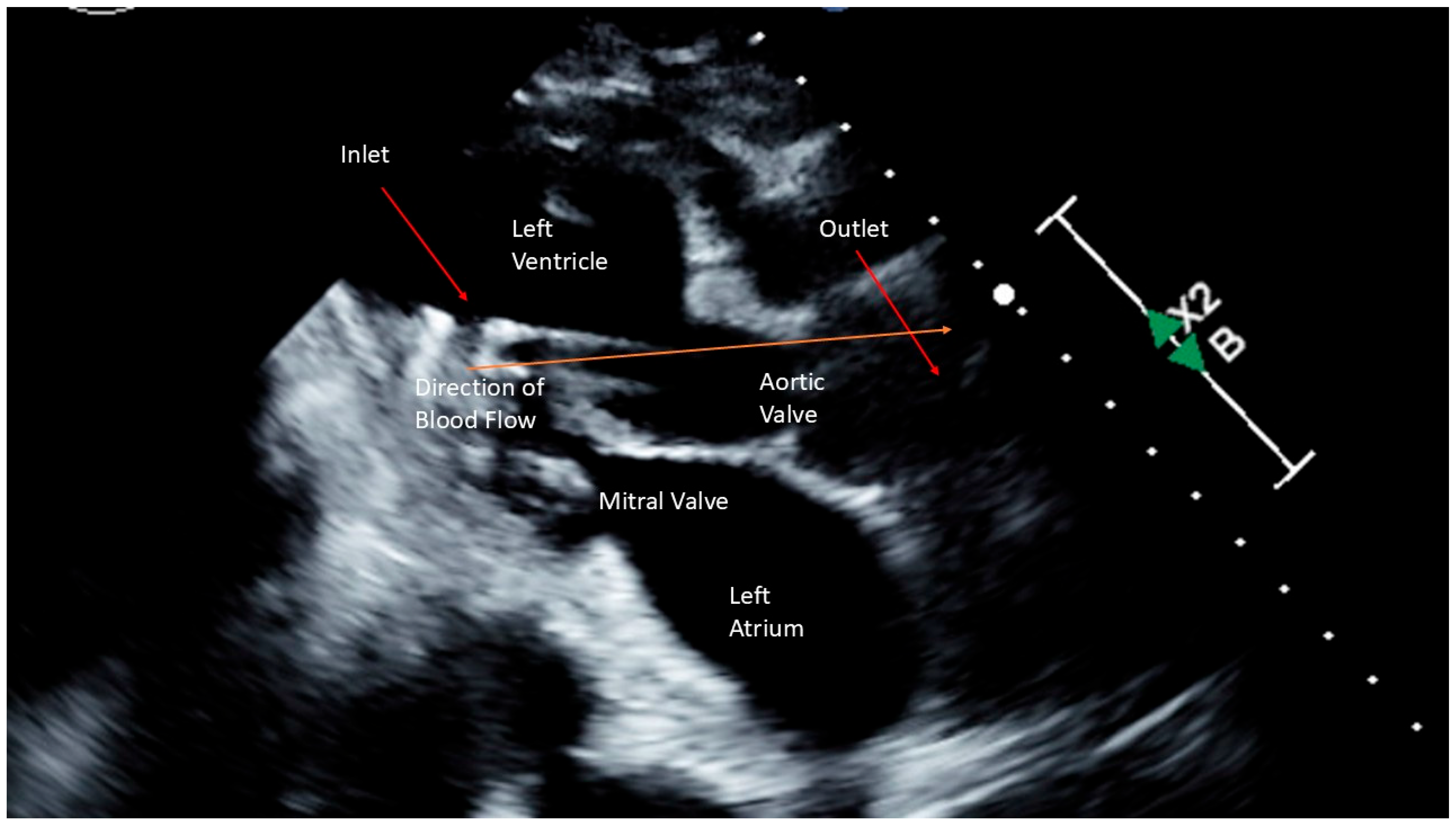
2.2. Case 2

| Characteristic | Patient 1 | Patient 2 |
|---|---|---|
| Age/Sex | 67/Female | 77/Female |
| LVOTO | No | No |
| Comorbid Conditions | Septic Shock, Small Bowel Obstruction, Colonic Perforation | Severe Mitral Regurgitation |
| Mechanical Ventilation | Yes | Yes |
| Hemodynamics | ||
| Before HPTP | ||
| AO (mmHg) | 63/45 (54) | 95/71 (79) |
| PA (mmHg) | 45/32 (38) | 49/32 (39) |
| PCWP (mmHg) | 31 | 31 |
| CI (L/min/m2) | 1.8 | 1.2 |
| SVR (dynes*s/cm5) | 1175 | 4500 |
| Type and Dose of Inotrope/Vasopressor | Dobutamine 10 mcg/kg/min Epinephrine 0.3 mcg/kg/min Norepinephrine 0.3 mcg/kg/min Vasopressin 0.06 mcg/kg/min Milrinone 0.375 mcg/kg/min Phenylephrine 180 mcg/min | Dobutamine 5 mcg/kg/min Epinephrine 0.08 mcg/kg/min Norepinephrine 0.13 mcg/kg/min |
| After HPTP | ||
| AO (mmHg) | 88/70 (75) | 108/65 (79) |
| PA (mmHg) | 30/16 (23) | 35/15 (22) |
| PCWP (mmHg) | 5 | 6 |
| CI (L/min/m2) | 2.7 | 2.5 |
| SVR (dynes*s/cm5) | 1588 | 1477 |
| Type and Dose of Inotrope/Vasopressor | 24 h After Placement Dobutamine 5 mcg/kg/min Epinephrine 0.04 mcg/kg/min Angiotensin II 8 ng/kg/min | 24 h After Placement Dobutamine 5 mcg/kg/min |
3. Discussion
4. Conclusions
Author Contributions
Funding
Institutional Review Board Statement
Informed Consent Statement
Data Availability Statement
Conflicts of Interest
References
- Rihal, C.S.; Naidu, S.S.; Givertz, M.M.; Szeto, W.Y.; Burke, J.A.; Kapur, N.K.; Kern, M.; Garratt, K.N.; Goldstein, J.A.; Dimas, V.; et al. 2015 scai/acc/hfsa/sts clinical expert consensus statement on the use of percutaneous mechanical circulatory support devices in cardiovascular care: Endorsed by the american heart assocation, the cardiological society of india, and sociedad latino americana de cardiologia intervencion; affirmation of value by the canadian association of interventional cardiology-association canadienne de cardiologie d’intervention. J. Am. Coll. Cardiol. 2015, 65, e7–e26. [Google Scholar] [PubMed]
- Peura, J.L.; Colvin-Adams, M.; Francis, G.S.; Grady, K.L.; Hoffman, T.M.; Jessup, M.; John, R.; Kiernan, M.S.; Mitchell, J.E.; O’Connell, J.B.; et al. Recommendations for the use of mechanical circulatory support: Device strategies and patient selection: A scientific statement from the American Heart Association. Circulation 2012, 126, 2648–2667. [Google Scholar] [CrossRef] [PubMed]
- Reeder, G.; Prasad, A. Management and prognosis of stress (takotsubo) cardiomyopathy. In Wolters Kluwer; Connor, R.F., Ed.; UpToDate: Waltham, MA, USA, 2020. [Google Scholar]
- Sattar, Y.; Siew, K.S.W.; Connerney, M.; Ullah, W.; Alraies, M.C. Management of takotsubo syndrome: A comprehensive review. Cureus 2020, 12, e6556. [Google Scholar] [CrossRef] [PubMed]
- Madias, J.E. Takotsubo cardiomyopathy: Current treatment. J. Clin. Med. 2021, 10, 3440. [Google Scholar] [CrossRef] [PubMed]
- Pelliccia, F.; Kaski, J.C.; Crea, F.; Camici, P.G. Pathophysiology of takotsubo syndrome. Circulation 2017, 135, 2426–2441. [Google Scholar] [CrossRef] [PubMed]
- Uribarri, A.; Vazirani, R.; Delia, M.A.; Tomasino, M.; Fernández-Cordón, C.; Martín, A.; Blanco-Ponce, E.; Salamanca, J.; Corbí-Pascual, M.; Vedia, O.; et al. Impact of mechanical circulatory support on outcomes in Takotsubo syndrome complicated by cardiogenic shock: Insights from the RETAKO registry. Int. J. Cardiol. 2025, 419, 132681. [Google Scholar] [CrossRef] [PubMed]
- Syed, M.; Khan, M.Z.; Osman, M.; Alharbi, A.; Khan, M.U.; Munir, M.B.; Balla, S. Comparison of outcomes in patients with takotsubo syndrome with-vs-without cardiogenic shock. Am. J. Cardiol. 2020, 136, 24–31. [Google Scholar] [CrossRef] [PubMed]
- Mohammedzein, A.; Taha, A.; Salwan, A.; Nambiar, R. Impella use in cardiogenic shock due to takotsubo cardiomyopathy with left ventricular outflow tract obstruction. JACC Case Rep. 2019, 1, 161–165. [Google Scholar] [CrossRef] [PubMed]
- von Mackensen, J.K.R.; Shazly, A.E.; Schoenrath, F.; Kempfert, J.; Starck, C.T.; Potapov, E.V.; Jacobs, S.; Falk, V.; Wert, L. Successful treatment of cardiogenic shock due to Takotsubo syndrome with implantation of a temporary microaxial left ventricular assist device in transaxillary approach. J. Cardiothorac. Surg. 2023, 18, 343. [Google Scholar] [CrossRef] [PubMed]
- Mariani, S.; Richter, J.; Pappalardo, F.; Bělohlávek, J.; Lorusso, R.; Schmitto, J.D.; Bauersachs, J.; Napp, L.C. Mechanical circulatory support for Takotsubo syndrome: A systematic review and meta-analysis. Int. J. Cardiol. 2020, 316, 31–39. [Google Scholar] [CrossRef] [PubMed]
- Napp, L.C.; Westenfeld, R.; Møller, J.E.; Pappalardo, F.; Ibrahim, K.; Bonello, L.; Wilkins, C.; Pershad, A.; Mannino, S.F.; Schreiber, T.L.; et al. Impella mechanical circulatory support for takotsubo syndrome with shock: A retrospective multicenter analysis. Cardiovasc. Revasc. Med. 2022, 40, 113–119. [Google Scholar] [CrossRef] [PubMed]
- Di Vece, D.; Citro, R.; Cammann, V.L.; Kato, K.; Gili, S.; Szawan, K.A.; Micek, J.; Jurisic, S.; Ding, K.J.; Bacchi, B.; et al. Outcomes associated with cardiogenic shock in takotsubo syndrome. Circulation 2019, 139, 413–415. [Google Scholar] [CrossRef] [PubMed]
- Nishikawa, R.; Nagano, N.; Kokubu, N.; Hashimoto, K.; Nakata, J.; Kishiue, N.; Takahashi, R.; Otomo, S.; Tsuchihashi, K.; Yano, T. Favorable effects of impella on takotsubo syndrome complicated with cardiogenic shock. Int. Heart J. 2021, 62, 1430–1435. [Google Scholar] [CrossRef] [PubMed]
- Karami, M.; den Uil, C.A.; Ouweneel, D.M.; Scholte, N.T.; Engström, A.E.; Akin, S.; Lagrand, W.K.; Vlaar, A.P.; Jewbali, L.S.; Henriques, J.P. Mechanical circulatory support in cardiogenic shock from acute myocardial infarction: Impella CP/5.0 versus ECMO. Eur. Heart J. Acute Cardiovasc. Care 2020, 9, 164–172. [Google Scholar] [CrossRef] [PubMed]
- von Mackensen, J.K.R.; Zwaans, V.I.T.; El Shazly, A.; Van Praet, K.M.; Heck, R.; Starck, C.T.; Schoenrath, F.; Potapov, E.V.; Kempfert, J.; Jacobs, S.; et al. Mechanical circulatory support strategies in takotsubo syndrome with cardiogenic shock: A systematic review. J. Clin. Med. 2024, 13, 473. [Google Scholar] [CrossRef] [PubMed]
- Trimarchi, G.; Teresi, L.; Licordari, R.; Pingitore, A.; Pizzino, F.; Grimaldi, P.; Calabrò, D.; Liotta, P.; Micari, A.; de Gregorio, C.; et al. Transient left ventricular dysfunction from cardiomyopathies to myocardial viability: When and why cardiac function recovers. Biomedicines 2024, 12, 1051. [Google Scholar] [CrossRef] [PubMed]


Disclaimer/Publisher’s Note: The statements, opinions and data contained in all publications are solely those of the individual author(s) and contributor(s) and not of MDPI and/or the editor(s). MDPI and/or the editor(s) disclaim responsibility for any injury to people or property resulting from any ideas, methods, instructions or products referred to in the content. |
© 2025 by the authors. Licensee MDPI, Basel, Switzerland. This article is an open access article distributed under the terms and conditions of the Creative Commons Attribution (CC BY) license (https://creativecommons.org/licenses/by/4.0/).
Share and Cite
Young, J.; McGrade, P.; Hernandez-Montfort, J.; Fan, J. High Profile Transvalvular Pump Assisted Recovery for Takotsubo Cardiomyopathy: A Case Series. J. Clin. Med. 2025, 14, 3225. https://doi.org/10.3390/jcm14093225
Young J, McGrade P, Hernandez-Montfort J, Fan J. High Profile Transvalvular Pump Assisted Recovery for Takotsubo Cardiomyopathy: A Case Series. Journal of Clinical Medicine. 2025; 14(9):3225. https://doi.org/10.3390/jcm14093225
Chicago/Turabian StyleYoung, Jordan, Patrick McGrade, Jaime Hernandez-Montfort, and Jerry Fan. 2025. "High Profile Transvalvular Pump Assisted Recovery for Takotsubo Cardiomyopathy: A Case Series" Journal of Clinical Medicine 14, no. 9: 3225. https://doi.org/10.3390/jcm14093225
APA StyleYoung, J., McGrade, P., Hernandez-Montfort, J., & Fan, J. (2025). High Profile Transvalvular Pump Assisted Recovery for Takotsubo Cardiomyopathy: A Case Series. Journal of Clinical Medicine, 14(9), 3225. https://doi.org/10.3390/jcm14093225