Article

Ventilatory Response to Exercise in HFrEF-COPD: Importance of Exercise Modality

by
Marta Íscar Urrutia
1,
Julia Herrero Huertas
2,*,
Marina Acebo Castro
1,
Ramón Fernández Álvarez
1,
Beatriz Díaz Molina
3 and
Marta García Clemente
1
1
Department of Pneumology, Asturias Central University Hospital, 33011 Oviedo, Spain
2
Department of Pneumology, Fundación Jiménez Díaz University Hospital, 28040 Madrid, Spain
3
Department of Cardiology, Asturias Central University Hospital, 33011 Oviedo, Spain
*
Author to whom correspondence should be addressed.
J. Clin. Med. 2025, 14(8), 2538; https://doi.org/10.3390/jcm14082538
Submission received: 3 February 2025 / Revised: 3 April 2025 / Accepted: 5 April 2025 / Published: 8 April 2025

Abstract

:
Background: Heart failure with reduced ejection fraction (HFrEF) frequently coexists with chronic obstructive pulmonary disease (COPD), and both conditions share symptoms such as exertional dyspnea. The cardiopulmonary exercise test (CPET) is an essential tool for assessing ventilatory and cardiovascular function and plays a key role in the differential diagnosis of dyspnea. However, the impact of exercise modality on the ventilatory and cardiovascular parameters obtained remains unclear in these groups. Our aim is to compare the oxygen consumption (V·O2) and breathing reserve (BR) values obtained from CPET on a treadmill and a cycle ergometer in patients with HFrEF-COPD and those with HFrEF alone. Methods: A prospective observational study included 65 patients with HFrEF (LVEF ≤ 40%), 18 of whom had COPD. Two CPETs were performed, the first on a treadmill and the second 48–72 h later on a cycle ergometer. Results: In the group with HFrEF-COPD, peak oxygen consumption (VO2/kg) and maximum ventilation (VE) values were significantly higher on the treadmill (20 ± 5 vs. 17 ± 4 mL/kg/min, p < 0.001 and 55 ± 19 vs. 45 ± 11 L/min, p < 0.001, respectively), while breathing reserve (BR%) was lower on the treadmill (16 ± 21 vs. 33 ± 20, p < 0.001). Compared to the HFrEF group, patients with HFrEF-COPD had a lower BR in both exercise modalities (p = 0.01). Conclusions: Treadmill CPET demonstrates greater oxygen consumption and a more pronounced ventilatory response. BR is consolidated as a differential parameter in ventilatory limitation. The choice of exercise modality should be considered based on the underlying pathologies and the objective of the test.

1. Introduction

Heart failure with reduced ejection fraction (HFrEF) is a highly prevalent condition with significant morbidity and mortality, characterized by the marked impairment of cardiac function and patient quality of life. Chronic obstructive pulmonary disease (COPD) frequently coexists with HFrEF, largely due to shared risk factors such as smoking, advanced age, and a sedentary lifestyle. Oxidative stress, chronic systemic inflammation, and hyperactivity of the sympathetic nervous system are pathogenic mechanisms shared by both conditions [1].
The coexistence of COPD worsens the prognosis of HFrEF, increases clinical complications, and presents additional challenges in diagnosis. According to studies carried out with different European cohorts, the prevalence of COPD varies from 9 to 41% among patients with heart failure. Conversely, the prevalence of heart failure increases significantly in hospitalized COPD patients, reaching up to 70% in those requiring mechanical ventilation [2].
Dyspnea on exertion is a predominant symptom shared by both conditions, posing a diagnostic challenge in distinguishing the underlying cause in patients with concomitant COPD and HFrEF [3]. Cardiopulmonary exercise testing (CPET) is valuable for functional assessment, prognosis, and exercise prescription across various pathologies. It is especially useful for the differential diagnosis of dyspnea, given its capacity to evaluate both the cardiocirculatory and ventilatory systems [4].
Exercise intolerance in HFrEF is based on an alteration of both the supply of oxygen to skeletal muscle and the alteration of its utilization by myocytes. This imbalance between oxygen supply and demand becomes evident during progressive exercise testing, leading to a reduced maximal workload. This limitation reflects the lack of enough adenosine triphosphate to maintain skeletal muscle contraction, with a primary manifestation being reduced oxygen consumption (V·O2) [5]. V·O2 is widely recognized as the gold standard for evaluating functional capacity.
In COPD, reduced ventilatory capacity is characterized by diminished breathing reserve (BR) at maximum exercise, which results in a decrease in functional capacity, manifested by a decreased peak V·O2. In contrast, patients with HFrEF typically preserve ventilatory reserve, making BR a clinically significant variable for distinguishing between cardiac and pulmonary disorders [5].
To improve exercise intolerance, in addition to pharmacological treatment and devices (pacemakers, endobronchial valves…), exercise training for both pathologies must be taken into account [6,7]. The most common exercise modalities are the cycle ergometer and the treadmill with a wide variety of protocols [5]. Both exercise modalities present distinct advantages. The treadmill induces greater systemic stress on the cardiovascular and ventilatory systems, while the cycle ergometer allows for the precise quantification of external work [8,9,10]. Our group carried out a comparative study of both ergometers in 65 patients with HFrEF, demonstrating a 20% higher V·O2 max with better ventilatory efficiency and lower BR for the treadmill compared to the cycle ergometer, using protocols of a similar exercise load and test duration [11].
The limited studies conducted on patients with HFrEF-COPD have been exclusively performed using a cycle ergometer. These studies showed that patients with both conditions, despite having similar resting limitations, exhibit lower exercise tolerance compared to those with either COPD or HFrEF alone [12,13,14,15], without considering the differential impact of the exercise modality on assessing functional limitation. Our research fills this gap by directly comparing ventilatory and cardiovascular responses during treadmill and cycle ergometer cardiopulmonary exercise testing.
We hypothesize that the choice of exercise modality, whether treadmill or cycle ergometer, plays a critical role in assessing whether exercise limitation is predominantly cardiac or ventilatory in patients with HFrEF-COPD, as it significantly influences the ventilatory and cardiovascular parameters obtained. The primary aim of this study was to evaluate the variables obtained from CPET performed on a treadmill and a cycle ergometer in patients with HFrEF alone and HFrEF-COPD.

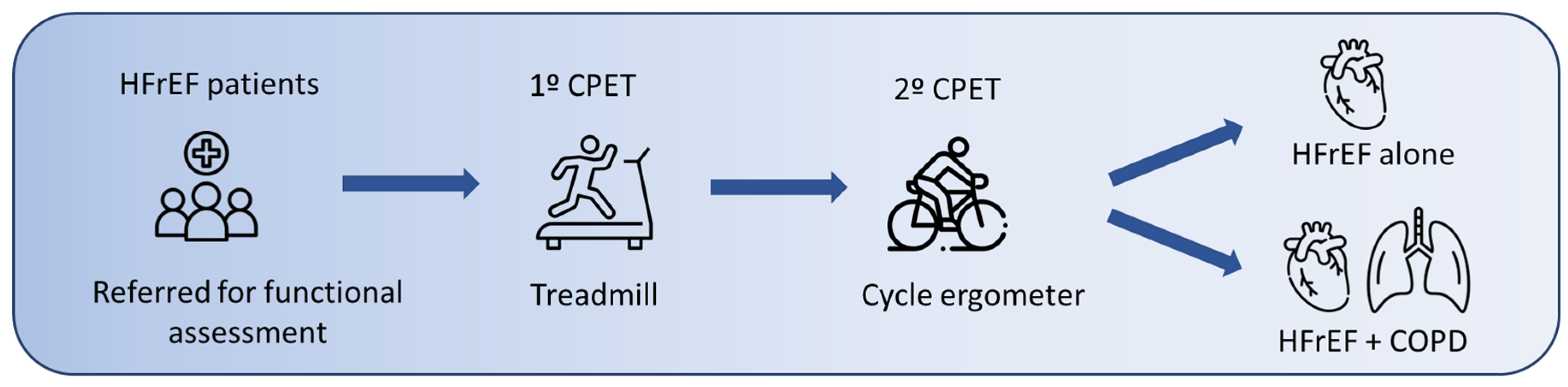
2. Materials and Methods

A prospective study was conducted on patients diagnosed with chronic HFrEF referred for functional assessment in the Exercise Unit from the Advanced Heart Failure and Heart Transplant Unit of the Central University Hospital of Asturias between July 2019 and March 2020. Medical follow-up was conducted until October 2024.
All patients had a documented diagnosis of chronic heart failure with reduced ejection fraction (≤40%) using the criteria of the ESC 2021 [6] in a stable situation. Patients with spirometry with a FEV1/FVC ratio < post-bronchodilator LIN [16] were classified as obstructive, and the diagnosis of COPD was considered when the criteria of the GOLD 2019 guideline were met [17].
Exclusion criteria included an inability to perform stress tests, hospitalization, chronic respiratory failure, disease exacerbation, or uncontrolled atrial fibrillation (AF) within three months before study enrollment.
The study was approved by the Research Ethics Committee of the Principality of Asturias with registration number N174/19. All patients included in the study received the study information sheet and signed the consent form accepting their participation in the study.
Each patient underwent two CPETs performed to their maximum tolerance. The first CPET was conducted on a treadmill (HP Cosmos Pulsar 2002, Nußdorf, Germany) using a modified version of the Bruce protocol [18]. This protocol consisted of 1 min stages with progressively increasing speeds ranging from 2.7 to 6 km/h and an incline gradient rising from 0% to 16%. The second CPET was carried out 72 h later using a cycle ergometer (Corival Lode BV, Groninga, The Netherlands), with workload increments of 5–20 W/min adjusted to the functional limitation of each patient. Before the second test, a clinical evaluation confirmed the absence of changes in symptoms, physical findings, body weight, or medication. Test termination criteria included the maximum perceived effort reported by the supervised patient, the occurrence of severe cardiovascular events, or the presence of limiting symptoms. Monitoring was extended into the recovery phase, which lasted 3 min [19].
Demographic, anthropometric, and clinical data, such as smoking status, comorbidities, treatment, NYHA functional classification, dyspnea (mMRC), and echocardiography findings, were collected. Standard respiratory and cardiorespiratory parameters were measured on a breath-by-breath basis during the two CPETs, following ATS/ACCP standards [19,20].
Data from the HFrEF-COPD group were analyzed and subsequently compared with the HFrEF-alone group (Figure 1). This subanalysis has not been previously published.

Statistical Analysis

Statistical analysis was performed using Stata v15.4.2 (Stata Corp LLC., College Station, TX, USA). Quantitative variables were expressed as the mean ± standard deviation (SD) for those following a normal distribution and as the median ± interquartile range (IQR) for those not following a normal distribution, while qualitative variables were presented as percentages. Initially, a descriptive analysis of the sample was carried out. The normality of the sample distribution was assessed using the Shapiro-Wilk test. For variables following a normal distribution, the Student’s t-test was applied for mean comparison, whereas the Wilcoxon test and the U–Mann–Whitney test was used for variables that did not meet the normality assumption. Categorical variables were analyzed using the chi-square test or Fisher’s exact test, when applicable. To assess the correlation of variables, the Pearson correlation coefficient was used. A value of p < 0.05 was considered statistically significant.

3. Results

3.1. Sample Characteristics

Sixty-five patients with HFrEF were included, 18 of whom had coexisting COPD. Table 1 presents the general characteristics of both groups (patients with and without COPD).
The HFrEF-COPD subgroup had a higher mean age than the group without COPD (61.6 ± 7.6 vs. 56.4 ± 9.6 years; p = 0.04) and a lower BMI (27.1 ± 4.4 vs. 30.0 ± 4.2 kg/m2; p = 0.01). There were no significant differences in LVEF or sex distribution (Table 1).
According to the GOLD classification, 8 patients (44.4%) in the HFrEF-COPD group had mild COPD, 9 (50%) had moderate COPD, and 1 patient (5.5%) had severe COPD. 94% of patients in this group had a history of smoking compared to 68% of patients without COPD. As expected, lung function was significantly lower in the HFrEF-COPD group.
Regarding the functional class, in the HFrEF-COPD group, we observed a slight increase in patients in the NYHA class II compared to the group without COPD. Regarding the etiology of heart failure, ischemic heart disease was more prevalent in the HFrEF-COPD group (56% vs. 39%), followed by idiopathic etiology (39% vs. 44%).
The treatment of heart failure in both groups is detailed in Table 2.
Patients in both groups were treated with beta-blockers and Renin-Angiotensin-Aldosterone System inhibitors (RAASi) with different diuretic combinations. There were no significant differences in the treatment of both groups except with implantable cardioverter defibrillators.
Regarding inhaled treatment, only four patients (22.2%) used inhaled anticholinergics and two (11.1%) used inhaled corticosteroids. The remaining patients (88.8%) used inhaled treatments as needed, such as salbutamol, LABA + LAMA, or LAMA.
The percentage of patients with an implantable cardioverter-defibrillator (ICD) was significantly higher in the HFrEF-COPD group compared to the percentage of patients without COPD (77.8% vs. 40.4%).

3.2. Cardiopulmonary Exercise Test (CPET), Group HFrEF-COPD

The time interval between the two CPETs performed was 3.0 ± 1.9 days. The parameters are detailed in Table 3.

3.2.1. Effort Level

The exercise duration and maximum respiratory ratio were similar in both exercise modalities. The RQ achieved in both protocols (0.9 (IQR: 0.9–1) vs. 1 (IQR: 0.9–1) (p = 0.880)) did not show significant differences, suggesting a similar degree of effort.
The early interruption of the test was necessary in two patients (11.1%) during the treadmill test due to dyspnea, while in the cycle ergometer, it was suspended in five patients (27.7%) due to fatigue in the lower limbs. In total, seven patients (38.9%) finished the test before reaching the ventilatory threshold (VT).

3.2.2. Oxygen Consumption (V·O2)

In the behavior of V·O2 throughout the CPET, there were no differences between both modalities in basal consumption (subject at rest); however, a significant difference was evident both in VT and in the maximum exercise point reached (V·O2 peak) (p < 0.001).
The peak V·O2kg value (mL/kg/min−1) was significantly higher in the treadmill test compared to the cycle ergometer: 20 (IQR: 17–23) vs. 17 (IQR: 15–19); p < 0.001).
A difference of 18.9% in the V·O2 peak and 18% in the V·O2/kg peak was observed in favor of the treadmill (p < 0.001). Both parameters showed an excellent correlation between the two tests (r = 0.93 y r = 0.89, respectively; p < 0.001).

3.2.3. Ventilatory Response and Gas Exchange

A more pronounced ventilatory response was observed on the treadmill, with a higher maximum respiratory rate ((RR) 30.9 (IQR: 26.5–36.5) vs. 25.5 (IQR: 21–28.5) bpm; p_0.017)) and maximum ventilation ((V·E) 59 (IQR: 49–62.5) vs. 45.9 (IQR: 38.6–46.8) L/min; p < 0.001)). Although EqO2 and EqCO2 were higher on the treadmill (38 (IQR: 34–39) vs. 36.5 (IQR: 33–39) and 38 (IQR: 35.5–39) vs. 37 (IQR: 33.5–39.5), respectively), these differences did not reach statistical significance.
The baseline PETCO2 difference minus the threshold PETCO2 was higher in the cycle ergometer without reaching statistical significance.
The lower V·E Slope (32 (IQR: 29–37) vs. 35 (IQR: 31–38.6); p = 0.042) on the treadmill reflects better ventilatory efficiency in this exercise modality.
It should be noted that the BR at the maximum effort achieved was significantly lower on the treadmill (12 (IQR: 2–35) vs. 25.5 (IQR: 22–48) 3; p < 0.001)), which demonstrates a greater degree of air trapping in standing.
Oscillating ventilation (EOV) was not observed in any patient on the treadmill and was only identified in two patients (11.1%) on the cycle ergometer.
No desaturation was observed during exercise in any of the exercise modalities.

3.2.4. Cardiovascular Response

The maximum HR was higher on the treadmill (119 (IQR: 102.5–132) vs. 108.5 (IQR: 100–120.5)) without reaching statistical significance. The heart rate recovery rate was similar for both ergometers in the 14 patients in sinus rhythm.
Regarding the blood pressure response, starting from similar baselines, the systolic blood pressure response was greater on the treadmill (170 (IQR: 150–185) vs. 162.5 (IQR: 140–180)) without reaching statistical significance, unlike thaat of diastolic blood pressure, which reached higher values on the cycle ergometer (82.5 (IQR: 80–90) vs. 80 (IQR: 70–90) (p = 0.037)), reaching statistical significance in this case.
The Oxygen Pulse (V·O2/FC) in maximum exercise reached higher values on the treadmill (14.1 (IQR: 11.9–17.7) vs. 12.7 (IQR: 10.3–15.5); p < 0.001)), which, together with the higher HR achieved, clearly indirectly reflects a greater cardiac output in the exercise performed on the treadmill.

3.3. Comparison of Patients with HFrEF with and Without COPD

When analyzing the results of both CPETs in both groups, we observed the absence of significant differences in most of the parameters studied (Table 4).
The maximum oxygen consumption (V·O2 peak) and V·O2/kg peak were slightly lower in patients with COPD, although the differences did not reach statistical significance. This could reflect a more pronounced functional limitation in this subgroup.
Patients with HFrEF-COPD presented significantly lower BR during maximum effort on both types of ergometers, although this was more pronounced on the treadmill (12% (IQR: 2–35) vs. 30% (IQR: 25.3–34.9); p = 0.006)) compared to the cycle ergometer (30% (IQR: 22–48) vs. 27% (IQR: 24.9–32.5); p = 0.02)). These results demonstrate the value of BR as a key differentiating characteristic between the two groups and reflect the greater ventilatory effort on the treadmill, associated with a higher air trapping degree when standing.
Regarding ventilatory efficiency, the HFrEF-COPD group presented worse efficiency in both types of exercise, reflected in a higher V·E slope, although it only acquired statistical significance in the cycle ergometer (35% (IQR: 31–38.6) vs. 32% (IQR: 27–35.8); p = 0.042)). Concerning oscillating breathing, this was observed more frequently in patients without COPD, both on the treadmill (41% vs. 0%; p = 0.001) and on the cycle ergometer (46% vs. 12%; p = 0.01).
There were no significant differences in maximum heart rate (HR), HR recovery index, or oxygen pulse, which reinforces the performance of a comparable effort level between both groups.

3.3.1. Symptoms and Preference

Patients in both groups presented a greater degree of dyspnea on the treadmill and more pronounced lower limb fatigue on the cycle ergometer, with no significant differences between both groups.
Regarding the preference for the type of ergometer, the majority of patients in both groups opted for the cycle ergometer, due to the increased feeling of safety during its use.

3.3.2. Mortality

During follow-up until October 2024, mortality was higher in the HFrEF-COPD group (27.8% vs. 6.4%).
Causes of mortality in patients with COPD included COVID-19 pneumonia (two cases) and cardiovascular causes (three cases). In the group of patients without COPD, three patients died due to lung neoplasia, COVID-19 pneumonia, and post-heart transplant complications.

4. Discussion

This is the first study that evaluates the physiological variables of exercise in patients with COPD coexisting with heart failure with reduced ejection fraction (HFrEF) using two types of ergometers, treadmill and cycle ergometer.
The main finding of this study is that the functional assessment of patients with HFrEF-COPD on a treadmill reveals increased oxygen consumption and an improvement in gas exchange, despite inducing greater air trapping in patients with airway obstruction.
Peak V·O2 is the most important variable of CPET, and it is usually decreased in both cardiac and respiratory pathology. Depending on the type of ergometer used, the difference in peak V·O2 is generally considered to be 5–10% greater on a treadmill [3]. In our study, with a similar effort level, there was a difference of 19.8% in peak V·O2/kg for HFrEF and of 18% in peak V·O2/kg for HFrFE-COPD, confirming the superiority of the treadmill, following previous literature, in healthy subjects and patients with or without COPD. Furthermore, in patients with mixed pathology, this higher V·O2 is maintained despite the coexistence of COPD. We did not find in the literature reviewed to date any studies that used both ergometers in mixed pathology (HFrEF-COPD).
The impact of the difference in the V·O2 obtained is determined by the prognostic scales in both groups. Prognostic assessment is of crucial importance in providing measures that improve quality of life and survival and reduce hospitalizations and healthcare costs [21].
Breathing reserve (BR) has been studied as a key parameter for distinguishing exercise limitation in patients with HFrEF-COPD. Our study confirms its usefulness, showing a significant decrease in BR in patients with COPD during exercise, secondary to hyperinflation and a lower maximum voluntary ventilation compared to patients without COPD [5].
In the functional assessment of patients with COPD, CPET on a treadmill is clearly recommended due to its superior sensitivity in detecting exertional desaturation and a greater decrease in BR [5]. Our findings reinforce this recommendation, demonstrating that the treadmill provides a more accurate characterization of ventilatory limitation than the cycle ergometer in these patients, making it a more effective tool for functional evaluation.
In our study, all patients with COPD had mixed disease with different degrees of ventilatory impairment (GOLD I–III). Despite the demonstrated benefits of bronchodilator treatment in patients with COPD, regardless of its association with heart failure [7], the poor adherence to medication prescribed for COPD compared to medication for HFrEF is striking. This finding, previously described in the literature [22], highlights the need for greater effort among pulmonologists and cardiologists in motivating treatment adherence.
The ventilatory response was greater on the treadmill, consistent with the study by Hsia et al. [23], which compared two linear protocols on a treadmill and cycle ergometer in 16 patients with COPD, noting lower BR and more pronounced desaturation on the treadmill. In our study, desaturation was not observed in any patient, probably due to the lower severity of the obstruction in our sample, with the behavior of V·E max being similar for both groups.
Ventilatory efficiency, assessed by V·E Slope, is a parameter widely used in clinical practice [5,21]. Both heart and lung diseases affect gas exchange at the lung level, which can be evaluated through the relationship or V·E to V·CO2 using a linear regression line. Our study demonstrated better ventilatory efficiency on the treadmill compared to the cycle ergometer, worsening for patients with mixed disease due to a greater deterioration in gas exchange. These findings are consistent with those of Hsia et al. [23] and Corrà et al. [24], but they differ from those of Beckers et al. [25], who compared 21 patients with HFrEF using both ergometers, finding a higher peak V·O2 on the treadmill, but without differences in ventilatory efficiency.
Due to the lack of inspiratory capacity maneuvers in the exercise modalities, the magnitude of dynamic hyperinflation could not be evaluated. In clinical practice, it is common to reserve the analysis of flow–volume curves and inspiratory capacity maneuvers for patients with unexplained dyspnea or patients with chronic respiratory diseases. The systematic quantification of dynamic hyperinflation is becoming increasingly important in the performance and interpretation of CPET. This phenomenon can occur in both vascular and respiratory pathologies, being of utmost importance in the latter group [26,27].
The differences in the hemodynamic response between exercise modalities in HF have been described by Kim et al. [28], who showed that treadmill exercise generates a higher cardiac output, reflected in increased V·O2 values. In our study, no significant differences were found between both groups (with and without COPD), probably due to a similar limitation in ejection fraction.
During follow-up, higher mortality was observed in the HFrEF-COPD group. COPD is independently associated with an increased risk of all-cause mortality and hospitalization in both heart failure with preserved ejection fraction and HFrEF, as demonstrated in previous studies [22,29].
Given the increase in the coexistence of COPD and HFrEF, it is crucial to manage patients with HFrFE-COPD via multidisciplinary healthcare teams, involving pulmonologists and cardiologists for optimal patient management.
The limitations of this study include the small number of patients, which is explained by the difficulty in selecting individuals with mixed pathology capable of performing a stress test. The differences in age and treatment between the two groups are another limitation to take into account. It should be noted that the initial test was performed for clinical purposes, which represents real life. Another limitation is the single-center nature of the study.
Among the strengths of our study is that this is the first study to investigate the response to effort in HFrEF-COPD patients based on the type of exercise. Despite this, we believe that the need to investigate cardiopulmonary interaction, their influence on exercise, and the potential clinical implications in sick populations justifies this study, with the aim of developing further research involving a greater number of patients and specialists.

5. Conclusions

In patients with HFrEF-COPD, treadmill CPET shows greater oxygen consumption and a lower ventilatory reserve, with a more pronounced ventilatory and cardiovascular response compared to the cycle ergometer. Cycle ergometer CPETs underestimate the ventilatory response to exercise, even in patients with confirmed respiratory pathology, which suggests the need to implement protocols that include the analysis of inspiratory capacity during the test.
When choosing the CPET and the exercise modality, it is key to assess the underlying pathologies and the clinical reason for the test.
Additionally, CPET should be adapted and its results interpreted by considering not only age, sex, and comorbidities but also the type of ergometer used.

Author Contributions

M.G.C. and M.Í.U. proposed the study, conducted the data collection, and composed the manuscript; J.H.H., M.A.C. and R.F.Á. performed the statistical analysis, interpreted the results, and composed the manuscript; B.D.M. contributed expertly to the interpretation of the results and the preparation of the manuscript. All authors have read and agreed to the published version of the manuscript.

Funding

This research received no external funding.

Institutional Review Board Statement

The study was conducted in accordance with the guidelines of the Declaration of Helsinki and was approved by the Research Ethics Committee of the Principality of Asturias (protocol code 174/19 and approval date 13 June 2019).

Informed Consent Statement

Informed consent was obtained from all subjects involved in the study.

Data Availability Statement

The original contributions presented in this study are included in the article. Further inquiries can be directed to the corresponding author(s).

Conflicts of Interest

The authors declare no conflicts of interest.

References

  1. Natali, D.; Cloatre, G.; Hovette, P.; Cochrane, B. Screening for Comorbidities in COPD. Breathe 2020, 16, 190315. [Google Scholar] [CrossRef] [PubMed]
  2. Papaporfyriou, A.; Bartziokas, K.; Gompelmann, D.; Idzko, M.; Fouka, E.; Zaneli, S.; Bakakos, P.; Loukides, S.; Papaioannou, A.I. Cardiovascular Diseases in COPD: From Diagnosis and Prevalence to Therapy. Life 2023, 13, 1299. [Google Scholar] [CrossRef]
  3. Myers, L.C.; Quint, J.K.; Hawkins, N.M.; Putcha, N.; Hamilton, A.; Lindenauer, P.; Wells, J.M.; Witt, L.J.; Shah, S.P.; Lee, T.; et al. A Research Agenda to Improve Outcomes in Patients with Chronic Obstructive Pulmonary Disease and Cardiovascular Disease: An Official American Thoracic Society Research Statement. Am. J. Respir. Crit. Care Med. 2024, 210, 715–729. [Google Scholar] [CrossRef] [PubMed]
  4. Barron, A.; Francis, D.P.; Mayet, J.; Ewert, R.; Obst, A.; Mason, M.; Elkin, S.; Hughes, A.D.; Wensel, R. Oxygen Uptake Efficiency Slope and Breathing Reserve, Not Anaerobic Threshold, Discriminate Between Patients with Cardiovascular Disease Over Chronic Obstructive Pulmonary Disease. JACC Heart Fail. 2016, 4, 252–261. [Google Scholar] [CrossRef]
  5. Sietsema, K.E.; Sue, D.Y.; Stringer, W.W.; Ward, S.A. Wasserman & Whipp’s Principles of Exercise Testing and Interpretation, 6th ed.; Wolters Kluwer: Philadelphia, PA, USA, 2021. [Google Scholar]
  6. McDonagh, T.A.; Metra, M.; Adamo, M.; Gardner, R.S.; Baumbach, A.; Böhm, M.; Burri, H.; Butler, J.; Čelutkienè, J.; Chioncel, O.; et al. 2021 ESC Guidelines for the Diagnosis and Treatment of Acute and Chronic Heart Failure. Eur. Heart J. 2021, 42, 3599–3726, Erratum in Eur. Heart J. 2021, 42, 4901. [Google Scholar] [CrossRef]
  7. Global Initiative for Chronic Obstructive Lung Disease. Global Strategy for Prevention, Diagnosis and Management of COPD: 2025 Report; Global Initiative for Chronic Obstructive Lung Disease: Philadelphia, PA, USA, 2025. [Google Scholar]
  8. Hermansen, L.; Saltin, B. Oxygen Uptake during Maximal Treadmill and Bicycle Exercise. J. Appl. Physiol. 1969, 26, 31–37. [Google Scholar] [CrossRef] [PubMed]
  9. Miyamara, M.; Honda, Y. Oxygen Intake and Cardiac Output during Maximal Treadmill and Bicycle Exercise. J. Appl. Physiol. 1972, 32, 185–188. [Google Scholar] [CrossRef]
  10. Lockwood, P.A.; Yoder, J.E.; Deuster, P.A. Comparison and Cross-Validation of Cycle Ergometry Estimates of VO2max. Med. Sci. Sports Exerc. 1997, 29, 1513–1520. [Google Scholar] [CrossRef]
  11. Herrero Huertas, J.; García Clemente, M.; Díaz Molina, B.; Lambert Rodríguez, J.L.; Íscar Urrutia, M. Heart Failure with Reduced Ejection Fraction and Prognostic Scales: The Impact of Exercise Modality in Cardiopulmonary Exercise Tests. J. Clin. Med. 2022, 11, 3122. [Google Scholar] [CrossRef]
  12. Guazzi, M.; Myers, J.; Vicenzi, M.; Bensimhon, D.; Chase, P.; Pinkstaff, S.; Arena, R. Cardiopulmonary Exercise Testing Characteristics in Heart Failure Patients with and without Concomitant Chronic Obstructive Pulmonary Disease. Am. Heart J. 2010, 160, 900–905. [Google Scholar] [CrossRef]
  13. Oliveira, M.F.; Arbex, F.F.; Alencar, M.C.; Souza, A.; Sperandio, P.A.; Medeiros, W.M.; Borghi-Silva, A.; Medina, L.A.; Santos, R.; Hirai, D.M.; et al. Heart Failure Impairs Muscle Blood Flow and Endurance Exercise Tolerance in COPD. COPD 2016, 13, 407–415. [Google Scholar] [CrossRef] [PubMed]
  14. Arbex, F.F.; Alencar, M.C.; Souza, A.; Mazzuco, A.; Sperandio, P.A.; Rocha, A.; Berton, D.C.; Borghi-Silva, A.; Almeida, D.R.; O’Donnell, D.E.; et al. Exercise Ventilation in COPD: Influence of Systolic Heart Failure. COPD 2016, 13, 693–699. [Google Scholar] [CrossRef] [PubMed]
  15. Rocha, A.; Arbex, F.F.; Sperandio, P.A.; Souza, A.; Biazzim, L.; Mancuso, F.; Berton, D.C.; Hochhegger, B.; Alencar, M.C.N.; Nery, L.E.; et al. Excess Ventilation in Chronic Obstructive Pulmonary Disease-Heart Failure Overlap: Implications for Dyspnea and Exercise Intolerance. Am. J. Respir. Crit. Care Med. 2017, 196, 1264–1274. [Google Scholar] [CrossRef]
  16. Quanjer, P.H.; Stanojevic, S.; Cole, T.J.; Baur, X.; Hall, G.L.; Culver, B.H.; Enright, P.L.; Hankinson, J.L.; Ip, M.S.M.; Zheng, J.; et al. Multi-Ethnic Reference Values for Spirometry for the 3–95-Year Age Range: The Global Lung Function 2012 Equations. Eur. Respir. J. 2012, 40, 1324–1343. [Google Scholar] [CrossRef]
  17. Singh, D.; Agusti, A.; Anzueto, A.; Barnes, P.J.; Bourbeau, J.; Celli, B.R.; Criner, G.J.; Frith, P.; Halpin, D.M.G.; Han, M.; et al. Global Strategy for the Diagnosis, Management, and Prevention of Chronic Obstructive Lung Disease: The GOLD Science Committee Report 2019. Eur. Respir. J. 2019, 53, 1900164. [Google Scholar] [CrossRef] [PubMed]
  18. Pereiro Alonso, M.E. Capacidad Aeróbica en una Población Adulta Sana y Calidad de Vida Relacionada con la Salud. 2003. Available online: http://hdl.handle.net/10651/16219 (accessed on 4 November 2024).
  19. Balady, G.J.; Arena, R.; Sietsema, K.; Myers, J.; Coke, L.; Fletcher, G.F.; Forman, D.; Franklin, B.; Guazzi, M.; Gulati, M.; et al. Clinician’s Guide to Cardiopulmonary Exercise Testing in Adults: A Scientific Statement from the American Heart Association. Circulation 2010, 122, 191–225. [Google Scholar] [CrossRef]
  20. American Thoracic Society; American College of Chest Physicians. ATS/ACCP Statement on Cardiopulmonary Exercise Testing. Am. J. Respir. Crit. Care Med. 2003, 167, 211–277. [Google Scholar] [CrossRef]
  21. Paolillo, S.; Agostoni, P. Prognostic Role of Cardiopulmonary Exercise Testing in Clinical Practice. Ann. Am. Thorac. Soc. 2017, 14 (Suppl. 1), S53–S58. [Google Scholar] [CrossRef]
  22. Ramalho, S.H.R.; de Albuquerque, A.L.P. Chronic Obstructive Pulmonary Disease in Heart Failure: Challenges in Diagnosis and Treatment for HFpEF and HFrEF. Curr. Heart Fail. Rep. 2024, 21, 163–173. [Google Scholar] [CrossRef]
  23. Hsia, D.; Casaburi, R.; Pradhan, A.; Torres, E.; Porszasz, J. Physiological Responses to Linear Treadmill and Cycle Ergometer Exercise in COPD. Eur. Respir. J. 2009, 34, 605–615. [Google Scholar] [CrossRef]
  24. Corrà, U.; Agostoni, P.G.; Anker, S.D.; Coats, A.J.; Crespo Leiro, M.G.; de Boer, R.A.; Harjola, V.P.; Hill, L.; Lainscak, M.; Lund, L.H.; et al. Role of Cardiopulmonary Exercise Testing in Clinical Stratification in Heart Failure. A Position Paper from the Committee on Exercise Physiology and Training of the Heart Failure Association of the European Society of Cardiology. Eur. J. Heart Fail. 2018, 20, 3–15, Erratum in Eur. J. Heart Fail. 2018, 20, 1501. [Google Scholar] [CrossRef] [PubMed]
  25. Beckers, P.J.; Possemiers, N.M.; Van Craenenbroeck, E.M.; Van Berendoncks, A.M.; Wuyts, K.; Vrints, C.J.; Conraads, V.M. Impact of Exercise Testing Mode on Exercise Parameters in Patients with Chronic Heart Failure. Eur. J. Prev. Cardiol. 2012, 19, 389–395. [Google Scholar] [CrossRef] [PubMed]
  26. Neder, J.A.; Phillips, D.B.; Marillier, M.; Bernard, A.C.; Berton, D.C.; O’Donnell, D.E. Clinical Interpretation of Cardiopulmonary Exercise Testing: Current Pitfalls and Limitations. Front. Physiol. 2021, 12, 552000. [Google Scholar] [CrossRef]
  27. Haverkamp, H.C.; Balmain, B.N. Ventilatory Responses to Exercise by Age, Sex, and Health Status. Curr. Sports Med. Rep. 2024, 23, 79–85. [Google Scholar] [CrossRef]
  28. Kim, S.; Yamabe, H.; Yokoyama, M. Hemodynamic Characteristics during Treadmill and Bicycle Exercise in Chronic Heart Failure: Mechanism for Different Responses of Peak Oxygen Uptake. Jpn. Circ. J. 1999, 63, 965–970. [Google Scholar] [CrossRef] [PubMed]
  29. Boudestein, L.C.; Rutten, F.H.; Cramer, M.J.; Lammers, J.W.J.; Hoes, A.W. The Impact of Concurrent Heart Failure on Prognosis in Patients with Chronic Obstructive Pulmonary Disease. Eur. J. Heart Fail. 2009, 11, 1182–1188. [Google Scholar] [CrossRef]
Figure 1. Study design. Included patients underwent two cardiopulmonary exercise tests (CPET): the first on a treadmill and the second on a cycle ergometer. Participants were categorized into two groups based on comorbidities: HFrEF alone and HFrEF-COPD.
Jcm 14 02538 g001
Table 1. General characteristics of both groups.
VariablesHFrEF (n = 47)IHFrEF + COPD
(n = 18)
p
General Characteristics
   Age (years)56.4 ± 9.661.6 ± 7.60.04
   Male sex30 (70%)16 (89%)0.198
   BMI (kg/m2)30 ± 4.227.1 ± 4.40.01
   LVEF (%)29.9 ± 8.628.6 ± 8.60.599
   Lung function
FEV1 (%)88.5 ± 14.173.3 ± 14.80.001
   FVC (%)90.6 ± 14.4 89.7 ± 19.30.864
   FEV 1/ FVC (%)75.3 ± 3.862.4 ± 4.30.001
   Functional class
   NYHA class I10 (21%)3 (17%)0.678
   NYHA class II29 (62%)12 (66%)0.711
   NYHA class III8 (17%)3 (17%)1000
   Cause of HFrEF
   Ischemic18 (39%)10 (56%)0.209
   Idiopathic20 (44%)7 (39%)0.788
   Hereditary6 (13%)0 (0%)0.175
   Others2 (4%)1 (5%)0.823
NS: not significant; BMI: body mass index; LVEF: left ventricular ejection fraction; FEV1(%): expiratory flow in the first second, percentage of predicted; FVC: forced vital capacity, percentage of predicted; NYHA: New York Heart Association.
Table 2. Chronic treatment for heart failure.
Chronic TreatmentHFrEF (n = 47)HFrEF-COPD
(n = 18)
p
Beta-blocker45 (95.8%)18 (100%)0.520
Amiodarone1 (2.1%)2 (11.1%)0.183
RAASi32 (68%)8 (44.4%)0.411
MRA25 (53.2%)12 (66.7%)0.407
Valsartan/Sacubitril13 (27.7%)9 (50%)0.142
Ivabradine5 (10.6%)2 (11.1%)0.956
Loop diuretics30 (63.8%)11 (61.1%)0.839
Thiazides47 (100%)1 (5.6%)0.277
Implantable cardioverter
defibrillator
Single chamber7 (15%)4 (22%)0.693
CRT12 (25.5%)10 (56%)0.03
No device28 (59.6%)4 (22%)0.006
RAASi: renin-angiotensin-aldosterone system inhibitors, MRA: mineralocorticoid receptor antagonist, CRT: cardiac resynchronization therapy.
Table 3. Results of cardiopulmonary exercise test (CPET) parameters on the treadmill and cycle ergometer in patients with HFrEF-COPD.
Treadmill
Median (IQR)
Cycle Ergometer
Median (IQR)
p
Test duration (min−1)11 (7.5–12)9 (8–10)0.216
V·O2 baseline ( mL/min−1)420 (330–550)450 (370–545)0.580
V·O2 peak ( mL/min−1)1550 (1180–1820)1225 (1040–1420)<0.001
V·O2 kg peak ( mL/kg/ min−1)20 (17–23)17 (15–19)<0.001
V·O2 at VT ( mL/min−1)1630 (1480–1710)1290 (1070–1410)<0.001
V·CO2 at VT ( mL/min−1)1610 (1465–1685)1275 (1040–1410)<0.001
RQ máx.0.9 (0.9–1)1 (0.9–1)0.880
V·Emax (L/min−1)59 (48–62.5)45.9 (38.6–46.8)<0.001
RR max (bpm−1)30.9 (26.5–36.4)25.5 (21–28.5)0.017
BR%12 (2–35)30 (22–48)<0.001
EQO2 at VT38 (34–39)36.5 (33–39)0.999
EQCO2 at VT38 (35.5–39)37 (33.5–39.5)0.999
VEslope32 (29–37)35 (31–38.6)0.042
PETCO2 baseline32 (30–36)32.5 (31–34)0.412
PETCO2 at VT (mmHg)35 (34–37)37.5 (34–39)0.249
SatpO2 initial (%)96.5 (96–98)98 (96–98)0.007
SatpO2 at the end (%)96 (95–98)98 (97–98)0.001
HR baseline (lpm−1)71.5 (66.5–75)68.5 (66.5–73.5)0.348
HR at the end (lpm−1)119 (102.5–132)108.5 (100–120.5)0.067
HR recovery index (bpm−1)17 (12–25)16 (14–19)0.079
V·O2 /FC (L/bpm/ min−1)14.1 (11.9–17.7)12.7 (10.3–15.5)<0.001
SBP initial (mmHg)120 (110–140)120 (115–140)0.702
SBP at the end (mmHg)170 (150–185)162.5 (140–180)0.562
DBP initial (mmHg)70 (60–80)80 (70–90)0.075
DBP at the end (mmHg)80 (70–90)82.5 (80–90)0.037
Borg dyspnea at the end5.5 (5–7)3.5 (2–4)0.001
Borg lower limbs at the end4 (0–7)4 (3–5)0.545
VT: ventilatory threshold; RQ: respiratory quotient; HR: heart rate; SBP: systolic blood pressure; DBP: diastolic blood pressure; V·O2: oxygen consumption; V·CO2: CO2 production; V·E: ventilation; RR: respiratory rate; EqO2: oxygen equivalent; EqCO2: carbon dioxide equivalent; PETCO2: partial pressure end-tidal carbon dioxide; BR: breathing reserve; NS: not significant.
Table 4. Results of the cardiopulmonary exercise test in both groups (with and without COPD).
ParametersHFrEF
(n = 47)
Median (IQR)
HFrEF+ COPD
(n = 18)
Median (IQR)
p
Test Duration (minutes)
Treadmill 11 (9.1–12.5)11 (7.5–12)0.284
Cycle Ergometer 11 (9–12)9 (8–10)0.089
Reaches VT (%)
Treadmill95.7%88.9%0.305
Cycle Ergometer 78.7%72.2%0.578
HR max. (bpm)
Treadmill119 (108–131)119 (102.5–132)0.537
Cycle Ergometer 107 (99.5–120)108.5 (100–120.5)0.922
HR recovery index (bpm)
Treadmill20.5 (14–31)17 (12–25)0.463
Cycle Ergometer 15 (10–25)16 (14–19)0.902
SBP at the End of Exercise (mmHg)
Treadmill150 (137.5–170)170 (150–185)0.352
Cycle Ergometer 160 (140–180)162.5 (140–180)0.824
DBP at the End of Exercise (mmHg)
Treadmill80 (70–90)80 (70–90)0.549
Cycle Ergometer 90 (80–100)82.5 (80–90)0.417
Final O2 Saturation (%)
Treadmill97 (96–98)98 (97–98)0.380
Cycle Ergometer 97 (96–98)96 (95–98)0.294
V·O2 peak (ml/min)
Treadmill1690 (1425–2140)1550 (1180–1820)0.101
Cycle Ergometer 1390 (1190–1680)1225 (1040–1420)0.125
V·O2 /HR max. (ml/bpm/ min−1)
Treadmill14.1 (11.8–17.1)14.1 (11.9–17.7)0.675
Cycle Ergometer 12.1 (9.7–15.2)12.7 (10.3–15.5)0.806
V·E Slope
Treadmill29 (27–34)32 (29–37)0.066
Cycle Ergometer 32 (27–35.8)35 (31–38.6)0.042
V·E max. (L/ min−1)
Treadmill53.3 (46.5–65.5)59 (48–62.5)0.765
Cycle Ergometer 45.8 (37.9–51.8)45.9 (38.6–46.8)0.686
RR max (rpm−1)
Treadmill30 (25.3–34.9)30.9 (26.5–36.4)0.804
Cycle Ergometer 27 (24.9–32.5)25.5 (21–28.5)0.076
EqO2 at VT
Treadmill35 (32–38)38 (34–39)0.049
Cycle Ergometer 36.5 (33–41)36.5 (33–39)0.919
EQCO2 at VT
Treadmill35.5 (32.5–38.5)38.8 (35.5–39)0.042
Cycle Ergometer 37 (33–41.5)37 (33.5–39.5)0.671
PET CO2 at VT (mmHg)
Treadmill36.5 (33.5–39.5)35 (34–37)0.075
Cycle Ergometer 35.5 (32–39)37.5 (34–39)0.958
ΔPETCO2 (mmHg)
Treadmill4.5 (1–6)3.5 (3–5.5)0.518
Cycle Ergometer 2 (0–5)3 (1.5–3.5)0.730
BR at max. exercise (%)
Treadmill30 (25.3–34.9)12 (2–35)0.006
Cycle Ergometer 27 (24.9–32.5)30 (22–48)0.020
EOV (%)
Treadmill41.3%0%0.001
Cycle Ergometer 45.6%11.8%0.010
Final Borg Dyspnea (1–10)
Treadmill6 (3.5–7)5.5 (5–7)0.567
Cycle Ergometer 4 (2–5.5)3.5 (2–4)0.561
Final Borg Lower Limbs (1–10)
Treadmill3 (0.5–7)4 (0–7)0.861
Cycle Ergometer5 (4–7)4 (3–5)0.178
NS: not significant; COPD: chronic obstructive pulmonary disease; VT: Ventilatory Threshold; RQ: respiratory quotient; HR: heart rate; SBP: systolic blood pressure; DBP: diastolic blood pressure; V·O2: oxygen consumption; V·CO2: CO2 production; EqO2: oxygen equivalent; EqCO2: carbon dioxide equivalent; PET CO2: partial pressure end-tidal carbon dioxide; ΔPETCO2: PETCO2 increase from start of test to VT; BR: breathing reserve; EOV: exercise oscillatory ventilation.
Disclaimer/Publisher’s Note: The statements, opinions and data contained in all publications are solely those of the individual author(s) and contributor(s) and not of MDPI and/or the editor(s). MDPI and/or the editor(s) disclaim responsibility for any injury to people or property resulting from any ideas, methods, instructions or products referred to in the content.

