Autoantibodies in Primary Biliary Cholangitis: From Classical Markers to Emerging Targets

 , , , , , ,
Abstract
1. Introduction
2. Anti-Mitochondrial Antibodies (AMAs)
2.1. Historical Background and Diagnostic Significance
2.2. Target Antigens of AMAs
2.3. Evolution of AMA Detection Methods
3. Antinuclear Antibodies (ANAs) in Primary Biliary Cholangitis
3.1. Basis of ANA Testing
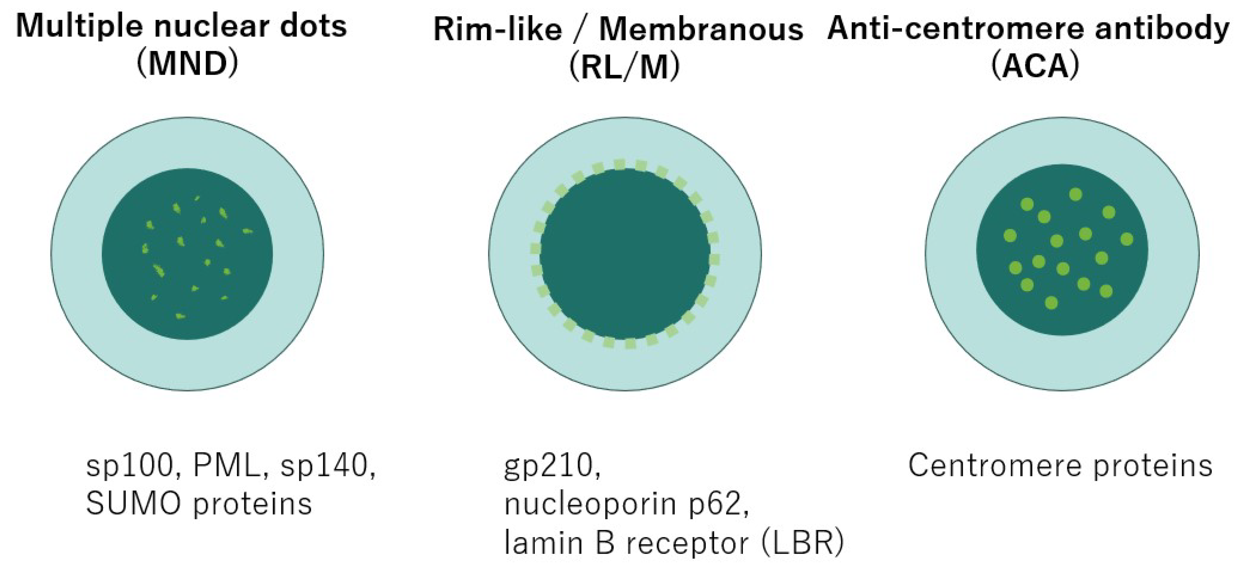
3.2. Frequency and Characteristic ANA Patterns in PBC
- Multiple Nuclear Dots (MND) pattern:
- Rim-like/Membranous (RL/M) pattern:
- Anti-centromere antibody (ACA) pattern:
3.3. Additional High-Specificity Antibodies
4. Emerging Autoantibodies
4.1. Anti-Kelch-like 12(KLHL12) Antibody
4.2. Anti-RPL30 Antibody
4.3. Anti-HK1 Antibody
5. Pathogenesis of PBC
5.1. Autoantigens and Neoantigen Formation in PBC
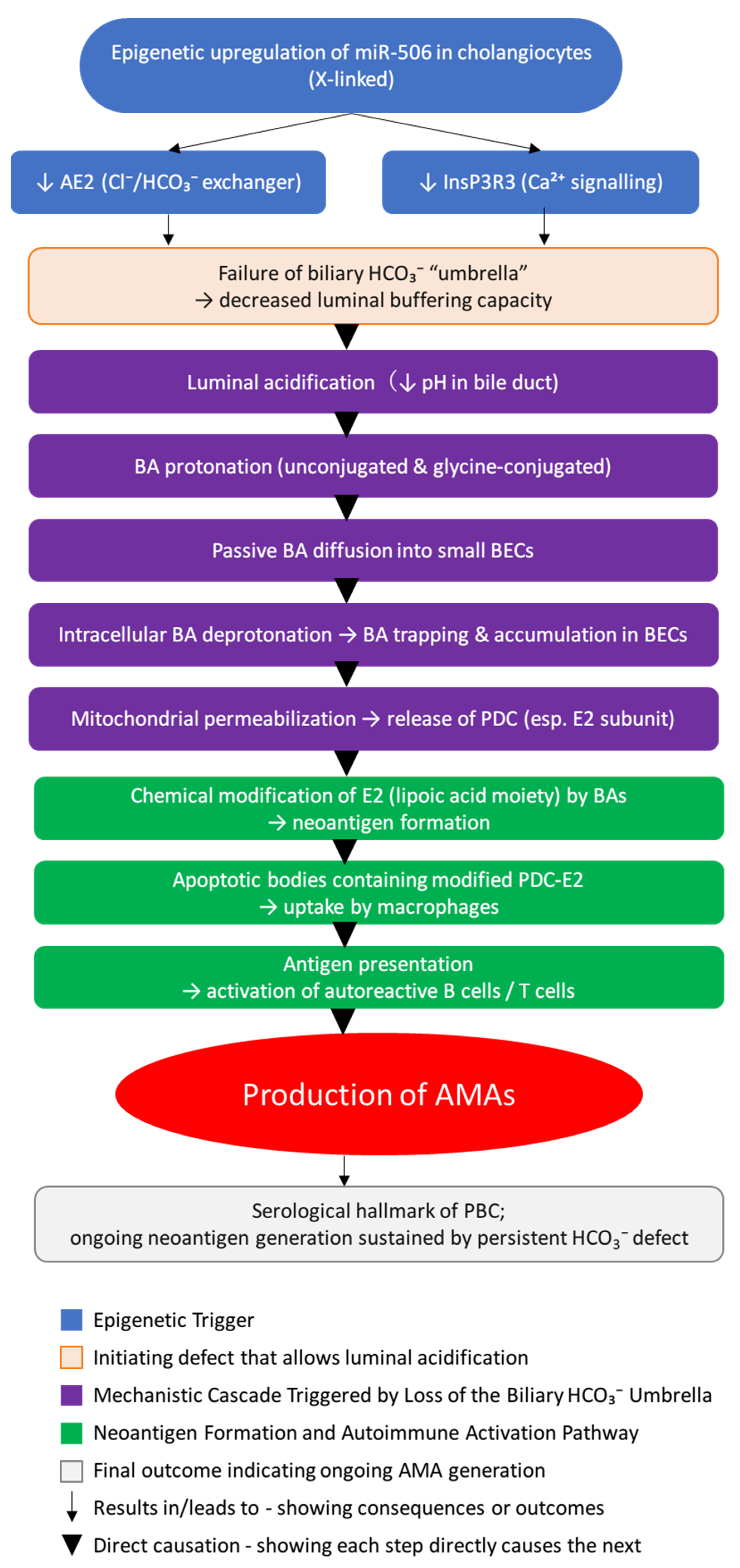
5.2. Epigenetic Dysregulation of miR-506 and Failure of the Biliary HCO3− Umbrella
5.3. Localized BEC Damage and Bile Acid Accumulation
5.4. Neoantigen Formation and AMA Pathophysiology
6. AMA Selectivity and the PDC-Fatigue Axis
6.1. Non-Systemic Pathology Driven by AMA Selectivity
6.2. PDC Dysfunction and the First Clinical Sign
7. Diagnostic and Prognostic Significance of Autoantibodies
7.1. Clinical Interpretation of PBC-Related Autoantibodies and Diagnostic Decision-Making
7.2. Autoantibody Profiles and Disease Stratification in PBC
8. Autoantibodies and Therapeutic Response in Primary Biliary Cholangitis
9. Limitations of Immunosuppressive Therapy
10. Serological Profiles of AILDs Overlap Syndromes
10.1. Overview of Overlap Syndromes in AILDs
- Hepatocellular injury pattern (e.g., ALT > 5 times the Upper Limit of Normal (ULN))
- Elevated Immunoglobulin G (IgG) levels (>2 times ULN)
- Positive ANA and/or Anti-Smooth Muscle Antibody (ASMA)
- Cholestatic injury pattern (e.g., ALP > 2 times ULN or GGT > 5 times ULN)
- Presence of AMA
- Florid bile duct lesions on liver biopsy
10.2. Serological Characteristics of AIH-PBC Overlap Syndrome
10.3. Perinuclear Antineutrophil Cytoplasmic Antibody (p-ANCA) in AILDs
11. Conclusions
Author Contributions
Funding
Institutional Review Board Statement
Informed Consent Statement
Data Availability Statement
Conflicts of Interest
References
- Hirschfield, G.M.; Beuers, U.; Corpechot, C.; Invernizzi, P.; Jones, D.; Marzioni, M.; Schramm, C. EASL Clinical Practice Guidelines: The Diagnosis and Management of Patients with Primary Biliary Cholangitis. J. Hepatol. 2017, 67, 145–172. [Google Scholar] [CrossRef]
- Lv, T.; Chen, S.; Li, M.; Zhang, D.; Kong, Y.; Jia, J. Regional Variation and Temporal Trend of Primary Biliary Cholangitis Epidemiology: A Systematic Review and Meta-Analysis. J. Gastroenterol. Hepatol. 2021, 36, 1423–1434. [Google Scholar] [CrossRef]
- Lu, M.; Zhou, Y.; Haller, I.V.; Romanelli, R.J.; VanWormer, J.J.; Rodriguez, C.V.; Anderson, H.; Boscarino, J.A.; Schmidt, M.A.; Daida, Y.G.; et al. Increasing Prevalence of Primary Biliary Cholangitis and Reduced Mortality with Treatment. Clin. Gastroenterol. Hepatol. 2018, 16, 1342–1350.e1. [Google Scholar] [CrossRef]
- Murillo Perez, C.F.; Goet, J.C.; Lammers, W.J.; Gulamhusein, A.; van Buuren, H.R.; Ponsioen, C.Y.; Carbone, M.; Mason, A.; Corpechot, C.; Invernizzi, P.; et al. Milder Disease Stage in Patients with Primary Biliary Cholangitis over a 44-Year Period: A Changing Natural History. Hepatology 2018, 67, 1920–1930. [Google Scholar] [CrossRef] [PubMed]
- European Association for the Study of the Liver. EASL Clinical Practice Guidelines: Management of Cholestatic Liver Diseases. J. Hepatol. 2009, 51, 237–267. [Google Scholar] [CrossRef] [PubMed]
- Lindor, K.D.; Bowlus, C.L.; Boyer, J.; Levy, C.; Mayo, M. Primary Biliary Cholangitis: 2018 Practice Guidance from the American Association for the Study of Liver Diseases. Hepatology 2019, 69, 394–419. [Google Scholar] [CrossRef]
- Mackay, I.R. Primary Biliary Cirrhosis Showing a High Titer of Autoantibody; Report of a Case. N. Engl. J. Med. 1958, 258, 185–188. [Google Scholar] [CrossRef]
- Walker, J.G.; Doniach, D.; Roitt, I.M.; Sherlock, S. Serological tests in diagnosis of primary biliary cirrhosis. Lancet 1965, 285, 827–831. [Google Scholar] [CrossRef] [PubMed]
- Whyte, J.; Hough, D.; Maddison, P.J.; McHugh, N.J. The Association of Primary Biliary Cirrhosis and Systemic Sclerosis Is Not Accounted for by Cross Reactivity Between Mitochondrial and Centromere Antigens. J. Autoimmun. 1994, 7, 413–424. [Google Scholar] [CrossRef]
- Vergani, D.; Alvarez, F.; Bianchi, F.B.; Cançado, E.L.R.; MacKay, I.R.; Manns, M.P.; Nishioka, M.; Penner, E. Liver Autoimmune Serology: A Consensus Statement from the Committee for Autoimmune Serology of the International Autoimmune Hepatitis Group. J. Hepatol. 2004, 41, 677–683. [Google Scholar] [CrossRef]
- Liang, E.Y.; Liu, M.; Ke, P.F.; Han, G.; Zhang, C.; Deng, L.; Wang, Y.X.; Huang, H.; Huang, W.J.; Liu, R.P.; et al. A Population-Based Characterization Study of Anti-Mitochondrial M2 Antibodies and Its Consistency with Anti-Mitochondrial Antibodies. Lab. Med. 2023, 54, 618–625. [Google Scholar] [CrossRef]
- Abe, M.; Onji, M. Natural History of Primary Biliary Cirrhosis. Hepatol. Res. 2008, 38, 639–645. [Google Scholar] [CrossRef] [PubMed]
- Mitchison, H.C.; Bassendine, M.F.; Hendrick, A.; Bennett, M.K.; Bird, G.; Watson, A.J.; James, O.F.W. Positive Antimitochondrial Antibody but Normal Alkaline Phosphatase: Is This Primary Biliary Cirrhosis? Hepatology 1986, 6, 1279–1284. [Google Scholar] [CrossRef]
- Duan, W.; Chen, S.; Li, S.; Lv, T.; Li, B.; Wang, X.; Wang, Y.; Zhao, X.; Ma, H.; Ou, X.; et al. The Future Risk of Primary Biliary Cholangitis (PBC) Is Low among Patients with Incidental Anti-mitochondrial Antibodies but without Baseline PBC. Hepatol. Commun. 2022, 6, 3112–3119. [Google Scholar] [CrossRef]
- Berg, P.A.; Klein, R. Mitochondrial Antigens and Autoantibodies: From Anti-M1 to Anti-M9. Klin. Wochenschr. 1986, 64, 897–909. [Google Scholar] [CrossRef]
- Hu, C.J.; Zhang, F.C.; Li, Y.Z.; Zhang, X. Primary Biliary Cirrhosis: What Do Autoantibodies Tell Us? World J. Gastroenterol. WJG 2010, 16, 3616–3629. [Google Scholar] [CrossRef] [PubMed]
- Gershwin, M.E.; Ansari, A.A.; Mackay, I.R.; Nakanuma, Y.; Nishio, A.; Rowley, M.J.; Coppel, R.L. Primary Biliary Cirrhosis: An Orchestrated Immune Response against Epithelial Cells. Immunol. Rev. 2000, 174, 210–225. [Google Scholar] [CrossRef] [PubMed]
- Reynoso-Paz, S.; Leung, P.S.C.; Van De Water, J.; Tanaka, A.; Munoz, S.; Bass, N.; Lindor, K.; Donald, P.J.; Coppel, R.L.; Ansari, A.A.; et al. Evidence for a Locally Driven Mucosal Response and the Presence of Mitochondrial Antigens in Saliva in Primary Biliary Cirrhosis. Hepatology 2000, 31, 24–29. [Google Scholar] [CrossRef]
- Hirschfield, G.M.; Dyson, J.K.; Alexander, G.J.M.; Chapman, M.H.; Collier, J.; Hübscher, S.; Patanwala, I.; Pereira, S.P.; Thain, C.; Thorburn, D.; et al. The British Society of Gastroenterology/UK-PBC Primary Biliary Cholangitis Treatment and Management Guidelines. Gut 2018, 67, 1568–1594. [Google Scholar] [CrossRef]
- Bogdanos, D.P.; Invernizzi, P.; Mackay, I.R.; Vergani, D. Autoimmune Liver Serology: Current Diagnostic and Clinical Challenges. World J. Gastroenterol. 2008, 14, 3374–3387. [Google Scholar] [CrossRef]
- Zhang, L.; Weetman, A.P.; Jayne, D.R.W.; Turner, I.; Yeaman, S.J.; Bassendine, M.F.; Oliveira, D.B.G. Anti-mitochondrial Antibody IgG Subclass Distribution and Affinity in Primary Biliary Cirrhosis. Clin. Exp. Immunol. 1992, 88, 56–61. [Google Scholar] [CrossRef]
- Dähnrich, C.; Pares, A.; Caballeria, L.; Rosemann, A.; Schlumberger, W.; Probst, C.; Mytilinaiou, M.; Bogdanos, D.; Vergani, D.; Stöcker, W.; et al. New ELISA for Detecting Primary Biliary Cirrhosis-Specific Antimitochondrial Antibodies. Clin. Chem. 2009, 55, 978–985. [Google Scholar] [CrossRef]
- Gabeta, S.; Norman, G.L.; Liaskos, C.; Papamichalis, P.A.; Zografos, T.; Garagounis, A.; Rigopoulou, E.I.; Dalekos, G.N. Diagnostic Relevance and Clinical Significance of the New Enhanced Performance M2 (MIT3) ELISA for the Detection of IgA and IgG Antimitochondrial Antibodies in Primary Biliary Cirrhosis. J. Clin. Immunol. 2007, 27, 378–387. [Google Scholar] [CrossRef]
- Kadokawa, Y.; Omagari, K.; Hazama, H.; Ohba, K.; Masuda, J.I.; Kinoshita, H.; Hayashida, K.; Isomoto, H.; Mizuta, Y.; Murase, K.; et al. Evaluation of Newly Developed ELISA Using “MESACUP-2 Test Mitochondrial M2” Kit for the Diagnosis of Primary Biliary Cirrhosis. Clin. Biochem. 2003, 36, 203–210. [Google Scholar] [CrossRef]
- Bogdanos, D.P.; Komorowski, L. Disease-Specific Autoantibodies in Primary Biliary Cirrhosis. Clin. Chim. Acta 2011, 412, 502–512. [Google Scholar] [CrossRef] [PubMed]
- Moteki, S.; Leung, P.S.C.; Coppel, R.L.; Dickson, E.R.; Kaplan, M.M.; Munoz, S.; Gershwin, M.E. Use of a Designer Triple Expression Hybrid Clone for Three Different Lipoyl Domains for the Detection of Antimitochondrial Autoantibodies. Hepatology 1996, 24, 97–103. [Google Scholar] [CrossRef] [PubMed]
- Muratori, P.; Muratori, L.; Gershwin, M.E.; Czaja, A.J.; Pappas, G.; Maccariello, S.; Granito, A.; Cassani, F.; Loria, P.; Lenzi, M.; et al. “True” Antimitochondrial Antibody-Negative Primary Biliary Cirrhosis, Low Sensitivity of the Routine Assays, or Both? Clin. Exp. Immunol. 2004, 135, 154–158. [Google Scholar] [CrossRef] [PubMed]
- Oertelt, S.; Rieger, R.; Selmi, C.; Invernizzi, P.; Ansari, A.A.; Coppel, R.L.; Podda, M.; Leung, P.S.C.; Gershwin, M.E. A Sensitive Bead Assay for Antimitochondrial Antibodies: Chipping Away at AMA-Negative Primary Biliary Cirrhosis. Hepatology 2007, 45, 659–665. [Google Scholar] [CrossRef]
- Öztürk, Ö.; Demirci Dos Santos Duarte, S.; Yasemin Balaban, H.; Şimşek, H.; Şener, B. Performance of Indirect Immunofluorescence Test and Immunoblot Tests in the Evaluation of Antinuclear and Antimitochondrial Antibodies. Turk. J. Med. Sci. 2022, 52, 1697–1703. [Google Scholar] [CrossRef]
- Iżycka-Świeszewska, E.; Walkusz, N.; Zieliński, P.; Sikorska, K. Autoimmune Hepatitis: Clinicopathological Characteristics and Histopathological Diagnosis in the Light of Current Views. Clin. Exp. Hepatol. 2025, 11, 14–24. [Google Scholar] [CrossRef]
- Wang, Z.; Li, Y.; Ren, L.; Li, Y.; Xu, T.; Li, W.; Gao, W.; Sun, G.; Liu, M. Clinical Performance of AMA-M2, Anti-gp210 and Anti-sp100 Antibody Levels in Primary Biliary Cholangitis: When Detected by Multiplex Bead-based Flow Fluorescent Immunoassay. Immun. Inflamm. Dis. 2024, 12, e1161. [Google Scholar] [CrossRef]
- Jayabalan, D.; Huang, Y.; Calzadilla-Bertot, L.; Janjua, M.; de Boer, B.; Joseph, J.; Cheng, W.; Hazeldine, S.; Smith, B.W.; MacQuillan, G.C.; et al. Predictors of Survival in Autoimmune Liver Disease Overlap Syndromes. World J. Hepatol. 2024, 16, 1269–1277. [Google Scholar] [CrossRef]
- Satoh, M.; Chan, E.K.L.; Sobel, E.S.; Kimpel, D.L.; Yamasaki, Y.; Narain, S.; Mansoor, R.; Reeves, W.H. Clinical Implication of Autoantibodies in Patients with Systemic Rheumatic Diseases. Expert Rev. Clin. Immunol. 2007, 3, 721–738. [Google Scholar] [CrossRef] [PubMed]
- Chan, E.K.L.; Damoiseaux, J.; Carballo, O.G.; Conrad, K.; de Melo Cruvinel, W.; Francescantonio, P.L.C.; Fritzler, M.J.; Garcia-De La Torre, I.; Herold, M.; Mimori, T.; et al. Report of the First International Consensus on Standardized Nomenclature of Antinuclear Antibody HEp-2 Cell Patterns (ICAP) 2014–2015. Front. Immunol. 2015, 6, 412. [Google Scholar] [CrossRef] [PubMed]
- Zeng, X.; Lv, T.; Li, S.; Chen, S.; Li, B.; Lu, Z.; Wang, Y.; Ou, X.; Zhao, X.; You, H.; et al. Patients with AMA/Anti-Sp100/Anti-Gp210 Positivity and Cholestasis Can Manifest Conditions Beyond Primary Biliary Cholangitis. J. Clin. Transl. Hepatol. 2025, 13, 200–206. [Google Scholar] [CrossRef] [PubMed]
- Rigopoulou, E.I.; Dalekos, G.N. Molecular Diagnostics of Primary Billary Cirrhosis. Expert Opin. Med. Diagn. 2008, 2, 621–634. [Google Scholar] [CrossRef]
- Granito, A.; Muratori, P.; Quarneti, C.; Pappas, G.; Cicola, R.; Muratori, L. Antinuclear Antibodies as Ancillary Markers in Primary Biliary Cirrhosis. Expert Rev. Mol. Diagn. 2012, 12, 65–74. [Google Scholar] [CrossRef]
- Invernizzi, P.; Selmi, C.; Ranftler, C.; Podda, M.; Wesierska-Gadek, J. Antinuclear Antibodies in Primary Biliary Cirrhosis. Semin. Liver Dis. 2005, 25, 298–310. [Google Scholar] [CrossRef]
- Rigopoulou, E.I.; Bogdanos, D.P. Role of Autoantibodies in the Clinical Management of Primary Biliary Cholangitis. World J. Gastroenterol. 2023, 29, 1795–1810. [Google Scholar] [CrossRef]
- Shi, H.; Wang, Q.; Liu, H.; Xu, B.; Liu, Y.; Zhao, J.; Sun, L.; Chen, D.; Huang, C.; Jin, R. Gastroesophageal Varices in Primary Biliary Cholangitis with Anti-Centromere Antibody Positivity: Early Onset? Open Life Sci. 2024, 19, 20220979. [Google Scholar] [CrossRef]
- Favoino, E.; Grapsi, E.; Barbuti, G.; Liakouli, V.; Ruscitti, P.; Foti, C.; Giacomelli, R.; Perosa, F. Systemic Sclerosis and Primary Biliary Cholangitis Share an Antibody Population with Identical Specificity. Clin. Exp. Immunol. 2023, 212, 32. [Google Scholar] [CrossRef] [PubMed]
- Hirschfield, G.M.; Heathcote, E.J. Antimitochondrial Antibody-Negative Primary Biliary Cirrhosis. Clin. Liver Dis. 2008, 12, 323–331. [Google Scholar] [CrossRef]
- Bauer, A.; Habior, A. Detection of Autoantibodies Against Nucleoporin P62 in Sera of Patients with Primary Biliary Cholangitis. Ann. Lab. Med. 2019, 39, 291–298. [Google Scholar] [CrossRef]
- Bauer, A.; Habior, A.; Gawel, D. Diagnostic and Clinical Value of Specific Autoantibodies against Kelch-like 12 Peptide and Nuclear Envelope Proteins in Patients with Primary Biliary Cholangitis. Biomedicines 2022, 10, 801. [Google Scholar] [CrossRef] [PubMed]
- Norman, G.L.; Yang, C.Y.; Ostendorff, H.P.; Shums, Z.; Lim, M.J.; Wang, J.; Awad, A.; Hirschfield, G.M.; Milkiewicz, P.; Bloch, D.B.; et al. Anti-Kelch-like 12 and Anti-Hexokinase 1: Novel Autoantibodies in Primary Biliary Cirrhosis. Liver Int. 2014, 35, 642–651. [Google Scholar] [CrossRef]
- Jin, L.; Pahuja, K.B.; Wickliffe, K.E.; Gorur, A.; Baumgärtel, C.; Schekman, R.; Rape, M. Ubiquitin-Dependent Regulation of COPII Coat Size and Function. Nature 2012, 482, 495–500. [Google Scholar] [CrossRef]
- Rondou, P.; Haegeman, G.; Vanhoenacker, P.; Van Craenenbroeck, K. BTB Protein KLHL12 Targets the Dopamine D4 Receptor for Ubiquitination by a Cul3-Based E3 Ligase. J. Biol. Chem. 2008, 283, 11083–11096. [Google Scholar] [CrossRef] [PubMed]
- Gupta, V.A.; Beggs, A.H. Kelch Proteins: Emerging Roles in Skeletal Muscle Development and Diseases. Skelet Muscle 2014, 4, 11. [Google Scholar] [CrossRef]
- Hu, C.J.; Song, G.; Huang, W.; Liu, G.Z.; Deng, C.W.; Zeng, H.P.; Wang, L.; Zhang, F.C.; Zhang, X.; Jeong, J.S.; et al. Identification of New Autoantigens for Primary Biliary Cirrhosis Using Human Proteome Microarrays. Mol. Cell. Proteom. 2012, 11, 669–680. [Google Scholar] [CrossRef]
- Zeng, Z.Y.; Huang, Z.X.; Wang, Y.R.; Xie, L.K.; Lin, Y.P.; Liang, Y.; Liu, Z.Y.; Li, D.L.; Zhang, X.Y. Anti-RPL30 as a Novel Biomarker for Enhanced Diagnosis of Autoantibody-Negative Primary Biliary Cholangitis. World J. Gastroenterol. 2025, 31, 104891. [Google Scholar] [CrossRef]
- Saraiva, L.M.; Seixas da Silva, G.S.; Galina, A.; da-Silva, W.S.; Klein, W.L.; Ferreira, S.T.; de Felice, F.G. Amyloid-β Triggers the Release of Neuronal Hexokinase 1 from Mitochondria. PLoS ONE 2010, 5, e15230. [Google Scholar] [CrossRef]
- Robey, R.B.; Hay, N. Mitochondrial Hexokinases: Guardians of the Mitochondria. Cell Cycle 2005, 4, 654–658. [Google Scholar] [CrossRef]
- Reshetnyak, V.I.; Maev, I.V. New Insights into the Pathogenesis of Primary Biliary Cholangitis Asymptomatic Stage. World J. Gastroenterol. 2023, 29, 5292–5304. [Google Scholar] [CrossRef]
- Smyk, D.S.; Rigopoulou, E.I.; Lleo, A.; Abeles, R.D.; Mavropoulos, A.; Billinis, C.; Invernizzi, P.; Bogdanos, D.P. Immunopathogenesis of Primary Biliary Cirrhosis: An Old Wives’ Tale. Immun. Ageing 2011, 8, 12. [Google Scholar] [CrossRef] [PubMed]
- Coppel, R.L.; McNeilage, L.J.; Surh, C.D.; Van De Water, J.; Spithill, T.W.; Whittingham, S.; Gershwin, M.E. Primary Structure of the Human M2 Mitochondrial Autoantigen of Primary Biliary Cirrhosis: Dihydrolipoamide Acetyltransferase. Proc. Natl. Acad. Sci. USA 1988, 85, 7317–7321. [Google Scholar] [CrossRef]
- Zhao, Y.; Wei, S.; Chen, L.; Zhou, X.; Ma, X. Primary Biliary Cholangitis: Molecular Pathogenesis Perspectives and Therapeutic Potential of Natural Products. Front. Immunol. 2023, 14, 1164202. [Google Scholar] [CrossRef]
- Assassi, S.; Fritzler, M.J.; Arnett, F.C.; Norman, G.L.; Shah, K.R.; Gourh, P.; Manek, N.; Perry, M.; Ganesh, D.; Rahbar, M.H.; et al. Primary Biliary Cirrhosis (PBC), PBC Autoantibodies, and Hepatic Parameter Abnormalities in a Large Population of Systemic Sclerosis Patients. J. Rheumatol. 2009, 36, 2250–2256. [Google Scholar] [CrossRef] [PubMed]
- Akimoto, S.; Ishikawa, O.; Muro, Y.; Takagi, H.; Tamura, T.; Miyachi, Y. Clinical and Immunological Characterization of Patients with Systemic Sclerosis Overlapping Primary Biliary Cirrhosis: A Comparison with Patients with Systemic Sclerosis Alone. J. Dermatol. 1999, 26, 18–22. [Google Scholar] [CrossRef] [PubMed]
- Kita, H.; Matsumura, S.; He, X.S.; Ansari, A.A.; Lian, Z.X.; Van de Water, J.; Coppel, R.L.; Kaplan, M.M.; Gershwin, M.E. Analysis of TCR Antagonism and Molecular Mimicry of an HLA-A*0201-Restricted CTL Epitope in Primary Biliary Cirrhosis. Hepatology 2002, 36, 918–926. [Google Scholar] [CrossRef]
- Reshetnyak, V.I.; Maev, I.V. Mechanism of Formation and Significance of Antimitochondrial Autoantibodies in the Pathogenesis of Primary Biliary Cholangitis. Explor. Immunol. 2024, 4, 624–639. [Google Scholar] [CrossRef]
- Ananthanarayanan, M.; Banales, J.M.; Guerra, M.T.; Spirli, C.; Munoz-Garrido, P.; Mitchell-Richards, K.; Tafur, D.; Saez, E.; Nathanson, M.H. Post-Translational Regulation of the Type III Inositol 1,4,5-Trisphosphate Receptor by MiRNA-506. J. Biol. Chem. 2015, 290, 184–196. [Google Scholar] [CrossRef]
- Chang, J.C.; Go, S.; Verhoeven, A.J.; Beuers, U.; Oude Elferink, R.P.J. Role of the Bicarbonate-Responsive Soluble Adenylyl Cyclase in Cholangiocyte Apoptosis in Primary Biliary Cholangitis; a New Hypothesis. Biochim. Biophys. Acta Mol. Basis Dis. 2018, 1864, 1232–1239. [Google Scholar] [CrossRef]
- Afroze, S.; Meng, F.; Jensen, K.; McDaniel, K.; Rahal, K.; Onori, P.; Gaudio, E.; Alpini, G.; Glaser, S.S. The Physiological Roles of Secretin and Its Receptor. Ann. Transl. Med. 2013, 1, 8–29. [Google Scholar] [CrossRef]
- Trampert, D.C.; Nathanson, M.H. Regulation of Bile Secretion by Calcium Signaling in Health and Disease. Biochim. Biophys. Acta (BBA)-Mol. Cell Res. 2018, 1865, 1761–1770. [Google Scholar] [CrossRef]
- Banales, J.M.; Sáez, E.; Úriz, M.; Sarvide, S.; Urribarri, A.D.; Splinter, P.; Tietz Bogert, P.S.; Bujanda, L.; Prieto, J.; Medina, J.F.; et al. Upregulation of Mir-506 Leads to Decreased AE2 Expression in Biliary Epithelium of Patients with Primary Biliary Cirrhosis. Hepatology 2012, 56, 687–697. [Google Scholar] [CrossRef]
- Amelsberg, A.; Schteingart, C.D.; Ton-Nu, H.T.; Hofmann, A.F. Carrier-Mediated Jejunal Absorption of Conjugated Bile Acids in the Guinea Pig. Gastroenterology 1996, 110, 1098–1106. [Google Scholar] [CrossRef]
- Floreani, A.; Gabbia, D.; De Martin, S. Primary Biliary Cholangitis: Primary Autoimmune Disease or Primary Secretory Defect. Expert Rev. Gastroenterol. Hepatol. 2023, 17, 863–870. [Google Scholar] [CrossRef]
- Halestrap, A.P.; Richardson, A.P. The Mitochondrial Permeability Transition: A Current Perspective on Its Identity and Role in Ischaemia/Reperfusion Injury. J. Mol. Cell. Cardiol. 2015, 78, 129–141. [Google Scholar] [CrossRef] [PubMed]
- Erice, O.; Munoz-Garrido, P.; Vaquero, J.; Perugorria, M.J.; Fernandez-Barrena, M.G.; Saez, E.; Santos-Laso, A.; Arbelaiz, A.; Jimenez-Agüero, R.; Fernandez-Irigoyen, J.; et al. MicroRNA-506 Promotes Primary Biliary Cholangitis-like Features in Cholangiocytes and Immune Activation. Hepatology 2018, 67, 1420–1440. [Google Scholar] [CrossRef] [PubMed]
- Saipiyeva, D.; Askarov, M.; Tuganbekov, T.; Rustemova, K.; Grigorevsky, V.; Dossatayeva, G.; Tostanovskaya, N. Antimitochindrial and Antinuclear Antibodies in Primary Biliary Cholangitis. J. Clin. Med. Kazakhstan 2019, 2, 16–22. [Google Scholar] [CrossRef]
- Surh, C.D.; Roche, T.E.; Danner, D.J.; Ansari, A.; Coppel, R.L.; Prindiville, T.; Dickson, E.R.; Gershwin, M.E. Antimitochondrial Autoantibodies in Primary Biliary Cirrhosis Recognize Cross-Reactive Epitope(s) on Protein X and Dihydrolipoamide Acetyltransferase of Pyruvate Dehydrogenase Complex. Hepatology 1989, 10, 127–133. [Google Scholar] [CrossRef] [PubMed]
- Odin, J.A.; Huebert, R.C.; Casciola-Rosen, L.; LaRusso, N.F.; Rosen, A. Bcl-2-Dependent Oxidation of Pyruvate Dehydrogenase-E2, a Primary Biliary Cirrhosis Autoantigen, during Apoptosis. J. Clin. Investig. 2001, 108, 223–232. [Google Scholar] [CrossRef]
- Ma, W.T.; Chen, D.K. Immunological Abnormalities in Patients with Primary Biliary Cholangitis. Clin. Sci. 2019, 133, 741–760. [Google Scholar] [CrossRef]
- Neuberger, J.; Thomson, R. PBC and AMA—What Is the Connection? Hepatology 1999, 29, 271–276. [Google Scholar] [CrossRef]
- Sogolow, E.D.; Lasker, J.N.; Short, L.M. Fatigue as a Major Predictor of Quality of Life in Women with Autoimmune Liver Disease: The Case of Primary Biliary Cirrhosis. Women’s Health Issues 2008, 18, 336–342. [Google Scholar] [CrossRef]
- Kremer, A.E.; Mayo, M.J.; Hirschfield, G.; Levy, C.; Bowlus, C.L.; Jones, D.E.; Steinberg, A.; McWherter, C.A.; Choi, Y.J. Seladelpar Improved Measures of Pruritus, Sleep, and Fatigue and Decreased Serum Bile Acids in Patients with Primary Biliary Cholangitis. Liver Int. 2022, 42, 112–123. [Google Scholar] [CrossRef]
- Galoosian, A.; Hanlon, C.; Zhang, J.; Holt, E.W.; Yimam, K.K. Clinical Updates in Primary Biliary Cholangitis: Trends, Epidemiology, Diagnostics, and New Therapeutic Approaches. J. Clin. Transl. Hepatol. 2020, 8, 49–60. [Google Scholar] [CrossRef]
- Reshetnyak, V.I.; Maev, I.V. Mechanism for Development of Malnutrition in Primary Biliary Cholangitis. World J. Meta-Anal. 2022, 10, 81–98. [Google Scholar] [CrossRef]
- Zhu, Y.J.; Li, J.; Liu, Y.G.; Jiang, Y.; Cheng, X.J.; Han, X.; Wang, C.Y.; Li, J. Role of Biochemical Markers and Autoantibodies in Diagnosis of Early-Stage Primary Biliary Cholangitis. World J. Gastroenterol. 2023, 29, 5075–5081. [Google Scholar] [CrossRef] [PubMed]
- Akbar, H.O. Autoimmune Liver Disease—Are There Spectra That We Do Not Know? Comp. Hepatol. 2011, 10, 9. [Google Scholar] [CrossRef]
- Wesierska-Gadek, J.; Penner, E.; Battezzati, P.M.; Selmi, C.; Zuin, M.; Hitchman, E.; Worman, H.J.; Gershwin, M.E.; Podda, M.; Invernizzi, P. Correlation of Initial Autoantibody Profile and Clinical Outcome in Primary Biliary Cirrhosis. Hepatology 2006, 43, 1135–1144. [Google Scholar] [CrossRef]
- Yang, F.; Yang, Y.; Wang, Q.; Wang, Z.; Miao, Q.; Xiao, X.; Wei, Y.; Bian, Z.; Sheng, L.; Chen, X.; et al. The Risk Predictive Values of UK-PBC and GLOBE Scoring System in Chinese Patients with Primary Biliary Cholangitis: The Additional Effect of Anti-Gp210. Aliment. Pharmacol. Ther. 2017, 45, 733–743. [Google Scholar] [CrossRef]
- Huang, C.; Han, W.; Wang, C.; Liu, Y.; Chen, Y.; Duan, Z. Early Prognostic Utility of Gp210 Antibody-Positive Rate in Primary Biliary Cholangitis: A Meta-Analysis. Dis. Markers 2019, 2019, 9121207. [Google Scholar] [CrossRef]
- Haldar, D.; Janmohamed, A.; Plant, T.; Davidson, M.; Norman, H.; Russell, E.; Serevina, O.; Chung, K.; Qamar, K.; Gunson, B.; et al. Antibodies to Gp210 and Understanding Risk in Patients with Primary Biliary Cholangitis. Liver Int. 2021, 41, 535–544. [Google Scholar] [CrossRef]
- Nakamura, M.; Takii, Y.; Ito, M.; Komori, A.; Yokoyama, T.; Shimizu-Yoshida, Y.; Koyabu, M.; Matsuyama, M.; Mori, T.; Kamihira, T.; et al. Increased Expression of Nuclear Envelope Gp210 Antigen in Small Bile Ducts in Primary Biliary Cirrhosis. J. Autoimmun. 2006, 26, 138–145. [Google Scholar] [CrossRef] [PubMed]
- Nakamura, M.; Shimizu-Yoshida, Y.; Takii, Y.; Komori, A.; Yokoyama, T.; Ueki, T.; Daikoku, M.; Yano, K.; Matsumoto, T.; Migita, K.; et al. Antibody Titer to Gp210-C Terminal Peptide as a Clinical Parameter for Monitoring Primary Biliary Cirrhosis. J. Hepatol. 2005, 42, 386–392. [Google Scholar] [CrossRef]
- Muratori, P.; Muratori, L.; Ferrari, R.; Cassani, F.; Bianchi, G.; Lenzi, M.; Rodrigo, L.; Linares, A.; Fuentes, D.; Bianchi, F.B. Characterization and Clinical Impact of Antinuclear Antibodies in Primary Biliary Cirrhosis. Am. J. Gastroenterol. 2003, 98, 431–437. [Google Scholar] [CrossRef]
- Muratori, P.; Muratori, L.; Cassani, F.; Terlizzi, P.; Lenzi, M.; Rodrigo, L.; Bianchi, F.B. Anti-Multiple Nuclear Dots (Anti-MND) and Anti-SP100 Antibodies in Hepatic and Rheumatological Disorders. Clin. Exp. Immunol. 2002, 127, 172–175. [Google Scholar] [CrossRef]
- Züchner, D.; Sternsdorf, T.; Szostecki, C.; Heathcote, E.J.; Cauch-Dudek, K.; Will, H. Prevalence, Kinetics, and Therapeutic Modulation of Autoantibodies against Sp100 and Promyelocytic Leukemia Protein in a Large Cohort of Patients with Primary Biliary Cirrhosis. Hepatology 1997, 26, 1123–1130. [Google Scholar] [CrossRef] [PubMed]
- Himoto, T.; Yamamoto, S.; Morimoto, K.; Tada, S.; Mimura, S.; Fujita, K.; Tani, J.; Morishita, A.; Masaki, T. Clinical Impact of Antibodies to Sp100 on a Bacterial Infection in Patients with Primary Biliary Cholangitis. J. Clin. Lab. Anal. 2021, 35, e24040. [Google Scholar] [CrossRef] [PubMed]
- Van Norstrand, M.D.; Malinchoc, M.; Lindor, K.D.; Therneau, T.M.; Gershwin, M.E.; Leung, P.S.C.; Dickson, E.R.; Homburger, H.A. Quantitative Measurement of Autoantibodies to Recombinant Mitochondrial Antigens in Patients with Primary Biliary Cirrhosis: Relationship of Levels of Autoantibodies to Disease Progression. Hepatology 1997, 25, 6–11. [Google Scholar] [CrossRef]
- Kim, K.A.; Jeong, S.H. The Diagnosis and Treatment of Primary Biliary Cirrhosis. Korean J. Hepatol. 2011, 17, 173–179. [Google Scholar] [CrossRef]
- Leuschner, U. Primary Biliary Cirrhosis—Presentation and Diagnosis. Clin. Liver Dis. 2003, 7, 741–758. [Google Scholar] [CrossRef]
- Rigopoulou, E.I.; Davies, E.T.; Bogdanos, D.P.; Liaskos, C.; Mytilinaiou, M.; Koukoulis, G.K.; Dalekos, G.N.; Vergani, D. Antimitochondrial Antibodies of Immunoglobulin G3 Subclass Are Associated with a More Severe Disease Course in Primary Biliary Cirrhosis. Liver Int. 2007, 27, 1226–1231. [Google Scholar] [CrossRef] [PubMed]
- Invernizzi, P.; Crosignani, A.; Battezzati, P.M.; Covini, G.; De Valle, G.; Larchi, A.; Zuin, M.; Podda, M. Comparison of the Clinical Features and Clinical Course of Antimitochondrial Antibody-Positive and -Negative Primary Biliary Cirrhosis. Hepatology 1997, 25, 1090–1095. [Google Scholar] [CrossRef]
- Juliusson, G.; Imam, M.; Björnsson, E.S.; Talwalkar, J.A.; Lindor, K.D. Long-Term Outcomes in Antimitochondrial Antibody Negative Primary Biliary Cirrhosis. Scand. J. Gastroenterol. 2016, 51, 745–752. [Google Scholar] [CrossRef]
- Wang, C.; Qin, Z.; Zhang, M.; Dai, Y.; Zhang, L.; Tian, W.; Gong, Y.; Chen, S.; Yang, C.; Xu, P.; et al. Autoantibodes to GP210 Are a Metric for UDCA Responses in Primary Biliary Cholangitis. J. Transl. Autoimmun. 2024, 8, 100239. [Google Scholar] [CrossRef] [PubMed]
- Gatselis, N.K.; Zachou, K.; Norman, G.L.; Gabeta, S.; Papamichalis, P.; Koukoulis, G.K.; Dalekos, G.N. Clinical Significance of the Fluctuation of Primary Biliary Cirrhosis-Related Autoantibodies during the Course of the Disease. Autoimmunity 2013, 46, 471–479. [Google Scholar] [CrossRef] [PubMed]
- Tang, L.; Zhong, R.; He, X.; Wang, W.; Liu, J.; Zhu, Y.; Li, Y.; Hou, J. Evidence for the Association between IgG-Antimitochondrial Antibody and Biochemical Response to Ursodeoxycholic Acid Treatment in Primary Biliary Cholangitis. J. Gastroenterol. Hepatol. 2017, 32, 659–666. [Google Scholar] [CrossRef]
- Kisand, K.E.; Karvonen, A.L.; Vuoristo, M.; Färkkilä, M.; Lehtola, J.; Inkovaara, J.; Miettinen, T.; Krohn, K.; Uibo, R. Ursodeoxycholic Acid Treatment Lowers the Serum Level of Antibodies against Pyruvate Dehydrogenase and Influences Their Inhibitory Capacity for the Enzyme Complex in Patients with Primary Biliary Cirrhosis. J. Mol. Med. 1996, 74, 269–274. [Google Scholar] [CrossRef]
- Benson, G.D.; Kikuchi, K.; Miyakawa, H.; Tanaka, A.; Watnik, M.R.; Gershwin, M.E. Serial Analysis of Antimitochondrial Antibody in Patients with Primary Biliary Cirrhosis. Clin. Dev. Immunol. 2004, 11, 129–133. [Google Scholar] [CrossRef]
- Kikuchi, K.; Hsu, W.; Hosoya, N.; Moritoki, Y.; Kajiyama, Y.; Kawai, T.; Takai, A.; Hayami, E.; Selmi, C.; Gershwin, M.E.; et al. Ursodeoxycholic Acid Reduces CpG-Induced IgM Production in Patients with Primary Biliary Cirrhosis. Hepatol. Res. 2009, 39, 448–454. [Google Scholar] [CrossRef]
- Tana, M.M.; Shums, Z.; Milo, J.; Norman, G.L.; Leung, P.S.; Gershwin, M.E.; Noureddin, M.; Kleiner, D.E.; Zhao, X.; Heller, T.; et al. The Significance of Autoantibody Changes Over Time in Primary Biliary Cirrhosis. Am. J. Clin. Pathol. 2015, 144, 601–606. [Google Scholar] [CrossRef]
- Tang, R.; Wei, Y.; Li, Y.; Chen, W.; Chen, H.; Wang, Q.; Yang, F.; Miao, Q.; Xiao, X.; Zhang, H.; et al. Gut Microbial Profile Is Altered in Primary Biliary Cholangitis and Partially Restored after UDCA Therapy. Gut 2018, 67, 534–541. [Google Scholar] [CrossRef]
- Nakamura, M.; Kondo, H.; Tanaka, A.; Komori, A.; Ito, M.; Yamamoto, K.; Ohira, H.; Zeniya, M.; Hashimoto, E.; Honda, M.; et al. Autoantibody Status and Histological Variables Influence Biochemical Response to Treatment and Long-Term Outcomes in Japanese Patients with Primary Biliary Cirrhosis. Hepatol. Res. 2015, 45, 846–855. [Google Scholar] [CrossRef] [PubMed]
- Nakamura, M.; Kondo, H.; Mori, T.; Komori, A.; Matsuyama, M.; Ito, M.; Takii, Y.; Koyabu, M.; Yokoyama, T.; Migita, K.; et al. Anti-Gp210 and Anti-Centromere Antibodies Are Different Risk Factors for the Progression of Primary Biliary Cirrhosis. Hepatology 2007, 45, 118–127. [Google Scholar] [CrossRef] [PubMed]
- Lleo, A.; Leung, P.S.C.; Hirschfield, G.M.; Gershwin, E.M. The Pathogenesis of Primary Biliary Cholangitis: A Comprehensive Review. Semin. Liver Dis. 2020, 40, 34–48. [Google Scholar] [CrossRef] [PubMed]
- van Niekerk, J.; Kersten, R.; Beuers, U. Role of Bile Acids and the Biliary HCO3− Umbrella in the Pathogenesis of Primary Biliary Cholangitis. Clin. Liver Dis. 2018, 22, 457–479. [Google Scholar] [CrossRef]
- Beuers, U.; Hohenester, S.; Maillette de Buy Wenniger, L.J.; Kremer, A.E.; Jansen, P.L.M.; Oude Elferink, R.P.J. The Biliary HCO(3)(-) Umbrella: A Unifying Hypothesis on Pathogenetic and Therapeutic Aspects of Fibrosing Cholangiopathies. Hepatology 2010, 52, 1489–1496. [Google Scholar] [CrossRef]
- Sohal, A.; Nikzad, N.; Kowdley, K.V. Overlap Syndromes in Autoimmune Liver Disease: A Review. Transl. Gastroenterol. Hepatol. 2025, 10, 33. [Google Scholar] [CrossRef]
- Rust, C.; Beuers, U.H. Overlap Syndromes among Autoimmune Liver Diseases. World J. Gastroenterol. 2008, 14, 3368–3373. [Google Scholar] [CrossRef]
- Wang, K.; Li, Y.; Pan, J.; He, H.; Zhao, Z.; Guo, Y.; Zhang, X. Noninvasive Diagnosis of AIH/PBC Overlap Syndrome Based on Prediction Models. Open Med. 2022, 17, 1550–1558. [Google Scholar] [CrossRef]
- El Mitri, I.; Kaaouch, H.; Ouboks, M.; Ballil, O. The Contribution of Antinuclear Antibodies in Primary Biliary Cholangitis (PBC): An Experience from the Immunology Laboratory at University Hospital Center Hassan II, Fes, Morocco. Qatar Med. J. 2023, 2023, 27. [Google Scholar] [CrossRef]
- Gatselis, N.K.; Zachou, K.; Loza, A.J.M.; Cançado, E.L.R.; Arinaga-Hino, T.; Muratori, P.; Efe, C.; Floreani, A.; Invernizzi, P.; Takahashi, A.; et al. Prevalence and Significance of Antimitochondrial Antibodies in Autoimmune Hepatitis (AIH): Results from a Large Multicentre Study of the International AIH Group. Eur. J. Intern. Med. 2023, 116, 43–50. [Google Scholar] [CrossRef] [PubMed]
- Preuß, B.; Frank, A.; Terjung, B.; Spengler, U.; Berg, C.; Klein, R. Autoantibodies to Beta Tubulin in Autoimmune Liver Diseases—Relation to PANCA and Clinical Relevance. Clin. Exp. Immunol. 2023, 216, 146–158. [Google Scholar] [CrossRef] [PubMed]
- Allard-Chamard, H.; Liang, P. Antineutrophil Cytoplasmic Antibodies Testing and Interpretation. Clin. Lab. Med. 2019, 39, 539–552. [Google Scholar] [CrossRef] [PubMed]
- Kugiyama, Y.; Abe, M.; Namisaki, T.; Yoshiji, H.; Abe, K.; Ohira, H.; Shirahashi, R.; Yokoyama, K.; Fukunaga, A.; Kawata, K.; et al. Clinical Characteristics and Long-Term Prognosis of Primary Biliary Cholangitis in Japan: Results of the 2nd-Generation Nationwide Survey. Hepatol. Res. 2025; early view. [Google Scholar] [CrossRef]



| Method | Detection Target/Antigen Source | Format & Result Type | Key Advantages & Role | Limitations & Interpretation Variability |
|---|---|---|---|---|
| Indirect Immunofluorescence (IIFE) | AMA (M2 pattern); ANA (on HEp-2 cells) [29]. Substrates include distal renal tubules, gastric mucosa, and liver tissue for AMA [30]. | Qualitative and Pattern-based. Result is titer-dependent (AMA ≥ 1:40 is considered positive) [31]. | Historically the “gold standard” for AMA detection. Essential for observing and classifying PBC-specific ANA patterns (MND, RL/M, ACA) [30]. | Requires highly skilled personnel for interpretation [29]. Routine conventional methods may have low sensitivity, potentially leading to “false” negative AMA results [32]. ANA testing on HEp-2 cells is not recommended as a first diagnostic method [29,30]. |
| Enzyme-Linked Immunosorbent Assay (ELISA)/LIA | Specific recombinant or purified antigens. Notably: MIT3 (fused PDC-E2, BCOADC-E2, OGDC-E2 epitopes); Anti-M2-3E [26,27]. | Quantitative. Highly standardized [25]. | Offers high levels of standardization and automation, eliminating the need for highly skilled personnel. MIT3 ELISA markedly improved sensitivity, identifying AMA positivity in 30–50% of cases previously classified as negative by conventional methods. The novel anti-M2-3E ELISA further improved diagnostic accuracy [26,27]. | |
| Multiplex/Bead-Based Fluorescent Assays | Multiple specific recombinant autoantigens simultaneously (e.g., PDC-E2, BCOADC-E2, OGDC-E2) [22]. Used for AMA-M2, anti-gp210, and anti-sp100 detection [31]. | Quantitative; allows for real-time antibody level assessment [31]. | Highly sensitive; bead-based techniques detected AMAs in 20% of patients negative by IIFL (all of whom were also ANA-positive) [31]. Quantitative assessment of anti-gp210 is valuable, as high levels are linked to cirrhosis and poor prognosis [31]. |
| Diagnostic Step | Action (Test) | Result and Interpretation | Rationale/Source Citations |
|---|---|---|---|
| I. Clinical Suspicion/Initial Screening | Diagnosis of PBC is established when two or more of the three standard criteria are met: (1) Biochemical evidence of cholestasis; (2) Presence of AMAs or other PBC-specific ANAs (such as anti-sp100 or anti-gp210); and (3) Histologic evidence of non-suppurative destructive cholangitis [1]. | ||
| 1. Biochemical Evidence | Positive findings (Cholestasis indicators, mainly elevated ALP and GGT, with exclusion of extrahepatic obstruction [79] | In early-stage PBC, GGT is often more robustly elevated (up to 29.2% of patients had GGT > 10× ULN), and ALP levels may be normal in some patients (29.2% in early stages) [79] | |
| 2. Primary Antibody Screening: AMA (Usually via Indirect Immunofluorescence (IIF)) | AMA Positive (IIF titer ≥ 1:40 is typically considered positive) [31] | AMA is the characteristic serological hallmark for PBC, present in approximately 90–95% of patients. AMA-M2 is the most relevant subtype [70]. | |
| II. Evaluation of Negative/Equivocal AMA Results | If AMA is negative (AMA-negative PBC is approximately 5–10% of cases) [80]. | AMA Negative (need confirmation/secondary markers) | PBC diagnosis relies on secondary markers if AMA is negative [79]. Routine IIFE assays can have low sensitivity, leading to potential “false” negatives [32]. |
| 1. Confirm AMA-M2/M2-3E Status (Using molecular assays like Immunoblot (IBT) or Multiplex Bead-Based Flow Fluorescent Immunoassay (MBFFI)). | AMA-M2/M2-3E Positive | Determination of the anti-M2-3E antibody significantly increases diagnostic accuracy in PBC patients. MBFFI demonstrated high sensitivity (85.71%) for AMA-M2 detection [31]. | |
| 2. Screen for PBC-Specific ANAs (Using IIF HEp-2 method, titer ≥ 1:100). | PBC-Specific ANA Positive | PBC-specific ANAs (anti-sp100 or anti-gp210) are criteria for PBC diagnosis in AMA-negative patients [31]. | |
| III. Interpretation of Specific ANA Patterns and Prognosis | Assess for specific ANA patterns characteristic of PBC using IIF or molecular assays. | Anti-gp210 Positive (Rim-like/Membranous (RL/M) pattern) | |
| Anti-sp100 Positive (Multiple Nuclear Dots (MND) pattern) | Patients with stage IV PBC. | ||
| ACA Positive (Centromere pattern). | ACA positivity (38.5%) was the highest ANA pattern found in early-stage AMA- and AMA-M2-negative PBC patients [79]. This suggests the possibility of early PBC. It is also associated with systemic sclerosis overlap [79]. | ||
| IV. Advanced Diagnostic Strategies and Monitoring | Combined Detection (AMA-M2 + anti-gp210 + anti-sp100) | Highest Sensitivity | Combining these three antibodies resulted in a sensitivity of up to 98.32% in MBFFI detection [31] |
| Final Confirmation | Liver Biopsy is Essential | Liver biopsy is essential when PBC-specific antibodies are absent or when an overlap syndrome (such as AIH–PBC) is suspected. The histological diagnostic criterion for PBC is the presence of non-suppurative destructive cholangitis [31]. |
| Autoantibody | Prevalence in PBC | Key Prognostic/Disease Progression Associations | References |
|---|---|---|---|
| AMA | 90–95% [1,9,10] | Diagnostic hallmark | [8] |
| Present years before symptoms/biochemical abnormalities | [12] | ||
| Titer not clearly predictive of prognosis | [91] | ||
| IgG3 AMA associated with advanced histology and higher cirrhosis frequency; correlates with Mayo risk score | [94] | ||
| Anti-gp210 (ANA, RL/M pattern) | 30–50% [26,27] | Strong predictor of poor prognosis: higher risk of cirrhosis, severe cholestasis, hepatic failure, and mortality | [31,81,83,84] |
| 20% of anti-gp210–positive patients lose their seroreactivity under UDCA therapy | [85,86,97] | ||
| Persistence of anti-gp210 or high gp210 expression in bile ducts associated with end-stage liver failure | [85,86] | ||
| Anti-sp100 (ANA, MND pattern) | 8.7–40.0% [88,89,90] | No significant difference in the frequency of anti-sp100 was observed between AMA-positive and AMA-negative PBC patients | [87,88,89,90] |
| Anti-centromere antibody (ACA) | 10–30% [40] | Not PBC-specific; associated with Raynaud’s phenomenon, sicca symptoms, and overlap with systemic sclerosis | [40,41] |
| Correlated with improvement of the Mayo risk score (p = 0.025) and with a favorable response to UDCA | [98] | ||
| Anti-p62 (ANA, RL/M subtype) | detected infrequently [43] | Highly specific; useful in AMA-negative PBC; diagnostic adjunct; prognostic role not yet fully established | [43] |
| Anti-LBR (ANA) | 15% [43,44] | Highly specific for PBC; associated with liver fibrosis but not with overall survival | [43] |
| Anti-KLHL12 | 40% [45] | Associated with higher bilirubin, fibrosis; suggested as a risk factor for poor prognosis | [44,45] |
| Anti-RPL30 | Correlates with INR and MELD score; potential marker of disease severity | [35,50] |
| Syndrome | Primary Type of Liver Injury | Key Serological Hallmarks | Additional Serological/Biochemical Features |
|---|---|---|---|
| AIH-PBC Overlap | Mixed Hepatocellular & Cholestatic | AMA Positive (Often 100%) [113] | Elevated IgG (AIH component) [110] |
| ANA and/or SMA Positive [110] | Elevated IgM (PBC component) [110] | ||
| High rate of Anti-gp210 and Anti-Sp100 positivity [113] | |||
| Anti-F-actin positive in some cases (40%) [113] | |||
| AMA-Negative PBC/AIH-Cholestatic Syndrome | Cholestatic (without classic markers) | AMA Negative [31] | Diagnosis relies heavily on PBC-specific ANAs (Anti-gp210, Anti-Sp100) or ANA Centromere (suggesting early PBC) [80] |
| AMA-M2 Negative [79] | |||
| AIH-PSC Overlap | Mixed Hepatocellular & Cholestatic | AMA Negative [110] | p-ANCA is the most frequently encountered autoantibody in PSC [80] |
| Often SMA and/or ANA Positive (AIH component) | Requires Cholangiographic evidence of strictures [110] |
Disclaimer/Publisher’s Note: The statements, opinions and data contained in all publications are solely those of the individual author(s) and contributor(s) and not of MDPI and/or the editor(s). MDPI and/or the editor(s) disclaim responsibility for any injury to people or property resulting from any ideas, methods, instructions or products referred to in the content. |
© 2025 by the authors. Licensee MDPI, Basel, Switzerland. This article is an open access article distributed under the terms and conditions of the Creative Commons Attribution (CC BY) license (https://creativecommons.org/licenses/by/4.0/).
Share and Cite
Mimura, S.; Morishita, A.; Oura, K.; Yano, R.; Nakahara, M.; Tadokoro, T.; Fujita, K.; Tani, J.; Tatsuta, M.; Himoto, T.; et al. Autoantibodies in Primary Biliary Cholangitis: From Classical Markers to Emerging Targets. J. Clin. Med. 2025, 14, 8503. https://doi.org/10.3390/jcm14238503
Mimura S, Morishita A, Oura K, Yano R, Nakahara M, Tadokoro T, Fujita K, Tani J, Tatsuta M, Himoto T, et al. Autoantibodies in Primary Biliary Cholangitis: From Classical Markers to Emerging Targets. Journal of Clinical Medicine. 2025; 14(23):8503. https://doi.org/10.3390/jcm14238503
Chicago/Turabian StyleMimura, Shima, Asahiro Morishita, Kyoko Oura, Rie Yano, Mai Nakahara, Tomoko Tadokoro, Koji Fujita, Joji Tani, Miwa Tatsuta, Takashi Himoto, and et al. 2025. "Autoantibodies in Primary Biliary Cholangitis: From Classical Markers to Emerging Targets" Journal of Clinical Medicine 14, no. 23: 8503. https://doi.org/10.3390/jcm14238503
APA StyleMimura, S., Morishita, A., Oura, K., Yano, R., Nakahara, M., Tadokoro, T., Fujita, K., Tani, J., Tatsuta, M., Himoto, T., & Kobara, H. (2025). Autoantibodies in Primary Biliary Cholangitis: From Classical Markers to Emerging Targets. Journal of Clinical Medicine, 14(23), 8503. https://doi.org/10.3390/jcm14238503