Review

Patient with Inflammatory Bowel Disease in a Dental Office—Which Antibiotic to Choose?—Narrative Review

by
Stanisław Niemczyk
1,
Wojciech Niemczyk
2,3,*,
Katarzyna Bąk-Drabik
4,
Katarzyna Latusek-Kotyczka
3,
Anna Zawilska
5,
Rafał Wiench
3,
Jakub Hadzik
2,* and
Marzena Dominiak
2
1
Municipal Hospital No. 4 in Gliwice, Zygmunta Starego 20, 44-100 Gliwice, Poland
2
Department of Dental Surgery, Faculty of Dentistry, Wroclaw Medical University, Krakowska 26, 50-425 Wroclaw, Poland
3
Department of Periodontal Diseases and Oral Mucosa Diseases, Faculty of Medical Sciences in Zabrze, Medical University of Silesia, pl. Traugutta 2, 41-800 Zabrze, Poland
4
Department of Pediatrics, School of Medicine with the Division of Dentistry in Zabrze, Medical University of Silesia, 40-752 Katowice, Poland
5
Department of Conservative Dentistry with Endodontics, Faculty of Medical Sciences in Zabrze, Medical University of Silesia, PlacAkademicki 17, 41-902 Bytom, Poland
*
Authors to whom correspondence should be addressed.
J. Clin. Med. 2025, 14(23), 8392; https://doi.org/10.3390/jcm14238392
Submission received: 29 October 2025 / Revised: 23 November 2025 / Accepted: 24 November 2025 / Published: 26 November 2025

Abstract

Background/Objectives: The rising global prevalence of inflammatory bowel disease (IBD), encompassing Crohn’s disease and ulcerative colitis, has resulted in an increase in the number of affected patients requiring dental care. The heightened risk of Clostridioides difficile infection (CDI) in IBD patients, particularly when exposed to commonly used dental antibiotics, is attributable to their altered gut microbiota and frequent immunosuppressive therapy. The objective of this review is to evaluate current antibiotic strategies for dental management in IBD and to identify safe and effective alternatives that minimise CDI risk. Methods: A narrative review was conducted in accordance with the SANRA guidelines. A comprehensive analysis of literature sourced from PubMed, Embase, Scopus, and Google Scholar was conducted. Results: The available evidence suggests that first- and second-line dental antibiotics—amoxicillin, ampicillin, and clindamycin—carry the highest risk of CDI. In contrast, metronidazole, which exhibits a comparable antimicrobial spectrum, has been shown to possess significantly reduced CDI potential and minimal disruption of gut microbiota. The utilisation of emerging local delivery systems, such as platelet-rich fibrin (PRF), has the potential to further reduce systemic antibiotic exposure. The adjunctive use of probiotics, prebiotics and synbiotics has been demonstrated to have the capacity to maintain microbial balance during therapy. Conclusions: Tailored, microbiome-conscious antibiotic strategies are essential in dental management of IBD patients. Further clinical research is needed to develop evidence-based guidelines and validate promising adjunctive approaches.

1. Introduction

Inflammatory Bowel Disease (IBD) is a group of disorders that include Crohn’s Disease (CD) and Ulcerative Colitis (UC). These are complex, multifactorial autoimmune diseases characterised by chronic inflammation of the gastrointestinal tract caused by a disturbed structure of the mucosa and an altered composition of intestinal microorganisms. CD and UC are differentiated based on clinical symptoms and the location of histopathological changes [1,2]. All over the world, there is a continuous increase in the incidence of these diseases [3,4,5,6]. The incidence of IBD has traditionally been highest in North America and Western Europe, with many cohorts suggesting a substantial secular increase over the second half of the twentieth century. However, the incidence of IBD is also increasing in emerging populations such as Asia, suggesting that changing environmental factors play a significant role in disease pathogenesis. Lifestyle, diet, and behaviour, as well as perturbations of the gut microbiota through antibiotic therapy, may also have important roles in disease pathogenesis [7].
The common clinical features ofUC include abdominal pain, diarrhoea, rectal bleeding and other gastrointestinal symptoms. In addition to these prevalent symptoms of the gastrointestinal tract, 6–40% of patients suspected of having IBD may also manifest various extraintestinal manifestations. It is important to note that extraintestinal manifestations may occur prior to the definitive diagnosis of IBD [8]. It has been established that up to 9% of patients diagnosed with IBD also exhibit oral manifestations [9].
Clostridioides difficile infection (CDI) has been increasing in frequency and severity in patients with inflammatory bowel disease. The potential for progression from colonisation to symptomatic CDI is contingent upon exposure to risk factors, including but not limited to contact with the healthcare environment, antibiotic use, and advanced age [10]. The incidence of CDI in patients with IBD is 32.9 per 1000 patients, while in patients with no IBD, the incidence of CDI was 9.3 per 1000 [11]. Studies have shown worse clinical outcomes in patients with concomitant CDI and IBD [12]. In patients with IBD, CDI is associated with increased morbidity, including escalation in IBD medical therapy, urgent colectomy, increased hospitalisation, and excess mortality [13]. The exacerbation of diarrhoea resulting from an underlying flare-up of IBD can frequently be clinically indistinguishable from the presence of superimposed CDI. The management of an IBD flare entails the administration of immunosuppressive therapy, whereas the management of CDI involves the administration of antibiotics. The infection itself is a contributing factor to IBD flare management, which renders the process of treatment challenging [14].
Disturbingly, the first- and second-line antibiotics used in dentistry are among the group of antibiotics with the highest risk of CDI [15,16]. Despite this, current dental guidelines do not provide specific recommendations for antibiotic use in patients with IBD, treating them instead as part of a broader “non-immunocompromised” category, which does not reflect their unique risk profile. Therefore, the aim of this study was to review current recommendations and clinical evidence on antibiotic therapy in dentistry and to assess their applicability to patients with inflammatory bowel disease. In particular, this review sought to determine which antibiotics can be used safely and effectively in this population while minimising the risk of Clostridioides difficile infection and preserving gut microbiota stability. The important goal of this review is to highlight the lack of appropriate guidelines and shed light on the direction of future research, emphasising the significance of the problem.

2. Materials and Methods

Due to the nature of the issue, the authors chose to do a narrative review, but in ensuring its reliability, they followed the Scale for the Assessment of Narrative Review Articles (SANRA) guidelines [17].

2.1. Search Strategy

The PubMed, Google Scholar, Embase and Scopus databases were searched for articles published from inception to October 2025 in English and Polish using keywords such as (“Dentistry” OR “Oral health” OR “Stomatology”) AND (“CDI” OR “Clostridium difficile” OR “Clostridioides difficile” OR “periprocedural antibiotic therapy” OR “antibiotic therapy”) AND (“IBD” OR “Crohn disease” OR “inflammatory bowel disease” OR “Ulcerative Colitis”). The abstracts resulting from the searches underwent aninitial review to see whether they fit with the search requirements. Articles were selected based upon their relevancy to general medicine, prioritising original works, clinical trials, and clinical practice guidelines based upon the quality ofthe journal and the authors’ experience in the area. Additionally, a forward/backward search strategy was performed. The exact search phrases for specific databases, along with the number of results obtained, are presented in Table 1.

2.2. Inclusion and Exclusion Criteria

The selection of studies was conducted in accordance with a predetermined set of inclusion and exclusion criteria, which were established prior to the commencement of the review process. Eligible publications included original research articles, clinical trials, observational studies, systematic reviews, meta-analyses, and relevant narrative reviews that addressed antibiotic therapy in dentistry, oral health considerations in IBD, the risk of CDI, or microbiome-related safety concerns associated with dental antibiotics. The present study has drawn upon a range of research reports that have documented data on adult and paediatric patients diagnosed with Crohn’s disease, ulcerative colitis, or indeterminate colitis. The corpus of articles selected for inclusion encompassed those delineating microbiological profiles of odontogenic infections, the pharmacology of antibiotics employed in dental practice, and adjunctive microbiome-supportive therapies (e.g., probiotics, local antibiotic delivery systems). These were included if they were deemed to be pertinent to clinical decision-making in the context of dental care for patients with IBD. Publications in English or Polish were eligible for inclusion in the review due to the reviewers’ proficient command of the respective languages.
Exclusion criteria encompassed studies not related to dental management, antibiotics, or IBD; papers lacking clinical relevance (e.g., animal studies without translational applicability); articles exclusively focusing on gastrointestinal therapy without any reference to dental implications; and studies assessing antibiotics not used in dentistry. Case reports, conference abstracts, correspondence, and non-peer-reviewed materials were excluded unless they provided clinically meaningful insights not available elsewhere. Duplicate publications or studies with insufficient methodological detail were removed. In instances where multiple papers reported overlapping data from the same cohort, the most comprehensive or recent publication was included. Following the removal of duplicates, 1112 unique records remained for screening.

2.3. Bias Minimization

In order to enhance the methodological rigour and reduce the risk of bias, several measures were implemented throughout the search and selection process. Firstly, four major databases (i.e., PubMed, Embase, Scopus and Google Scholar) were utilisedin order to ensure comprehensive coverage and to minimise database-specific bias. Search terms and eligibility criteria were defined a priori in order to prevent subjective selection during screening.
Secondly, all titles, abstracts and full texts were screened independently by two reviewers. Discrepancies were resolved through discussion, thereby minimising selection bias and increasing consistency. In order to ensure accurate interpretation whilst maintaining broad international representation, language restrictions were limited to English and Polish, the respective languages in which the reviewers were fluent. In order to circumvent the potential for duplication bias, a meticulous examination of the overlapping datasets was undertaken; in such instances, the most recent or comprehensive publication was selected for inclusion.
Thirdly, the focus was exclusively on peer-reviewed literature and high-quality sources, encompassing original studies, clinical trials, systematic reviews, and meta-analyses. Non-reviewed materials, conference abstracts, and anecdotal reports were excluded, unless they contributed unique, clinically relevant insights. This approach served to limit the influence of studies of a low quality or those which were methodologically unclear. Finally, emphasis was placed on transparent reporting of the screening process, the use of explicit inclusion and exclusion criteria, and the synthesis of findings to reduce interpretative and reporting bias.

3. Pathophysiology of IBD Relevant to Dental Practice

3.1. Immune Dysregulation and Microbiome Alterations

The aetiology of immune dysregulation in IBD is attributable to a bidirectional interaction between host immunity and the gut microbiota. Alterations in microbial composition are a consequence of, and a perpetuating factor in, the process of chronic intestinal inflammation. Single-cell transcriptomic profiling has been demonstrated to reveal widespread reprogramming across epithelial, stromal and immune compartments in Crohn’s disease. This reprogramming is characterised by the activation of myeloid subsets, the expansion of inflammatory fibroblasts, and the dysregulation of cytokine networks, which collectively shape a pro-inflammatory tissue environment [18].
In addition to the observed immune abnormalities, patients exhibit characteristic gut dysbiosis, characterised by a reduction in butyrate-producing Firmicutes and an expansion of Proteobacteria. This disruption of colonocyte metabolism and impairment of epithelial hypoxia results in the promotion of the growth of facultative anaerobes associated with inflammation [19].
The loss of beneficial microbial metabolites, such as short-chain fatty acids, as well as alterations to tryptophan metabolism and disrupted bile-acid processing, have been demonstrated to further weaken mucosal barrier integrity and impair regulatory immune pathways. Collectively, these alterations create a self-reinforcing loop in which microbial imbalance enhances mucosal immune activation, while immune-mediated tissue injury further reshapes the gut microbial ecosystem [20].

3.2. Malabsorption and Nutritional Deficiencies

Malabsorption is a common occurrence in cases of IBD, arising from a combination of factors including, but not limited to, chronic mucosal inflammation, a reduced absorptive surface, ileal involvement or resection, and disturbances in bile acid metabolism. Consequently, nutritional imbalances frequently contribute to or exacerbate oral lesions observed in IBD [21,22,23].
The pathophysiology of iron deficiency is multifactorial, arising from chronic intestinal bleeding, inflammation-driven impaired absorption, and inadequate intake. The condition is closely associated with the presence of oral signs, including angular cheilitis, atrophic glossitis, mucosal pallor, and delayed healing. The presence of iron-related oral changes in malnourished IBD patients has been well documented, and such changes frequently coexist with other micronutrient deficits [24].
Vitamin B12 deficiency has been observed to develop predominantly in cases of Crohn’s disease involving the ileum or following ileal resection and may also be associated with bacterial overgrowth [25]. B12 depletion has been demonstrated to result in the onset of glossitis, burning mouth symptoms, mucosal atrophy, and impaired epithelial regeneration. Its association with elevated genomic instability in IBD further substantiates its pertinence to oral mucosal fragility [26]. Insufficient folate has been demonstrated to be a contributing factor to the development of oral ulcers, erythematous mucosa, and impaired healing capacity, with the potential to exacerbate recurrent aphthous lesions [27,28,29].
Vitamin D deficiency is a prevalent comorbidity in patients suffering from IBD, due to a combination of factors including impaired fat absorption, reduced intake, inflammation, and limited sun exposure. A deficiency of this vitamin has been associated with an increased susceptibility to oral infections, periodontal inflammation, and impaired mucosal healing [30,31,32,33].

4. Oral Manifestations of IBD and Treatment

4.1. IBD Group Diseases and the Condition of the Oral Cavity

Oral manifestations of IBD can be specific or non-specific and can be caused by intestinal malabsorption or induced by pharmacological treatments [34]. It has been demonstrated that all drug classes employed in the treatment of IBD, including immunosuppressants and corticosteroids [35], can result in the formation of lesions within the oral cavity [24,34,36]. Additionally, individuals with inflammatory bowel disease are at an elevated risk of developing periodontitis, which can be attributed to alterations in their oral microbiota and the presence of common inflammatory processes [37,38].
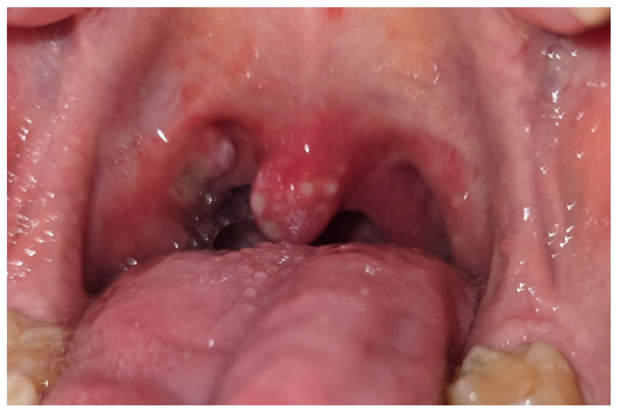
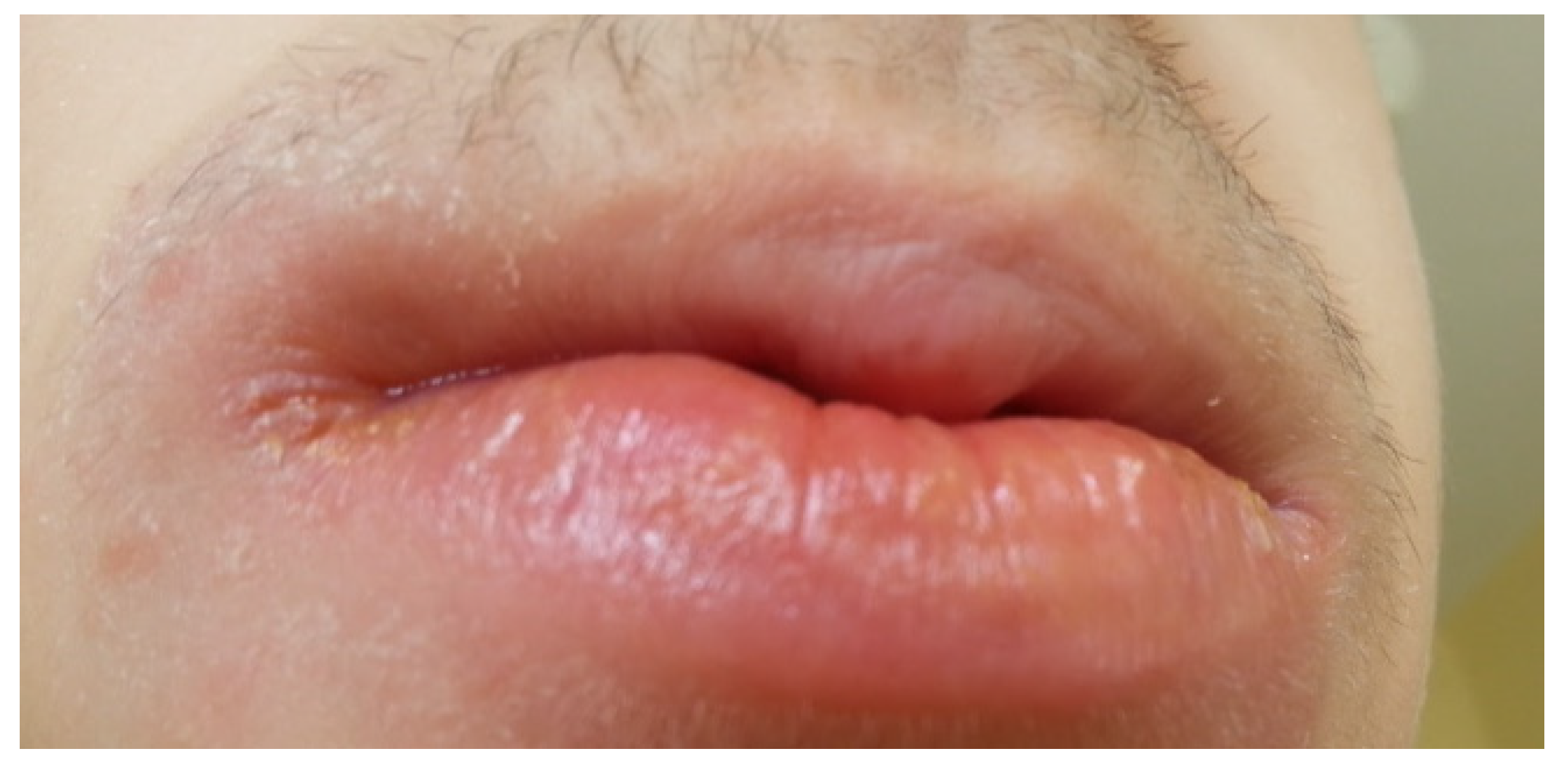
A significant relationship has been found between the use of azathioprine and mesalazine by patients and the occurrence of oral manifestations of the disease [39]. Some of these manifestations, such as aphthae (Figure 1 and Figure 2), buccal and labial mucosal swelling (Figure 3), mucosal tags, deep linear ulcerations, and cobblestoned oral mucosa, are more indicative of CD, while pyostomatitisvegetans is correlated to UC [34,40].
The therapy for patients with IBD involves modulating the body’s inflammatory response. The drugs used are immunosuppressive and include immunosuppressants (azathioprine, mercaptopurine, cyclosporine) and corticosteroids (prednisone, budesonide) [35].

4.2. Treatment of the Oral Lesions

The primary guiding principle for managing oral lesions in IBD involves pinpointing their root cause. These may stem directly from IBD, malnutrition or the use of concurrent medications.
The objective in managing oral lesions among individuals with IBD is to alleviate pain, expedite lesion healing, and forestall secondary infections. Treatment approaches vary based on the underlying cause, severity of clinical manifestation, and the specific symptoms of the oral lesions. Options for treatment encompass both topical and/or systemic medications [24].
Treating particular oral lesions in CD and pyostomatitis vegetans, conditions that can arise in both diseases, consistently focuses on addressing and managing the underlying disease. Typically, lesions show positive responses to treatment for IBD [41,42]. When this outcome is not achieved, the treatment for oral manifestations mirrors the approaches used in gastroenterological care. Pharmacological options encompass topical or systemic corticosteroids, immunosuppressive agents, and biologics, particularly anti-Tumour Necrosis Factor (TNF)-α drugs. Topical treatments may involve intralesional injections, mouthwashes, and ointments, typically starting with corticosteroid ointments and/or mouthwashes, along with nonsteroidal anti-inflammatory pastes. If there is no response, intralesional corticosteroid injections may be administered. If symptoms persist, a systemic approach involving corticosteroids becomes necessary [42,43].

5. Dental Infection Susceptibility in IBD

The examination of purulent content collected from odontogenic foci has revealed the presence of Streptococcus viridans, Peptostreptococcus, as well as bacteria of the Prevotella, Bacteroides, and Actinomyces species [44]. Recent studies indicate that anaerobic bacteria are increasingly playing a significant role in odontogenic head and neck infections [45,46,47]. Compared to the healthy population in a similar age range, patients with IBD have a higher incidence of caries, and thus odontogenic inflammation, which is most often caused by it [24,34]. The caries lesions are mainly caused by limitations in optimal nutrition (low-fibre diet) [24,48,49]. This may also be due to the sometimes-used form of treatment for flare-ups of the disease in the form of a liquid diet or parenteral nutrition, which affects the lack of mechanical cleaning of the teeth from dental plaque by food [50,51,52].

6. Clostridioides Difficile Infection in IBD Patients

6.1. Why Are Patients with IBD a High-Risk Group?

Clostridioides difficile is a Gram-Positive anaerobic bacillus that produces endospores. It is commonly found in the digestive tract of humans, animals, and the environment [53]. It is one of the most common nosocomial infections and the leading cause of healthcare-associated diarrhoea worldwide [53,54]. The most important risk factors include: antibiotic treatment, old age, hospitalisation, or stay in a nursing home [55]. Clinical presentation can vary from asymptomatic carriage to varying degrees of diarrhoea, and even to the most severe life-threatening colitis leading to death [53,55]. It is important to minimise the risk of Clostridioides difficile infection as 20–30% of patients with CDI may develop recurrent infections and up to 60% of patients may develop further episodes after a first relapse [54]. Patients with IBD in complete remission who are not exposed to antibiotics or hospitalisation are much more likely to be carriers of CDI than healthy people (8.2% vs. 1.0%, respectively) [56,57]. IBD patients treated with immunomodulatory drugs and antibiotics are at a significantly higher risk of developing CDI than those treated with antibiotics alone. Shomron Ben-Horin Et Al. [58] found that 12% of patients on combination therapy developed CDI compared to no CDI patients on antibiotics alone.

6.2. Consequences of CDI in This Population

Studies over the years have shown that the main and most common infectious cause of exacerbations in IBD are Clostridioides difficile infection [59]. In addition, the long-term colectomy risk was significantly higher for IBD patients with CDI compared to those without CDI (odds ratio [OR]: 2.22, 95% confidence interval [CI]: 1.17, 4.18). Significantly higher mortality was found for CDI in IBD patients both short-term [OR: 3.84, 95% CI: 2.62, 5.61] and long-term [OR: 3.65, 95% CI: 1.58, 8.44] [60]. Therefore, it is important to reduce the incidence of CDI by avoiding unnecessary antibiotic treatments or choosing appropriate antibiotics, which can reduce the risk of mortality in IBD [61].

6.3. Antibiotic Risks

More than 90% of healthcare-associated C. difficile infections are linked to antibiotic therapy. The risk of infection increases with prolonged antibiotic use [62], but even a single dose can increase the risk. CDI may also appear up to several weeks after completion of antiobiotic therapy [63]. Different antibiotics have varying levels of association with C. difficile infection; they can be classified into three groups: high, moderate, and low risk. (Table 2) [64,65].
In dentistry, amoxicillin and ampicillin, as well as clindamycin, are commonly used as first- and second-line therapeutic and prophylactic antibiotics [16,66]. However, all three of these antibiotics have been shown to have a high risk of causing C. difficile infections in patients [15,64]. According to a meta-analysis by Brown Et Al. [67], clindamycin has been identified as having the highest risk of causing C. difficile infections among all antibiotics tested. Due to this fact, the American Dental Association has discontinued the recommendation of clindamycin in preoperative prophylaxis in dentistry [68]. Actis Et Al. posit the hypothesis that non-steroidal anti-inflammatory drugs (NSAIDs) and antibiotics (specifically those of the macrolide structure) have the capacity to induce intestinal and hepatic damage, thereby significantly enhancing co-morbidities in gastroenterological outpatients, with the result that cost-containment guidelines are no longer adhered to [69].

7. Management Strategies for Safe Antibiotic Use in Dentistry

7.1. Guidelines of Global Associations

The current recommendations of the; American Dental Association, American Association of Endodontists, National Institute for Health and Care Excellence, Polish Dental Association and National Programme To Protect Antibiotics Working Group, European Society of Endodontology regarding the use of antibiotics in dentistry do not provide specific guidance for antibiotic therapy in patients with IBD [16,70,71,72,73,74,75,76,77]. Only general recommendations for non-immunocompromised patients are provided, which include patients with Crohn’s disease and ulcerative colitis. However, due to the nature of these diseases, individual recommendations should be considered for them. Additionally, such recommendations have not been developed by the European Crohn’s and Colitis Organisation [76].

7.2. Metronidazole

One potential solution worth examining is the use of metronidazole, as it have the advantage of a lower risk of CDI compared to first- and second-line antibiotics in the dental treatment of patients with IBD.
Metronidazole has bactericidal activity and targets anaerobic microorganisms by inhibiting nucleic acid synthesis. It also has antiprotozoal activity and does not disrupt the protective aerobic microbiota [78,79,80].
It is commonly used in perioperative prophylaxis in procedures involving the large intestine and some procedures with access through the oral mucosa. The therapeutic properties of metronidazole in the treatment of exacerbations of CD and in maintaining remission are well-known [81,82]. Metronidazole is not an antibiotic, but a chemotherapeutic agent that belongs to the low-risk group for causing CDI [15]. It is also used as first-line treatment for C. difficile infection, although current guidelines indicate that vancomycin is more effective than metronidazole in IBD [58,76].
A study conducted by Ingham et al. to investigated the efficacy of metronidazole in the treatment of acute dental infections. This investigation compared the effectiveness of metronidazole with that of parenteral penicillin in a controlled trial. The study revealed that all 37 patients exhibited a positive response, suggesting that metronidazole is comparable in effectiveness to parenteral penicillin. A further 24 patients treated with metronidazole also exhibited satisfactory responses [83]. A meta-analysis was conducted by Raghavan et al. [84], which concluded with the recommendation of the pre- and post-operative use of metronidazole in patients undergoing third molar surgery to reduce the incidence of dry socket.
A single dose of metronidazole before surgery has been shown to be more effective for C. difficile prophylaxis than multiple doses of metronidazole plus cefuroxime [85]. In contrast to amoxicillin, cefotaxime, and vancomycin, metronidazoledid not exert a notable influence on the composition or diversity of the gut microbiota. This is presumably due to the fact that is readily absorbed in the small intestine, thereby limiting its impact on the lower gut, where the majority of microbiota are located [86].

7.3. Local Administration of Antibiotic Carriers

The administration of antibiotics Via advanced carrier systems has garnered mounting attention as a promising approach to mitigate systemic adverse effects and the risk of CDI, a matter of particular pertinence in patients with IBD [87]. Local drug delivery enables high concentrations of the active compound directly at the site of infection while significantly reducing systemic exposure. This concept is particularly advantageous in the field of dentistry, where infections are typically localised and confined to the soft or hard tissues of the oral cavity [88].
In recent studies, the potential of autologous platelet concentrates, such as platelet-rich fibrin (PRF) and concentrated growth factors (CGF), as innovative drug carriers has been investigated. The fibrin matrix, abundant in platelets and leukocytes, functions as a natural scaffold for the controlled release of drugs and enhances tissue regeneration through growth factors such as PDGF, TGF-β, and VEGF. A multitude of in vitro investigations have substantiated that PRF and CGF possess the capacity to effectively incorporate and steadily release antibiotics, including amoxicillin, metronidazole, and clindamycin, thereby preserving antimicrobial efficacy over an extended duration [89,90]. This prolonged, localised release profile has the potential to enhance infection control while minimising systemic antibiotic exposure, which is a pivotal benefit for IBD patients who are at risk of developing intestinal dysbiosis and CDI recurrence [91].
Recent findings indicate that PRF and CGF mixed with metronidazole or fluconazole exhibit potent antimicrobial and antifungal effects against common oral pathogens such as Streptococcus viridans, Enterococcus faecalis, Candida albicans, and Prevotella intermedia. Notably, the biological properties of these autologous biomaterials remain unaltered by the incorporation of antimicrobial agents, thereby enabling concurrent tissue regeneration and infection prevention. This renders them particularly well-suited for utilisation in periodontal surgery, implantology, and regenerative endodontic procedures in patients diagnosed with IBD [89,92].
The potential of alternative carrier systems, including hydrogels, nanofibers, microspheres, and biodegradable membranes, to deliver antibiotics topically in the oral cavity has been a subject of investigation. However, autologous fibrin matrices have been demonstrated to exhibit superior biocompatibility, availability, and cost-effectiveness, with the added benefit of reducing the reliance on synthetic materials and preservatives that could potentially induce hypersensitivity or inflammatory responses [88,93,94,95,96,97].
For patients diagnosed with IBD, local administration of antibiotics may offer a safe and effective adjunctive treatment, with benefits that include minimising systemic antibiotic load, protecting gut microbiota, and thereby reducing the risk of CDI. Further clinical trials are warranted to standardise preparation protocols, determine optimal drug concentrations, and confirm long-term safety and efficacy in both immunocompromised and systemically burdened populations.

7.4. Microbiome-Supportive Adjuncts

Adjunctive therapies such as probiotics, prebiotics, and synbiotics may support microbiome stability during and after antibiotic use. Although widely discussed in gastroenterology, their application in dentistry requires clear, evidence-based context. In patients with inflammatory bowel disease, these adjuncts may help reduce dysbiosis, mitigate antibiotic-associated gastrointestinal symptoms, and potentially lower the risk of Clostridioides difficile infection. However, their use must be carefully tailored to dental procedures and patient immunological status [98].
Probiotics, defined as live microorganisms that confer a health benefit to the host when administered in adequate amounts, have been the most widely studied biotics in this context. A substantial body of research, evidenced by numerous randomised clinical trials, has demonstrated that specific probiotic strains, including Lactobacillus rhamnosus GG, Bifidobacterium longum, and Saccharomyces boulardii, have the potential to reduce the incidence of antibiotic-associated diarrhoea and CDI in both healthy and immunocompromised populations. In patients diagnosed with IBD, the utilisation of probiotics has been a subject of investigation for its potential to contribute to the maintenance of remission and the reduction in disease activity. Studies have yielded more encouraging outcomes in UC than in CD. The multi-strain probiotic VSL#3 appears to provide the most consistent clinical benefit, although not all studies confirm its efficacy. Multi-strain formulations have demonstrated a greater potential for efficacy in comparison to single-strain probiotics [98,99,100].
In circumstances where systemic antibiotics are deemed necessary within the context of dental practice, the judicious administration of probiotic strains may contribute to the maintenance of gut microbiota stability, thereby mitigating the risk of developing antibiotic-associated diarrhoea in patients diagnosed with IBD. Evidence-supported strains include Saccharomyces boulardii, which has demonstrated benefit in preventing antibiotic-associated diarrhoea and reducing recurrence of C. difficile infection, as well as several Lactobacillus and Bifidobacterium species commonly used in clinical trials, such as Lactobacillus rhamnosus GG, L. plantarum, L. acidophilus, L. casei, Bifidobacterium longum, and B. breve. These strains may be considered as adjuncts during or immediately after antibiotic therapy, particularly in IBD patients with a history of dysbiosis or gastrointestinal sensitivity [101,102,103]. However, it is imperative to note that the utilisation of probiotics must be tailored to the individual, as their efficacy is not universally applicable, particularly in cases of immunosuppression. Contraindications for this treatment include severe immunosuppression, high-dose corticosteroid therapy, neutropenia, the presence of central venous catheters, critical illness, and severe active colitis with mucosal barrier disruption. In such cases, the risk of probiotic-related bacteremia or fungemia, particularly with S. boulardii, is considered to exceed the potential benefits. For patients receiving biologics or thiopurines, probiotics may be used cautiously and ideally in consultation with the patient’s gastroenterologist. Consequently, while certain strains may offer beneficial effects during dental antibiotic therapy, their use in IBD must be meticulously tailored to the patient’s immune status and overall clinical condition [100,104].
Prebiotics, such as inulin, fructooligosaccharides, and galactooligosaccharides, are non-digestible substrates that selectively stimulate the growth of beneficial bacteria like Bifidobacterium and Lactobacillus. While prebiotics are generally considered safe, their clinical efficacy in IBD remains unproven. The existing controlled trials are limited in scope and underpowered, demonstrating inconsistent or minimal benefits in modulating disease activity. While not inherently detrimental, prebiotics have been observed to induce mild gastrointestinal adverse effects, including bloating or discomfort. However, the administration of prebiotics during the early stages of life may have a significant impact on the development of the gut microbiome, which could subsequently influence an individual’s susceptibility to or protection from IBD in later life [100,105].
Synbiotics, a combination of probiotics and prebiotics, seek to enhance microbial balance in a synergistic manner. Preliminary evidence suggests that synbiotics may improve mucosal healing, reduce inflammation, and enhance gastrointestinal tolerance during antibiotic therapy. However, given the variability observed in probiotic–prebiotic combinations and dosages, it is premature to draw definitive conclusions regarding their efficacy [106,107].
Recent research has introduced paraprobiotics and postbiotics as promising alternatives. Paraprobiotics—non-viable microbial cells or their components—offer several advantages, including safety in immunocompromised individuals, absence of risk for bacterial translocation or antibiotic resistance gene transfer, and greater stability in production and storage. In a similar vein, postbiotics, comprising microbial metabolites such as short-chain fatty acids, have exhibited promising benefits on epithelial integrity and inflammation modulation. However, it should be noted that the current state of research is preliminary and heterogeneous [108,109].

8. Discussion

8.1. General Interpretation

Notwithstanding the rising prevalence of individuals afflicted with IBD who are concurrently afflicted with dental ailments, there persists an absence of formal guidelines to assist dental practitioners in the judicious selection of antibiotic therapy for this demographic. This discrepancy is of clinical significance, as numerous conventional dental antibiotics carry a high risk of Clostridioides difficile infection and gut microbiota disruption. Within this context, metronidazole offers significant potential due to its low CDI risk and robust anaerobic coverage; however, its limitations include its ineffectiveness against aerobic pathogens, the necessity for combination therapy in numerous odontogenic infections, and safety concerns such as alcohol interactions and neuropathy.
In order to further minimise systemic antibiotic exposure, local antimicrobial delivery systems, including PRF and CGF, represent promising adjuncts. These approaches have the potential to reduce the necessity for broad-spectrum systemic antibiotics by enhancing localised infection control and promoting tissue healing.
Furthermore, probiotics have been shown to provide a supportive benefit for selected IBD patients during or after systemic antibiotic therapy, particularly in terms of preventing antibiotic-associated gastrointestinal disturbances. However, the utilisation of these agents is not without caveats; the efficacy of these agents varies according to the specific strain of the pathogen, their benefits are largely supplementary rather than therapeutic, and their use may be contraindicated in individuals with compromised immune systems due to the risk of translocation or fungemia.
Collectively, these findings underscore the necessity for individualised, microbiome-conscious antibiotic strategies in the dental management of IBD patients and emphasise the importance of developing formal guidelines to support clinical decision-making.

8.2. Strengths

This review addresses a significant yet understudied clinical intersection: the safe utilisation of systemic antibiotics in the dental management of patients with inflammatory bowel disease. A key strength of the study is the comprehensive, multi-database search strategy, which captured a wide spectrum of medical, dental, microbiological, and pharmacological literature relevant to antibiotic decision-making in this high-risk population. The review also incorporates emerging topics, such as the potential role of local delivery systems (PRF, CGF) and microbiome-supportive adjuncts (probiotics), which are seldom discussed in dentistry despite growing clinical relevance. Finally, the review emphasises practical applicability, synthesising available evidence into clear clinical considerations that can support dentists in contexts where formal guidelines are lacking.

8.3. Limitations

It is important to note that this review is subject to several significant limitations. Firstly, there is a notable absence of studies specifically evaluating antibiotic therapy for IBD patients in the dental office, which restricts the ability to draw strong, evidence-based conclusions tailored to this population. The majority of available data are extrapolated from general medical or gastroenterological contexts, rather than from dental clinical trials involving patients with IBD.
Secondly, although PRF and CGF show promise as local antimicrobial carriers, the majority of evidence supporting their antibacterial activity and drug-delivery potential comes from in vitro studies. This limits the reliability and direct clinical applicability of these findings. In order to confirm the effectiveness and safety of the aforementioned treatment in a dental context, it is necessary to conduct high-quality in vivo or clinical studies.
In conclusion, due to the breadth and heterogeneity of the topic, it was only possible to synthesise the included literature in a narrative manner. The heterogeneity of the studies, in terms of their design, methodology and outcomes, precluded the application of standardised risk-of-bias tools or quantitative synthesis (e.g., meta-analysis). Consequently, the conclusions derived from this study should be interpreted with a degree of caution, taking into account the inherent limitations of the methodology employed in narrative reviews.

8.4. Future Directions

It is recommended that future research endeavours seek to address the significant evidence gaps that have been identified in this review. The necessity for clinical studies specifically involving IBD patients in dental settings is evident, particularly in the context of evaluating the efficacy and safety of antibiotics, the incidence of CDI, and the outcomes of microbiome interventions. In order to validate the antimicrobial and drug-delivery potential of PRF and CGF, it is essential to conduct high-quality in vivo and clinical trials, thus building upon preliminary in vitro findings. Furthermore, the potential of probiotics and synbiotics as adjunctive therapies during dental antibiotic treatment warrants exploration, with particular attention to strain-specific effects and safety in immunosuppressed individuals. Ultimately, multidisciplinary collaboration between dentistry, gastroenterology, microbiology, and pharmacology is essential to develop evidence-based, IBD-specific antibiotic guidelines that support safe and personalised dental care.

9. Conclusions

Patients suffering from inflammatory bowel disease present a unique set of challenges in the context of dental antibiotic management, due to their elevated risk of Clostridioides difficile infection and vulnerability to microbiome disruption. Notwithstanding these concerns, there are currently no dedicated guidelines to support dentists in selecting safe and effective antibiotic regimens for this population. This review underscores the potential role of metronidazole as a lower-risk option in selected cases, while acknowledging its limitations—including insufficient aerobic coverage and safety considerations—and emphasising the need for judicious, case-specific use rather than universal preference.
The utilisation of emerging alternatives, such as local antimicrobial delivery systems (PRF, CGF), has the potential to reduce reliance on systemic antibiotics. However, the current body of evidence is predominantly confined to in vitro studies and necessitates clinical validation. The use of probiotics and synbiotics has been demonstrated to provide a supportive benefit in certain IBD patients undergoing systemic antibiotic treatment. However, it is imperative to emphasise that the utilisation of these substances must be tailored to the individual, taking into account the strain-specific efficacy and contraindications in immunosuppressed individuals.
In conclusion, the extant evidence supports the implementation of a more personalised and microbiome-conscious approach to antibiotic therapy in dentistry for patients suffering from inflammatory bowel disease (IBD). There are still significant knowledge gaps, and well-designed clinical studies are urgently needed to establish evidence-based protocols that balance antimicrobial effectiveness with patient safety.

Funding

This research received no external funding.

Institutional Review Board Statement

Narrative review did not require formal ethical approval.

Informed Consent Statement

Informed consent for publication was obtained.

Conflicts of Interest

The authors declare no conflicts of interest.

References

  1. Elzayat, H.; Mesto, G.; Al-Marzooq, F. Unraveling the Impact of Gut and Oral Microbiome on Gut Health in Inflammatory Bowel Diseases. Nutrients 2023, 15, 3377. [Google Scholar] [CrossRef] [PubMed]
  2. Agrawal, M.; Jess, T. Implications of the Changing Epidemiology of Inflammatory Bowel Disease in a Changing World. UEG J. 2022, 10, 1113–1120. [Google Scholar] [CrossRef]
  3. Mosli, M.; Alawadhi, S.; Hasan, F.; Abou Rached, A.; Sanai, F.; Danese, S. Incidence, Prevalence, and Clinical Epidemiology of Inflammatory Bowel Disease in the Arab World: A Systematic Review and Meta-Analysis. Inflamm. Intest. Dis. 2021, 6, 123–131. [Google Scholar] [CrossRef]
  4. Kaplan, G.G.; Windsor, J.W. The Four Epidemiological Stages in the Global Evolution of Inflammatory Bowel Disease. Nat. Rev. Gastroenterol. Hepatol. 2021, 18, 56–66. [Google Scholar] [CrossRef]
  5. Lewis, J.D.; Parlett, L.E.; Jonsson Funk, M.L.; Brensinger, C.; Pate, V.; Wu, Q.; Dawwas, G.K.; Weiss, A.; Constant, B.D.; McCauley, M.; et al. Incidence, Prevalence, and Racial and Ethnic Distribution of Inflammatory Bowel Disease in the United States. Gastroenterology 2023, 165, 1197–1205.e2. [Google Scholar] [CrossRef]
  6. Park, J.; Cheon, J.H. Incidence and Prevalence of Inflammatory Bowel Disease across Asia. Yonsei Med. J. 2021, 62, 99. [Google Scholar] [CrossRef] [PubMed]
  7. Ananthakrishnan, A.N. Epidemiology and Risk Factors for IBD. Nat. Rev. Gastroenterol. Hepatol. 2015, 12, 205–217. [Google Scholar] [CrossRef]
  8. Li, C.; Wu, Y.; Xie, Y.; Zhang, Y.; Jiang, S.; Wang, J.; Luo, X.; Chen, Q. Oral Manifestations Serve as Potential Signs of Ulcerative Colitis: A Review. Front. Immunol. 2022, 13, 1013900. [Google Scholar] [CrossRef]
  9. Zhang, Y.; Qiao, D.; Chen, R.; Zhu, F.; Gong, J.; Yan, F. The Association between Periodontitis and Inflammatory Bowel Disease: A Systematic Review and Meta-analysis. BioMed Res. Int. 2021, 2021, 6692420. [Google Scholar] [CrossRef]
  10. Del Vecchio, L.E.; Fiorani, M.; Tohumcu, E.; Bibbò, S.; Porcari, S.; Mele, M.C.; Pizzoferrato, M.; Gasbarrini, A.; Cammarota, G.; Ianiro, G. Risk Factors, Diagnosis, and Management of Clostridioides Difficile Infection in Patients with Inflammatory Bowel Disease. Microorganisms 2022, 10, 1315. [Google Scholar] [CrossRef] [PubMed]
  11. Hourigan, S.K.; Oliva-Hemker, M.; Hutfless, S. The Prevalence of Clostridium Difficile Infection in Pediatric and Adult Patients with Inflammatory Bowel Disease. Dig. Dis. Sci. 2014, 59, 2222–2227. [Google Scholar] [CrossRef] [PubMed]
  12. Berg, A.M.; Kelly, C.P.; Farraye, F.A. Clostridium Difficile Infection in the Inflammatory Bowel Disease Patient. Inflamm. Bowel Dis. 2013, 19, 194–204. [Google Scholar] [CrossRef]
  13. D’Aoust, J.; Battat, R.; Bessissow, T. Management of Inflammatory Bowel Disease with Clostridium Difficile Infection. World J. Gastroenterol. 2017, 23, 4986. [Google Scholar] [CrossRef]
  14. Sehgal, K.; Yadav, D.; Khanna, S. The Interplay of Clostridioides Difficile Infection and Inflammatory Bowel Disease. Ther. Adv. Gastroenterol. 2021, 14, 17562848211020285. [Google Scholar] [CrossRef]
  15. Martirosian, G.; Hryniewicz, W.; Ozorowski, T.; Pawlik, K.; Deptuła, A. Zakażenia Clostridioides (Clostridium) Difficile: Epidemiologia, Diagnostyka, Terapia, Profilaktyka; Wydanie Drugie; Narodowy Instytut Leków: Warszawa, Poland, 2018; ISBN 978-83-949636-0-6. [Google Scholar]
  16. Kaczmarzyk, T.; Babiuch, K.; Bołtacz-Rzepkowska, E.; Dominiak, M.; Konopka, T.; Lipski, M.; Olczak-Kowalczyk, D.; Szeląg, A.; Szuta, M.; Hryniewicz, W. Polish Dental Association and National Programme to Protect Antibiotics Working Group Recommendations for Administration of Antibiotics in Dentistry. J. Stomatol. 2018, 71, 457–465. [Google Scholar] [CrossRef]
  17. Baethge, C.; Goldbeck-Wood, S.; Mertens, S. SANRA—A Scale for the Quality Assessment of Narrative Review Articles. Res. Integr. Peer Rev. 2019, 4, 5. [Google Scholar] [CrossRef] [PubMed]
  18. Kong, L.; Pokatayev, V.; Lefkovith, A.; Carter, G.T.; Creasey, E.A.; Krishna, C.; Subramanian, S.; Kochar, B.; Ashenberg, O.; Lau, H.; et al. The Landscape of Immune Dysregulation in Crohn’s Disease Revealed through Single-Cell Transcriptomic Profiling in the Ileum and Colon. Immunity 2023, 56, 444–458.e5. [Google Scholar] [CrossRef]
  19. Andoh, A.; Nishida, A. Alteration of the Gut Microbiome in Inflammatory Bowel Disease. Digestion 2023, 104, 16–23. [Google Scholar] [CrossRef]
  20. Xu, X.; Ocansey, D.K.W.; Hang, S.; Wang, B.; Amoah, S.; Yi, C.; Zhang, X.; Liu, L.; Mao, F. The Gut Metagenomics and Metabolomics Signature in Patients with Inflammatory Bowel Disease. Gut Pathog. 2022, 14, 26. [Google Scholar] [CrossRef] [PubMed]
  21. Mustafi, S.; Sinha, R.; Sarkar, S.; Giri, D.; Saha, A.P.; Yadav, P. Comparative Estimation of Serum Levels of Vitamin A, Vitamin B12, Vitamin D and Vitamin E in Patients with Recurrent Aphthous Stomatitis and Normal Individuals—A Case-Control Study. J. Indian. Acad. Oral Med. Radiol. 2021, 33, 442–446. [Google Scholar] [CrossRef]
  22. Valvano, M.; Capannolo, A.; Cesaro, N.; Stefanelli, G.; Fabiani, S.; Frassino, S.; Monaco, S.; Magistroni, M.; Viscido, A.; Latella, G. Nutrition, Nutritional Status, Micronutrients Deficiency, and Disease Course of Inflammatory Bowel Disease. Nutrients 2023, 15, 3824. [Google Scholar] [CrossRef]
  23. Valvano, M.; Faenza, S.; Cortellini, F.; Vinci, A.; Ingravalle, F.; Calabrò, M.; Scurti, L.; Di Nezza, M.; Valerio, S.; Viscido, A.; et al. The Relationship Between Nutritional Status, Micronutrient Deficiency, and Disease Activity in IBD Patients: A Multicenter Cross-Sectional Study. Nutrients 2025, 17, 2690. [Google Scholar] [CrossRef]
  24. Muhvić-Urek, M.; Tomac-Stojmenović, M.; Mijandrušić-Sinčić, B. Oral Pathology in Inflammatory Bowel Disease. World J. Gastroenterol. 2016, 22, 5655. [Google Scholar] [CrossRef]
  25. Alkhouri, R.H.; Hashmi, H.; Baker, R.D.; Gelfond, D.; Baker, S.S. Vitamin and Mineral Status in Patients with Inflammatory Bowel Disease. J. Pediatr. Gastroenterol. Nutr. 2013, 56, 89–92. [Google Scholar] [CrossRef]
  26. Kim, S.Y.; Mun, E.C.; Chung, J.-W.; Ha, M.; Ahn, S.-M.; Han, M.-D.; Han, S.-H.; Yun, S.-C.; Kim, J.H.; Kim, K.O.; et al. Increased Genomic Damage and Vitamin B Status in Inflammatory Bowel Disease Patients: A Case-Control, Prospective, Pilot Study. Mutat. Res./Genet. Toxicol. Environ. Mutagen. 2019, 837, 42–47. [Google Scholar] [CrossRef]
  27. Le, T. Micronutrient Deficiencies in Patients with Inflammatory Bowel Disease. Cutis 2024, 113, 159–166. [Google Scholar] [CrossRef] [PubMed]
  28. Rosa, A.; Cianconi, G.; De Angelis, R.; Pujia, A.M.; Arcuri, C. Hypovitaminosis and Its Association with Recurrent Aphthous Stomatitis: A Comprehensive Review of Clinical Correlations and Diagnostic Considerations. Front. Oral Health 2025, 6, 1520067. [Google Scholar] [CrossRef]
  29. Esquivel-Pedraza, L.; Fernández-Cuevas, L.; Delgado-Martínez, I.; Cicero-Casarrubias, A.; Milke-García, M.D.P.; Chang-Bool, E.M.; Barragán-Heredia, L.M.; Maldonado-Molina, J.; Rivera-Flores, R.L.; Yamamoto-Furusho, J.K.; et al. Oral Mucosal Findings in Ambulatory Patients with Inflammatory Bowel Disease. Braz. Oral Res. 2025, 39, e095. [Google Scholar] [CrossRef] [PubMed]
  30. Margulies, S.L.; Kurian, D.; Elliott, M.S.; Han, Z. Vitamin D Deficiency in Patients with Intestinal Malabsorption Syndromes—Think in and Outside the Gut. J. Dig. Dis. 2015, 16, 617–633. [Google Scholar] [CrossRef]
  31. Vernia, F.; Valvano, M.; Longo, S.; Cesaro, N.; Viscido, A.; Latella, G. Vitamin D in Inflammatory Bowel Diseases. Mechanisms of Action and Therapeutic Implications. Nutrients 2022, 14, 269. [Google Scholar] [CrossRef] [PubMed]
  32. Schäffler, H.; Schmidt, M.; Huth, A.; Reiner, J.; Glass, Ä.; Lamprecht, G. Clinical Factors Are Associated with Vitamin D Levels in IBD Patients: A Retrospective Analysis. J. Dig. Dis. 2018, 19, 24–32. [Google Scholar] [CrossRef]
  33. Al-Maweri, S.A.; Al-Qadhi, G.; Halboub, E.; Alaizari, N.; Almeslet, A.; Ali, K.; Osman, S.A.A. Vitamin D Deficiency and Risk of Recurrent Aphthous Stomatitis: Updated Meta-Analysis with Trial Sequential Analysis. Front. Nutr. 2023, 10, 1132191. [Google Scholar] [CrossRef]
  34. Lauritano, D.; Boccalari, E.; Stasio, D.; Vella, D.F.; Carinci, F.; Lucchese, A.; Petruzzi, M. Prevalence of Oral Lesions and Correlation with Intestinal Symptoms of Inflammatory Bowel Disease: A Systematic Review. Diagnostics 2019, 9, 77. [Google Scholar] [CrossRef] [PubMed]
  35. Saeid Seyedian, S.; Nokhostin, F.; Dargahi Malamir, M. A Review of the Diagnosis, Prevention, and Treatment Methods of Inflammatory Bowel Disease. JMedLife 2019, 12, 113–122. [Google Scholar] [CrossRef] [PubMed]
  36. Niemczyk, S.; Niemczyk, W.; Prokurat, M.; Grudnik, K.; Kuleszyński, M.; Niciejewska, E.; Lau, K.; Kasperczyk, J. Impact of E-Cigarettes on the Oral Health-Literature Review. Polski Merkur. Lek. 2023, 51, 13–17. [Google Scholar] [CrossRef] [PubMed]
  37. Tan, C.X.W.; Brand, H.S.; Iqbal, S.; De Boer, N.K.H.; Forouzanfar, T.; De Visscher, J.G.A.M. A Self-Reported Survey on Oral Health Problems in Patients with Inflammatory Bowel Disease with a Stoma. Oral Surg. Oral Med. Oral Pathol. Oral Radiol. 2020, 130, e80–e86. [Google Scholar] [CrossRef]
  38. Agossa, K.; Roman, L.; Gosset, M.; Yzet, C.; Fumery, M. Periodontal and Dental Health in Inflammatory Bowel Diseases: A Systematic Review. Expert. Rev. Gastroenterol. Hepatol. 2021, 15, 1–15. [Google Scholar] [CrossRef]
  39. Khozeimeh, F.; Shakerin, H.; Daghaghzadeh, H.; Najarzadegan, F.; Golestannejad, Z.; Adibi, P. Oral Manifestations in Inflammatory Bowel Disease: A Cross-Sectional Study in Isfahan. Dent. Res. J. 2021, 18, 4. [Google Scholar] [CrossRef]
  40. Tan, C.X.W.; Brand, H.S.; De Boer, N.K.H.; Forouzanfar, T. Gastrointestinal Diseases and Their Oro-Dental Manifestations: Part 2: Ulcerative Colitis. Br. Dent. J. 2017, 222, 53–57. [Google Scholar] [CrossRef]
  41. Rogler, G.; Singh, A.; Kavanaugh, A.; Rubin, D.T. Extraintestinal Manifestations of Inflammatory Bowel Disease: Current Concepts, Treatment, and Implications for Disease Management. Gastroenterology 2021, 161, 1118–1132. [Google Scholar] [CrossRef]
  42. Ribaldone, D.G.; Brigo, S.; Mangia, M.; Saracco, G.M.; Astegiano, M.; Pellicano, R. Oral Manifestations of Inflammatory Bowel Disease and the Role of Non-Invasive Surrogate Markers of Disease Activity. Medicines 2020, 7, 33. [Google Scholar] [CrossRef] [PubMed]
  43. Katsanos, K.H.; Torres, J.; Roda, G.; Brygo, A.; Delaporte, E.; Colombel, J.-F. Review Article: Non-malignant Oral Manifestations in Inflammatory Bowel Diseases. Aliment. Pharmacol. Ther. 2015, 42, 40–60. [Google Scholar] [CrossRef]
  44. Jundt, J.S.; Gutta, R. Characteristics and Cost Impact of Severe Odontogenic Infections. Oral Surg. Oral Med. Oral Pathol. Oral Radiol. 2012, 114, 558–566. [Google Scholar] [CrossRef]
  45. Piekoszewska-Ziętek, P.; Turska-Szybka, A.; Olczak-Kowalczyk, D. Odontogenic Infections—Review of the Literature. Nowa Stomatol. 2016, 21, 120–134. [Google Scholar] [CrossRef]
  46. Tkaczyk, M.; Kuśka-Kielbratowska, A.; Fiegler-Rudol, J.; Niemczyk, W.; Mertas, A.; Skaba, D.; Wiench, R. The Prevalence and Drug Susceptibility of Candida Species and an Analysis of Risk Factors for Oral Candidiasis—A Retrospective Study. Antibiotics 2025, 14, 876. [Google Scholar] [CrossRef]
  47. Fiegler-Rudol, J.; Niemczyk, W.; Janik, K.; Zawilska, A.; Kępa, M.; Tanasiewicz, M. How to Deal with Pulpitis: An Overview of New Approaches. Dent. J. 2025, 13, 25. [Google Scholar] [CrossRef]
  48. Marruganti, C.; Discepoli, N.; Gaeta, C.; Franciosi, G.; Ferrari, M.; Grandini, S. Dental Caries Occurrence in Inflammatory Bowel Disease Patients: A Systematic Review and Meta-Analysis. Caries Res. 2021, 55, 485–495. [Google Scholar] [CrossRef]
  49. Nijakowski, K.; Gruszczyński, D.; Surdacka, A. Oral Health Status in Patients with Inflammatory Bowel Diseases: A Systematic Review. Int. J. Environ. Res. Public Health 2021, 18, 11521. [Google Scholar] [CrossRef]
  50. Marafini, I.; Salvatori, S.; Troncone, E.; Scarozza, P.; Fantini, E.; Monteleone, G. No Effect of a Liquid Diet in the Management of Patients with Stricturing Crohn’s Disease. Int. J. Color. Dis. 2020, 35, 1881–1885. [Google Scholar] [CrossRef] [PubMed]
  51. Adamina, M.; Gerasimidis, K.; Sigall-Boneh, R.; Zmora, O.; De Buck Van Overstraeten, A.; Campmans-Kuijpers, M.; Ellul, P.; Katsanos, K.; Kotze, P.G.; Noor, N.; et al. Perioperative Dietary Therapy in Inflammatory Bowel Disease. J. Crohn’s Colitis 2020, 14, 431–444. [Google Scholar] [CrossRef] [PubMed]
  52. Fiegler-Rudol, J.; Niemczyk, W.; Netkowska, M.; Demel, K.; Talaska, J.; Klimczak, T.; Hochuł, D. The Influence of Parenteral Nutrition on the Condition of the Oral Cavity: Literature Review. Wiad. Lek. 2024, 77, 1069–1073. [Google Scholar] [CrossRef] [PubMed]
  53. Czepiel, J.; Dróżdż, M.; Pituch, H.; Kuijper, E.J.; Perucki, W.; Mielimonka, A.; Goldman, S.; Wultańska, D.; Garlicki, A.; Biesiada, G. Clostridium Difficile Infection: Review. Eur. J. Clin. Microbiol. Infect. Dis. 2019, 38, 1211–1221. [Google Scholar] [CrossRef] [PubMed]
  54. Rodríguez, T.; Esther, L. Análisis de La Producción de Factores de Virulencia y Resistencia a Antibióticos En Biopelícula de Clostridioides (Clostridium) Difficile En Infecciones Recurrentes y No Recurrentes; Universidad Autónoma Nuevo León: San Nicolas de los Garza, Mexico, 2019. [Google Scholar]
  55. Smits, W.K.; Lyras, D.; Lacy, D.B.; Wilcox, M.H.; Kuijper, E.J. Clostridium Difficile Infection. Nat. Rev. Dis. Primers 2016, 2, 16020. [Google Scholar] [CrossRef]
  56. Clayton, E.M.; Rea, M.C.; Shanahan, F.; Quigley, E.M.M.; Kiely, B.; Hill, C.; Ross, R.P. The Vexed Relationship Between Clostridium Difficile and Inflammatory Bowel Disease: An Assessment of Carriage in an Outpatient Setting Among Patients in Remission. Am. J. Gastroenterol. 2009, 104, 1162–1169. [Google Scholar] [CrossRef]
  57. Singh, H.; Nugent, Z.; Yu, B.N.; Lix, L.M.; Targownik, L.E.; Bernstein, C.N. Higher Incidence of Clostridium Difficile Infection Among Individuals with Inflammatory Bowel Disease. Gastroenterology 2017, 153, 430–438.e2. [Google Scholar] [CrossRef]
  58. Ben-Horin, S.; Margalit, M.; Bossuyt, P.; Maul, J.; Shapira, Y.; Bojic, D.; Chermesh, I.; Al-Rifai, A.; Schoepfer, A.; Bosani, M.; et al. Combination Immunomodulator and Antibiotic Treatment in Patients with Inflammatory Bowel Disease and Clostridium Difficile Infection. Clin. Gastroenterol. Hepatol. 2009, 7, 981–987. [Google Scholar] [CrossRef]
  59. Khanna, S. Management of Clostridioides Difficile Infection in Patients with Inflammatory Bowel Disease. Intest. Res. 2021, 19, 265–274. [Google Scholar] [CrossRef] [PubMed]
  60. Balram, B.; Battat, R.; Al-Khoury, A.; D’Aoust, J.; Afif, W.; Bitton, A.; Lakatos, P.L.; Bessissow, T. Risk Factors Associated with Clostridium Difficile Infection in Inflammatory Bowel Disease: A Systematic Review and Meta-Analysis. J. Crohn’s Colitis 2019, 13, 27–38. [Google Scholar] [CrossRef]
  61. Burisch, J.; Munkholm, P. Inflammatory Bowel Disease Epidemiology. Curr. Opin. Gastroenterol. 2013, 29, 357–362. [Google Scholar] [CrossRef]
  62. Sunenshine, R.H.; McDonald, L.C. Clostridium Difficile-Associated Disease: New Challenges from an Established Pathogen. Clevel. Clin. J. Med. 2006, 73, 187–197. [Google Scholar] [CrossRef]
  63. Pattani, R.; Palda, V.A.; Hwang, S.W.; Shah, P.S. Probiotics for the Prevention of Antibiotic-Associated Diarrhea and Clostridium Difficile Infection among Hospitalized Patients: Systematic Review and Meta-Analysis. Open Med. 2013, 7, e56–e67. [Google Scholar] [PubMed]
  64. Kukla, M.; Cisek, K. Diagnosis, Prophylaxis and Treatment of Clostridium Difficile Infection According to 2017 Guidelines of Infectious Diseases Society of America (IDSA) and Society for Healthcare Epidemiology of America (SHEA). Gastroenterol. Klin. 2019, 11, 43–54. [Google Scholar]
  65. Flynn, T.R. Evidence-Based Principles of Antibiotic Therapy. In Evidence-Based Oral Surgery; Ferneini, E.M., Goupil, M.T., Eds.; Springer International Publishing: Cham, Switzerland, 2019; pp. 283–316. ISBN 978-3-319-91360-5. [Google Scholar]
  66. Krasuska-Sławińska, E. Zakażenia Zębopochodne, Zapalenie Tkanek Przyzębia Oraz Profilaktyka Zakażeń Odległych–Wskazania Do Antybiotykoterapii. Med. Faktów 2020, 13, 322–330. [Google Scholar] [CrossRef]
  67. Brown, K.A.; Khanafer, N.; Daneman, N.; Fisman, D.N. Meta-Analysis of Antibiotics and the Risk of Community-Associated Clostridium Difficile Infection. Antimicrob. Agents Chemother. 2013, 57, 2326–2332. [Google Scholar] [CrossRef]
  68. Tong, D.C.; Rothwell, B.R. Antibiotic Prophylaxis in Dentistry: A Review and Practice Recommendations. J. Am. Dent. Assoc. 2000, 131, 366–374. [Google Scholar] [CrossRef]
  69. Actis, G.; Pellicano, R.; Fadda, M.; Rosina, F. Antibiotics and Non-Steroidal Anti-Inflammatory Drugs in Outpatient Practice: Indications and Unwanted Effects in a Gastroenterological Setting. Curr. Drug Saf. 2014, 9, 133–137. [Google Scholar] [CrossRef]
  70. Diogenes, C.; Cha, B. AAE Guidance on Antibiotic Prophylaxis for Patients at Risk of Systemic Disease. Am. Assoc. Endodontists 2017, 43, 1409–1413. [Google Scholar]
  71. Segura-Egea, J.J.; Gould, K.; Hakan Şen, B.; Jonasson, P.; Cotti, E.; Mazzoni, A.; Sunay, H.; Tjäderhane, L.; Dummer, P.M.H. European Society of Endodontology Position Statement: The Use of Antibiotics in Endodontics. Int. Endod. J. 2018, 51, 20–25. [Google Scholar] [CrossRef] [PubMed]
  72. Thornhill, M.H.; Dayer, M.; Lockhart, P.B.; McGurk, M.; Shanson, D.; Prendergast, B.; Chambers, J.B. A Change in the NICE Guidelines on Antibiotic Prophylaxis. Br. Dent. J. 2016, 221, 112–114. [Google Scholar] [CrossRef]
  73. Lockhart, P.B.; Tampi, M.P.; Abt, E.; Aminoshariae, A.; Durkin, M.J.; Fouad, A.F.; Gopal, P.; Hatten, B.W.; Kennedy, E.; Lang, M.S.; et al. Evidence-Based Clinical Practice Guideline on Antibiotic Use for the Urgent Management of Pulpal- and Periapical-Related Dental Pain and Intraoral Swelling. J. Am. Dent. Assoc. 2019, 150, 906–921.e12. [Google Scholar] [CrossRef]
  74. American Academy of Pediatric Dentistry. Antibiotic pro-Phylaxis for Dental Patients at Risk for Infection. The Reference Manualof Pediatric Dentistry. Chicago, III. Am. Acad. Pediatr. Dent. 2022, 500–506. [Google Scholar]
  75. Thornhill, M.H.; Gibson, T.B.; Yoon, F.; Dayer, M.J.; Prendergast, B.D.; Lockhart, P.B.; O’Gara, P.T.; Baddour, L.M. Antibiotic Prophylaxis Against Infective Endocarditis Before Invasive Dental Procedures. J. Am. Coll. Cardiol. 2022, 80, 1029–1041. [Google Scholar] [CrossRef]
  76. Kucharzik, T.; Ellul, P.; Greuter, T.; Rahier, J.F.; Verstockt, B.; Abreu, C.; Albuquerque, A.; Allocca, M.; Esteve, M.; Farraye, F.A.; et al. ECCO Guidelines on the Prevention, Diagnosis, and Management of Infections in Inflammatory Bowel Disease. J. Crohn’s Colitis 2021, 15, 879–913. [Google Scholar] [CrossRef]
  77. Bishop, E.J.; Tiruvoipati, R. Management of Clostridioides Difficile Infection in Adults and Challenges in Clinical Practice: Review and Comparison of Current IDSA/SHEA, ESCMID and ASID Guidelines. J. Antimicrob. Chemother. 2023, 78, 21–30. [Google Scholar] [CrossRef]
  78. Smith, A. Metronidazole Resistance: A Hidden Epidemic? Br. Dent. J. 2018, 224, 403–404. [Google Scholar] [CrossRef]
  79. Mitchell, D.A. Metronidazole: Its Use in Clinical Dentistry. J. Clin. Periodontol. 1984, 11, 145–158. [Google Scholar] [CrossRef]
  80. Ahmadi, H.; Ebrahimi, A.; Ahmadi, F. Antibiotic Therapy in Dentistry. Int. J. Dent. 2021, 2021, 6667624. [Google Scholar] [CrossRef]
  81. D’Haens, G.R.; Vermeire, S.; Van Assche, G.; Noman, M.; Aerden, I.; Van Olmen, G.; Rutgeerts, P. Therapy of Metronidazole with Azathioprine to Prevent Postoperative Recurrence of Crohn’s Disease: A Controlled Randomized Trial. Gastroenterology 2008, 135, 1123–1129. [Google Scholar] [CrossRef] [PubMed]
  82. Ceruelos, A.H.; Romero-Quezada, L.C.; Ruvalcaba, J.C.; Contreras, L.L. Therapeutic Uses of Metronidazole and Its Side Effects: An Update. Eur. Rev. Med. Pharmacol. Sci. 2019, 23, 397–401. [Google Scholar]
  83. Ingham, H.R.; Hood, F.J.C.; Bradnum, P.; Tharagonnet, D.; Selkon, J.B. Metronidazole Compared with Penicillin in the Treatment of Acute Dental Infections. Br. J. Oral. Surg. 1977, 14, 264–269. [Google Scholar] [CrossRef] [PubMed]
  84. Raghavan, R.; Shah, S.; Rudagi, B.; Ticku, S.; Gaikwad, S.; Shitole, D. Metronidazole for the Prophylaxis of Alveolar Osteitis: A Systematic Review and Meta-Analysis. J. Maxillofac. Oral Surg. 2024, 24, 258–266. [Google Scholar] [CrossRef] [PubMed]
  85. Fernandes, R.; Robinson, P.; Rangarajan, K.; Scott, S.; Angco, L. The Role of Single-Shot Metronidazole in the Prevention of Clostridium Difficile Infection Following Ileostomy Reversal Surgery. Int. J. Color. Dis. 2017, 32, 733–736. [Google Scholar] [CrossRef] [PubMed]
  86. Tulstrup, M.V.-L.; Christensen, E.G.; Carvalho, V.; Linninge, C.; Ahrné, S.; Højberg, O.; Licht, T.R.; Bahl, M.I. Antibiotic Treatment Affects Intestinal Permeability and Gut Microbial Composition in Wistar Rats Dependent on Antibiotic Class. PLoS ONE 2015, 10, e0144854. [Google Scholar] [CrossRef]
  87. Reymunde Duran, D.A.; Chung, S.S.; DuPont, A.W. S2118 Recurrent Clostridioides Difficile Infections Caused by Topical Clindamycin. Am. J. Gastroenterol. 2022, 117, e1439. [Google Scholar] [CrossRef]
  88. Niemczyk, W.; Żurek, J.; Niemczyk, S.; Kępa, M.; Zięba, N.; Misiołek, M.; Wiench, R. Antibiotic-Loaded Platelet-Rich Fibrin (AL-PRF) as a New Carrier for Antimicrobials: A Systematic Review of In Vitro Studies. Int. J. Mol. Sci. 2025, 26, 2140. [Google Scholar] [CrossRef]
  89. Niemczyk, W.; Kępa, M.; Żurek, J.; Aboud, A.; Skaba, D.; Wiench, R. Comparative Evaluation of Platelet-Rich Fibrin (PRF) and Concentrated Growth Factor (CGF) as Carriers for Antibiotics—In Vitro Study. Int. J. Mol. Sci. 2025, 26, 4303. [Google Scholar] [CrossRef]
  90. Niemczyk, W.; Niemczyk, S.; Odrzywolska, O.; Doroz, P.; Hochuł, D.; Zawadzka, K. Application of I-PRF in Dentistry. Wiadomości Lek. 2024, 77, 2348–2352. [Google Scholar] [CrossRef]
  91. Nitzan, O. Role of Antibiotics for Treatment of Inflammatory Bowel Disease. World J. Gastroenterol. 2016, 22, 1078. [Google Scholar] [CrossRef]
  92. Niemczyk, W.; Kępa, M.; Żurek, J.; Aboud, A.; Skaba, D.; Wiench, R. Application of Platelet-Rich Fibrin and Concentrated Growth Factors as Carriers for Antifungal Drugs—In Vitro Study. J. Clin. Med. 2025, 14, 5111. [Google Scholar] [CrossRef]
  93. Ghazwani, M.; Vasudevan, R.; Kandasamy, G.; Hani, U.; Niharika, G.; Naredla, M.; Devanandan, P.; Puvvada, R.C.; Almehizia, A.A.; Hakami, A.R.; et al. Development and In Vitro Characterization of Antibiotic-Loaded Nanocarriers for Dental Delivery. Molecules 2023, 28, 2914. [Google Scholar] [CrossRef] [PubMed]
  94. Kida, D.; Zakrzewska, A.; Zborowski, J.; Szulc, M.; Karolewicz, B. Polymer-Based Carriers in Dental Local Healing—Review and Future Challenges. Materials 2021, 14, 3948. [Google Scholar] [CrossRef]
  95. Tahmasebi, E.; Ardestani, A.K.; Hassani, Z.; Alam, M.; Abbasi, K.; Kahrizi, S.; Madihi, N.; Abbasiparashkouh, Z.; Mohammadi, A.; Shahab, P.; et al. The Current Novel Drug Delivery System (Natural and Chemical Composites) in Dental Infections for Antibiotics Resistance: A Narrative Review. Cell. Mol. Biol. 2022, 68, 141–160. [Google Scholar] [CrossRef]
  96. Trusek, A.; Kijak, E. Drug Carriers Based on Graphene Oxide and Hydrogel: Opportunities and Challenges in Infection Control Tested by Amoxicillin Release. Materials 2021, 14, 3182. [Google Scholar] [CrossRef] [PubMed]
  97. Hakim, L.K.; Yazdanian, M.; Alam, M.; Abbasi, K.; Tebyaniyan, H.; Tahmasebi, E.; Khayatan, D.; Seifalian, A.; Ranjbar, R.; Yazdanian, A. Biocompatible and Biomaterials Application in Drug Delivery System in Oral Cavity. Evid.-Based Complement. Altern. Med. 2021, 2021, 9011226. [Google Scholar] [CrossRef]
  98. Lê, A.; Mantel, M.; Marchix, J.; Bodinier, M.; Jan, G.; Rolli-Derkinderen, M. Inflammatory Bowel Disease Therapeutic Strategies by Modulation of the Microbiota: How and When to Introduce Pre-, pro-, Syn-, or Postbiotics? Am. J. Physiol.-Gastrointest. Liver Physiol. 2022, 323, G523–G553. [Google Scholar] [CrossRef] [PubMed]
  99. Smolinska, S.; Popescu, F.-D.; Zemelka-Wiacek, M. A Review of the Influence of Prebiotics, Probiotics, Synbiotics, and Postbiotics on the Human Gut Microbiome and Intestinal Integrity. J. Clin. Med. 2025, 14, 3673. [Google Scholar] [CrossRef]
  100. Martyniak, A.; Medyńska-Przęczek, A.; Wędrychowicz, A.; Skoczeń, S.; Tomasik, P.J. Prebiotics, Probiotics, Synbiotics, Paraprobiotics and Postbiotic Compounds in IBD. Biomolecules 2021, 11, 1903. [Google Scholar] [CrossRef]
  101. Al Sharaby, A.; Abugoukh, T.M.; Ahmed, W.; Ahmed, S.; Elshaikh, A.O. Do Probiotics Prevent Clostridium Difficile-Associated Diarrhea? Cureus 2022, 14, e27624. [Google Scholar] [CrossRef]
  102. Heil, E.L.; Harris, A.D.; Brown, C.; Seung, H.; Thom, K.A.; Von Rosenvinge, E.; Sorongon, S.; Pineles, L.; Goodman, K.E.; Leekha, S. A Multicenter Evaluation of Probiotic Use for the Primary Prevention of Clostridioides Difficile Infection. Clin. Infect. Dis. 2021, 73, 1330–1337. [Google Scholar] [CrossRef] [PubMed]
  103. Goodman, C.; Keating, G.; Georgousopoulou, E.; Hespe, C.; Levett, K. Probiotics for the Prevention of Antibiotic-Associated Diarrhoea: A Systematic Review and Meta-Analysis. BMJ Open 2021, 11, e043054. [Google Scholar] [CrossRef]
  104. Zhang, X.-F.; Guan, X.-X.; Tang, Y.-J.; Sun, J.-F.; Wang, X.-K.; Wang, W.-D.; Fan, J.-M. Clinical Effects and Gut Microbiota Changes of Using Probiotics, Prebiotics or Synbiotics in Inflammatory Bowel Disease: A Systematic Review and Meta-Analysis. Eur. J. Nutr. 2021, 60, 2855–2875. [Google Scholar] [CrossRef] [PubMed]
  105. Akutko, K.; Stawarski, A. Probiotics, Prebiotics and Synbiotics in Inflammatory Bowel Diseases. J. Clin. Med. 2021, 10, 2466. [Google Scholar] [CrossRef] [PubMed]
  106. Li, H.-Y.; Zhou, D.-D.; Gan, R.-Y.; Huang, S.-Y.; Zhao, C.-N.; Shang, A.; Xu, X.-Y.; Li, H.-B. Effects and Mechanisms of Probiotics, Prebiotics, Synbiotics, and Postbiotics on Metabolic Diseases Targeting Gut Microbiota: A Narrative Review. Nutrients 2021, 13, 3211. [Google Scholar] [CrossRef]
  107. Parigi, T.L.; Vieujean, S.; Paridaens, K.; Dalgaard, K.; Peyrin-Biroulet, L.; Danese, S. Efficacy, Safety, and Concerns on Microbiota Modulation, Antibiotics, Probiotics, and Fecal Microbial Transplant for Inflammatory Bowel Disease and Other Gastrointestinal Conditions: Results from an International Survey. Microorganisms 2023, 11, 2806. [Google Scholar] [CrossRef]
  108. Lee, N.; Park, Y.-S.; Kang, D.-K.; Paik, H.-D. Paraprobiotics: Definition, Manufacturing Methods, and Functionality. Food Sci. Biotechnol. 2023, 32, 1981–1991. [Google Scholar] [CrossRef]
  109. Siciliano, R.A.; Reale, A.; Mazzeo, M.F.; Morandi, S.; Silvetti, T.; Brasca, M. Paraprobiotics: A New Perspective for Functional Foods and Nutraceuticals. Nutrients 2021, 13, 1225. [Google Scholar] [CrossRef] [PubMed]
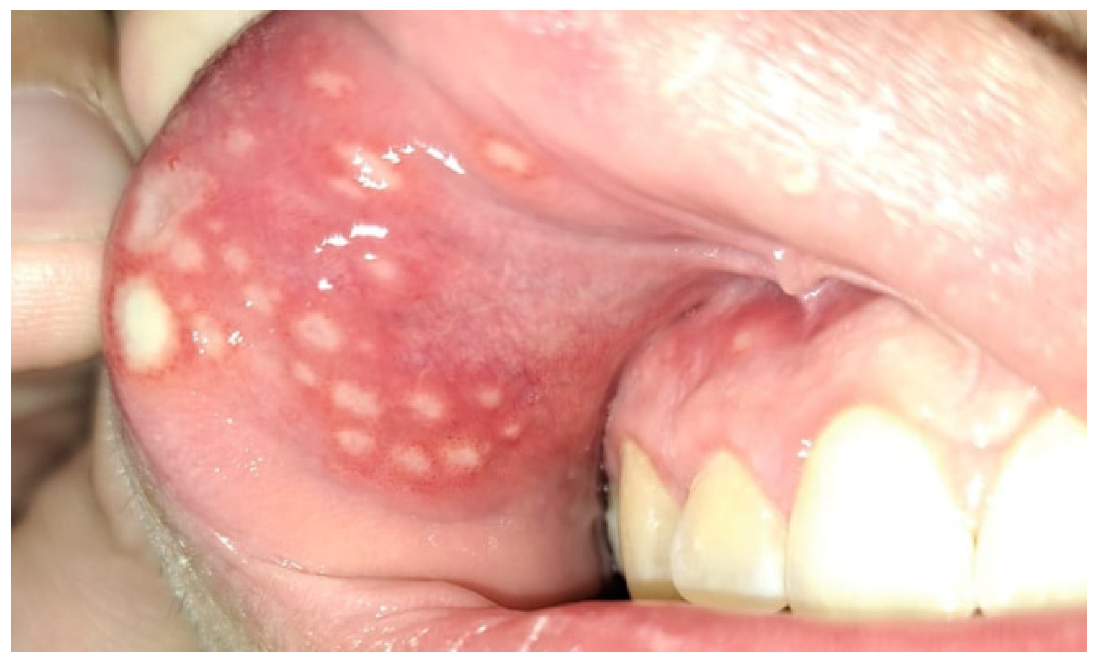
Figure 1. Patient aged 17 with diagnosed ulcerative colitis. Numerous aphthous-like lesions in the oral vestibule near the incisor teeth and canine, involving both the associated gingival mucosa and the mucosa of the upper lip.
Jcm 14 08392 g001
Figure 2. Patient aged 17 with diagnosed ulcerative colitis. Aphthous lesions in the soft palate, palatine tonsils and palatine uvula.
Jcm 14 08392 g002
Figure 3. Patient 16 years old diagnosed with Crohn’s disease. Red labial inflammation with swelling.
Jcm 14 08392 g003
Table 1. Syntaxes for the various databases.
DatabaseSearch TermsNumber of Results
PubMed(“Dentistry” OR “Oral health” OR “Stomatology”) AND (“CDI” OR “Clostridium difficile” OR “Clostridioides difficile” OR “periprocedural antibiotic therapy” OR “antibiotic therapy”) AND (“IBD” OR “Crohn disease” OR “inflammatory bowel disease” OR “Ulcerative Colitis”).440
Embase(‘Dentistry’ OR ‘Oral Health’ OR ‘Stomatology’)
AND
(‘CDI’ OR ‘Clostridium difficile’ OR ‘Clostridioides difficile’ OR ‘Periprocedural Antibiotic Therapy’ OR ‘Antibiotic Therapy’)
AND
(‘IBD’ OR ‘Crohn Disease’ OR ‘Inflammatory Bowel Disease’ OR ‘Ulcerative Colitis’)
258
Scopus(“Oral”) AND (“CDI” OR “Clostridium difficile” OR “Clostridioides difficile” OR “Periprocedural Antibiotic Therapy” OR “Antibiotic Therapy”) AND (“IBD” OR “Crohn Disease” OR “Inflammatory Bowel Disease” OR “Ulcerative Colitis”)587
Google Scholar“Dentistry” OR “Oral Health” OR “Stomatology” AND “CDI” OR “Clostridium difficile” OR “Clostridioides difficile” OR “Periprocedural Antibiotic Therapy” OR “Antibiotic Therapy” AND “IBD” OR “Crohn Disease” OR “Inflammatory Bowel Disease” OR “Ulcerative Colitis”500 most accurate results were included in the review process
Table 2. Relationship between antibiotic group and risk of CDI.
High RiskModerate RiskLow Risk
-
Fluoroquinolones
-
Second generation cephalosporins
-
Third generation cephalosporins
-
Clindamycin
-
Ampicillin
-
Amoxicillin
-
Broad-spectrum penicillins with inhibitors (except ticarcillin/clavulanate and piperacillin/tazobactam
-
Macrolides
-
Trimethoprim/Sulfamethoxazole
-
Other penicillins
-
Sulfonamides
-
Aminoglycosides
-
Bacitracin
-
Metronidazole
-
Teicoplanin
-
Vancomycin
-
Rifampicin
-
Chloramphenicol
-
Tetracycline
-
Carbapenems
-
Daptomycin
-
Tigecycline
Disclaimer/Publisher’s Note: The statements, opinions and data contained in all publications are solely those of the individual author(s) and contributor(s) and not of MDPI and/or the editor(s). MDPI and/or the editor(s) disclaim responsibility for any injury to people or property resulting from any ideas, methods, instructions or products referred to in the content.