Article

Cognitive Age Delta as a Marker of Healthy and Pathological Cognitive Aging: The Role of Lifestyle, Cognitive Reserve, and Vascular Risk

by
Ainara Estanga
1,*,†,
Iñigo Tellaetxe-Elorriaga
2,3,†,
Mirian Ecay-Torres
1,
Jorge García Condado
2,3,
Maite García-Sebastián
1,
Maria Arriba
1,
Carolina López
1,
Naia Ros
1,4,
Ane Iriondo
1,
Imanol Reparaz-Escudero
1,5,
Asier Erramuzpe
2,6,
Pablo Martínez-Lage
1 and
Miren Altuna
1,7,*
1
Center for Research and Memory Clinic, CITA-Alzheimer Foundation, 20009 Donostia-San Sebastián, Spain
2
Computational Neuroimaging Laboratory, Biobizkaia Health Research Institute, 48903 Barakaldo, Spain
3
Biomedical Research Doctorate Program, Universidad Del País Vasco (EHU), 48940 Leioa, Spain
4
Department of Clinical and Health Psychology and Research Methods, University of the Basque Country UPV/EHU, 20018 Donostia-San Sebastián, Spain
5
Navarrabiomed, Hospital Universitario de Navarra (HUN), Universidad Pública de Navarra (UPNA), IdiSNA, 31008 Pamplona, Spain
6
Ikerbasque, the Basque Foundation for Science, 48009 Bilbao, Spain
7
Department of Neurology, Bioaraba Health Research Institute, Araba University Hospital-Txagorritxu, 01009 Vitoria-Gasteiz, Spain
*
Authors to whom correspondence should be addressed.
These authors contributed equally to this work.
J. Clin. Med. 2025, 14(22), 8176; https://doi.org/10.3390/jcm14228176
Submission received: 12 October 2025 / Revised: 14 November 2025 / Accepted: 14 November 2025 / Published: 18 November 2025
(This article belongs to the Special Issue New Insights in Cognitive Aging and Mild Cognitive Impairment)

Abstract

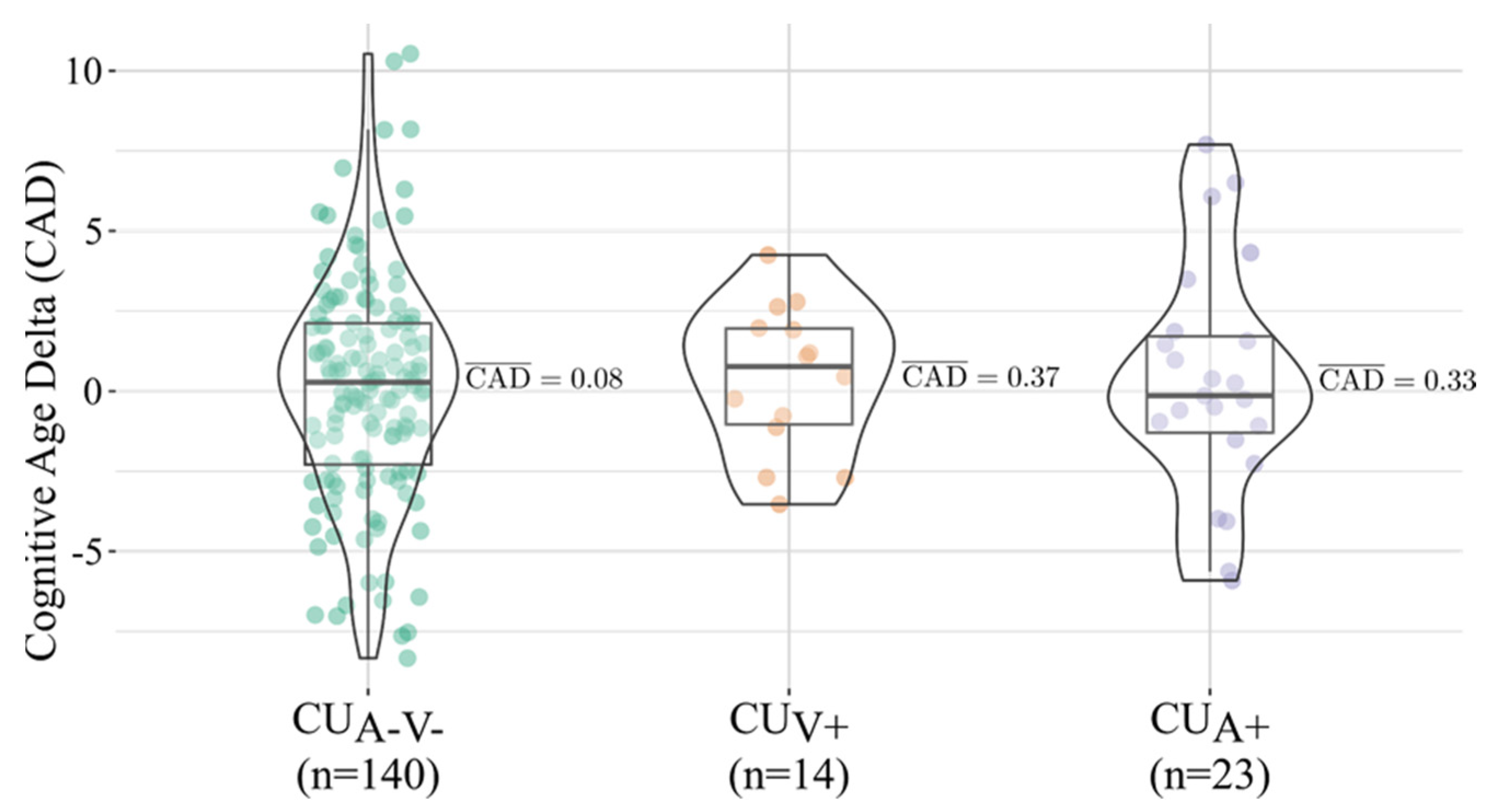
Background: Chronological age is an imprecise proxy for cognitive aging. The Cognitive Age Delta (CAD)—the difference between predicted cognitive age and chronological age—offers a scalable, individualized marker of functional brain aging. We examined determinants of CAD in cognitively unimpaired (CU) adults stratified by Alzheimer’s disease (AD) and vascular biomarkers. Methods: We analyzed 177 CU participants from the Gipuzkoa Alzheimer Project (Basque Country, Northern Spain) classified as amyloid-negative/vascular-negative (CUA−V−, n = 140), amyloid-positive (CUA+, n = 23), or vascular-positive (CUV+, n = 14) using CSF and MRI criteria; vascular burden was defined as Fazekas ≥ 2 on T2-FLAIR or ≥4 microbleeds on SWI, excluding non-traumatic superficial siderosis and established ischemic lesions. MRI was used solely for vascular classification. Associations with demographic, genetic, lifestyle, and reserve measures were tested with General Linear Models. Results: CAD did not differ across biomarker groups (Kruskal–Wallis H(2) = 0.17, p = 0.91). Median (IQR) CAD values were 0.28 (−4.13, 4.69) for CUA−V−, −0.14 (−3.15, 2.87) for CUA+, and 0.77 (−2.22, 3.76) for CUV+, indicating comparable distributions. Higher vocabulary scores (proxy of cognitive reserve) related to a younger cognitive age in CUA−V− (β = −1.39, p < 0.001) and CUA+ (β = −2.08, p = 0.054). In CUA+, greater sedentary time—particularly computer-based sitting—was also associated with lower CAD (daily sitting β = −2.13, p = 0.009; workday computer sitting β = −2.32, p = 0.015). CAD showed no associations with CSF Aβ42, p-tau or t-tau, APOE ε4 load, or vascular risk factors (all p > 0.05). Conclusions: CAD captures interindividual resilience-related variability beyond classical AD biomarkers. Vocabulary, a marker of lifelong enrichment, emerged as a robust determinant of a younger cognitive age, while amyloid and vascular pathology exerted limited influence at preclinical stages. These findings support CAD as a sensitive, scalable endpoint for identifying protective factors and guiding personalized prevention in early Aging.

1. Introduction

The global increase in life expectancy is reshaping demographic structures worldwide, leading to a rapid growth in the proportion of older adults. This demographic shift is accompanied by a parallel rise in the prevalence of age-related cognitive decline and dementia, which represent major medical, social, and economic challenges [1]. Cognitive impairment not only diminishes quality of life and independence but also imposes an enormous burden on healthcare systems and caregivers. Consequently, the promotion of healthy cognitive aging and early detection of individuals at risk have become critical priorities for brain health initiatives in neurology and geriatrics.
There is increasing recognition that interventions targeting the earliest, ideally in preclinical phases of disease are likely to be the most effective for prevention. In this context, evaluating the impact of established risk factors, both biological (e.g., APOE genotype, amyloid pathology, vascular cerebral lesions) [2] and potentially modifiable behaviors (e.g., physical activity, sleep, diet, cognitive and social engagement) in cognitively unimpaired (CU) individuals is essential [1,3,4]. Such an approach enables us to disentangle mechanisms of healthy cognitive aging from preclinical disease processes and to identify determinants that accelerate or protect against decline, even before clinical symptoms emerge.
Reliance on chronological age as a measure of cognitive aging is limited because it does not capture the substantial interindividual variability in cognitive trajectories. Cognitive aging is a heterogeneous process, with domain-specific differences in memory, executive function, attention, and processing speed [5]. This variability is shaped by complex interactions among biology, lifestyle, and cognitive reserve or resilience [6,7]. Hence, there is a strong rationale for developing individualized, functional markers of cognitive aging that can go beyond chronological age and have the potential to detect subtle deviations earlier that would be apparent clinically.
One promising strategy is to adapt brain age models widely used in neuroimaging to the neuropsychological domain. Recently, regression-based approaches have been developed using brief cognitive test batteries to predict an individual’s cognitive age [8]. The difference between predicted cognitive age and actual chronological age, the Cognitive Age Delta (CAD), reflects whether a person’s cognitive performance is “older” or “younger” than expected. CAD has already shown clinical relevance: in mild cognitive impairment, higher CAD predicted progression to Alzheimer’s disease (AD), demonstrating that short neuropsychological assessments can yield a powerful, scalable biomarker [8]. Thus, cognitive screening tools, beyond their traditional diagnostic role, may provide added value as surrogate markers of functional brain aging.
The importance of CAD is further underscored by the shift toward biomarker-based definitions of AD. According to the most recent research framework, AD is defined strictly by amyloid and/or tau biomarkers, even in asymptomatic individuals [9,10]. At the same time, longitudinal evidence shows that much of the decline traditionally attributed to healthy aging is actually driven by undetected AD pathology or cerebrovascular lesions [11,12,13]. Examining CU older adults who are AD biomarker-negative defined by ATN system (A-) and cerebral vascular burden negative (V-) thus provides a unique opportunity to study genuine healthy cognitive aging largely unconfounded by covert pathology. This approach enhances the internal validity of CAD as a marker of cognitive aging and aligns with prevention-oriented research.
Beyond pathology, lifestyle and psychosocial factors are critical, modifiable targets for promoting late-life cognitive health [1]. Meta-analysis and large cohort studies consistently show that regular physical activity, healthy diet, adequate sleep, abstinence from smoking, moderation in alcohol use, and sustained intellectual and social engagement are associated with better cognitive outcomes and lower risk of dementia [6,14,15,16,17]. Nevertheless, most of this evidence relates to clinical outcomes such as incident dementia or average decline at the population level. Much less is known about how these exposures influence the individualized CAD metric in biomarker-defined CU populations. Identifying factors associated with a younger CAD could highlight actionable targets for primary prevention and provide mechanistic insight into resilience.
The present study builds on this framework by applying CAD modeling to a well-characterized cohort of cognitively unimpaired older adults. We first examine CU A-/V- individuals to characterize determinants of cognitive aging in the absence of amyloid and significant vascular pathology. In parallel, we extend analyses to CU participants stratified by distinct biological risk profiles, specifically those with amyloid positivity only (CUA+) and vascular positivity only (CUV+). This stratification allows us to test whether the same lifestyle and psychosocial factors modulate CAD across groups with distinct biological vulnerabilities.
Our overarching aim is twofold: first, to establish CAD as a sensitive, added-value marker of cognitive function in the biomarker era; and second, to identify modifiable lifestyle and psychosocial determinants of CAD across both pathology-free and pathology-positive CU individuals. By pinpointing factors that consistently relate to a younger cognitive age, we seek to highlight targets for personalized prevention strategies and provide novel insights into resilience mechanisms in aging.

2. Materials and Methods

2.1. Participants and Design

This cross-sectional study was conducted within the Gipuzkoa Alzheimer Project (GAP), led by CITA-Alzheimer Foundation, a longitudinal cohort on preclinical and prodromal phases of AD in the Basque Country (Northern Spain). Participants were recruited between 2011–2013 from the community through local media advertisements and presentations at the local Alzheimer’s Association (AFAGI—Alzheimer Gipuzkoa). The cohort enrolls 411 adults aged 40–80 years without dementia and without neurological, psychiatric or systemic conditions that could affect cognition. The inclusion/exclusion criteria and study procedures have been fully described earlier [18,19]. Each participant underwent a comprehensive clinical, neurological, and neuropsychological evaluation, complemented by assessments of diet and lifestyle factors, as well as structural magnetic resonance imaging (MRI) and biological sampling, and optional cerebrospinal fluid (CSF) collection by lumbar puncture. To minimize potential desynchronization across modalities, the neurological examination, neuropsychological testing, nursing/vascular measurements, and lifestyle data collection (nutrition and physical activity) were completed across two onsite visits scheduled in two consecutive weeks. For participants who consented to lumbar puncture, CSF collection occurred on average 3.92 months after the baseline visits (SD = 4.23 months), ensuring that all variables analyzed in the present study refer to the same baseline time window.
The study was conducted in accordance with international ethical recommendations for medical research in humans, adhering to the standards of the Declaration of Helsinki (1964 and subsequent revisions) and applicable Spanish legislation. The protocol, informed consent form, and all required documentation were reviewed and approved by the Clinical Research Ethics Committee of the Health Area of Gipuzkoa (approval date: 20 October 2010). Written informed consent was obtained from all participants prior to enrollment. Data confidentiality was strictly ensured in compliance with Organic Law 3/2018, of 5 December, on Data Protection and Digital Rights Guarantee; all files were stored in an independent, encrypted database.

2.2. Clinical and Neurological Evaluation

Participants underwent a standardized clinical and neurological evaluation that included medical history, systemic and neurological examination, and family history of dementia, including the degree of relatedness (first vs. non–first degree relative) and the dementia subtype, when possible. Motor performance was examined with the Unified Parkinson’s Disease Rating Scale motor section (UPDRS-III) and the Tinetti Gait and Balance scales [20]. Functional independence was measured with the Assessment of Instrumental Activities of Daily Living (ADCS-ADL) [21], and subjective cognitive complaints were recorded through structured interviews and the Memory Failures of Everyday Questionnaire (MFE) [22]. Neuropsychiatric and affective symptoms were evaluated using the Neuropsychiatric Inventory (NPI) [23] and the Hospital Anxiety and Depression Scale (HADS) [24]. Subjective sleep quality and perceived stress were assessed with the Pittsburgh Sleep Quality Index (PSQI) [25] and the Perceived Stress Questionnaire (PSQ) [26], respectively.
Vascular and anthropometric parameters were defined according to ENRICA criteria [27], including metabolic syndrome, obesity (based on body mass index (BMI) and waist-to-hip ratio), hypertension, diabetes, hypercholesterolemia (total, LDL, HDL), and hypertriglyceridemia. Systolic and diastolic blood pressure, heart rate, and right and left ankle–brachial index were also recorded. Anthropometric parameters included body mass index and waist-to-hip ratio. Blood biochemistry was analyzed under fasting conditions and included the following parameters: total cholesterol (mg/dL), HDL cholesterol (mg/dL), LDL cholesterol (mg/dL), triglycerides (mg/dL), C-reactive protein (mg/L), fasting glucose (mg/dL), fasting insulin (µU/mL), glycated hemoglobin (HbA1c, in both IFCC mmol/mol and NGSP % units), vitamin B12 (pg/mL), and folate (ng/mL).

2.3. Neuropsychological Assessment

A comprehensive neuropsychological battery was administered by trained neuropsychologists. The assessment covered global cognition and the major cognitive domains of memory, language, visuospatial ability, attention, processing speed, and executive function. Global cognition was measured with the Mini-Mental State Examination (MMSE) [28] and Memory Alteration Test (M@T) [29]. Memory was assessed using the Free and Cued Selective Reminding test (FCSRT) [30,31] and the Rey–Osterrieth Complex Figure Test (ROCF) immediate and delayed recall [31,32]. Language abilities were evaluated with the Boston Naming Test (BNT) [33,34] and verbal fluency tasks, including both semantic fluency (“animals”) and phonemic fluency (letter “P”) [35]. Visuospatial and visuoconstructive skills were examined using the Rey–Osterrieth Complex Figure copy [31,32], Judgement of Line Orientation (JLO) [36] and the 15-Objects Test [37]. Attention and processing speed were assessed with the Digit Symbol–Coding subtest from the Wechsler Adult Intelligence Scale, 3rd edition (WAIS-III) [38], Digits from the WAIS-III [38] and the Trail Making Test Part A (TMT-A) [39]. Executive functioning was measured using the Trail Making Test Part B (TMT-B) [39], the Stroop Word–Color Interference Test [40] and Letter and Number sequencing subtest from the WAIS-III [38]. All tests were administered according to standardized procedures, and raw scores were interpreted using validated normative data adjusted for age, sex, and education level [41].

2.4. Cognitive Reserve

Premorbid intellectual functioning was estimated using the Vocabulary subtest from WAIS-III. Cognitive reserve was further assessed with the Cognitive Reserve Questionnaire [42], developed to quantify experiences contributing to cognitive reserve such as years of formal education, occupational attainment, engagement in cognitively stimulating activities (e.g., reading, playing musical instruments, learning languages, playing chess). Engagement in everyday activities was quantified with the Leisure and Productive Activities Questionnaire [43], which summarizes intellectual, cultural and social activities (e.g., reading, theatre, meeting friends, travelling) and productive roles (e.g., childcare/eldercare, crafts, housework, shopping).

2.5. Lifestyle Variables

Lifestyle-related evaluation included physical activity through total score from the IPAQ questionnaire [44], and sedentary behavior assessed with a questionnaire from the “Seguimiento Universidad de Navarra” (SUN) Study (1999), which records average daily hours spent in different sedentary contexts (television, computer use, driving, and other activities sitting or lying down) on weekdays and weekends. In the present analyses, we focused on total sitting time, computer-based sitting, and time spent driving. Dietary habits (Mediterranean diet adherence), tobacco use (active smoker status and number of cigarettes per day), and alcohol consumption (weekly and daily alcohol units) were also collected.

2.6. Syndromic Cognitive Diagnosis

Syndromic cognitive diagnosis was established through consensus meetings between neurologists and neuropsychologists. Mild Cognitive Impairment (MCI) was defined as objective dysfunction using age, sex and education adjusted norms, in at least one cognitive domain together with subjective cognitive decline reported by the participant, a study partner, or the clinician. CU status required no objective cognitive impairment across domains and preserved functional independence. Participants with subjective cognitive complaints alone or isolated low performance on a single test, in the absence of subjective cognitive decline that did not meet impairment criteria were classified as CU.

2.7. Genetic Risk

Genotyping of APOE polymorphisms was performed on DNA extracted from peripheral blood leukocytes using polymerase chain reaction (PCR) amplification following established protocols [44]. Participants were classified into three categories according to APOE ε4 allele load: homozygotes (ε4/ε4), heterozygotes (one ε4), and non-carriers (no ε4 allele).

2.8. CSF AD Biomarker

CSF samples were obtained and collected under aseptic conditions using an atraumatic needle and without sedation, following international consensus recommendations as described previously [45]. Concentrations of amyloid-β42 (Aβ42), total tau (t-tau), and phosphorylated tau (p-tau) were quantified using INNOTEST® enzyme-linked immunosorbent assays (ELISA; Fujirebio Europe, Ghent, Belgium) according to the manufacturer’s instructions. All samples were analyzed at the Clinical Neurochemistry Laboratory of Hospital Sant Pau (Barcelona, Spain).
CSF amyloid and tau biomarkers were interpreted using the cutoff values established by Alcolea et al. [46]: Aβ42 < 550 pg/mL (A+), p-tau > 61 pg/mL (T+), and t-tau > 350 pg/mL (N+). For Aβ42, the threshold was adjusted to 580 pg/mL by adding a 5% confidence interval following previously described approach [47]. Based on these criteria, amyloid (A), tau pathology (T), and neurodegeneration (N) status were determined, and participants were categorized within the AT(N) framework [9,10,48].

2.9. MRI Vascular Pathology

MRI was acquired on a Siemens Magnetom Trio Tim 3T scanner. White matter hyperintensities (WMH) were assessed on axial T2-weighted FLAIR sequences, and cerebrovascular burden was defined by the presence of severe WMH (Fazekas score ≥ 2) [49], or four or more cerebral microbleeds (CMBs) identified on susceptibility-weighted imaging (SWI) sequences. In addition, participants presenting with cortical superficial siderosis (cSS) of non-traumatic origin or established ischemic lesions (either lacunar infarcts or territorial infarctions) were excluded from the analyses to minimize confounding effects of overt cerebrovascular injury.
MRI data were used exclusively to define vascular burden (V+), not to classify neurodegeneration (N+), given the limited sensitivity and specificity of structural MRI, particularly visual atrophy scales such as the Scheltens’ Medial Temporal Atrophy (MTA) scale, for detecting early AD related neurodegeneration in CU individuals [50]. This methodological decision is consistent with the recent biological definitions of Alzheimer’s disease proposed by Jack et al. [10] (NIA-AA) and Dubois et al. [9] (IWG-3), which no longer require the presence of neurodegeneration (N+) for the definition of AD, acknowledging that such changes typically occur at more advanced stages and that MRI-based atrophy markers are non-specific at the preclinical phase.
This operational definition of vascular positivity (V+) adheres to current STRIVE and STRIVE-2 recommendations [51,52] and mirrors the neuroimaging exclusion criteria applied in pivotal anti-amyloid monoclonal antibody trials (e.g., Lecanemab, Donanemab), where similar thresholds for CMBs and siderosis are used to identify individuals with vascular fragility and increased susceptibility to amyloid-related imaging abnormalities (ARIA) [53,54]. This alignment ensures both biological validity and clinical relevance of the V+ construct within the context of preclinical AD.

2.10. Biomarker Defined Classification of CU Participants

For the present study, analyses were restricted to CU individuals with available CSF and MRI data, enabling the examination of cognitive aging in both pathology-free and pathology-positive participants. This approach better isolates age-related variability from early neurodegenerative effects, and helps minimize potential circularity, as neuropsychological test scores contribute to both diagnostic adjudication and the features used in the CAD model. The participant selection process is illustrated in Figure 1.
Participants were subsequently assigned to one of three CU subgroups based on the A/T/N classification and cerebrovascular burden:
  • CU Biomarker negative (CUA-V-) (n = 142): A−T−N− and V−.
  • CU Amyloid pathology (CUA+) (n = 23): A+ (irrespective of T, N) and V-.
  • CU Vascular pathology (CUV+) (n = 14): V+ with otherwise biomarker-negative ATN profile (A−T−N−).

2.11. Cognitive Age Modeling and CAD

Feature preselection was guided by the association between chronological age and individual neuropsychological test scores within the CUA-V- group to identify age-sensitive measures. Specifically, we computed the Pearson correlation coefficients between chronological age and all primary scores from the neuropsychological battery covering global cognition and major domains. Features with correlation coefficients passing Bonferroni multiple comparisons correction were included in the model, trying to keep the number of features to the minimum and the widest cognitive domain representation, while prioritizing the highest correlation coefficients. Highly collinear ρ > 0.7 features were also discarded to mitigate the noise in the model. The final selection of features includes: Digit Symbol-Coding (WAIS-III) subtest, Stroop Color Naming Test, Stroop Word-Color Interference Test, 15-Objects Test, ROCF Delayed Recall, TMT-A, FCSRT Immediate Total Free Recall, TMT-B, Boston Naming Test, Phonemic Verbal Fluency Test (“P”), FCSRT Immediate Total Recall, Semantic Verbal Fluency Test (“Animals”).
The features were corrected for education years to remove the well-known confounding effect of this variable in neuropsychological test performances [41,55,56] and to prevent it from leaking into the subsequent association analyses. This was done first estimating the effect of the education years, sex, and APOE4 allele load [57,58,59] in the CADs with a General Linear Model (GLM) (CAD ~ education years + sex + APOE4 load) to also identify other potential covariates of interest. We found a moderate negative effect between education years and CADs (=−0.99, p = 2.09 × 10⁻⁴) but not with sex (=0.01, p = 0.98) nor APOE4 allele load (=0.03, p = 0.91). Then, the residuals of a linear model estimating the score using the years of education were added to the original mean of the scores to maintain their scale and ranges, as shown in Equation (1):
X c o r r e c t e d = X ¯ + β ^ X X ^
where:
X ¯ = mean of the original scores
X ^ = predicted values from the regression model with education
β ^ = estimated coefficient
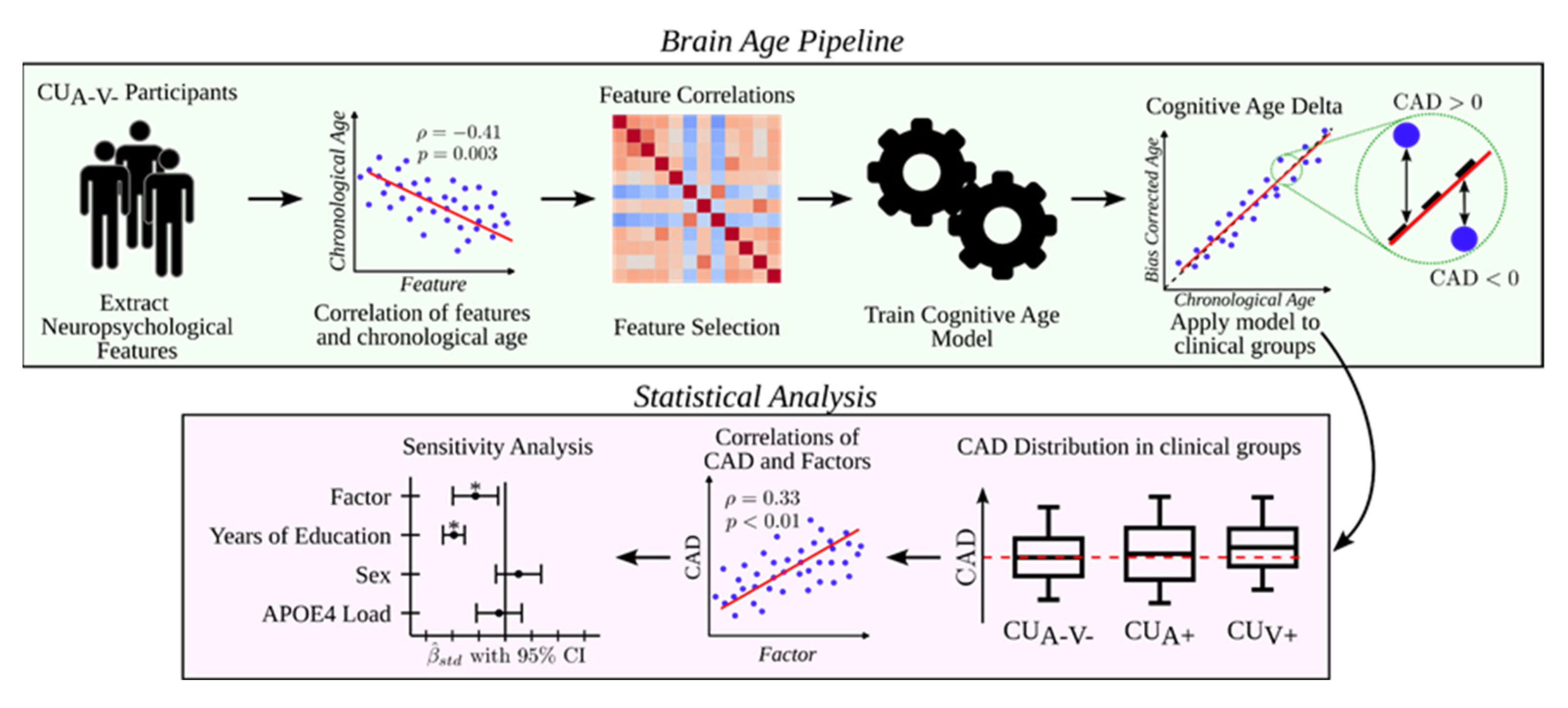
Cognitive age was then modeled via multiple linear regression, with the selected neuropsychological scores as predictors and chronological age as the dependent variable, following prior work [8]. For each participant, the regression model outputs their predicted cognitive age. However, well-known bias in age modeling is the tendency for younger participants to be assigned higher ages, while older participants are assigned lower ages than their actual ages. This phenomenon is attributed to a regression to the mean problem [60,61]. This bias can be conceptualized as our best estimate for a subject about whom we have no prior information being the mean age. The predicted age bias can be corrected [62], by considering the actual participant’s age through fitting a linear model as shown in AgeML [63], thus obtaining a bias-corrected predicted cognitive age.
The final model intercept and coefficients (original scale and standardized β) are reported in Appendix A Table A1. To ensure full transparency and reproducibility, the complete training and prediction pipeline, including the model weights and scripts used for the present analyses, is publicly available at https://github.com/itellaetxe/cognitive_age_delta_jcm (accessed on 8 November 2025). Specific details on the model training can be found in the Methods section of AgeML [63]. In summary, with y as age, X as our feature set and f() as our pipeline, we optimize the parameters of our pipeline f() by means of minimizing the mean squared error (MSE) of age on CUA-V- subjects. Our pipeline consists of a feature scaler and a linear regressor, and we use a 5-fold cross-validation scheme. Finally, the Cognitive Age Delta (CAD) was defined as in Equation (2):
CAD = ŷ − y
where:
ŷ = bias-corrected predicted cognitive age
y = chronological age

2.12. Statistical Analysis

All statistical analyses were performed in R version 4.5.1 (R Foundation for Statistical Computing, Vienna, Austria). Chronological Age and CADs distribution means across all clinical groups were compared using non-parametric Kruskal-Wallis tests. We analyzed the effect of the selected factors (modifiable and non-modifiable) with General Linear Models (GLMs) of the shape (CAD~Factor) for each clinical group (CUA-V-, CUA+, CUV+). If the factor showed a statistically significant association with the CADs within the clinical group, a sensitivity analysis was conducted adding sex, APOE4 allele load, and years of education to the model as covariates (CAD~Factor + sex + APOE4 Load + Years of Education). The latter covariate was included to consider its possible association with the factor of interest, thus isolating the effect of the factor. All input variables were standardized to allow comparing their effects. Factors were analyzed in groups to prevent multiple comparisons corrections from throwing off all possible associations, due to our modest sample size. A type I error rate of 5% (= 0.05) was set for all analyses, after correcting for multiple comparisons within each group using FDR. We analyzed these factor groups: neurological history, motor scales, cognitive symptoms, neuropsychiatric symptoms, stress and sleep questionnaires, cardiovascular parameters, cognitive reserve related factors, tobacco and alcohol consumption, physical activity and sedentarism, and adherence to Mediterranean diet. Associations between the available CSF markers and CADs were explored also using GLMs correcting for years of education, sex, and APOE4 allele load.
Figure 2 summarizes the analytical workflow, including CAD computation and subsequent statistical analyses.

3. Results

3.1. Sample Characteristics

A total of 177 CU participants were included in the analyses: 140 CUA−V−, 23 CUA+, and 14 CUV+. Two additional CUA−V− participants were excluded from the initial CU group (n = 179) due to missing data in the neuropsychological tests required for CAD computation. The three subgroups did not differ significantly in sex distribution (χ2 = 1.38, p = 0.50) or years of education (Kruskal–Wallis test, H(2) = 0.33, p = 0.84). However, significant age differences were observed among groups (Kruskal–Wallis test, H(2) = 14.39, p = 0.001), with CUA−V− participants being on average about 5 years younger than both CUA+ and CUV+ individuals. Given the mean cohort age (55–60 years), this difference—although statistically significant—is unlikely to be of major clinical relevance. The frequency of APOE ε4 carriers was higher in the CUA+ group compared to CUA−V− and CUV+ (χ2 = 7.69, p = 0.021). Median (interquartile range, IQR) and frequency (%) values are shown in Table 1.
Additionally, there were no significant differences in age, sex distribution, years of education, and MCI or APOE ε4 carrier frequency between participants who underwent lumbar puncture and those who did not (Appendix A Table A2), indicating that CSF data availability was not biased by major demographic or genetic factors.

3.2. CAD Distribution and Group Comparisons

No significant differences in CAD were observed among the three CU subgroups (Kruskal–Wallis test, H(2) = 0.17, p = 0.91). Median (IQR) CAD values were [0.28 (−4.13, 4.69)] for CUA-V-, [−0.14 (−3.15, 2.87)] for CUA+, and [0.77 (−2.22, 3.76)] for CUV+, indicating a comparable distribution of cognitive performance relative to chronological age across groups (Figure 3).

3.3. Associations Between CAD and Cognitive Reserve Related Variables

In the stratified analyses, a significant association was found between CAD and the Vocabulary subtest from the WAIS-III, independent of years of education, sex, and APOE4 allele load in the CUA-V- group. This association did not survive FDR correction in the amyloid positive group, although the 95% confidence interval (CI) did not include 0 (CUA-V-: β = −1.39, p <0.001); CUA+: β = −2.08, p = 0.054). All p-values are reported after FDR correction. Figure 4 shows the association observed in the CUA-V- group.
In addition, within the CUA+ group, the association between the Cognitive Reserve Questionnaire and CAD also did not survive FDR correction, despite the 95% CI not including 0 (β = −2.47, p = 0.054). No significant associations were found for the Leisure and Productive Activities Questionnaire or for bilingualism (p < 0.05 post FDR correction).

3.4. Associations Between CAD and Lifestyle Factors

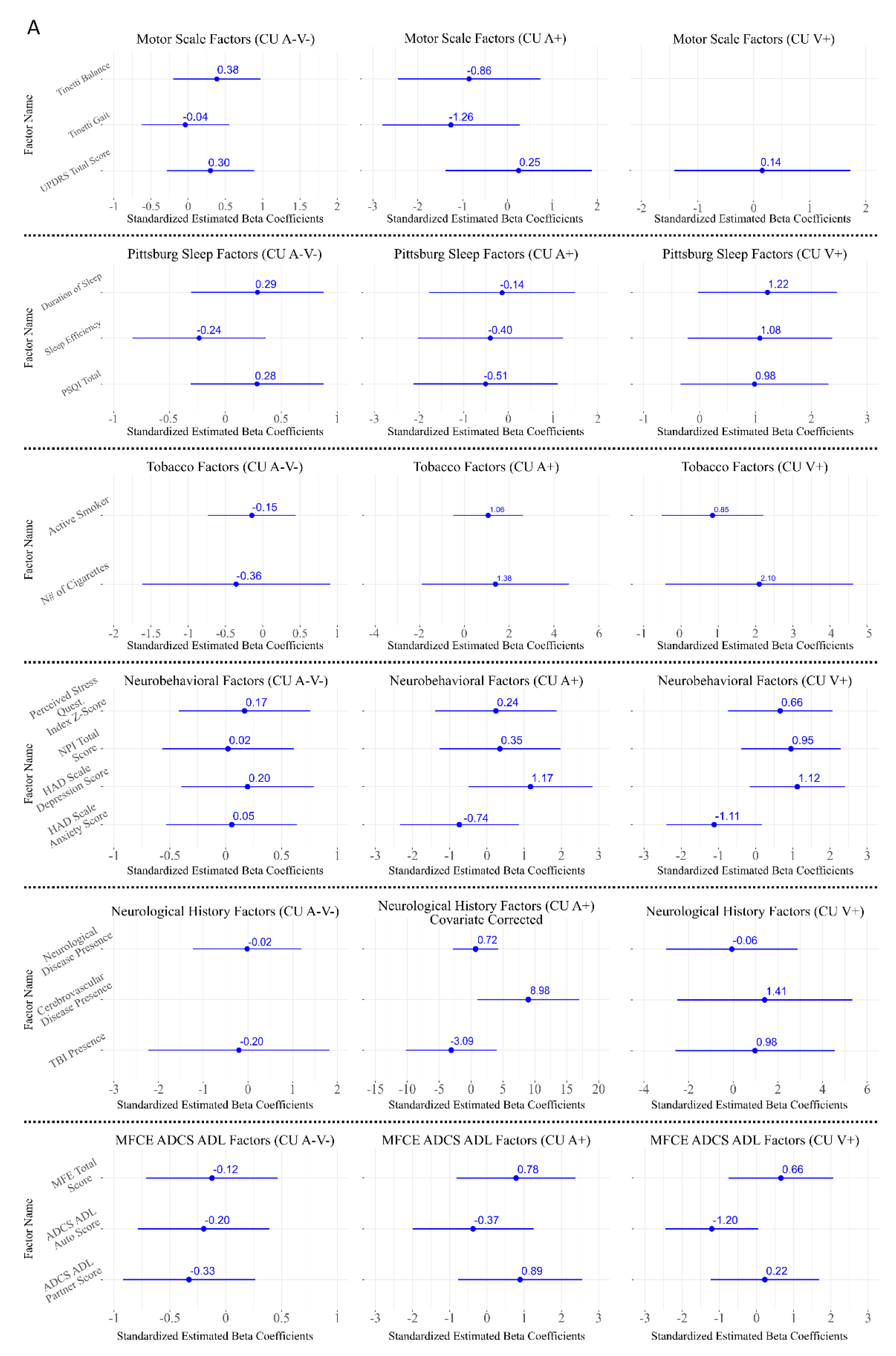
Regarding lifestyle-related variables, a significant association was observed between daily hours spent sitting in total and hours spent working seated from Monday to Friday at a computer and CAD only in the CUA+ group (β = −2.13, p = 0.046 and β = −2.32, p = 0.046, respectively) (Figure 5).
In contrast, no significant associations were found for physical activity, diet, tobacco and alcohol consumption in any of the groups (p < 0.05 post FDR correction) (Figure 5).

3.5. Associations Between CAD and Neurological Assessment

No significant associations were detected between CAD and clinical or neurological variables, including neurological history (any neurological disease, traumatic brain injury, or cerebrovascular disease including transit ischemic attack), motor scales (UPDRS-III and Tinetti), or subjective cognitive complaints across any subgroup categorized via a detailed neurological history (all p > 0.05) (Figure 5).

3.6. Associations Between CAD and Cardiovascular Risk

Similarly, CAD was not associated with vascular risk factors or vascular parameters, including cardiovascular disease, systolic and diastolic blood pressure, or heart rate, in any of the groups (all p > 0.05) (Figure 5).

3.7. Associations Between CAD and Neurobehavioral and Sleep-Related Factors

No significant associations were found between CAD and neurobehavioral variables, including depressive and anxiety symptoms measured with the HADS, neuropsychiatric symptoms assessed with the NPI, or perceived stress (PSQ). Perceived sleep quality, evaluated with the PSQI, was also not significantly related to CAD in any subgroup (all p > 0.05) (Figure 5).

3.8. Associations Between CAD and CSF Biomarkers

Finally, no significant associations were observed between CAD and CSF biomarker concentrations, including amyloid-β42 (Aβ42), phosphorylated tau (p-tau) or total tau (t-tau) (all p > 0.05).

4. Discussion

The present study leverages the CAD model—a scalable, individualized biomarker of functional brain aging—to investigate the influence of cognitive reserve, APOE genotype, sex, and lifestyle factors in a deeply phenotyped cohort of CU adults from the Basque Country (Northern Spain). Despite the absence of significant mean CAD differences among biomarker-defined subgroups (A−V−, A+, and V+), CAD captured differential associations with cognitive reserve and lifestyle factors, highlighting its sensitivity to early resilience mechanisms even before overt neurodegeneration.

4.1. The Value of Cognitive Age Delta in the Biomarker Era

Chronological age is an imprecise surrogate for biological aging, prompting the development of objective metrics such as Brain Age (a neuroimaging-derived structural metric) [64] and epigenetic clocks (DNA methylation-based cellular metrics) [65,66] to capture interindividual variability. While these biological clocks are crucial—accelerated brain or epigenetic age has been prospectively associated with worse cognitive outcomes and neuropathological burden—they primarily reflect structural integrity or cellular processes. In contrast, CAD provides a direct, low-cost, and non-invasive functional measure of age-related cognitive performance. Unlike molecular biomarkers, CAD directly captures interindividual variability in performance that likely reflects the integration of reserve, lifestyle, and subclinical pathology. This derived metric offers an immediately translatable snapshot of cognitive health that complements, rather than replaces, more resource-intensive neuroimaging or fluid biomarker assessments.
Our methodology extends previous CAD studies employing a comprehensive, multidomain neuropsychological battery [67,68] and stratifying CU participants according to both the AT(N) framework and cerebrovascular burden (V status) [10,69,70]. Prior research on cognitive age often relied on clinical diagnosis (e.g., MCI vs. CU), which overlooks the substantial impact of subclinical pathology. By focusing our baseline analyses on individuals without biomarker evidence of pathology (A−V−), we minimized confounding and enhanced the internal validity of CAD as a marker of genuine cognitive aging processes. This rigorous, biomarker-informed stratification allows testing whether determinants of cognitive aging differ across distinct biological risk profiles, an essential step toward precision prevention [11]. Furthermore, using a rich neuropsychological profile (NPS) helps mitigate potential misclassification errors that often occur with single-test screening or unidimensional markers [71,72].

4.2. APOE and Sex Effects

As expected, APOE ε4 carriers were more frequently A+, consistent with its role in promoting amyloid aggregation and impairing clearance [57,73]. Yet, APOE did not influence CAD, suggesting that genetic risk accelerates molecular pathology without necessarily translating into detectable functional aging differences prior to tau involvement. This may also relate to the low number of ε4 homozygotes, which limits power to detect genotype-dependent effects [59,74].
Sex differences were minimal overall, though growing evidence suggests women show enhanced resilience to amyloid-related changes until menopause, after which hormonal decline may amplify susceptibility to tau accumulation and cognitive aging [75,76]. Future CAD studies should explicitly model sex-by-pathology and sex-by-APOE interactions, as these represent critical modulators of vulnerability and resilience in aging.

4.3. CSF Biomarkers

Importantly, the A+ subgroup represents a very early preclinical phase (A+T−N−), in which amyloid accumulation occurs before tau deposition or neurodegeneration. According to the updated biological definitions of AD [9,10], tau aggregation is more closely linked to cognitive decline, explaining the absence of direct associations between CAD and CSF Aβ or p-tau. This supports the interpretation that CAD reflects resilience-related rather than pathology-driven variability

4.4. Rigorous Biomarker Definition for Vascular Pathology

A key methodological strength of this study lies in the rigorous and clinically meaningful definition of cerebrovascular pathology (V+), which was determined by the presence of severe white matter hyperintensities (Fazekas score ≥ 2) or ≥4 CMBs. This dual criterion was selected not only for its alignment with current clinical trial standards, as ≥4 CMBs represent the exclusion threshold in major anti-amyloid monoclonal antibody studies (e.g., Donanemab, Lecanemab) due to the increased risk of ARIA, but also because it reflects a threshold of clinically significant cerebrovascular frailty [51,52,53,54].
The selection of Fazekas score ≥ 2 (denoting confluent WMH) defines a high white matter hyperintensity burden, a parameter consistently associated with greater cognitive dysfunction and successful prediction of cognitive performance in aging and AD cohorts. Similarly, the consensus threshold of ≥4 CMBs defines a high-risk group. CMBs, which represent prior microscopic hemorrhages, are key imaging markers of underlying small vessel disease (SVD) and vessel wall integrity impairment, independently linked to increased risk of stroke, mortality, and cognitive deterioration. The co-occurrence of extensive WMH and/or multiple CMBs marks a state of heightened small-vessel vulnerability, associated with impaired vascular repair, endothelial dysfunction, and disrupted neurovascular coupling.
While our primary criteria focus on these two widely used markers, the V+ definition captures the broader spectrum of SVD, which includes other severe manifestations such as lacunar infarcts and cSS. By adopting high-burden thresholds for WMH and CMBs, we effectively select individuals with a clinically and biologically significant SVD burden.
The use of SWI for microbleed detection further enhances the precision of this definition. SWI provides superior sensitivity and spatial resolution for identifying small-vessel pathology, detecting substantially more CMBs than conventional T2*-weighted Gradient-Recalled Echo (GRE) sequences [77]. This methodological choice strengthens the accuracy of vascular burden classification, thereby providing a more nuanced framework for examining the interplay between vascular pathology, cognitive aging, and resilience mechanisms.

4.5. Differential Associations of Cognitive Reserve and Lifestyle Factors

The present study focused on the CUA−V−, CUA+, and CUV+ subgroups, deliberately omitting specific analysis of T+ groups (e.g., A+T+) due to the small sample size in these less-represented categories (n = 23 for A+, n = 14 for V+). Statistical power limitations preclude reliable inference in these combined pathology groups. Crucially, the CUA+ cohort, specifically in the absence of tau pathology (T−) or neurodegeneration (N−), aligns with the earliest conceptualization of the AD continuum, potentially capturing individuals decades before clinical onset. The ability of CAD to detect functional associations in this preclinical stage is a key contribution.
Our analysis revealed a significant negative association between Vocabulary score and CAD in the pathology-free group (CUA−V−: β^ = −1.39, p < 0.001), a finding that was replicated in the CUA+ group (β = −2.08, p = 0.054). Vocabulary performance is traditionally considered a robust indicator of verbal intelligence [38]. Beyond reflecting linguistic proficiency, it is often used as a proxy for cognitive reserve (CR) [7,78] capturing cumulative lifetime intellectual enrichment. Consistent with our results, prior work has shown that higher premorbid IQ (assessed with the Wechsler Test of Premorbid Functioning (TOPF)) is associated with a younger cognitive age estimate, even after covariate adjustment [68]. This robust inverse relationship, where richer vocabulary as an estimate of premorbid intelligence predicts a “younger” cognitive age (lower CAD), strongly supports the CR hypothesis. Within the updated conceptual framework proposed by [79], CR is defined as a brain property enabling better than expected cognitive performance given the degree of brain aging or pathology. Individuals with higher vocabulary scores may thus rely on more efficient or flexible neural networks that help maintain cognitive function despite age-related brain changes. Moreover, our results are aligned with evidence from previous studies showing that premorbid verbal ability, as indexed by reading or vocabulary tests, operates as an independent determinant of cognitive performance in both healthy individuals and those in the AD continuum [80,81]. Accordingly, vocabulary can be understood as a prototypical CR proxy, and its association with CAD supports the interpretation of CAD as a behavioral expression of functional resilience, the capacity to sustain efficient cognition despite age-related neural alterations. The persistence of this association in the CUA+ cohort is particularly relevant, suggesting that CR remains a critical factor modulating functional cognitive performance even in the presence of preclinical amyloid pathology.
In contrast, apart from a strong association with vocabulary, leisure and productive activities, the Cognitive Reserve Questionnaire, and bilingualism did not emerge as significant predictors. This may reflect the fact that lifestyle-based proxies of CR exert stronger effects on longitudinal trajectories than in cross-sectional analyses, and that the homogeneity and high educational attainment of this cohort may have limited the variability required to detect such associations. Nevertheless, previous research has consistently highlighted the role of these factors as contributors to cognitive reserve. Participation in cognitively stimulating leisure activities across the lifespan has been linked to delayed cognitive decline and reduced dementia risk [82,83], while standardized questionnaires such as the Cognitive Reserve Questionnaire capture the cumulative contribution of education, occupation, and enrichment activities to reserve [42,84]. Moreover, the protective impact of bilingualism is known to depend on factors such as proficiency, age of acquisition, and daily use [85], and recent reviews emphasize that while it may not reduce pathology incidence, bilingualism can delay symptom onset and enhance resilience to neuropathological burden [86]. Taken together, these findings suggest that CAD may be particularly sensitive to stable, crystallized proxies of reserve, such as vocabulary and educational attainment, whereas more dynamic or context-dependent indicators require longitudinal investigation to determine their contribution to functional cognitive aging.
An unexpected finding emerged in the CUA+ group, where more hours spent sitting daily was associated with a lower CAD (β^ = −2.13, p = 0.009). This finding appears counterintuitive, as high sedentary behavior is consistently linked to increased neurodegeneration and worse cognitive performance [87,88]. However, this paradoxical association should be interpreted cautiously within the context of our highly educated, research-volunteer cohort. The significant association was detected for both “daily hours spent sitting in total” and hours spent working seated at a computer. This suggests a potential confounding mechanism: high total sitting time in this academically privileged cohort may reflect time spent engaged in cognitively demanding, non-physical activities (e.g., computer-based intellectual work, reading) which themselves contribute positively to cognitive reserve and resilience. Indeed, recent studies have highlighted that the cognitive consequences of sedentary behavior differ according to its nature, with cognitively active sedentary behaviors showing neutral or even beneficial associations compared to passive activities such as television viewing [89,90,91].
Furthermore, it is important to note that the total sitting time assessed here includes time spent in bed at night that is not dedicated to sleeping, a metric that may correlate with underlying sleep problems, but is distinct from the formal sleep quality index (PSQI) which was non-significant. Sleep, in particular, represents a modifiable determinant tightly linked to amyloid clearance through glymphatic pathways and to vascular health [92,93]. Short sleep duration, sleep fragmentation, and untreated obstructive sleep apnea (OSA) are all associated with increased Aβ accumulation, endothelial dysfunction, and white matter damage, which may converge to accelerate cognitive aging [94].
Finally, the remaining factors investigated—including cardiovascular risk parameters, physical function, and socio-emotional status (e.g., perceived stress) —did not show significant associations with CAD in this cross-sectional, CU cohort. This absence of association contrasts with the compelling evidence from multidomain intervention trials, such as the FINGER study [17] and the CITA GO-ON trial (part of the World-Wide FINGERS network) [95], which directly target these very factors to prevent cognitive decline. The null findings in our cohort may reflect several possibilities: (a) CAD captures the functional state of resilience (CR, as evidenced by Vocabulary) but is less sensitive to the cross-sectional state of modifiable risk factors; (b) the modest sample size in the A+ and V+ subgroups limited the power to detect subtle, multifactorial effects; or (c) the impact of these factors primarily manifests over time, affecting the rate of cognitive decline rather than the baseline cross-sectional CAD score, emphasizing the necessity of longitudinal follow-up.

4.6. Strengths

Despite these limitations, the study presents several notable methodological strengths. Participants underwent extensive clinical and biological characterization using gold-standard biomarkers, including CSF analyses for amyloid and tau, and high-resolution 3T MRI. Critically, vascular pathology was assessed with superior diagnostic sensitivity, utilizing not only conventional Fazekas scoring but also SWI to identify cerebral microbleeds. This rigorous, multimodal approach and the strict biomarker-based classification of cognitively unimpaired individuals (A-T-N- and V-) enhance the internal validity of the findings. Fundamentally, the integration of CAD modeling offers an innovative framework for moving beyond chronological age, yielding a sensitive, scalable biomarker of functional brain aging. The added value of CAD lies in its potential for personalized medicine and prevention, enabling the early, individualized identification of modifiable determinants of cognitive resilience. This ability to track subtle deviations in cognitive performance before clinical symptoms emerge positions CAD as a highly useful outcome measure for preclinical AD clinical trials, including both pharmacological interventions and non-pharmacological lifestyle programs (such as FINGER-like trials), providing a precise and earlier metric for tracking intervention effects in high-risk populations

4.7. Limitations

The findings of this study must be considered within the context of several methodological constraints that should be acknowledged. First, the cross-sectional design precludes establishing causal inference regarding the directionality of the observed associations between cognitive age and lifestyle or biological factors, meaning longitudinal data will be essential to determine whether these variables truly influence the trajectory of cognitive aging or reflect reverse causation. While longitudinal follow-up data for the GAP clinical-biological cohort are available, we first prioritized a cross-sectional baseline analysis to generate preliminary hypotheses and establish the feasibility and added value of calculating CAD metric at a single time point. This metric is intended to detect subtle cognitive changes in relation to associated amyloid and/or vascular pathology, as well as exposure to modifiable and non-modifiable risk and protective factors. This decision is largely motivated by the baseline visit offering the most robust characterization of the cohort: it includes the largest sample size and the highest number of subjects with exhaustive clinical-biological phenotyping. Crucially, the baseline assessment maximizes the statistical power for our primary study focus—the cognitively unimpaired group—as subject attrition in later follow-up visits (common in similar cohorts) progressively reduces the available sample size and increases the proportion of participants with age-related cognitive impairment.
Second, participants were recruited from an ongoing research cohort rather than a representative population-based sample, which may limit the generalizability of the findings, as individuals who volunteer for biomarker-based studies often possess higher education and health literacy. Furthermore, the racial and ethnic composition of our sample was predominantly Caucasian, restricting extrapolation to more diverse populations. We must consider the potential for ceiling effects in the neuropsychological assessments, as the tests used were primarily designed for detecting mild cognitive impairment and dementia, which may reduce their sensitivity to subtle cognitive variations among cognitively unimpaired middle-aged adults. To minimize this potential confounding effect, we selected a battery of cognitive tests specifically designed to capture age- or aging-associated changes within the relevant cognitive domains. Furthermore, the use of pre-established specific cognitive composites was not initially planned, as there is currently no broad consensus regarding their optimal construction. Cognitive composites are highly context-dependent and must be tailored to several factors: the specific study group, the anticipated underlying pathology, the objective of the neuropsychological assessment (e.g., diagnosing cognitive impairment versus detecting subtle changes in cognitively unimpaired individuals), and the prior experience of the research team in their previous work.
Finally, the relatively modest sample size limits statistical power to detect small effects and precludes the exploration of potential synergistic interactions between amyloid and vascular pathology (A+V+), as well as possible associations with tau pathology.

4.8. Future Directions

Future research should prioritize the longitudinal validation of the CAD framework to determine its predictive value for cognitive decline with and without amyloid and vascular burden, as well as its responsiveness to preventive interventions. Indeed, from the Gipuzkoa Alzheimer Project (GAP), we have data for two additional follow-up visits over the next 6 years, following the baseline data used in this manuscript. In future works, we expect to explore specifically if (1) CAD could be a prognostic measure to detect those subjects that may convert from cognitively unimpaired to MCI, mainly in those at higher risk due to amyloid positivity and/or vascular pathology, (2) to recheck the proposed CAD assessment in a slightly older population, (3) to assess its sensitivity to detect intraindividual differences in the follow up, and (4) to specifically address even if it not a baseline, in the follow up, if exposure to modifiable risk and protective factors of dementia may have a higher impact in CAD differences. These future longitudinal analyses may help to confirm the best performing cognitive tests in middle age and early elderly population in relation to detecting subtle changes in cognitive performance about both healthy and pathological aging, and not only for diagnosis purposes of cognitive decline, and this could be the first step to create potential composite cognitive scores for this specific purpose that may facilitate more standardized assessment in different clinical contexts.
External validation in independent and more diverse cohorts is essential to confirm generalizability, ideally leveraging less invasive plasma biomarkers, such as p-tau217, which has demonstrated high diagnostic accuracy for defining amyloid positivity (A+), to enable larger-scale and more inclusive studies. Expanding CAD into a multimodal biological aging framework—for instance, integrating complementary markers such as brain age derived from MRI or epigenetic clocks based on DNA methylation—could provide a more comprehensive understanding of resilience and vulnerability mechanisms. Incorporating digital biomarkers from wearable devices and smartphone-based assessments will further enhance ecological validity and sensitivity to subtle, real-world cognitive changes. Finally, CAD holds strong translational potential as an individualized and sensitive outcome measure in both pharmacological and lifestyle-based prevention trials targeting preclinical AD and vascular dementia.

5. Conclusions

In summary, this study validates CAD as a sensitive and scalable index of functional brain aging that captures interindividual variability beyond traditional Alzheimer’s disease and vascular biomarkers. CAD effectively identifies mechanisms of cognitive resilience, as evidenced by the robust association between higher Vocabulary—a marker of cognitive reserve—and a younger cognitive age in both pathology-free (A−V−) and preclinical amyloid-positive (A+) participants. The absence of strong effects from amyloid or vascular pathology underscores that functional cognitive aging may be modulated earlier and more dynamically by lifelong intellectual enrichment and lifestyle factors than by static biological lesions. Although cross-sectional, these findings highlight the potential of CAD to identify individuals with preserved cognitive efficiency despite biological vulnerability. Future longitudinal and multimodal studies integrating CAD with neuroimaging, plasma biomarkers, sleep and cardiovascular profiles, and digital monitoring will be essential to delineate the temporal interplay between lifestyle, systemic health, and molecular pathology. Ultimately, this approach supports the transition toward precision prevention strategies aimed at sustaining brain maintenance and delaying the onset of cognitive impairment across the aging continuum.

Author Contributions

Conceptualization, A.E. (Ainara Estanga), I.T.-E., P.M.-L., M.A. (Miren Altuna); methodology, A.E. (Ainara Estanga), I.T.-E., P.M.-L., M.A. (Miren Altuna); validation, A.E. (Ainara Estanga), I.T.-E., M.E.-T., M.G.-S., J.G.C., A.I., A.E. (Asier Erramuzpe), C.L., M.A. (Maria Arriba), N.R., I.R.-E., P.M.-L. and M.A. (Miren Altuna); formal analysis, A.E. (Ainara Estanga), I.T.-E., M.A. (Miren Altuna); investigation, A.E. (Ainara Estanga), M.E.-T., M.G.-S., M.A. (Maria Arriba), N.R., C.L., I.R.-E., P.M.-L. and M.A. (Miren Altuna); resources, A.E. (Ainara Estanga), P.M.-L. and M.A. (Miren Altuna); data curation, A.E. (Ainara Estanga), P.M.-L. and M.A. (Miren Altuna); writing—original draft preparation, A.E. (Ainara Estanga), I.T.-E. and M.A. (Miren Altuna); writing—review and editing, A.E. (Ainara Estanga), I.T.-E., M.E.-T., M.G.-S., J.G.C., A.I., A.E. (Asier Erramuzpe), C.L., M.A. (Maria Arriba), N.R., I.R.-E., P.M.-L. and M.A. (Miren Altuna); visualization, A.E. (Ainara Estanga), I.T.-E. and M.A. (Miren Altuna); supervision, P.M.-L. and M.A. (Miren Altuna); project administration, A.E. (Ainara Estanga), P.M.-L. and M.A. (Miren Altuna); funding acquisition, A.E. (Ainara Estanga), M.G.-S., P.M.-L. and M.A. (Miren Altuna). All authors have read and agreed to the published version of the manuscript.

Funding

This study has been partially funded by Instituto de Salud Carlos III through the project “PI21/00718” (Co-funded by European Regional Development Fund; “A way to make Europe”); the Red guipuzcoana de Ciencia, Tecnología e Innovación—Gipuzkoa NEXT 2022 from the Gipuzkoa Provincial Council under Grant No. 2022-CIEN-000088-04-01; through the project PLEC2021-008171 funded by MICIN/AEI/10.13039/501100011033 and by the European Union NextGenerationEU/PRTR and supported by the Department of Health of the Basque Government and Fundación hna (www.fundacionhna.es) (accessed on 1 September 2025). The work of A.E. was supported in part by the Basque Country’s Department of Economic Development and Infrastructure (Elkartek Program) under Grant KK-2024/00028, the Spanish Ministry of Science and Innovation under Grant RYC2021-032390-I and Ikerbasque: The Basque Foundation for Science and the Spanish Ministry of Science under Grant PID2023-148012OB-I00.

Institutional Review Board Statement

The study was conducted in accordance with international ethical recommendations for medical research in humans, adhering to the standards of the Declaration of Helsinki (1964 and subsequent revisions) and applicable Spanish legislation. The protocol, informed consent form, and all required documentation were reviewed and approved by the Clinical Research Ethics Committee of the Health Area of Gipuzkoa (approval date: 20 October 2010). Data confidentiality was strictly ensured in compliance with Organic Law 3/2018, of December 5, on Data Protection and Digital Rights Guarantee; all files were stored in an independent, encrypted database.

Informed Consent Statement

Informed consent was obtained from all subjects involved in the study.

Data Availability Statement

The original data and analysis scripts used in this study are available from the authors upon request. Requests for access should be directed to the corresponding authors. The scripts used in this study can be found in https://github.com/itellaetxe/cognitive_age_delta_jcm. (accessed on 1 September 2025).

Acknowledgments

We gratefully acknowledge the participation of all volunteers in the Gipuzkoa Alzheimer Project (GAP). We also extend our appreciation to all the staff of the CITA-Alzheimer Foundation.

Conflicts of Interest

M.A. reported receiving speaker honoraria from Esteve Pharmaceuticals, Lilly, Neuraxpharm, Kern Pharma, Novo Nordisk, Zambon and Nutricia. P.M.-L. reports consulting fees from Eli Lilly, Roche, and Eisai; honoraria for lectures and educational activities from Eli Lilly, Roche, Eisai, Nutricia, and Almirall; and support for travel/meeting attendance from Eli Lilly, Roche, Eisai, and Nutricia. He declares no other financial or non-financial relationships relevant to the submitted work. A.E. reports receiving speaker honoraria for educational activities from Grupo Alter. All activities were outside the submitted work.

Abbreviations

The following abbreviations are used in this manuscript:
ADAlzheimer’s disease
ADCS-ADLAlzheimer’s Disease Cooperative Study–Activities of Daily Living
AFAGIAsociación de Familiares de Alzheimer y otras Demencias de Gipuzkoa
Aβ42Amyloid beta 1–42 peptide
APOEApolipoprotein E
ARIAAmyloid-Related Imaging Abnormalities
AT(N)Amyloid, Tau, and Neurodegeneration classification framework
A+/T+/N+/V+Positive for Amyloid/Tau/Neurodegeneration/Vascular burden
BMIBody Mass Index
BNTBoston Naming Test
CADCognitive Age Delta
CITACenter for Research and Memory Clinic, CITA-Alzheimer Foundation
CIConfidence Interval
CMBsCerebral Microbleeds
cSSCortical Superficial Siderosis
CSFCerebrospinal Fluid
CRCognitive Reserve
CUCognitively Unimpaired
CUA−V−/Cognitively Unimpaired Amyloid- (A-T-N-) and Vascular–
CUA+Cognitively Unimpaired Amyloid+ (A+ and T and N+ or -) and Vascular–
CUV+Cognitively Unimpaired Vascular+ and Amyloid- (A-T-N-)
DNADeoxyribonucleic Acid
ELISAEnzyme-Linked Immunosorbent Assay
ENRICAEstudio de Nutrición y Riesgo Cardiovascular (Spanish Study on Nutrition and Cardiovascular Risk)
FCSRTFree and Cued Selective Reminding Test
FDRFalse Discovery Rate
FLAIRFluid-Attenuated Inversion Recovery
GAPGipuzkoa Alzheimer Project
GLMGeneral Linear Model
GREGradient-Recalled Echo
HADSHospital Anxiety and Depression Scale
HbA1cGlycated Hemoglobin
HDLHigh-Density Lipoprotein
IPAQInternational Physical Activity Questionnaire
IQRInterquartile Range
IWG-3International Working Group Criteria, 3rd Revision
JLOJudgment of Line Orientation Test
LDLLow-Density Lipoprotein
M@TMemory Alteration Test
MCIMild Cognitive Impairment
MMSEMini-Mental State Examination
MRIMagnetic Resonance Imaging
MTAMedial Temporal Atrophy scale
NPINeuropsychiatric Inventory
NPSNeuropsychological Profile
OSAObstructive Sleep Apnea
PCRPolymerase Chain Reaction
p-tauPhosphorylated tau
t-tauTotal tau
PSQPerceived Stress Questionnaire
PSQIPittsburgh Sleep Quality Index
ROCFRey–Osterrieth Complex Figure Test
SVDSmall Vessel Disease
SWISusceptibility-Weighted Imaging
STRIVE/STRIVE-2Standards for Reporting Vascular Changes on Neuroimaging (1st and 2nd editions)
SUNSeguimiento Universidad de Navarra
TMT-A/TMT-BTrail Making Test Parts A and B
TOPFWechsler Test of Premorbid Functioning
UPDRS-IIIUnified Parkinson’s Disease Rating Scale, Part III (motor section)
VVascular status (burden)
WAIS-IIIWechsler Adult Intelligence Scale, 3rd Edition
WMHWhite Matter Hyperintensities

Appendix A

Table A1. Regression coefficients (B) and standardized weights (β) of neuropsychological test variables contributing to the Cognitive Age model after bias correction.
Standardized Coefficient (β)Original Scale Coefficient (B)
Digit Symbol WAIS-III−1.7630−0.1156
Stroop Color−0.8945−0.0741
Stroop Word-Color−1.1618−0.1138
15 Object test−0.6991−0.4875
ROCF Recall 30 min−1.3264−0.2507
TMT-A0.17970.0172
FCSRT Immediate Total Free Recall−0.6966−0.1280
TMT-B−0.96840.0393
Boston naming test−0.1727−0.0519
Phonological verbal fluency (“p”)−0.0218−0.0046
FCSRT Immediate Total Recall−0.2325−0.0595
Semantic verbal fluency (“animals”)0.08860.0150
Intercept = 96.25 (Original coefficient scale), Standardized Intercept = 55.77. Neuropsychological test scores used as predictors of chronological age in the Cognitive Age Model were adjusted for years of education prior to model training. Standardized coefficients β represent the change in age per 1 standard deviation increase in each neuropsychological test. Abbreviations: WAIS-III, Wechsler Adult Intelligence Scale, 3rd edition; ROCF, Rey–Osterrieth Complex Figure; FCSRT, Free and Cued Selective Reminding Test; TMT-A/B, Trail Making Test Part A/B.
Table A2. Characteristics of participants with and without CSF data.
With CSF Data
(n = 239)
Without CSF Data
(n = 172)
p-Value
Age, years, mean ± SD57 ± 757 ± 70.36
Sex, female, n (%)125 (52.3%)103 (59.9%)0.08
Years of education, mean ± SD14 ± 414 ± 40.71
MCI syndromic diagnosis, n (%)39 (16.3%)23 (13.4%)0.25
APOE ε4 carrier, n (%)60 (25.1%)44 (25.6%)0.39
Abbreviations: CSF = Cerebrospinal fluid; SD = Standard Deviation.

References

  1. Livingston, G.; Huntley, J.; Liu, K.Y.; Costafreda, S.G.; Selbæk, G.; Alladi, S.; Ames, D.; Banerjee, S.; Burns, A.; Brayne, C.; et al. Dementia Prevention, Intervention, and Care: 2024 Report of the Lancet Standing Commission. Lancet 2024, 404, 572–628. [Google Scholar] [CrossRef]
  2. Safiri, S.; Ghaffari Jolfayi, A.; Fazlollahi, A.; Morsali, S.; Sarkesh, A.; Daei Sorkhabi, A.; Golabi, B.; Aletaha, R.; Motlagh Asghari, K.; Hamidi, S.; et al. Alzheimer’s Disease: A Comprehensive Review of Epidemiology, Risk Factors, Symptoms Diagnosis, Management, Caregiving, Advanced Treatments and Associated Challenges. Front. Med. 2024, 11, 1474043. [Google Scholar] [CrossRef]
  3. Nianogo, R.A.; Rosenwohl-Mack, A.; Yaffe, K.; Carrasco, A.; Hoffmann, C.M.; Barnes, D.E. Risk Factors Associated with Alzheimer Disease and Related Dementias by Sex and Race and Ethnicity in the US. JAMA Neurol. 2022, 79, 584. [Google Scholar] [CrossRef]
  4. Zhang, X.-X.; Tian, Y.; Wang, Z.-T.; Ma, Y.-H.; Tan, L.; Yu, J.-T. The Epidemiology of Alzheimer’s Disease Modifiable Risk Factors and Prevention. J. Prev. Alzheimers Dis. 2021, 8, 313–321. [Google Scholar] [CrossRef]
  5. Salthouse, T.A. Trajectories of Normal Cognitive Aging. Psychol. Aging 2019, 34, 17–24. [Google Scholar] [CrossRef]
  6. Prince, J.B.; Davis, H.L.; Tan, J.; Muller-Townsend, K.; Markovic, S.; Lewis, D.M.G.; Hastie, B.; Thompson, M.B.; Drummond, P.D.; Fujiyama, H.; et al. Cognitive and Neuroscientific Perspectives of Healthy Ageing. Neurosci. Biobehav. Rev. 2024, 161, 105649. [Google Scholar] [CrossRef] [PubMed]
  7. Stern, Y.; Arenaza-Urquijo, E.M.; Bartrés-Faz, D.; Belleville, S.; Cantilon, M.; Chetelat, G.; Ewers, M.; Franzmeier, N.; Kempermann, G.; Kremen, W.S.; et al. Whitepaper: Defining and Investigating Cognitive Reserve, Brain Reserve, and Brain Maintenance. Alzheimer’s Dement. 2020, 16, 1305–1311. [Google Scholar] [CrossRef] [PubMed]
  8. Garcia Condado, J.; Cortes, J.M. NeuropsychBrainAge: A Biomarker for Conversion from Mild Cognitive Impairment to Alzheimer’s Disease. Alzheimer’s Dement. Diagn. Assess. Dis. Monit. 2023, 15, e12493. [Google Scholar] [CrossRef] [PubMed]
  9. Dubois, B.; Villain, N.; Schneider, L.; Fox, N.; Campbell, N.; Galasko, D.; Kivipelto, M.; Jessen, F.; Hanseeuw, B.; Boada, M.; et al. Alzheimer Disease as a Clinical-Biological Construct—An International Working Group Recommendation. JAMA Neurol. 2024, 81, 1304–1311. [Google Scholar] [CrossRef]
  10. Jack, C.R.; Andrews, J.S.; Beach, T.G.; Buracchio, T.; Dunn, B.; Graf, A.; Hansson, O.; Ho, C.; Jagust, W.; McDade, E.; et al. Revised Criteria for Diagnosis and Staging of Alzheimer’s Disease: Alzheimer’s Association Workgroup. Alzheimer’s Dement. 2024, 20, 5143–5169. [Google Scholar] [CrossRef]
  11. Bos, I.; Vos, S.J.B.; Jansen, W.J.; Vandenberghe, R.; Gabel, S.; Estanga, A.; Ecay-Torres, M.; Tomassen, J.; den Braber, A.; Lleó, A.; et al. Amyloid-β, Tau, and Cognition in Cognitively Normal Older Individuals: Examining the Necessity to Adjust for Biomarker Status in Normative Data. Front. Aging Neurosci. 2018, 10, 193. [Google Scholar] [CrossRef]
  12. Borland, E.; Stomrud, E.; van Westen, D.; Hansson, O.; Palmqvist, S. The Age-Related Effect on Cognitive Performance in Cognitively Healthy Elderly Is Mainly Caused by Underlying AD Pathology or Cerebrovascular Lesions: Implications for Cutoffs Regarding Cognitive Impairment. Alzheimers Res. Ther. 2020, 12, 30. [Google Scholar] [CrossRef]
  13. López-Martos, D.; Brugulat-Serrat, A.; Cañas-Martínez, A.; Canals-Gispert, L.; Marne, P.; Gramunt, N.; Suárez-Calvet, M.; Milà-Alomà, M.; Minguillon, C.; Fauria, K.; et al. Reference Data for Attentional, Executive, Linguistic, and Visual Processing Tests Obtained from Cognitively Healthy Individuals with Normal Alzheimer’s Disease Cerebrospinal Fluid Biomarker Levels. J. Alzheimer’s Dis. 2023, 95, 237–249. [Google Scholar] [CrossRef] [PubMed]
  14. Hyun, J.; Hall, C.B.; Katz, M.J.; Derby, C.A.; Lipnicki, D.M.; Crawford, J.D.; Guaita, A.; Vaccaro, R.; Davin, A.; Kim, K.W.; et al. Education, Occupational Complexity, and Incident Dementia: A COSMIC Collaborative Cohort Study. J. Alzheimer’s Dis. 2022, 85, 179–196. [Google Scholar] [CrossRef] [PubMed]
  15. Chen, H.; Dhana, K.; Huang, Y.; Huang, L.; Tao, Y.; Liu, X.; Melo van Lent, D.; Zheng, Y.; Ascherio, A.; Willett, W.; et al. Association of the Mediterranean Dietary Approaches to Stop Hypertension Intervention for Neurodegenerative Delay (MIND) Diet With the Risk of Dementia. JAMA Psychiatry 2023, 80, 630. [Google Scholar] [CrossRef] [PubMed]
  16. Kuiper, J.S.; Zuidersma, M.; Oude Voshaar, R.C.; Zuidema, S.U.; van den Heuvel, E.R.; Stolk, R.P.; Smidt, N. Social Relationships and Risk of Dementia: A Systematic Review and Meta-Analysis of Longitudinal Cohort Studies. Ageing Res. Rev. 2015, 22, 39–57. [Google Scholar] [CrossRef]
  17. Ngandu, T.; Lehtisalo, J.; Levälahti, E.; Laatikainen, T.; Lindström, J.; Peltonen, M.; Solomon, A.; Ahtiluoto, S.; Antikainen, R.; Hänninen, T.; et al. Recruitment and Baseline Characteristics of Participants in the Finnish Geriatric Intervention Study to Prevent Cognitive Impairment and Disability (FINGER)—A Randomized Controlled Lifestyle Trial. Int. J. Environ. Res. Public Health 2014, 11, 9345–9360. [Google Scholar] [CrossRef]
  18. Estanga, A.; Ecay-Torres, M.; Ibañez, A.; Izagirre, A.; Villanua, J.; Garcia-Sebastian, M.; Iglesias Gaspar, M.T.; Otaegui-Arrazola, A.; Iriondo, A.; Clerigue, M.; et al. Beneficial Effect of Bilingualism on Alzheimer’s Disease CSF Biomarkers and Cognition. Neurobiol. Aging 2017, 50, 144–151. [Google Scholar] [CrossRef]
  19. Ecay-Torres, M.; Estanga, A.; Tainta, M.; Izagirre, A.; Garcia-Sebastian, M.; Villanua, J.; Clerigue, M.; Iriondo, A.; Urreta, I.; Arrospide, A.; et al. Increased CAIDE Dementia Risk, Cognition, CSF Biomarkers, and Vascular Burden in Healthy Adults. Neurology 2018, 91, e217–e226. [Google Scholar] [CrossRef]
  20. Tinetti, M.E. Performance-Oriented Assessment of Mobility Problems in Elderly Patients. J. Am. Geriatr. Soc. 1986, 34, 119–126. [Google Scholar] [CrossRef]
  21. Galasko, D.; Bennett, D.A.; Sano, M.; Marson, D.; Kaye, J.; Edland, S.D. ADCS Prevention Instrument Project: Assessment of Instrumental Activities of Daily Living for Community-Dwelling Elderly Individuals in Dementia Prevention Clinical Trials. Alzheimer Dis. Assoc. Disord. 2006, 20, S152–S169. [Google Scholar] [CrossRef]
  22. Montejo, P.; Montenegro, M.; Sueiro, M.J.; Huertas, E. Cuestionario de Fallos de Memoria de La Vida Cotidiana (MFE). Análisis de Factores Con Población Española. An. Psicol. 2014, 30, 320–328. [Google Scholar] [CrossRef]
  23. Cummings, J.L.; Mega, M.; Gray, K.; Rosenberg-Thompson, S.; Carusi, D.A.; Gornbein, J. The Neuropsychiatric Inventory. Neurology 1994, 44, 2308. [Google Scholar] [CrossRef] [PubMed]
  24. Zigmond, A.S.; Snaith, R.P. The Hospital Anxiety and Depression Scale. Acta Psychiatr. Scand. 1983, 67, 361–370. [Google Scholar] [CrossRef] [PubMed]
  25. Buysse, D.J.; Reynolds, C.F.; Monk, T.H.; Berman, S.R.; Kupfer, D.J. The Pittsburgh Sleep Quality Index: A New Instrument for Psychiatric Practice and Research. Psychiatry Res. 1989, 28, 193–213. [Google Scholar] [CrossRef]
  26. Levenstein, S.; Prantera, C.; Varvo, V.; Scribano, M.L.; Berto, E.; Luzi, C.; Andreoli, A. Development of the Perceived Stress Questionnaire: A New Tool for Psychosomatic Research. J. Psychosom. Res. 1993, 37, 19–32. [Google Scholar] [CrossRef]
  27. Rodríguez-Artalejo, F.; Graciani, A.; Guallar-Castillón, P.; León-Muñoz, L.M.; Zuluaga, M.C.; López-García, E.; Gutiérrez-Fisac, J.L.; Taboada, J.M.; Aguilera, M.T.; Regidor, E.; et al. Justificación y Métodos Del Estudio Sobre Nutrición y Riesgo Cardiovascular En España (ENRICA). Rev. Esp. Cardiol. 2011, 64, 876–882. [Google Scholar] [CrossRef]
  28. Folstein, M.F.; Folstein, S.E.; McHugh, P.R. Mini-Mental State. J. Psychiatr. Res. 1975, 12, 189–198. [Google Scholar] [CrossRef]
  29. Rami, L.; Molinuevo, J.L.; Sanchez-Valle, R.; Bosch, B.; Villar, A. Screening for Amnestic Mild Cognitive Impairment and Early Alzheimer’s Disease with M@T (Memory Alteration Test) in the Primary Care Population. Int. J. Geriatr. Psychiatry 2007, 22, 294–304. [Google Scholar] [CrossRef]
  30. Buschke, H. Cued Recall in Amnesia. J. Clin. Neuropsychol. 1984, 6, 433–440. [Google Scholar] [CrossRef]
  31. Pena-Casanova, J.; Gramunt-Fombuena, N.; Quinones-Ubeda, S.; Sanchez-Benavides, G.; Aguilar, M.; Badenes, D.; Molinuevo, J.L.; Robles, A.; Barquero, M.S.; Payno, M.; et al. Spanish Multicenter Normative Studies (NEURONORMA Project): Norms for the Rey-Osterrieth Complex Figure (Copy and Memory), and Free and Cued Selective Reminding Test. Arch. Clin. Neuropsychol. 2009, 24, 371–393. [Google Scholar] [CrossRef] [PubMed]
  32. Rey, A. Test de Copia y de Reproduccion de Memoria de Figuras Geometricas Complejas, 9th ed.; TEA Ediciones S.A.: Madrid, Spain, 2009. [Google Scholar]
  33. Goodglass, H. Evaluacion de La Afasia y Trastornos Relacionados, 3rd ed.; Editorial Médica Paramericana: Madrid, Spain, 2005. [Google Scholar]
  34. Pena-Casanova, J.; Quinones-Ubeda, S.; Gramunt-Fombuena, N.; Aguilar, M.; Casas, L.; Molinuevo, J.L.; Robles, A.; Rodriguez, D.; Barquero, M.S.; Antunez, C.; et al. Spanish Multicenter Normative Studies (NEURONORMA Project): Norms for Boston Naming Test and Token Test. Arch. Clin. Neuropsychol. 2009, 24, 343–354. [Google Scholar] [CrossRef] [PubMed]
  35. Pena-Casanova, J.; Quinones-Ubeda, S.; Gramunt-Fombuena, N.; Quintana-Aparicio, M.; Aguilar, M.; Badenes, D.; Cerulla, N.; Molinuevo, J.L.; Ruiz, E.; Robles, A.; et al. Spanish Multicenter Normative Studies (NEURONORMA Project): Norms for Verbal Fluency Tests. Arch. Clin. Neuropsychol. 2009, 24, 395–411. [Google Scholar] [CrossRef] [PubMed]
  36. Benton, A.; Hannay, H.J.; Varney, N.R. Visual Perception of Line Direction in Patients with Unilateral Brain Disease. Neurology 1975, 25, 907. [Google Scholar] [CrossRef]
  37. Pillon, B.; Dubois, B.; Bonnet, A.-M.; Esteguy, M.; Guimaraes, J.; Vigouret, J.-M.; Lhermitte, F.; Agid, Y. Cognitive Slowing in Parkinson’s Disease Fails to Respond to Levodopa Treatment. Neurology 1989, 39, 762. [Google Scholar] [CrossRef]
  38. Wechsler, D. WAIS-III. Escala de Inteligencia de Wechsler Para Adultos-III, 2nd ed.; TEA Ediciones S.A.: Madrid, Spain, 2001. [Google Scholar]
  39. Reitan, R.; Wolfson, D. The Halstead—Reitan Neuropsychological Test Battery. Theory and Clinical Interpretation, 2nd ed.; Neuropsychology Press: Tucson, AZ, USA, 1985. [Google Scholar]
  40. Golden, C. Stroop: Test de Colores y Palabras, 5th ed.; TEA Ediciones S.A.: Madrid, Spain, 2007. [Google Scholar]
  41. Pena-Casanova, J.; Blesa, R.; Aguilar, M.; Gramunt-Fombuena, N.; Gomez-Anson, B.; Oliva, R.; Molinuevo, J.L.; Robles, A.; Barquero, M.S.; Antunez, C.; et al. Spanish Multicenter Normative Studies (NEURONORMA Project): Methods and Sample Characteristics. Arch. Clin. Neuropsychol. 2009, 24, 307–319. [Google Scholar] [CrossRef]
  42. Rami, L.; Valls-Pedret, C.; Bartrés-Faz, D.; Caprile, C.; Solé-Padullés, C.; Castellvi, M.; Olives, J.; Bosch, B.; Molinuevo, J.L. Cognitive Reserve Questionnaire. Scores Obtained in a Healthy Elderly Population and in One with Alzheimer’s Disease. Rev. Neurol. 2011, 52, 195–201. [Google Scholar]
  43. Fernández Ballesteros, R.; Zamarron Cassinello, M.D.; Diez Nicolas, J.; De Juan Espinosa, M.; Montero, P.; Lopez Bravo, M.; Hernandez Torres, A. Estudio Longitudinal Sobre Envejecimiento Activo (ELEA). Estudios I+D+I; IMSERSO: Madrid, Spain, 2006; Volume 37. [Google Scholar]
  44. Blázquez, L.; De Juan, D.; Ruiz-Martínez, J.; Emparanza, J.I.; Sáenz, A.; Otaegui, D.; Sistiaga, A.; Martínez-Lage, P.; Lamet, I.; Samaranch, L.; et al. Genes Related to Iron Metabolism and Susceptibility to Alzheimer’s Disease in Basque Population. Neurobiol. Aging 2007, 28, 1941–1943. [Google Scholar] [CrossRef]
  45. Duits, F.H.; Martinez-Lage, P.; Paquet, C.; Engelborghs, S.; Lleó, A.; Hausner, L.; Molinuevo, J.L.; Stomrud, E.; Farotti, L.; Ramakers, I.H.G.B.; et al. Performance and Complications of Lumbar Puncture in Memory Clinics: Results of the Multicenter Lumbar Puncture Feasibility Study. Alzheimer’s Dement. 2016, 12, 154–163. [Google Scholar] [CrossRef]
  46. Alcolea, D.; Martínez-Lage, P.; Sánchez-Juan, P.; Olazarán, J.; Antúnez, C.; Izagirre, A.; Ecay-Torres, M.; Estanga, A.; Clerigué, M.; Guisasola, M.C.; et al. Amyloid Precursor Protein Metabolism and Inflammation Markers in Preclinical Alzheimer Disease. Neurology 2015, 85, 626–633. [Google Scholar] [CrossRef]
  47. Landau, S.M.; Lu, M.; Joshi, A.D.; Pontecorvo, M.; Mintun, M.A.; Trojanowski, J.Q.; Shaw, L.M.; Jagust, W.J. Comparing Positron Emission Tomography Imaging and Cerebrospinal Fluid Measurements of Β-amyloid. Ann. Neurol. 2013, 74, 826–836. [Google Scholar] [CrossRef]
  48. Jack, C.R.; Bennett, D.A.; Blennow, K.; Carrillo, M.C.; Feldman, H.H.; Frisoni, G.B.; Hampel, H.; Jagust, W.J.; Johnson, K.A.; Knopman, D.S.; et al. A/T/N: An Unbiased Descriptive Classification Scheme for Alzheimer Disease Biomarkers. Neurology 2016, 87, 539–547. [Google Scholar] [CrossRef]
  49. Fazekas, F.; Chawluk, J.; Alavi, A.; Hurtig, H.; Zimmerman, R. MR Signal Abnormalities at 1.5 T in Alzheimer’s Dementia and Normal Aging. Am. J. Roentgenol. 1987, 149, 351–356. [Google Scholar] [CrossRef]
  50. Scheltens, P.; Leys, D.; Barkhof, F.; Huglo, D.; Weinstein, H.C.; Vermersch, P.; Kuiper, M.; Steinling, M.; Wolters, E.C.; Valk, J. Atrophy of Medial Temporal Lobes on MRI in “Probable” Alzheimer’s Disease and Normal Ageing: Diagnostic Value and Neuropsychological Correlates. J. Neurol. Neurosurg. Psychiatry 1992, 55, 967–972. [Google Scholar] [CrossRef]
  51. Wardlaw, J.M.; Smith, E.E.; Biessels, G.J.; Cordonnier, C.; Fazekas, F.; Frayne, R.; Lindley, R.I.; O’Brien, J.T.; Barkhof, F.; Benavente, O.R.; et al. Neuroimaging Standards for Research into Small Vessel Disease and Its Contribution to Ageing and Neurodegeneration. Lancet Neurol. 2013, 12, 822–838. [Google Scholar] [CrossRef]
  52. Duering, M.; Biessels, G.J.; Brodtmann, A.; Chen, C.; Cordonnier, C.; de Leeuw, F.-E.; Debette, S.; Frayne, R.; Jouvent, E.; Rost, N.S.; et al. Neuroimaging Standards for Research into Small Vessel Disease-Advances since 2013. Lancet Neurol. 2023, 22, 602–618. [Google Scholar] [CrossRef]
  53. van Dyck, C.H.; Swanson, C.J.; Aisen, P.; Bateman, R.J.; Chen, C.; Gee, M.; Kanekiyo, M.; Li, D.; Reyderman, L.; Cohen, S.; et al. Lecanemab in Early Alzheimer’s Disease. N. Engl. J. Med. 2023, 388, 9–21. [Google Scholar] [CrossRef]
  54. Rabinovici, G.D.; Selkoe, D.J.; Schindler, S.E.; Aisen, P.; Apostolova, L.G.; Atri, A.; Greenberg, S.M.; Hendrix, S.B.; Petersen, R.C.; Weiner, M.; et al. Donanemab: Appropriate Use Recommendations. J. Prev. Alzheimers Dis. 2025, 12, 100150. [Google Scholar] [CrossRef]
  55. Lövdén, M.; Fratiglioni, L.; Glymour, M.M.; Lindenberger, U.; Tucker-Drob, E.M. Education and Cognitive Functioning Across the Life Span. Psychol. Sci. Public Interest 2020, 21, 6–41. [Google Scholar] [CrossRef]
  56. Ganguli, M.; Snitz, B.E.; Lee, C.-W.; Vanderbilt, J.; Saxton, J.A.; Chang, C.-C.H. Age and Education Effects and Norms on a Cognitive Test Battery from a Population-Based Cohort: The Monongahela-Youghiogheny Healthy Aging Team. Aging Ment. Health 2010, 14, 100–107. [Google Scholar] [CrossRef]
  57. Liu, C.-C.; Kanekiyo, T.; Xu, H.; Bu, G. Apolipoprotein E and Alzheimer Disease: Risk, Mechanisms and Therapy. Nat. Rev. Neurol. 2013, 9, 106–118. [Google Scholar] [CrossRef]
  58. Wisdom, N.M.; Callahan, J.L.; Hawkins, K.A. The Effects of Apolipoprotein E on Non-Impaired Cognitive Functioning: A Meta-Analysis. Neurobiol. Aging 2011, 32, 63–74. [Google Scholar] [CrossRef]
  59. Fortea, J.; Pegueroles, J.; Alcolea, D.; Belbin, O.; Dols-Icardo, O.; Vaqué-Alcázar, L.; Videla, L.; Gispert, J.D.; Suárez-Calvet, M.; Johnson, S.C.; et al. APOE4 Homozygozity Represents a Distinct Genetic Form of Alzheimer’s Disease. Nat. Med. 2024, 30, 1284–1291. [Google Scholar] [CrossRef]
  60. de Lange, A.G.; Anatürk, M.; Rokicki, J.; Han, L.K.M.; Franke, K.; Alnæs, D.; Ebmeier, K.P.; Draganski, B.; Kaufmann, T.; Westlye, L.T.; et al. Mind the Gap: Performance Metric Evaluation in Brain-age Prediction. Hum. Brain Mapp. 2022, 43, 3113–3129. [Google Scholar] [CrossRef]
  61. de Lange, A.-M.G.; Cole, J.H. Commentary: Correction Procedures in Brain-Age Prediction. Neuroimage Clin. 2020, 26, 102229. [Google Scholar] [CrossRef]
  62. Butler, E.R.; Chen, A.; Ramadan, R.; Le, T.T.; Ruparel, K.; Moore, T.M.; Satterthwaite, T.D.; Zhang, F.; Shou, H.; Gur, R.C.; et al. Pitfalls in Brain Age Analyses. Hum. Brain Mapp. 2021, 42, 4092–4101. [Google Scholar] [CrossRef]
  63. Garcia Condado, J.G.; Tellaetxe-Elorriaga, I.; Cortes, J.M.; Erramuzpe, A. AgeML: Age Modeling with Machine Learning. IEEE J. Biomed. Health Inform. 2025, 29, 3772–3781. [Google Scholar] [CrossRef]
  64. More, S.; Antonopoulos, G.; Hoffstaedter, F.; Caspers, J.; Eickhoff, S.B.; Patil, K.R. Brain-Age Prediction: A Systematic Comparison of Machine Learning Workflows. Neuroimage 2023, 270, 119947. [Google Scholar] [CrossRef]
  65. Horvath, S. DNA Methylation Age of Human Tissues and Cell Types. Genome Biol. 2013, 14, 3156. [Google Scholar] [CrossRef]
  66. Levine, M.E.; Lu, A.T.; Quach, A.; Chen, B.H.; Assimes, T.L.; Bandinelli, S.; Hou, L.; Baccarelli, A.A.; Stewart, J.D.; Li, Y.; et al. An Epigenetic Biomarker of Aging for Lifespan and Healthspan. Aging 2018, 10, 573–591. [Google Scholar] [CrossRef]
  67. Statsenko, Y.; Habuza, T.; Charykova, I.; Gorkom, K.N.-V.; Zaki, N.; Almansoori, T.M.; Baylis, G.; Ljubisavljevic, M.; Belghali, M. Predicting Age from Behavioral Test Performance for Screening Early Onset of Cognitive Decline. Front. Aging Neurosci. 2021, 13, 661514. [Google Scholar] [CrossRef]
  68. Anatürk, M.; Kaufmann, T.; Cole, J.H.; Suri, S.; Griffanti, L.; Zsoldos, E.; Filippini, N.; Singh-Manoux, A.; Kivimäki, M.; Westlye, L.T.; et al. Prediction of Brain Age and Cognitive Age: Quantifying Brain and Cognitive Maintenance in Aging. Hum. Brain Mapp. 2021, 42, 1626–1640. [Google Scholar] [CrossRef]
  69. Chun, M.Y.; Jang, H.; Kim, S.-J.; Park, Y.H.; Yun, J.; Lockhart, S.N.; Weiner, M.; De Carli, C.; Moon, S.H.; Choi, J.Y.; et al. Emerging Role of Vascular Burden in AT(N) Classification in Individuals with Alzheimer’s and Concomitant Cerebrovascular Burdens. J. Neurol. Neurosurg. Psychiatry 2024, 95, 44–51. [Google Scholar] [CrossRef]
  70. Jack, C.R.; Bennett, D.A.; Blennow, K.; Carrillo, M.C.; Dunn, B.; Haeberlein, S.B.; Holtzman, D.M.; Jagust, W.; Jessen, F.; Karlawish, J.; et al. NIA-AA Research Framework: Toward a Biological Definition of Alzheimer’s Disease. Alzheimer’s Dement. 2018, 14, 535–562. [Google Scholar] [CrossRef]
  71. Kwak, S. Rethinking Neuropsychological Test Validity in Dementia Assessment: A Critical Review in the Age of Neuroimaging and Digital Markers. Front. Hum. Neurosci. 2025, 19, 1578648. [Google Scholar] [CrossRef]
  72. Kim, J.; Na, H.K.; Byun, J.; Shin, J.; Kim, S.; Lee, B.H.; Na, D.L. Tracking Cognitive Decline in Amnestic Mild Cognitive Impairment and Early-Stage Alzheimer Dementia: Mini-Mental State Examination versus Neuropsychological Battery. Dement. Geriatr. Cogn. Disord. 2017, 44, 105–117. [Google Scholar] [CrossRef]
  73. Verghese, P.B.; Castellano, J.M.; Holtzman, D.M. Apolipoprotein E in Alzheimer’s Disease and Other Neurological Disorders. Lancet Neurol. 2011, 10, 241–252. [Google Scholar] [CrossRef]
  74. Altmann, A.; Tian, L.; Henderson, V.W.; Greicius, M.D. Sex Modifies the APOE-related Risk of Developing Alzheimer Disease. Ann. Neurol. 2014, 75, 563–573. [Google Scholar] [CrossRef]
  75. Ferretti, M.T.; Iulita, M.F.; Cavedo, E.; Chiesa, P.A.; Schumacher Dimech, A.; Santuccione Chadha, A.; Baracchi, F.; Girouard, H.; Misoch, S.; Giacobini, E.; et al. Sex Differences in Alzheimer Disease—The Gateway to Precision Medicine. Nat. Rev. Neurol. 2018, 14, 457–469. [Google Scholar] [CrossRef]
  76. Lin, Y.; Lee, W.; Fuh, J. The Role of Cognitive Reserve in White Matter Hyperintensities: From Cognitive Aging to Alzheimer’s Spectrum. Alzheimer’s Dement. Diagn. Assess. Dis. Monit. 2025, 17, e70167. [Google Scholar] [CrossRef]
  77. Martínez Camblor, L.; Peña Suárez, J.M.; Martínez-Cachero García, M.; Santamarta Liébana, E.; Rodríguez Castro, J.; Saiz Ayala, A. Microhemorragias Cerebrales. Utilidad de Las Secuencias de Susceptibilidad Magnética (SWI). Radiologia 2023, 65, 362–375. [Google Scholar] [CrossRef]
  78. Bartrés-Faz, D.; Arenaza-Urquijo, E.M. Structural and Functional Imaging Correlates of Cognitive and Brain Reserve Hypotheses in Healthy and Pathological Aging. Brain Topogr. 2011, 24, 340–357. [Google Scholar] [CrossRef]
  79. Stern, Y.; Albert, M.; Barnes, C.A.; Cabeza, R.; Pascual-Leone, A.; Rapp, P.R. A Framework for Concepts of Reserve and Resilience in Aging. Neurobiol. Aging 2023, 124, 100–103. [Google Scholar] [CrossRef] [PubMed]
  80. Vemuri, P.; Weigand, S.D.; Przybelski, S.A.; Knopman, D.S.; Smith, G.E.; Trojanowski, J.Q.; Shaw, L.M.; Decarli, C.S.; Carmichael, O.; Bernstein, M.A.; et al. Cognitive Reserve and Alzheimer’s Disease Biomarkers Are Independent Determinants of Cognition. Brain 2011, 134, 1479–1492. [Google Scholar] [CrossRef] [PubMed]
  81. Rentz, D.M.; Locascio, J.J.; Becker, J.A.; Moran, E.K.; Eng, E.; Buckner, R.L.; Sperling, R.A.; Johnson, K.A. Cognition, Reserve, and Amyloid Deposition in Normal Aging. Ann. Neurol. 2010, 67, 353–364. [Google Scholar] [CrossRef]
  82. Valenzuela, M.J.; Sachdev, P. Brain Reserve and Dementia: A Systematic Review. Psychol. Med. 2006, 36, 441–454. [Google Scholar] [CrossRef]
  83. Corbo, I.; Marselli, G.; Di Ciero, V.; Casagrande, M. The Protective Role of Cognitive Reserve in Mild Cognitive Impairment: A Systematic Review. J. Clin. Med. 2023, 12, 1759. [Google Scholar] [CrossRef]
  84. Kartschmit, N.; Mikolajczyk, R.; Schubert, T.; Lacruz, M.E. Measuring Cognitive Reserve (CR)–A Systematic Review of Measurement Properties of CR Questionnaires for the Adult Population. PLoS ONE 2019, 14, e0219851. [Google Scholar] [CrossRef]
  85. Perani, D.; Abutalebi, J. Bilingualism, Dementia, Cognitive and Neural Reserve. Curr. Opin. Neurol. 2015, 28, 618–625. [Google Scholar] [CrossRef]
  86. Berkes, M.; Bialystok, E. Bilingualism as a Contributor to Cognitive Reserve: What It Can Do and What It Cannot Do. Am. J. Alzheimers Dis. Other Demen. 2022, 37. [Google Scholar] [CrossRef]
  87. Raichlen, D.A.; Aslan, D.H.; Sayre, M.K.; Bharadwaj, P.K.; Ally, M.; Maltagliati, S.; Lai, M.H.C.; Wilcox, R.R.; Klimentidis, Y.C.; Alexander, G.E. Sedentary Behavior and Incident Dementia Among Older Adults. JAMA 2023, 330, 934. [Google Scholar] [CrossRef] [PubMed]
  88. Gogniat, M.A.; Won, J.; Cruz, C.; Aranda, A.; Verma, A.; Gujral, S.; Weinstein, A.M.; Zaheed, A.B.; Cole, K.R.; Full, K.M.; et al. Sedentary Behavior, Cognition, and Brain Health in Older Adults: A Systematic Review. Front. Aging Neurosci. 2025, 17, 1622049. [Google Scholar] [CrossRef] [PubMed]
  89. Zou, L.; Herold, F.; Cheval, B.; Wheeler, M.J.; Pindus, D.M.; Erickson, K.I.; Raichlen, D.A.; Alexander, G.E.; Müller, N.G.; Dunstan, D.W.; et al. Sedentary Behavior and Lifespan Brain Health. Trends Cogn. Sci. 2024, 28, 369–382. [Google Scholar] [CrossRef] [PubMed]
  90. Wingood, M.; Gell, N.M.; Rosenberg, D.E.; Stoddard, G.J.; Bouldin, E.D. Associations of Cognitively Active Versus Passive Sedentary Behaviors and Cognition in Older Adults. J. Phys. Act. Health 2024, 21, 928–938. [Google Scholar] [CrossRef]
  91. Rizvi, N.; Lin, H.; Beiser, A.S.; Spartano, N.L. How Do Occupational Sedentary Behavior and Occupational Cognitive Complexity Relate to Cognitive Function? A Cross-Sectional Study. Health Sci. Rep. 2025, 8, e70949. [Google Scholar] [CrossRef]
  92. Xie, L.; Kang, H.; Xu, Q.; Chen, M.J.; Liao, Y.; Thiyagarajan, M.; O’Donnell, J.; Christensen, D.J.; Nicholson, C.; Iliff, J.J.; et al. Sleep Drives Metabolite Clearance from the Adult Brain. Science 2013, 342, 373–377. [Google Scholar] [CrossRef]
  93. Lucey, B.P.; McCullough, A.; Landsness, E.C.; Toedebusch, C.D.; McLeland, J.S.; Zaza, A.M.; Fagan, A.M.; McCue, L.; Xiong, C.; Morris, J.C.; et al. Reduced Non–Rapid Eye Movement Sleep Is Associated with Tau Pathology in Early Alzheimer’s Disease. Sci. Transl. Med. 2019, 11, 474. [Google Scholar] [CrossRef]
  94. Altuna, M.; García-Sebastián, M.; Cipriani, R.; Capetillo-Zarate, E.; Alberdi, E.; Estanga, A.; Ecay-Torres, M.; Iriondo, A.; Saldias, J.; Cañada, M.; et al. Stepwise Approach to Alzheimer’s Disease Diagnosis in Primary Care Using Cognitive Screening, Risk Factors, Neuroimaging and Plasma Biomarkers. Sci. Rep. 2025, 15, 31526. [Google Scholar] [CrossRef]
  95. Tainta, M.; Ecay-Torres, M.; Barandiaran, M.; Estanga, A.; López, C.; Altuna, M.; Iriondo, A.; Saldias, J.; Garcia-Sebastian, M.; Cañada, M.; et al. CITA GO-ON Study. A Community Based Multidomain Lifestyle Intervention to Prevent Cognitive Decline. Protocol Design and Recruitment Process. Front. Aging Neurosci. 2025, 17, 1539711. [Google Scholar] [CrossRef]
Figure 1. Participant Flow Diagram CAD Study. Abbreviations: MCI = Mild Cognitive Impairment; CU = Cognitively Unimpaired; CSF = Cerebrospinal fluid; GAP = Gipuzkoa Alzheimer Project; A = Amyloid pathology; V = Vascular pathology.
Jcm 14 08176 g001
Figure 2. Workflow of CAD computation and subsequent statistical analyses. The figure illustrates the main processing steps used to obtain the Cognitive Age Delta (CAD) from neuropsychological data, including age bias correction, and the subsequent analyses exploring associations between CAD and clinical, biological, and lifestyle factors. * indicates significant difference (p < 0.05).
Jcm 14 08176 g002
Figure 3. Comparison of Cognitive Age Delta (CAD) values among CU subgroups (CUA−V−, CUA+, and CUV+).
Jcm 14 08176 g003
Figure 4. Association between Cognitive Age Delta (CAD) and WAIS-III Vocabulary z-scores in the CUA−V− group. The scatterplot shows the linear relationship between CAD and Vocabulary performance in the biomarker-negative clinical group, after correcting for years of education, sex, and APOE ε4 load. The fitted regression line (solid) and the 95% confidence interval (shaded area) are displayed. Higher Vocabulary scores were associated with a lower (younger) predicted cognitive age.
Jcm 14 08176 g004
Figure 5. Associations between Cognitive Age Delta (CAD) and the studied factors across Cognitively Unimpaired (CU) subgroups. (A) Includes Motor Scales, Pittsburg Sleep Questionnaire, tobacco use, neurobehavioral symptoms, neurological history, Memory Failures of Everyday Questionnaire (MFCE) and Assessment of Instrumental Activities of Daily Living (ADCS-ADL). (B) Includes alcohol use, cognitive reserve, cardiovascular disease, vascular parameters, diet, and International Physical Activity Questionnaire (IPAQ) and sedentary behavior. Standardized beta coefficients (β) and their 95% confidence intervals are shown for each CU subgroup (CUA−V−, CUA+, and CUV+). If the group title includes “covariate-corrected”, the plot represents the results of the sensitivity analyses after including sex, APOE ε4 load, and years of education as covariates in the GLM. Only Cognitive Reserve– and Sedentarism–related factors remained significant after sensitivity and multiple-comparison corrections (p < 0.05 uncorrected; p < 0.05 FDR-corrected).
Jcm 14 08176 g005aJcm 14 08176 g005b
Table 1. Characteristics of the study participants by study subgroups.
CUA-V-
(n = 140)
CUA+
(n = 23)
CUV+
(n = 14)
p-Value
Age, years, median (IQR)55 (52–60)61 (55–66)62 (58.25–66.25)0.001
Sex, female, n (%)79 (55.6%)11 (47.8%)6 (42.9%)0.08
Years of education, median (IQR)14 (12–16)14 (10–16)13 (11–18.25)0.82
APOE ε4 carrier, n (%)29 (20.7%)11 (47.8%)3 (21.4%)0.02
IQR = interquartile range; n = sample size.
Disclaimer/Publisher’s Note: The statements, opinions and data contained in all publications are solely those of the individual author(s) and contributor(s) and not of MDPI and/or the editor(s). MDPI and/or the editor(s) disclaim responsibility for any injury to people or property resulting from any ideas, methods, instructions or products referred to in the content.